Embed Size (px)
Citation preview
Med & Health 2010; 5(2): 86-92
86
ORIGINAL ARTICLE
Retrospective Review of Facial Nerve Schwannomas: The Universiti Kebangsaan Malaysia Medical Centre Experience Mazita A1, Zahirrudin Z2, Saim L1, Asma A1
1 Department of Otorhinolaryngology, Faculty of Medicine, Universiti Kebangsaan Malaysia, Jalan Yaacob Latif, Bandar Tun Razak, 56000 Cheras Kuala Lumpur, Malaysia
2 Department of Otorhinolaryngology, Hospital Pulau Pinang, Jalan Residensi 10990 Pulau Pinang, Malaysia
ABSTRAK Schwannoma saraf kranial ke VII (fasial) merupakan ketumbuhan yang perlahan tum-besarannya dan jarang berlaku, yang terhasil dari sel Schwann pada neurilemma. Ka-jian retrospektif ke atas 6 pesakit yang didiagnosa dengan Schwannoma saraf kranial fasial antara tahun 1998 dan 2008 telah dijalankan. Kajian menunjukkan 3 pesakit adalah perempuan dan selebihnya lelaki. Purata umur mereka adalah 42 tahun (dari 19 hingga 66 tahun). Ketumbuhan tersebut berasal dari bahagian kanal auditori inter-nal (2 pesakit), intra-temporal (3 pesakit) dan intraparotid (1pesakit). Kesemua ketum-buhan tersebut telah dibedah dengan jayanya dan kesinambungan saraf kranial fasial dapat dikekalkan dalam 2 pesakit. Kajian menunjukkan bahawa gejala Schwannoma saraf kranial fasial boleh menyerupai ketumbuhan jenis lain yang melibatkan bahagian saraf kranial fasial yang sama. Pakar bedah mesti sentiasa peka dengan kewujudan-nya terutamanya bila pesakit datang dengan gejala kelemahan saraf fasial yang pro-gresif. Pesakit pula sentiasa perlu diberitahu mengenai kemungkinan komplikasi saraf fasial lumpuh kerana diagnosa Schwannoma saraf kranial fasial hanya dapat dikenal-pasti semasa waktu pembedahan. Kata kunci: Schwannoma saraf kranial fasial, tulang temporal, kelenjar parotid
ABSTRACT Facial nerve schwannoma is a rare slow growing benign tumour which arises from the Schwann cell of the neurilemma. A retrospective review of 6 patients who had been diagnosed with facial nerve schwannoma between 1998 and 2008 was conducted. There was equal distribution of male and female patients. The mean age was 42 years (range 19 to 66 years). The tumour originated in the internal auditory canal (2 pa-tients), intra-temporal (3 patients) and intraparotid (1 patient) segments of the facial nerve. All tumours were successfully removed and facial nerve continuity was pre-
Address for correspondence and reprint requests: Dr Mazita Ami, Department of Otorhinolaryngology, Faculty of Medicine, Universiti Kebangsaan Malaysia, Jalan Yaacob Latif, Bandar Tun Razak, 56000 Cheras Kuala Lumpur. Tel: 603-91456046. Fax: 603-91456675. Email: [email protected]
Facial nerve schwannomas Med & Health 2010; 5(2): 86-92
87
served in 2 cases. The presenting symptoms of facial nerve schwannoma are non specific and dependent on the site of tumour origin. It is a great mimicker of other le-sions that can present at the same location. The surgeon should have a high index of suspicion when patients present with progressive facial nerve palsy. Patients should always be counselled regarding risk of facial paralysis because the diagnosis of facial nerve schwannoma is often confirmed intra-operatively. Key words: facial nerve schwannoma, temporal bone, parotid gland INTRODUCTION Facial nerve schwannoma (FNS) is a rare slow growing benign tumour. Schwannomas arise from the Schwann cell of the neurilemma and can involve the cranial nerve, spinal nerve and sym-pathetic nerve. In 60% of cases, schwannoma usually arises from the cranial nerves, commonly of the sensory type (Sarma et al. 2002). Of all the possi-ble cranial nerves involved, vestibular schwannomas are the most common. In a reported series of 46 patients the commonest nonvestibular schwannomas are trigeminal (56.5%), followed by facial schwannomas (15.2%), glossopharyn-geal nerve (15.2%), vagus and spinal accessory nerve (4.4% patients), hypog-lossal schwannomas (6.5%) and oculo-motor (2.2%). Involvement of other cranial nerves such as trochlear and ab-ducens nerves are rare.
Facial nerve schwannoma can involve any segment of the nerve where each has a unique presentation. It is the com-monest tumour among primary facial nerve tumours. This case series review aims to provide data on the clinical pres-entation of FNS and its management. MATERIALS AND METHODS A retrospective analysis of 6 consecutive cases of FNS managed between 1998 and 2008 was conducted. All these pa-tients had histological confirmation of schwannoma from their surgical speci-
mens. The location of the tumor was de-termined by imaging pre-operatively and confirmed intra-operatively. Tumours arising within the tympanic and mastoid segment were grouped as intratemporal tumours. The facial nerve function was evaluated with the House-Brackmann facial nerve grading system. The post-operative evaluation for recurrence was performed either clinically or in patients with intracanalicular FNS, and evaluation was by magnetic resonance imaging.
RESULTS Six patients (3 females, 3 males) with an average age of 42 years (age range 19 to 66 years) were included in this study (Table 1). The mean follow up period was 2 years. The FNS originated in the internal auditory canal (IAC) (2 cases, 33%), intratemporal (3 cases, 50%) and intraparotid (1 case, 16.6%) segments of the facial nerve. The commonest initial presenting symptom was reduced hear-ing and tinnitus (5 out of 6 cases). Pro-gressive facial nerve palsy occurred in 3 patients during the course of the disease (2 with intratemporal FNS and 1 with IAC FNS). The patient with intraparotid FNS only presented with parotid swelling and did not have facial nerve (FN) palsy or hearing loss. In this current series we observed that tumours which involved the tympanic segment would initially present with tinnitus, ear blockage and conductive hearing loss followed by FN palsy. However, there was one patient
Med & Health 2010; 5(2): 86-92 Mazita A. et al.
88
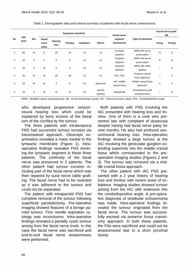
Table 1: Demographic data and clinical summary of patients with facial nerve schwannoma.
Symptoms (months) Facial nerve grade
HB No
Age (yrs)
Sex Facial nerve palsy
Hearing loss
Tinnitus Imbalance Others
Facial nerve segment
involvement Type of Operation
Preop Postop
1 66 M 8 10 10 nil nil Tympanic
segment
MRM with nerve
preservation IV IV
2 43 F 4 8 8 nil nil Tympanic
segment
MRM with nerve
preservation III III
3 48 F nil 6 3 nil nil Tympanic
segment
MRM with cable
grafting I IV
4 49 M nil 48 48 24 nil IAC, CPA Posterior cranial fossa approach
I V
5 28 M 12 3 6 nil dysacusia IAC, middle cranial fossa
Middle cranial fossa approach
V V
6 19 F nil nil nil nil parotid
swelling Intraparotid
Parotidectomy with
reanastomoses I III
MRM - Modified radical mastoidectomy; HB - House-Brackmann grade; IAC - Internal acoustic canal; CPA - Cerebellopontine angle
who developed progressive sensori-neural hearing loss which could be explained by bony erosion of the basal turn of the cochlea by the tumour.
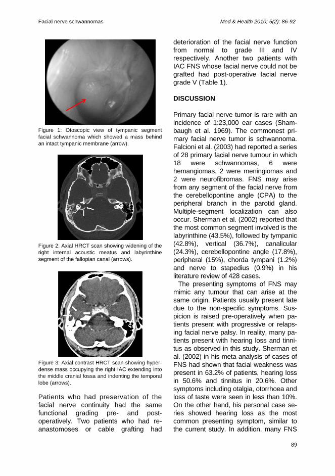
The three patients with intratemporal FNS had successful tumour excision via transmastoid approach. Otoscopic ex-amination revealed a mass medial to the tympanic membrane (Figure 1). Intra-operative findings revealed FNS involv-ing the tympanic segment in these three patients. The continuity of the facial nerve was preserved in 2 patients. The other patient had tumour excision in-cluding part of the facial nerve which was then repaired by sural nerve cable graft-ing. The facial nerve had to be resected as it was adherent to the tumour and could not be separated.
The patient with intraparotid FNS had complete removal of the tumour following superficial parotidectomy. Pre-operative imaging showed features of a benign pa-rotid tumour. Fine needle aspiration cy-tology was inconclusive. Intra-operative findings revealed a partially cystic tumour arising from the facial nerve trunk. In this case the facial nerve was sacrificed and end-to-end facial nerve anastomoses were performed.
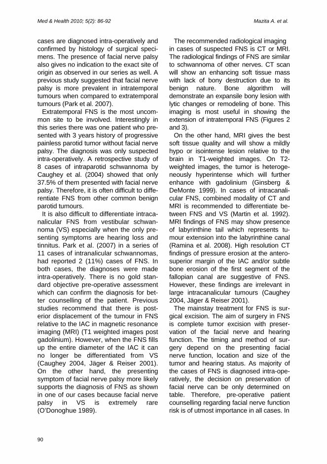
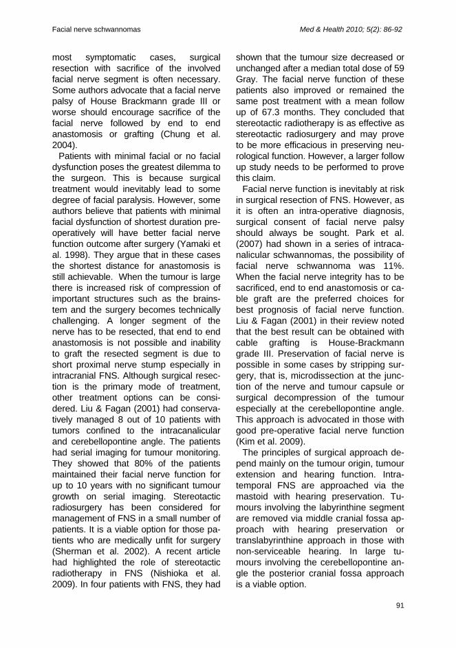
Both patients with FNS involving the IAC presented with hearing loss and tin-nitus. One of them is a cook who pre-sented late with complaint of dysacusia despite having had facial nerve palsy for nine months. He also had profound sen-sorineural hearing loss. Intra-operative findings showed a large tumour at the IAC involving the geniculate ganglion ex-tending superiorly into the middle cranial fossa which corresponded to the pre-operative imaging studies (Figures 2 and 3). The tumour was removed via a mid-dle cranial fossa approach.
The other patient with IAC FNS pre-sented with a 2 year history of hearing loss and tinnitus with recent onset of im-balance. Imaging studies showed tumour arising from the IAC with extension into the cerebellopontine angle. A pre-opera-tive diagnosis of vestibular schwannoma was made. Intra-operative findings re-vealed the tumour originated from the facial nerve. The tumour was success-fully excised via posterior fossa craniot-omy approach. In both these patients, the FNs were sacrificed and could not be anastomosed due to a short proximal stump.
Facial nerve schwannomas Med & Health 2010; 5(2): 86-92
89
Figure 1: Otoscopic view of tympanic segment facial schwannoma which showed a mass behind an intact tympanic membrane (arrow). Figure 2: Axial HRCT scan showing widening of the right internal acoustic meatus and labyrinthine segment of the fallopian canal (arrows). Figure 3: Axial contrast HRCT scan showing hyper-dense mass occupying the right IAC extending into the middle cranial fossa and indenting the temporal lobe (arrows). Patients who had preservation of the facial nerve continuity had the same functional grading pre- and post-operatively. Two patients who had re-anastomoses or cable grafting had
deterioration of the facial nerve function from normal to grade III and IV respectively. Another two patients with IAC FNS whose facial nerve could not be grafted had post-operative facial nerve grade V (Table 1). DISCUSSION Primary facial nerve tumor is rare with an incidence of 1:23,000 ear cases (Sham-baugh et al. 1969). The commonest pri-mary facial nerve tumor is schwannoma. Falcioni et al. (2003) had reported a series of 28 primary facial nerve tumour in which 18 were schwannomas, 6 were hemangiomas, 2 were meningiomas and 2 were neurofibromas. FNS may arise from any segment of the facial nerve from the cerebellopontine angle (CPA) to the peripheral branch in the parotid gland. Multiple-segment localization can also occur. Sherman et al. (2002) reported that the most common segment involved is the labyrinthine (43.5%), followed by tympanic (42.8%), vertical (36.7%), canalicular (24.3%), cerebellopontine angle (17.8%), peripheral (15%), chorda tympani (1.2%) and nerve to stapedius (0.9%) in his literature review of 428 cases.
The presenting symptoms of FNS may mimic any tumour that can arise at the same origin. Patients usually present late due to the non-specific symptoms. Sus-picion is raised pre-operatively when pa-tients present with progressive or relaps-ing facial nerve palsy. In reality, many pa-tients present with hearing loss and tinni-tus as observed in this study. Sherman et al. (2002) in his meta-analysis of cases of FNS had shown that facial weakness was present in 63.2% of patients, hearing loss in 50.6% and tinnitus in 20.6%. Other symptoms including otalgia, otorrhoea and loss of taste were seen in less than 10%. On the other hand, his personal case se-ries showed hearing loss as the most common presenting symptom, similar to the current study. In addition, many FNS
Med & Health 2010; 5(2): 86-92 Mazita A. et al.
90
cases are diagnosed intra-operatively and confirmed by histology of surgical speci-mens. The presence of facial nerve palsy also gives no indication to the exact site of origin as observed in our series as well. A previous study suggested that facial nerve palsy is more prevalent in intratemporal tumours when compared to extratemporal tumours (Park et al. 2007).
Extratemporal FNS is the most uncom-mon site to be involved. Interestingly in this series there was one patient who pre-sented with 3 years history of progressive painless parotid tumor without facial nerve palsy. The diagnosis was only suspected intra-operatively. A retrospective study of 8 cases of intraparotid schwannoma by Caughey et al. (2004) showed that only 37.5% of them presented with facial nerve palsy. Therefore, it is often difficult to diffe-rentiate FNS from other common benign parotid tumours.
It is also difficult to differentiate intraca-nalicular FNS from vestibular schwan-noma (VS) especially when the only pre-senting symptoms are hearing loss and tinnitus. Park et al. (2007) in a series of 11 cases of intranalicular schwannomas, had reported 2 (11%) cases of FNS. In both cases, the diagnoses were made intra-operatively. There is no gold stan-dard objective pre-operative assessment which can confirm the diagnosis for bet-ter counselling of the patient. Previous studies recommend that there is post-erior displacement of the tumour in FNS relative to the IAC in magnetic resonance imaging (MRI) (T1 weighted images post gadolinium). However, when the FNS fills up the entire diameter of the IAC it can no longer be differentiated from VS (Caughey 2004, Jäger & Reiser 2001). On the other hand, the presenting symptom of facial nerve palsy more likely supports the diagnosis of FNS as shown in one of our cases because facial nerve palsy in VS is extremely rare (O’Donoghue 1989).
The recommended radiological imaging in cases of suspected FNS is CT or MRI. The radiological findings of FNS are similar to schwannoma of other nerves. CT scan will show an enhancing soft tissue mass with lack of bony destruction due to its benign nature. Bone algorithm will demonstrate an expansile bony lesion with lytic changes or remodeling of bone. This imaging is most useful in showing the extension of intratemporal FNS (Figures 2 and 3).
On the other hand, MRI gives the best soft tissue quality and will show a mildly hypo or isointense lesion relative to the brain in T1-weighted images. On T2-weighted images, the tumor is heteroge-neously hyperintense which will further enhance with gadolinium (Ginsberg & DeMonte 1999). In cases of intracanali-cular FNS, combined modality of CT and MRI is recommended to differentiate be-tween FNS and VS (Martin et al. 1992). MRI findings of FNS may show presence of labyrinthine tail which represents tu-mour extension into the labyrinthine canal (Ramina et al. 2008). High resolution CT findings of pressure erosion at the antero-superior margin of the IAC and/or subtle bone erosion of the first segment of the fallopian canal are suggestive of FNS. However, these findings are irrelevant in large intracanalicular tumours (Caughey 2004, Jäger & Reiser 2001).
The mainstay treatment for FNS is sur-gical excision. The aim of surgery in FNS is complete tumor excision with preser-vation of the facial nerve and hearing function. The timing and method of sur-gery depend on the presenting facial nerve function, location and size of the tumor and hearing status. As majority of the cases of FNS is diagnosed intra-ope-ratively, the decision on preservation of facial nerve can be only determined on table. Therefore, pre-operative patient counselling regarding facial nerve function risk is of utmost importance in all cases. In
Facial nerve schwannomas Med & Health 2010; 5(2): 86-92
91
most symptomatic cases, surgical resection with sacrifice of the involved facial nerve segment is often necessary. Some authors advocate that a facial nerve palsy of House Brackmann grade III or worse should encourage sacrifice of the facial nerve followed by end to end anastomosis or grafting (Chung et al. 2004).
Patients with minimal facial or no facial dysfunction poses the greatest dilemma to the surgeon. This is because surgical treatment would inevitably lead to some degree of facial paralysis. However, some authors believe that patients with minimal facial dysfunction of shortest duration pre-operatively will have better facial nerve function outcome after surgery (Yamaki et al. 1998). They argue that in these cases the shortest distance for anastomosis is still achievable. When the tumour is large there is increased risk of compression of important structures such as the brains-tem and the surgery becomes technically challenging. A longer segment of the nerve has to be resected, that end to end anastomosis is not possible and inability to graft the resected segment is due to short proximal nerve stump especially in intracranial FNS. Although surgical resec-tion is the primary mode of treatment, other treatment options can be consi-dered. Liu & Fagan (2001) had conserva-tively managed 8 out of 10 patients with tumors confined to the intracanalicular and cerebellopontine angle. The patients had serial imaging for tumour monitoring. They showed that 80% of the patients maintained their facial nerve function for up to 10 years with no significant tumour growth on serial imaging. Stereotactic radiosurgery has been considered for management of FNS in a small number of patients. It is a viable option for those pa-tients who are medically unfit for surgery (Sherman et al. 2002). A recent article had highlighted the role of stereotactic radiotherapy in FNS (Nishioka et al. 2009). In four patients with FNS, they had
shown that the tumour size decreased or unchanged after a median total dose of 59 Gray. The facial nerve function of these patients also improved or remained the same post treatment with a mean follow up of 67.3 months. They concluded that stereotactic radiotherapy is as effective as stereotactic radiosurgery and may prove to be more efficacious in preserving neu-rological function. However, a larger follow up study needs to be performed to prove this claim.
Facial nerve function is inevitably at risk in surgical resection of FNS. However, as it is often an intra-operative diagnosis, surgical consent of facial nerve palsy should always be sought. Park et al. (2007) had shown in a series of intraca-nalicular schwannomas, the possibility of facial nerve schwannoma was 11%. When the facial nerve integrity has to be sacrificed, end to end anastomosis or ca-ble graft are the preferred choices for best prognosis of facial nerve function. Liu & Fagan (2001) in their review noted that the best result can be obtained with cable grafting is House-Brackmann grade III. Preservation of facial nerve is possible in some cases by stripping sur-gery, that is, microdissection at the junc-tion of the nerve and tumour capsule or surgical decompression of the tumour especially at the cerebellopontine angle. This approach is advocated in those with good pre-operative facial nerve function (Kim et al. 2009).
The principles of surgical approach de-pend mainly on the tumour origin, tumour extension and hearing function. Intra-temporal FNS are approached via the mastoid with hearing preservation. Tu-mours involving the labyrinthine segment are removed via middle cranial fossa ap-proach with hearing preservation or translabyrinthine approach in those with non-serviceable hearing. In large tu-mours involving the cerebellopontine an-gle the posterior cranial fossa approach is a viable option.
Med & Health 2010; 5(2): 86-92 Mazita A. et al.
92
CONCLUSION The presenting symptoms of FNS are commonly hearing loss and progressive facial nerve palsy which are non specific. Pre-operative diagnosis still remains a diagnostic dilemma especially when the facial nerve function is normal. Patients should always be counselled regarding facial nerve paralysis before any ear sur-gery because the diagnosis of FNS is often made intra-operatively. The integr-ity of the facial nerve should be pre-served whenever possible. If part of the nerve is sacrificed, end to end anatomo-sis or cable grafting gives the best out-come for facial nerve function. REFERENCES
Caughey, R.J., May, M. & Schaitkin, B.M. 2004.
Intraparotid facial nerve schwannoma: Diagnosis and management. Otolaryngol Head Neck Surg. 130(5):586-592
Chung, J.W., Ahn, J.H., Kim, J.H., Nam, S.Y., Kim, C.J. & Lee, K.S. 2004. Facial nerve schwannomas: different manifestations and outcomes. Surg Neurol. 62(3):245-252.
Falcioni, M., Russo, A., Taibah, A. & Sanna, M. 2003. Facial nerve tumors. Otol Neurotol 24:942–947
Ginsberg, L.E. & DeMonte, F. 1999. Case 16: Facial nerve schwannoma with middle cranial fossa involvement. Radiology 213(2):364-368
Jäger, L. & Reiser, M. 2001. Computed tomography and magnetic resonance imaging of the normal and pathologic conditions of the facial nerve. European Journal of Radiology 40:133-146
Kim, J., Moon, I.S., Lee, J.D., Shim, D.B. & Lee, W.S. 2009. Useful surgical techniques for facial nerve preservation in tumorous intra-
temporal lesions. Auris Nasus Larynx, in press.
Liu, R. & Fagan, P. 2001. Facial nerve schwannoma: Surgical excision versus conservative management. Ann Otol Rhinol Laryngol 110(11):1025-1029
Martin, N., Sterkers, O., Mompoint, D. & Nahum, H. 1992. Facial nerve neuromas: MR imaging Report of four cases. Neuroradiology 34:62-67.
Nishioka, K., Abo, D., Aoyama, H., Furuta, Y., Onimaru, R., Onodera, S., Sawamura, Y., Ishikawa, M., Fukuda, S. & Shirato, H. 2009. Stereotactic radiotherapy for intracranial nonacoustic schwannoma including facial nerve schwannoma. Int J Radiation Oncology Biol Phys, 75(5):1415-1419
O’Donoghue, G.M., Brackmann, D.E., House, J.W. & Jackler, R.K. 1989. Neuromas of the facial nerve. Am J Otolaryngol 10:49-54
Park, H.Y., Kim, S.H., Son, E.J., Lee, H.K., Lee, W.S. 2007. Intracanalicular facial nerve schwannoma. Otol Neurotol 28(3):376-380
Ramina, R., Neto, M.C., da Silva Junior, E.B., Vosgerau, R., Leal, A.G. & Fernandes, Y.B. 2008. Facial nerve schwannomas. In: Samii’s Essentials in Neurosurgery. 1st edition. Edited by Tatagiba M, Aguiar PHP. Springer Publication; 165-174.
Sarma, S., Sekhar, L.N. & Schessel, D.A. 2002. Nonvestibular schwannomas of the brain: A 7-year experience. Neurosurgery 50(3):437-48
Shambaugh, G.E.Jr., Arenberg, I.K., Barney, P.L. & Valvassori, G.E. 1969. Facial neurilemmomas. A study of four diverse cases. Arch Otolaryngol 90:742-755
Sherman, J.D., Dagnew, E., Pensak, M.L., van Loveren, H.R. & Tew, J.M.Jr. 2002. Facial nerve neuromas: Report of 10 cases and review of the literature. Neurosurgery 50(3):450-456
Yamaki, T., Morimoto, S., Ontaki, M., Sakatani, K., Sakai, J., Himi, T., Harabuchi, Y., Sumiyoshi, T., Hashi, K. 1998. Intracranial facial nerve neuroma: Surgical strategy of tumour removal and functional reconstruction. Surg Neurol 49:538-546


![Neurological Disorders - OMICS Publishing Group...Schwannomas are generally benign tumors of the peripheral nerve sheath and account for 8% of all primary intracranial tumors [1,2]](https://img.pdfslide.net/doc/110x75/5f7ff1f90cbb51524d18b287/neurological-disorders-omics-publishing-group-schwannomas-are-generally-benign.jpg)