Embed Size (px)
Citation preview

ITALIAN JOURNALOF PEDIATRICS
Milani et al. Italian Journal of Pediatrics (2015) 41:4 DOI 10.1186/s13052-015-0110-1
DEBATE Open Access
Rubinstein-Taybi syndrome: clinical features,genetic basis, diagnosis, and managementDonatella Milani1, Francesca Maria Paola Manzoni1, Lidia Pezzani1, Paola Ajmone2, Cristina Gervasini3,Francesca Menni1 and Susanna Esposito1*
Abstract
Background: Rubinstein-Taybi syndrome (RSTS) is an extremely rare autosomal dominant genetic disease, with anestimated prevalence of one case per 125,000 live births. RSTS is characterized by typical facial features,microcephaly, broad thumbs and first toes, intellectual disability, and postnatal growth retardation. However, nostandard diagnostic criteria are available for RSTS. In this review, we summarized the clinical features and geneticbasis of RSTS and highlighted areas for future studies on an appropriate diagnostic protocol and follow-up care forRSTS.
Discussion: RSTS is primarily characterized by delayed growth in height and weight, microcephaly, dysmorphicfacial features, and broad thumbs and big toe. Over 90% RSTS individuals with disabilities survive to adulthood, buthealthcare for these patients is particularly complex, time-consuming, and costly. In addition, no standard diagnosticcriteria and follow-up care guidelines are available for RSTS. It has been shown that mutations in the genes encodingthe cyclic-AMP-regulated enhancer binding protein (CREBBP) and the E1A-binding protein p300 (EP300) contributed tothe development of RSTS. Therefore, genetic tests are useful for the diagnosis of RSTS, although most RSTS cases arecurrently diagnosed based on clinical features.
Summary: The clinical features of RSTS have been extensively studied, which significantly contributes to the diagnosisof this extremely rare syndrome. However, the pathogenesis and genotype-phenotype associations of RSTS are largelyunknown. Therefore, multicenter studies and international cooperation are highlighted for better understanding of thisdisease, establishing standard diagnostic criteria, and providing professional management and follow-up care of RSTS.
Keywords: CREBBP, Intellectual disability, Plurimalformative syndrome, Rubinstein syndrome, Rubinstein-Taybi syndrome
BackgroundPlurimalformative syndromes, which are named accordingto their low prevalence and incidence in the population,consist of a large group of rare diseases. Rubinstein-Taybisyndrome (RSTS, OMIM #180849, #613684) is an ex-tremely rare disease and was first described in 1963 [1].The incidence of RSTS is 1 in 100,000 to 125,000 livebirths. Currently, no precise diagnostic criteria are avail-able, although RSTS is primarily characterized by poorpostnatal height-weight growth, intellectual disability,microcephaly, dysmorphic facial features, broad thumbs,and big first toes. While a number of major malformations
* Correspondence: [email protected] Highly Intensive Care Unit, Department of Pathophysiology andTransplantation, Università degli Studi di Milano, Fondazione IRCCS Ca’Granda Ospedale Maggiore Policlinico, Via Commenda 9, 20122 Milano, ItalyFull list of author information is available at the end of the article
© 2015 Milani et al.; licensee BioMed Central.Commons Attribution License (http://creativecreproduction in any medium, provided the orDedication waiver (http://creativecommons.orunless otherwise stated.
and clinical complications are associated with RSTS, thesesigns and symptoms cannot be considered pathogno-monic to RSTS. Until the 90s, diagnosis has remained ex-clusively clinical and radiological (x-ray of hands and feet).The genetic bases were first identified in 1991, demon-strating a de novo reciprocal translocation with break-points in chromosomal region 16p13.3 in some patients[2-4]. Subsequently, affected subjects were analyzed usingFluorescent In Situ Hybridization (FISH). In six cases, thehybridization signal was present on only one allele on16p13.3, confirming that the absence of this region lead toRSTS [5,6]. Additional research has led to the discovery ofmutations in the gene encoding cyclic-AMP-regulated en-hancer binding protein (CREBBP) in 16p13.3 in RSTS pa-tients [7]. Mutations of CREBBP gene were reported in
This is an Open Access article distributed under the terms of the Creativeommons.org/licenses/by/4.0), which permits unrestricted use, distribution, andiginal work is properly credited. The Creative Commons Public Domaing/publicdomain/zero/1.0/) applies to the data made available in this article,

Milani et al. Italian Journal of Pediatrics (2015) 41:4 Page 2 of 9
approximately half of RSTS patients [8,9]. CREBBP geneand its homolog, E1A binding protein p300 (EP300) onchromosome 22, are involved in a number of basic cellularactivities, such as DNA repair, growth, differentiation,apoptosis of cells, and tumor suppression by serving astranscriptional co-activators in different signaling path-ways [10]. As CREBBP and EP300 interact closely, studieshave been conducted to investigate whether mutations inEP300 were associated with RSTS. The results of EP300sequencing in a group of six subjects revealed threemutations [11]. Subsequently, the incidence of EP300mutations was estimated as about 5-8% [12-16]. Generally,55-70% clinically diagnosed RSTS cases were confirmedthrough genetic testing [17]. In this review, we discussedthe clinical features and genetic studies of RSTS and we tryto outline future directions for an appropriate clinical diag-nosis and follow-up of this condition.
DiscussionTypical featuresRSTS is characterized by slow development of heightand weight, microcephaly, dysmorphic facial features,broad thumbs, and big toes [18]. The prenatal develop-ment is normal, with average or near-normal growth pa-rameters at birth. The growth charts typically approachthe lower limits of normality in the first postnatal period,primarily reflecting hypo-feeding exacerbated by gastro-esophageal reflux. Subsequently, the tendency of over-weight or obesity (earlier in males than females) can beobserved during adolescence. Specific and recently reviewedgrowth charts are essential for appropriate assessment of thegrowth of affected individuals [18]. Facial features are pri-marily characterized by low frontal hairline, arched/thickeyebrows, downslanting of palpebral fissures, a protrudingbeaked nose with columella below alae nasi, dysplastic andlow-set ears, an arched palate, mild micrognathia, dentalanomalies (altered conformation, malocclusion, and over-crowding of teeth), and atypical smile (“grimacing”) withnearly completely closed eyes (Figure 1). The feet and handstypically present an enlarged first finger and clinodactyly ofthe fifth finger (Figure 2), whereas polydactyly with bifidthumbs and first toes is rarely observed. Other skeletalanomalies include abducted thumbs, vertebral anomalies,ligamentous laxity, severe and prolonged aseptic inflamma-tion of the femur head, anomalies similar with Perthes dis-ease (3%), and occasionally slipped capital femoral epiphysis[19,20]. Particularly, high risk of cervical vertebral abnormal-ities (instability of C1–C2, os odontoideum, hypoplasia ofthe dens, fusion of the cervical vertebrae) has been reported[21-23], with possible stenosis at the craniovertebral junc-tion, which may cause cervical myelopathy. Complex neuro-radiological issues including corpus callosum dysgenesis(17%) [24,25], Chiari type I malformation with or withoutsyringomyelia [25-28], Dandy Walker malformation and
hydrocephalus [29,30], and tethered cord [27,31] have beenreported and are still under investigation. Cerebrovascularabnormalities such as spontaneous dissection of thesupraaortic arteries [32] and cerebral infarction due todissecting aneurysm of the anterior cerebral arteryhave also been reported [33]. However, any organ canbe affected in RSTS patients. Possible malformations,medical problems, and complications include (Table 1):
– conductive and/or sensorineural deafness, recurrentmiddle ear infections, recurrent respiratoryinfections, immune deficiencies [34-36];
– nonspecific abnormalities of electroencephalography(EEG) (57-66%) and seizures (25%) [37,38];
– cataract, unilateral or bilateral iris/retinal/opticnerve coloboma (9-11%), glaucoma, lacrimal ductobstructions (38-47%), refractive errors (41-56%),and strabismus (60-71%) [39-41]. In addition, Jacobset al. described for the first time peripheralavascularity with fluorescein angiography in 2012 [42];
– dental problems: talon cusps (73%), enamelhypoplasia, and abnormal tooth number [43,44];
– congenital heart diseases: atrial septal defect,ventricular septal defect, patent ductus arteriosus,coarctation of the aorta, pulmonic stenosis, bicuspidaortic valve, pseudotruncus, aortic stenosis,dextrocardia, vascular rings, and conductiondisorders (24-38%) [45]. Occasional association ofhypoplastic left heart with RSTS has also beenreported [46];
– renal malformations (52%) and cryptorchidism(78-100%) [47];
– endocrine disorders: congenital hypothyroidism[48,49], thyroid hypoplasia, GH deficiency, andpituitary hypoplasia [28];
– gastrointestinal disorders: gastroesophageal reflux,constipation (40-74%), and megacolon/Hirschsprungdisease [47,50];
– obstructive sleep apnea, anesthetic and intubationcomplications [51,52];
– skin problems including pilomatrixomas, ingrowntoenails, paronychia, and the tendency to formkeloids (24%) [53,54];
– cancers, particularly of neural and developmentalorigins (neuroblastoma, medulloblastoma,oligodendroglioma, meningeoma,pheochromocytoma, rhabdomyosarcoma,leiomyosarcoma, seminoma, odontoma, choristoma,and pilomatrixomas [55-63]. Leukemia andlymphoma have also been reported [55];
– hirsutism.
The neonatal period of individuals with RSTS is typ-ically characterized by hypotonia and delayed psycho-
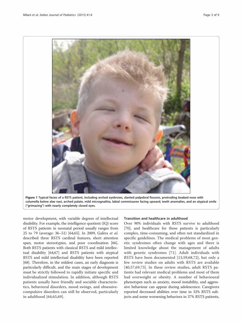
Figure 1 Typical facies of a RSTS patient, including arched eyebrows, slanted palpebral fissures, protruding beaked nose withcolumella below alae nasi, arched palate, mild micrognathia, labial commissures facing upward, teeth anomalies, and an atypical smile(“grimacing”) with nearly completely closed eyes.
Milani et al. Italian Journal of Pediatrics (2015) 41:4 Page 3 of 9
motor development, with variable degrees of intellectualdisability. For example, the intelligence quotient (IQ) scoreof RSTS patients in neonatal period usually ranges from25 to 79 (average: 36–51) [64,65]. In 2009, Galèra et al.described three RSTS cardinal features, short attentionspan, motor stereotypies, and poor coordination [66].Both RSTS patients with classical RSTS and mild intellec-tual disability [64,67] and RSTS patients with atypicalRSTS and mild intellectual disability have been reported[68]. Therefore, in the mildest cases, an early diagnosis isparticularly difficult, and the main stages of developmentmust be strictly followed to rapidly initiate specific andindividualized stimulation. In addition, although RSTSpatients usually have friendly and sociable characteris-tics, behavioral disorders, mood swings, and obsessive-compulsive disorders can still be observed, particularlyin adulthood [64,65,69].
Transition and healthcare in adulthoodOver 90% individuals with RSTS survive to adulthood[70], and healthcare for these patients is particularlycomplex, time-consuming, and often not standardized inspecific guidelines. The medical problems of most gen-etic syndromes often change with ages and there islimited knowledge about the management of adultswith genetic syndromes [71]. Adult individuals withRSTS have been documented [13,59,68,72], but only afew review studies on adults with RSTS are available[40,57,69,73]. In these review studies, adult RSTS pa-tients had relevant medical problems and most of themhad overweight or obesity. A number of behaviouralphenotypes such as anxiety, mood instability, and aggres-sive behaviour can appear during adolescence. Caregiversreported decreased abilities over time in 32% RSTS sub-jects and some worsening behaviors in 37% RSTS patients,
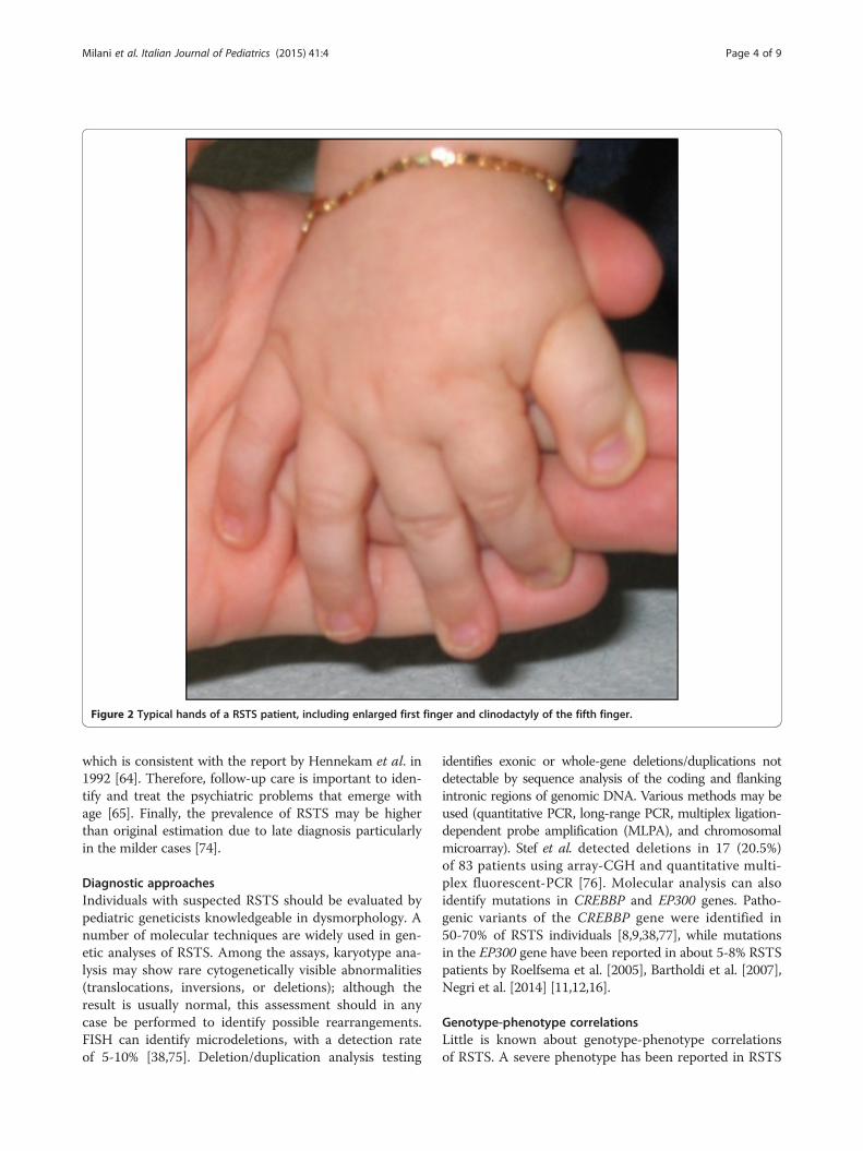
Figure 2 Typical hands of a RSTS patient, including enlarged first finger and clinodactyly of the fifth finger.
Milani et al. Italian Journal of Pediatrics (2015) 41:4 Page 4 of 9
which is consistent with the report by Hennekam et al. in1992 [64]. Therefore, follow-up care is important to iden-tify and treat the psychiatric problems that emerge withage [65]. Finally, the prevalence of RSTS may be higherthan original estimation due to late diagnosis particularlyin the milder cases [74].
Diagnostic approachesIndividuals with suspected RSTS should be evaluated bypediatric geneticists knowledgeable in dysmorphology. Anumber of molecular techniques are widely used in gen-etic analyses of RSTS. Among the assays, karyotype ana-lysis may show rare cytogenetically visible abnormalities(translocations, inversions, or deletions); although theresult is usually normal, this assessment should in anycase be performed to identify possible rearrangements.FISH can identify microdeletions, with a detection rateof 5-10% [38,75]. Deletion/duplication analysis testing
identifies exonic or whole-gene deletions/duplications notdetectable by sequence analysis of the coding and flankingintronic regions of genomic DNA. Various methods may beused (quantitative PCR, long-range PCR, multiplex ligation-dependent probe amplification (MLPA), and chromosomalmicroarray). Stef et al. detected deletions in 17 (20.5%)of 83 patients using array-CGH and quantitative multi-plex fluorescent-PCR [76]. Molecular analysis can alsoidentify mutations in CREBBP and EP300 genes. Patho-genic variants of the CREBBP gene were identified in50-70% of RSTS individuals [8,9,38,77], while mutationsin the EP300 gene have been reported in about 5-8% RSTSpatients by Roelfsema et al. [2005], Bartholdi et al. [2007],Negri et al. [2014] [11,12,16].
Genotype-phenotype correlationsLittle is known about genotype-phenotype correlationsof RSTS. A severe phenotype has been reported in RSTS
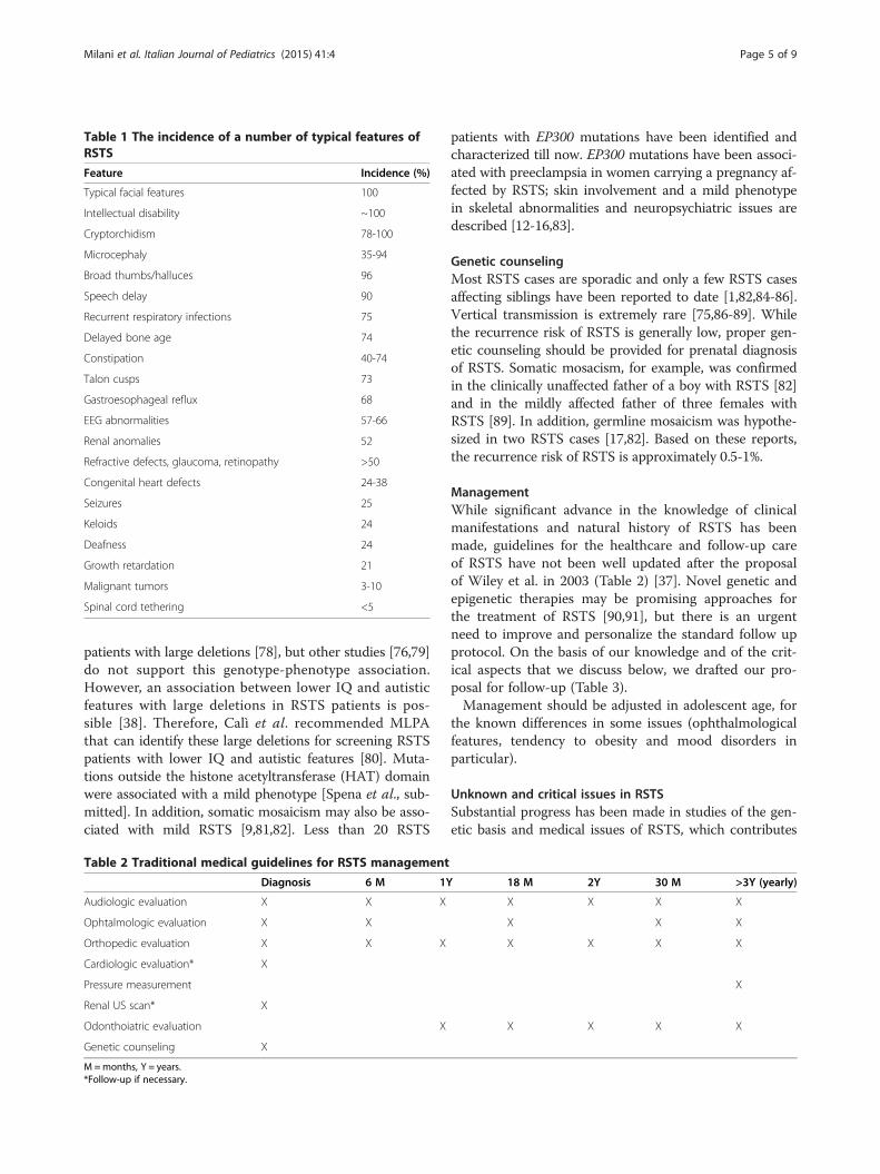
Table 1 The incidence of a number of typical features ofRSTS
Feature Incidence (%)
Typical facial features 100
Intellectual disability ~100
Cryptorchidism 78-100
Microcephaly 35-94
Broad thumbs/halluces 96
Speech delay 90
Recurrent respiratory infections 75
Delayed bone age 74
Constipation 40-74
Talon cusps 73
Gastroesophageal reflux 68
EEG abnormalities 57-66
Renal anomalies 52
Refractive defects, glaucoma, retinopathy >50
Congenital heart defects 24-38
Seizures 25
Keloids 24
Deafness 24
Growth retardation 21
Malignant tumors 3-10
Spinal cord tethering <5
Milani et al. Italian Journal of Pediatrics (2015) 41:4 Page 5 of 9
patients with large deletions [78], but other studies [76,79]do not support this genotype-phenotype association.However, an association between lower IQ and autisticfeatures with large deletions in RSTS patients is pos-sible [38]. Therefore, Calì et al. recommended MLPAthat can identify these large deletions for screening RSTSpatients with lower IQ and autistic features [80]. Muta-tions outside the histone acetyltransferase (HAT) domainwere associated with a mild phenotype [Spena et al., sub-mitted]. In addition, somatic mosaicism may also be asso-ciated with mild RSTS [9,81,82]. Less than 20 RSTS
Table 2 Traditional medical guidelines for RSTS management
Diagnosis 6 M 1Y
Audiologic evaluation X X X
Ophtalmologic evaluation X X
Orthopedic evaluation X X X
Cardiologic evaluation* X
Pressure measurement
Renal US scan* X
Odonthoiatric evaluation X
Genetic counseling X
M=months, Y = years.*Follow-up if necessary.
patients with EP300 mutations have been identified andcharacterized till now. EP300 mutations have been associ-ated with preeclampsia in women carrying a pregnancy af-fected by RSTS; skin involvement and a mild phenotypein skeletal abnormalities and neuropsychiatric issues aredescribed [12-16,83].
Genetic counselingMost RSTS cases are sporadic and only a few RSTS casesaffecting siblings have been reported to date [1,82,84-86].Vertical transmission is extremely rare [75,86-89]. Whilethe recurrence risk of RSTS is generally low, proper gen-etic counseling should be provided for prenatal diagnosisof RSTS. Somatic mosacism, for example, was confirmedin the clinically unaffected father of a boy with RSTS [82]and in the mildly affected father of three females withRSTS [89]. In addition, germline mosaicism was hypothe-sized in two RSTS cases [17,82]. Based on these reports,the recurrence risk of RSTS is approximately 0.5-1%.
ManagementWhile significant advance in the knowledge of clinicalmanifestations and natural history of RSTS has beenmade, guidelines for the healthcare and follow-up careof RSTS have not been well updated after the proposalof Wiley et al. in 2003 (Table 2) [37]. Novel genetic andepigenetic therapies may be promising approaches forthe treatment of RSTS [90,91], but there is an urgentneed to improve and personalize the standard follow upprotocol. On the basis of our knowledge and of the crit-ical aspects that we discuss below, we drafted our pro-posal for follow-up (Table 3).Management should be adjusted in adolescent age, for
the known differences in some issues (ophthalmologicalfeatures, tendency to obesity and mood disorders inparticular).
Unknown and critical issues in RSTSSubstantial progress has been made in studies of the gen-etic basis and medical issues of RSTS, which contributes
18 M 2Y 30 M >3Y (yearly)
X X X X
X X X
X X X X
X
X X X X
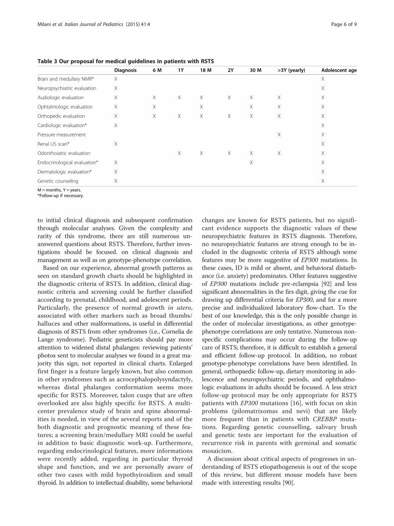
Table 3 Our proposal for medical guidelines in patients with RSTS
Diagnosis 6 M 1Y 18 M 2Y 30 M >3Y (yearly) Adolescent age
Brain and medullary NMR* X X
Neuropsychiatric evaluation X X
Audiologic evaluation X X X X X X X X
Ophtalmologic evaluation X X X X X X
Orthopedic evaluation X X X X X X X X
Cardiologic evaluation* X X
Pressure measurement X X
Renal US scan* X X
Odonthoiatric evaluation X X X X X X
Endocrinological evaluation* X X X
Dermatologic evaluation* X X
Genetic counseling X X
M=months, Y = years.*Follow-up if necessary.
Milani et al. Italian Journal of Pediatrics (2015) 41:4 Page 6 of 9
to initial clinical diagnosis and subsequent confirmationthrough molecular analyses. Given the complexity andrarity of this syndrome, there are still numerous un-answered questions about RSTS. Therefore, further inves-tigations should be focused. on clinical diagnosis andmanagement as well as on genotype-phenotype correlation.Based on our experience, abnormal growth patterns as
seen on standard growth charts should be highlighted inthe diagnostic criteria of RSTS. In addition, clinical diag-nostic criteria and screening could be further classifiedaccording to prenatal, childhood, and adolescent periods.Particularly, the presence of normal growth in utero,associated with other markers such as broad thumbs/halluces and other malformations, is useful in differentialdiagnosis of RSTS from other syndromes (i.e., Cornelia deLange syndrome). Pediatric geneticists should pay moreattention to widened distal phalanges: reviewing patients’photos sent to molecular analyses we found in a great ma-jority this sign, not reported in clinical charts. Enlargedfirst finger is a feature largely known, but also commonin other syndromes such as acrocephalopolysyndactyly,whereas distal phalanges conformation seems morespecific for RSTS. Moreover, talon cusps that are oftenoverlooked are also highly specific for RSTS. A multi-center prevalence study of brain and spine abnormal-ities is needed, in view of the several reports and of theboth diagnostic and prognostic meaning of these fea-tures; a screening brain/medullary MRI could be usefulin addition to basic diagnostic work-up. Furthermore,regarding endocrinological features, more informationswere recently added, regarding in particular thyroidshape and function, and we are personally aware ofother two cases with mild hypothyiroidism and smallthyroid. In addition to intellectual disability, some behavioral
changes are known for RSTS patients, but no signifi-cant evidence supports the diagnostic values of theseneuropsychiatric features in RSTS diagnosis. Therefore,no neuropsychiatric features are strong enough to be in-cluded in the diagnostic criteria of RSTS although somefeatures may be more suggestive of EP300 mutations. Inthese cases, ID is mild or absent, and behavioral disturb-ance (i.e. anxiety) predominates. Other features suggestiveof EP300 mutations include pre-eclampsia [92] and lesssignificant abnormalities in the firs digit, giving the cue fordrawing up differential criteria for EP300, and for a moreprecise and individualized laboratory flow-chart. To thebest of our knowledge, this is the only possible change inthe order of molecular investigations, as other genotype-phenotype correlations are only tentative. Numerous non-specific complications may occur during the follow-upcare of RSTS; therefore, it is difficult to establish a generaland efficient follow-up protocol. In addition, no robustgenotype-phenotype correlations have been identified. Ingeneral, orthopaedic follow-up, dietary monitoring in ado-lescence and neuropsychiatric periods, and ophthalmo-logic evaluations in adults should be focused. A less strictfollow-up protocol may be only appropriate for RSTSpatients with EP300 mutations [16], with focus on skinproblems (pilomatrixomas and nevi) that are likelymore frequent than in patients with CREBBP muta-tions. Regarding genetic counselling, salivary brushand genetic tests are important for the evaluation ofrecurrence risk in parents with germinal and somaticmosaicism.A discussion about critical aspects of progresses in un-
derstanding of RSTS etiopathogenesis is out of the scopeof this review, but different mouse models have beenmade with interesting results [90].

Milani et al. Italian Journal of Pediatrics (2015) 41:4 Page 7 of 9
ConclusionsRSTS is an extremely rare condition for which someclinical aspects have been clearly identified, but a lot ofstudies are ongoing and needed. Multicenter studies areneeded to expand our knowledge on the clinical pheno-type, identify specific genotype-phenotype correlations,evaluate the presence of somatic mosaicisms to betterdefine mild phenotypes, and identify new candidategenes. The ultimate goal of these studies is to extendour current knowledge concerning this syndrome and todefine new international guidelines for diagnosis, careand treatment of patients with RSTS.
SummaryRSTS is an extremely rare multiple congenital anomaly/intellectual disability syndrome, with an estimated preva-lence of one case per 125,000 live births. No precisediagnostic criteria have been defined, although the dis-tinctive features include typical facial features, microceph-aly, broad thumbs and first toes, intellectual disability andpostnatal growth retardation. RSTS is mainly character-ized by poor growth in height and weight, microcephaly,dysmorphic facial features, broad thumbs and big toe. Sev-eral organs and systems may be affected, but none of othersigns or symptoms can be considered pathognomonic.More than 90% of individuals with disabilities survive intoadulthood, and health care for these patients is particularlycomplex, time-consuming, often not standardized in spe-cific guidelines. The gene most frequently involved iscyclic-AMP-regulated enhancer binding protein (CREBBP);alterations in the E1A-binding protein p300 (EP300) havealso been detected, but many cases have only been clinic-ally diagnosed. Future multicenter studies are necessary toexpand our knowledge on the clinical phenotype, iden-tify specific genotype-phenotype correlations, evaluatethe presence of somatic mosaicisms to better definemild phenotypes, and identify new candidate genes.The ultimate goal of these studies is to extend ourcurrent knowledge concerning this syndrome and todefine new international guidelines for diagnosis, careand treatment of patients with RSTS.
Ethical approvalThe follow-up studies of RSTS children performed bythe authors have been approved by the Ethics Commit-tee of Fondazione IRCCS Ca’ Granda Ospedale MaggiorePoliclinico, Milan, Italy.
ConsentWritten informed consent was obtained from the pa-tients’ parents for the publication of this report and anyaccompanying images.
AbbreviationsRSTS: Rubinstein-Taybi syndrome; CREBBP: Cyclic-AMP-regulated enhancerbinding protein; EP300: E1A binding protein p300; FISH: Fluorescent in situhybridization; HAT: Histone acetyltransferase; IQ: Intelligence quotient;MLPA: Multiplex ligation-dependent probe amplification.
Competing interestsThe authors declare that they have no competing interests.
Authors’ contributionsDM, FMPM, LP, PA, CG and FM drafted the manuscript and followed theRSTS patients at Fondazione IRCCS Ca’ Granda Ospedale MaggiorePoliclinico, Milan, Italy. SE, the Director of the Unit where the patients werefollowed, revised the manuscript and made substantial scientificcontributions. All authors have read and approved the final version of themanuscript.
AcknowledgmentsThe authors thank Italian Association RTS Una vita speciale for the preciousawareness work on public opinion.
FundingThis study was supported by grants from the Italian Ministry of Health(Bando Giovani Ricercatori 2009).
Author details1Pediatric Highly Intensive Care Unit, Department of Pathophysiology andTransplantation, Università degli Studi di Milano, Fondazione IRCCS Ca’Granda Ospedale Maggiore Policlinico, Via Commenda 9, 20122 Milano, Italy.2UO Neuropsichiatria dell’Infanzia e dell’Adolescenza, Fondazione IRCCS Ca’Granda Ospedale Maggiore Policlinico, Milano, Italy. 3Department of HealthScience, Medical Genetics, Università degli Studi di Milano, Milano, Italy.
Received: 19 October 2014 Accepted: 7 January 2015
References1. Rubinstein JH, Taybi H. Broad thumbs and toes and facial abnormalities. A
possible mental retardation syndrome. Am J Dis Child. 1963;105:588–608.2. Imaizumi K, Kuroki Y. Rubinstein-Taybi syndrome with de novo reciprocal
translocation t(2;16)(p13.3;p13.3). Am J Med Genet A. 1991;38:636–9.3. Lacombe D, Saura R, Taine L, Battin J. Confirmation of assignment of a locus
for Rubinstein- Taybi syndrome gene to 16p13.3. Am J Med Genet A.1992;44:126–8.
4. Tommerup N, van der Hagen CB, Heiberg A. Tentative assignment of alocus for Rubinstein-Taybi syndrome to 16p13.3 by a de novo reciprocaltranslocation, t(7;16)(q34;p13.3). Am J Med Genet A. 1992;44:237–41.
5. Breuning MH, Dauwerse HG, Fugazza G, Saris JJ, Spruit L, Wijnen H, et al.Rubinstein-Taybi syndrome caused by submicroscopic deletions within16p13.3. Am J Hum Genet. 1993;52:249–54.
6. Hennekam RC, Tilanus M, Hamel BC, Voshart-van Heeren H, Mariman EC,van Beersum SE, et al. Deletion at chromosome 16p13.3 as a cause ofRubinstein-Taybi syndrome: clinical aspects. Am J Hum Genet. 1993;52:255–62.
7. Petrij F, Giles RH, Dauwerse HG, Saris JJ, Hennekam RC, Masuno M,Tommerup N, van Ommen GJ, Goodman RH, Peters DJ. Rubinstein-Taybisyndrome caused by mutations in the transcriptional co-activator CBP.Nature. 1995;376:348–51.
8. Coupry I, Roudaut C, Stef M, Delrue MA, Marche M, Burgelin I, et al.Molecular analysis of the CBP gene in 60 patients with Rubinstein-Taybisyndrome. J Med Genet. 2002;39:415–21.
9. Bentivegna A, Milani D, Gervasini C, Castronovo P, Mottadelli F, Manzini S,et al. Rubinstein-Taybi syndrome: spectrum of CREBBP mutations in Italianpatients. BMC Med Genet. 2006;7:77.
10. Goodman RH, Smolik S. CBP/p300 in cell growth, transformation, anddevelopment. Genes Dev. 2000;14:1553–77.
11. Roelfsema JH, White SJ, Ariyürek Y, Bartholdi D, Niedrist D, Papadia F, et al.Genetic heterogeneity in Rubinstein-Taybi syndrome: mutations in both theCBP and EP300 genes cause disease. Am J Hum Genet. 2005;76:572–80.
12. Bartholdi D, Roelfsema JH, Papadia F, Breuning MH, Niedrist D, HennekamRC, et al. Genetic heterogeneity in Rubinstein-Taybi syndrome: delineation
Milani et al. Italian Journal of Pediatrics (2015) 41:4 Page 8 of 9
of the phenotype of the first patients carrying mutations in EP300. J MedGenet. 2007;44:327–33.
13. Bartsch O, Labonté J, Albrecht B, Wieczorek D, Lechno S, Zechner U, et al.Two patients with EP300 mutations and facial dysmorphism differentfrom the classic Rubinstein–Taybi syndrome. Am J Med Genet A.2010;152A:181–4.
14. Tsai AC, Dossett CJ, Walton CS, Cramer AE, Eng PA, Nowakowska BA, et al.Exon deletions of the EP300 and CREBBP genes in two children with Rubinstein-Taybi syndrome detected by aCGH. Eur J Hum Genet. 2011;19(1):43–9.
15. Woods SA, Robinson HB, Kohler LJ, Agamanolis D, Sterbenz G, Khalifa M.Exome sequencing identifies a novel EP300 frame shift mutation in apatient with features that overlap Cornelia de Lange syndrome. Am J MedGenet A. 2014;164A:251–8.
16. Negri G, Milani D, Colapietro P, Forzano F, Della Monica M, Rusconi D, et al.Clinical and molecular characterization of Rubinstein-Taybi syndrome patientscarrying distinct novel mutations of the EP300 gene. Clin Genet. 2014.doi:10.1111/cge.12348.
17. Tajir M, Fergelot P, Lancelot G, Elalaoui SC, Arveiler B, Lacombe D, et al.Germline mosaicism in Rubinstein–Taybi syndrome. Gene. 2013;518:476–8.
18. Beets L, Rodrıguez-Fonseca C, Hennekam RC. Growth charts for individualswith Rubinstein–Taybi syndrome. Am J Med Genet A. 2014;164(9):2300–9.
19. Bonioli E, Bellini C, Sénès FM, Palmieri A, Di Stadio M, Pinelli G. Slippedcapital femoral epiphysis associated with Rubinstein-Taybi syndrome. ClinGenet. 1993;44(2):79–81.
20. Shah H, Singh G, Vijayan S, Girisha KM. Second report of slipped capitalfemoral epiphysis in Rubinstein-Taybi syndrome. Clinical Dysmorphology.2011;20:55–7.
21. Robson MJ, Brown LM, Sharrard WJ. Cervical spondylolisthesis and otherskeletal abnormalities in Rubinstein-Taybi syndrome. J Bone Joint Surg Br.1980;62:297–9.
22. Yamamoto T, Kurosawa K, Masuno M, Okuzumi S, Kondo S, Miyama S, et al.Congenital anomaly of cervical vertebrae is a major complication ofRubinstein-Taybi syndrome. Am J Med Genet A. 2005;135:130–3.
23. Marzuillo P, Grandone A, Luongo C, Cantelmi G, Polito C, del Giudice EM, etal. Brain magnetic resonance in the routine management of Rubinstein-Taybisyndrome (RTS) can prevent lifethreatening events and neurological deficits.Am J Med Genet A. 2014;164A:2129–32.
24. Rubinstein JH. Broad thumb-hallux (Rubinstein-Taybi) syndrome 1957–1988.Am J Med Genet Suppl. 1990;6:3–16.
25. Wojcik C, Volz K, Ranola M, Kitch K, Karim T, O’Neil J, et al. Rubinstein–Taybisyndrome associated with Chiari type I malformation caused by a large16p13.3 microdeletion: A contiguous gene syndrome? Am J Med Genet A.2010;152A:479–83.
26. Parsley L, Bellus G, Handler M, Tsai AC-H. Identical twin sisters with Rubinstein–Taybi syndrome associated with Chiari malformations and syrinx. Am J MedGenet A. 2011;155:2766–770.
27. Giussani C, Selicorni A, Fossati C, Ingelmo P, Canonico F, Landi A, et al.The association of neural axis and craniovertebral junction anomalieswith scoliosis in Rubinstein–Taybi syndrome. Child Nerv Syst.2012;28:2163–8.
28. Marzuillo Grandone A, Coppola R, Cozzolino D, Festa A, Messa F, Luongo C,et al. Novel cAMP binding protein-BP (CREBBP) mutation in a girl withRubinstein-Taybi syndrome, GH deficiency, Arnold Chiari malformation andpituitary hypoplasia. BMC Medical Genetics. 2013;14:28.
29. Barson AJ. Proceedings: Rubinstein-Taybi syndrome. Arch Dis Child.1974;49(6):495.
30. Agarwal R, Aggarwal R, Kabra M, Deorari AK. Dandy-Walker malformationin Rubinstein-Taybi syndrome: a rare association. Clin Dysmorphol.2002;11(3):223–4.
31. Tanaka T, Ling BC, Rubinstein JH, Crone KR. Rubinstein-Taybi syndrome inchildren with tethered spinal cord. J Neurosurg. 2006;105(4 Suppl):261–4.
32. Fischer S, Bäzner H, Henkes H. Cervical artery dissection in a young patientwith Rubinstein-Taybi syndrome. Clin Neuroradiol. 2013;23:41–4.
33. Ishizaka S, Sou G, Morofuji Y, Hayashi K, Kitagawa N, Tateishi Y, et al.Dissecting aneurysm of the anterior cerebral artery with Rubinstein-Taybisyndrome–a case report. Brain Nerve. 2010;62:1083–8.
34. Peñaranda A, Cerón M. Rubinstein-Taybi syndrome and mixed bilateralhypoacousia case report. Otol Neurotol. 2007;28:501–3.
35. Naimi DR, Munoz J, Rubinstein J, Hostoffer Jr RW. Rubinstein-Taybi syndrome:an immune deficiency as a cause for recurrent infections. Allergy Asthma Proc.2006;27:281–4.
36. Torres LC, Sugayama SM, Arslanian C, Sales MM, Carneiro-Sampaio M. Evaluationof the immune humoral response of Brazilian patients with Rubinstein-Taybisyndrome. Braz J Med Biol Res. 2010;43:1215–24.
37. Wiley S, Swayne S, Rubinstein JH, Lanphear NE, Stevens CA. Rubinstein-Taybisyndrome medical guidelines. Am J Med Genet A. 2003;119A:101–10.
38. Schorry EK, Keddache M, Lanphear N, Rubinstein JH, Srodulski S, Fletcher D,et al. Genotype-phenotype correlations in Rubinstein-Taybi syndrome. Am JMed Genet A. 2008;146A:2512–9.
39. Marabotti A, Giannecchini G, Cariello A, Cappelli C, Giannecchini I, Bedei A.Stenosis of the lachrymal system in Rubinstein-Taybi syndrome. Ophthalmol.2002;216:272–6.
40. Van Genderen MM, Kinds GF, Riemslag FCC, Hennekam RCM. Ocularfeatures in Rubinstein-Taybi syndrome: investigation of 24 patients andreview of the literature. Br J Ophthalmol. 2000;84:1177–84.
41. Kosaki R, Fujita H, Takada H, Okada M, Torii C, Kosaki K. Monozygotic twinsof Rubinstein–Taybi syndrome discordant for glaucoma. Am J Med Genet A.2011;155:1189–91.
42. Jacobs DJ, Sein J, Berrocal AM, Grajewski AL, Hodapp E. Fluoresceinangiography findings in a case of Rubinstein-Taybi syndrome. Clin Ophthalmol.2012;6:1369–71.
43. Hennekam RC, Stevens CA, Van de Kamp JJ. Etiology and recurrence risk inRubinstein- Taybi syndrome. Am J Med Genet. 1990;6:56–64.
44. Bloch-Zupan A, Stachtou J, Emmanouil D, Arveiler B, Griffiths D, Lacombe D.Oro- dental features as useful diagnostic tool in Rubinstein-Taybi syndrome.Am J Med Genet A. 2007;143:570–3.
45. Stevens CA, Bhakta MG. Cardiac abnormalities in the Rubinstein-Taybisyndrome. Am J Med Genet A. 1995;59:346–8.
46. Hanauer D, Argilla M, Wallerstein R. Rubinstein-Taybi syndrome and hypoplasticleft heart. Am J Med Genet A. 2002;112:109–11.
47. Hennekam RCM. Rubinstein-Taybi syndrome. In: Cassidy SB, Allanson JE,editors. Management of genetic syndromes. 3rd ed. Hoboken, NJ: Wiley-Blackwell;2010. p. 705–15.
48. Olson DP, Koenig RJ. Thyroid function in Rubinstein-Taybi syndrome. J ClinEndocrinol Metab. 1997;82:3264–6.
49. Kurtoglu S, Akcakus M, Gunes T, Cetin N, Topaloglu N. Congenitalhypothyroidism associated with Rubinstein-Taybi syndrome. J PediatrEndocrinol Metab. 2003;16:457–9.
50. Isidor B, Podevin G, Camby C, Mosnier J-F, Chauty A, Lyet J-M, et al. Rubinstein–Taybi syndrome and Hirschsprung disease in a patient harbouring an intragenicdeletion of the CREBBP gene. Am J Med Genet A. 2010;152A:1847–8.
51. Zucconi M, Ferini-Strambi L, Erminio C, Pestalozza G, Smirne S.Obstructive sleep apnea in the Rubinstein-Taybi syndrome. Respiration.1993;60:127–32.
52. Choi HS. Pulmonary hypertension due to obstructive sleep apnea in a childwith Rubinstein-Taybi syndrome. Korean J Pediatr. 2012;55:212–14.
53. Bayle P, Bazex J, Lamant L, Lauque D, Durieu C, Albes B. Multiple perforatingand non perforating pilomatricomas in a patient with Churg-Strausssyndrome and Rubinstein-Taybi syndrome. J Eur Acad Dermatol Venereol.2004;18:607–10.
54. van de Kar AL, Houge G, Shaw AC, De Jong D, van Belzen MJ, Peters DJ,Hennekam RC. Keloids in Rubinstein-Taybi Syndrome: a clinical study. Br JDermatol. 2014 doi:10.1111/bjd.13124.
55. Siraganian PA, Rubinstein JH, Miller RW. Keloids and neoplasms in theRubinstein-Taybi syndrome. Med Pediatr Oncol. 1989;17:485–91.
56. de Kort E, Conneman N, Diderich K. A case of Rubinstein-Taybi syndromeand congenital neuroblastoma. Am J Med Genet A. 2014;164A:1332–3.
57. Miller RW, Rubinstein JH. Tumors in Rubinstein-Taybi syndrome. Am J MedGenet. 1995;56:112–5.
58. Ihara K, Kuromaru R, Takemoto M, Hara T. Rubinstein-Taybi syndrome: a girlwith a history of neuroblastoma and premature thelarche. Am J Med GenetA. 1999;83:365–6.
59. Roelfsema JH, Peters DJ. Rubinstein–Taybi syndrome: clinical and molecularoverview. Expert Rev Mol Med. 2007;9:1–16.
60. Bourdeaut F, Miquel C, Richer W, Grill J, Zerah M, Grison C, et al.Rubinstein-Taybi syndrome predisposing to non-WNT, non-SHH, group 3medulloblastoma. Pediatr Blood Cancer. 2014;61:383–6.
61. Evans G, Burnell L, Campbell R, Gattamaneni HR, Birch J. Congenitalanomalies and genetic syndromes in 173 cases of medulloblastoma. MedPediatr Oncol. 1993;21:433–4.
62. Skousen GJ, Wardinsky T, Chenaille P. Medulloblastoma in patient withRubinstein–Taybi syndrome. Am J Med Genet A. 1996;66:367.

Milani et al. Italian Journal of Pediatrics (2015) 41:4 Page 9 of 9
63. Taylor MD, Mainprize TG, Rutka JT, Becker L, Bayani J, Drake JM.Medulloblastoma in a child with Rubenstein–Taybi syndrome: Case reportand review of the literature. Pediatr Neurosurg. 2001;35:235–8.
64. Hennekam RC, Baselier AC, Beyaert E, Bos A, Blok JB, Jansma HB, et al.Psychological and speech studies in Rubinstein-Taybi syndrome. Am J MentRetard. 1992;96:645–60.
65. Yagihashi T, Kosaki K, Okamoto N, Mizuno S, Kurosawa K, Takahashi T, et al.Age-dependent change in behavioral feature in Rubinstein-Taybi syndrome.Congenit Anom. 2012;52:82–6.
66. Galéra C, Taupiac E, Fraisse S, Naudion S, Toussaint E, Rooryck-Thambo C, etal. Socio-behavioral characteristics of children with Rubinstein-Taybi syn-drome. J Autism Dev Disord. 2009;39:1252–60.
67. Bartsch O, Locher K, Meinecke P, Kress W, Seemanová E, Wagner A, et al.Molecular studies in 10 cases of Rubinstein-Taybi syndrome, including amild variant showing a missense mutation in codon 1175 of CREBBP. J MedGenet. 2002;39:496–501.
68. Wieczorek D, Bartsch O, Lechno S, Kohlhase J, Peters DJ, Dauwerse H, et al.Two adults with Rubinstein–Taybi syndrome with mild mental retardation,glaucoma, normal growth and skull circumference, and camptodactyly ofthird fingers. Am J Med Genet A. 2009;149A:2849–854.
69. Stevens CA, Pouncey J, Knowles D. Adults with Rubinstein–Taybi syndrome.Am J Med Genet A. 2011;155:1680–4.
70. Blum RW. Transition to adult health care: Setting the stage. J AdolescHealth. 1995;17:3–5.
71. Schrander-Stumpel CTRM, Williams MS. Adult dysmorphology. Seminars inmedical genetics. Am J Med Genet C. 2007;145:321.
72. Nakai K, Yoneda K, Moriue T, Kubota Y. Striate palmoplantar keratoderma ina patient with Rubinstein–Taybi syndrome. J Eur Acad Dermatol Venereol.2009;23:333–5.
73. Levitas AS, Reid CS. Rubinstein-Taybi syndrome and psychiatric disorders.J Intellect Disabil Res. 1998;42:284–92.
74. Li C, Szybowska M. A novel mutation c.4003 G > C in the CREBBP gene inan adult female with Rubinstein–Taybi syndrome presenting with subtledysmorphic features. Am J Med Genet A. 2010;152A:2939–41.
75. Petrij F, Dauwerse HG, Blough RI, Giles RH, van der Smagt JJ, Wallerstein R,et al. Diagnostic analysis of the Rubinstein-Taybi syndrome: five cosmidsshould be used for microdeletion detection and low number of proteintruncating mutations. J Med Genet. 2000;37:168–76.
76. Stef M, Simon D, Mardirossian B, Delrue MA, Burgelin I, Hubert C, et al.Spectrum of CREBBP gene dosage anomalies in Rubinstein-Taybi syndromepatients. Eur J Hum Genet. 2007;15:843–7.
77. Bartsch O, Schmidt S, Richter M, Morlot S, Seemanová E, Wiebe G, et al.DNA sequencing of CREBBP demonstrates mutations in 56% of patientswith Rubinstein-Taybi syndrome (RSTS) and in another patient with incompleteRSTS. Hum Genet. 2005;117:485–93.
78. Bartsch O, Rasi S, Delicado A, Dyack S, Neumann LM, Seemanová E, et al.Evidence for a new contiguous gene syndrome, the chromosome 16p13.3deletion syndrome alias severe Rubinstein-Taybi syndrome. Hum Genet.2006;120:179–86.
79. Lai AHM, Brett MS, Chin WH, Lim ECP, Ng JSH, Tan EC. A submicroscopicdeletion involving part of the CREBBP gene detected by array-CGH in apatient with Rubinstein–Taybi syndrome. Gene. 2012;499:182–5.
80. Calì F, Failla P, Chiavetta V, Ragalmuto A, Ruggeri G, Schinocca P, et al.Multiplex ligation-dependent probe amplification detection of anunknown large deletion of the CREB-binding protein gene in a patientwith Rubinstein-Taybi syndrome. Genetics and Molecular Research.2013;12(3):2809–15.
81. Gervasini C, Castronovo P, Bentivegna A, Mottadelli F, Faravelli F,Giovannucci-Uzielli ML, et al. High frequency of mosaic CREBBP deletions inRubinstein-Taybi syndrome patients and mapping of somatic and germ-linebreakpoints. Genomics. 2007;90:567–73.
82. Chiang PW, Lee NC, Chien N, Hwu WL, Spector E, Tsai AC. Somatic andgerm-line mosaicism in Rubinstein-Taybi syndrome. Am J Med Genet A.2009;149A:1463–67.
83. Foley P, Bunyan D, Stratton J, Dillon M, Lynch SA. Further case ofRubinstein-Taybi syndrome due to a deletion in EP300. Am J Med Genet A.2009;149A:997–1000.
84. Padfield CJ, Partington MW, Simpson NE. The Rubinstein-Taybi syndrome.Arch Dis Child. 1968;43(227):94–101.
85. Verma IC. Rubinstein Taybi syndrome. Case report. Indian Pediatr.1970;7:672–4.
86. Cotsirilos P, Taylor JC, Matalon R. Dominant inheritance of a syndromesimilar to Rubinstein-Taybi. Am J Med Genet A. 1987;26:85–93.
87. Hennekam RC, Lommen EJ, Strengers JL, Van Spijker HG, Jansen-Kokx TM.Rubinstein-Taybi syndrome in a mother and son. Eur J Pediatr. 1989;14:439–41.
88. Marion RW, Garcia DM, Karasik JB. Apparent dominant transmission of theRubinstein-Taybi syndrome. Am J Med Genet A. 1993;46:284–7.
89. Bartsch O, Kress W, Kempf O, Lechno S, Haaf T, Zechner U. Inheritance andvariable expression in Rubinstein–Taybi syndrome. Am J Med Genet A.2010;152A:2254–61.
90. Shim JH, Greenblatt MB, Singh A, Brady N, Hu D, Drapp R, et al.Administration of BMP2/7 in utero partially reverses Rubinstein-Taybisyndrome-like skeletal defects induced by Pdk1 or Cbp mutations in mice.Journal of Clin Invest. 2012;122:91–106.
91. Park E, Kim Y, Ryu H, Kowall NW, Lee J, Ryu H. Epigenetic Mechanisms ofRubinstein–Taybi Syndrome. Neuromol Med. 2014;16:16–24.
92. Milani D, Pezzani L, Negri G, Gervasini C, Esposito S. The potential impact offetal genotype on maternal blood pressure during pregnancy: the exampleof EP300. J Hypertens. in press.
Submit your next manuscript to BioMed Centraland take full advantage of:
• Convenient online submission
• Thorough peer review
• No space constraints or color figure charges
• Immediate publication on acceptance
• Inclusion in PubMed, CAS, Scopus and Google Scholar
• Research which is freely available for redistribution
Submit your manuscript at www.biomedcentral.com/submit


![A patient with ulcerated calcifying epithelioma of ... · Rubinstein-Taybi syndrome, Turner’s syndrome, xero-derma pigmentosum and basal cell naevus syndrome [12-14]. Case report](https://img.pdfslide.net/doc/110x75/5f566e4151c69a596e787065/a-patient-with-ulcerated-calcifying-epithelioma-of-rubinstein-taybi-syndrome.jpg)