Embed Size (px)
Citation preview
74 Copyright © 2011 Journal of Korean Neurotraumatology Society
CLINICAL ARTICLEJ Korean Neurotraumatol Soc 2011;7:74-77 ISSN 1738-8708
Introduction
Large cranial defects can result from decompressive craniectomy performed for rapid relief of intractable in-tracranial hypertension.8) Decompressive craniectomy re-duces the risk of death in patients experiencing severe brain edema. However, patients with large cranial defects may experience complications, including sinking flap syn-drome and syndrome of the trephined. A large cranial de-fect is one of the indications for cranioplasty, and, accord-
ing to prior practice, this procedure is commonly performed 3-6 months after craniectomy because of infection risks or unresolved brain swelling. Recently, the purpose of cra-nioplasty has changed, from cosmetic or protective effects to therapeutic effects. We performed early cranioplasty in an effort to diminish complications from large cranial de-fects. The aim of this study was to assess the safety and ef-ficacy of early cranioplasty after decompressive craniecto-my. Considering the hospitalization periods and complications that proceed from large cranial defects, another goal of this study is the prompt reintegration of these patients into their normal living environments.
Materials and Methods
From January 2009 to December 2010, decompressive craniectomies were performed on 82 patients in our de-
Safety and Efficacy of Early Cranioplasty after Decompressive Craniectomy in Traumatic Brain Injury Patients
Kwang-Chun Cho, MD, Sung-Choon Park, MD, PhD, Il-Seung Choe, MD, PhD and Dae-Hee Seo, MD, PhDDepartment of Neurosurgery, Myongji Hospital, Kwandong University College of Medicine, Goyang, Korea
Objective: Patients with large cranial defects after decompressive craniectomy have suffered from complications, including sinking flap syndrome and syndrome of the trephined. A large cranial defect is one of the indications for cranioplasty, and recently, early cranioplasty has been advanced. To assess the safety and efficacy of early cranioplasty, we performed ear-ly cranioplasty. Methods: From January 2009 to December 2010, a total of 36 patients who underwent cranioplasty were enrolled in this study. Group I included 15 patients who underwent early cranioplasty within 6 weeks. Group II included 21 patients who underwent delayed cranioplasty 6 weeks after decompressive craniectomy. In all patients, brain computed tomographic (CT) scans were performed and laboratory results were checked for identification of infections. Duraplasty with artificial dura, use of polymethylmethacrylate (PMMA) for reconstruction, and fixation materials were checked in order to evaluate the effect on complication after the cranioplasty procedure. Outcomes of the procedure were evaluated 1 month after cranioplasty using the Barthel index of activity of daily living (ADL). To evaluate the safety of early cra-nioplasty, we compared the ratio of infection, subdural fluid collection, and ventricle dilatation in the early cranioplasty group (Group I) and the delayed cranioplasty group (Group II). Results: Mean periods between decompressive craniec-tomy and cranioplasty of Groups I and II were 35.20±3.76 (29-42) and 62.95±14.82 (44-102) days. Mean Barthel index-es of ADL about 1 month after cranioplasty in Groups I and II were 65.67±5.30 (55-75) and 47.86±10.67 (30-75). Dif-ferences between the two groups were statistically significant (p<0.05). None of the patients suffered surgery related complications during the follow-up period. Conclusion: We suggest that with appropriate selection of patients, early cra-nioplasty for large cranial defects after decompressive craniectomy would be safe and helpful for improvement of neuro-logic function of patients with severe traumatic brain injury. (J Korean Neurotraumatol Soc 2011;7:74-77)
KEY WORDS: Early cranioplasty ㆍDecompressive craniectomy.
Received: June 10, 2011 / Revised: August 19, 2011Accepted: August 19, 2011Address for correspondence: Dae-Hee Seo, MD, PhDDepartment of Neurosurgery, Myongji Hospital, Kwandong Uni-versity College of Medicine, 697-24 Hwajeong-dong, Deogyang-gu, Goyang 412-270, KoreaTel: +82-31-810-6630, Fax: +82-31-969-0500E-mail: [email protected]
online © ML Comm
www.neurotrauma.or.kr 75
Kwang-Chun Cho, et al.
partment. Of these patients, 40 patients were excluded for the following reasons: 15 patients due to death or follow-up loss, 7 patients due to decompressive craniectomy for ma-lignant infarction, and 18 patients due to subarachnoid hemorrhage. Consequently, 42 patients with traumatic brain injury underwent cranioplasty in our department. Because 6 patients underwent decompressive craniectomy at other hospitals, a total of 36 patients were enrolled in this study. Group I included 15 patients who underwent early cranio-plasty within 6 weeks. Group II included 21 patients who underwent delayed cranioplasty 6 weeks after decompres-sive craniectomy.
Data on all patients were gathered upon enrollment. In all patients, brain computed tomographic (CT) scans were performed for evaluation of changes in brain swelling, fluid collection, and ventricle dilatation. In addition, labo-ratory results, including white blood cell counts, erythro-cyte sedimentation rate (ESR), and C-reactive protein (CRP) levels were assessed in order to identify infections. We also gauged on whether or not patients underwent duraplasty with artificial dura during decompressive craniectomy. All patients, except one, underwent decompressive craniecto-my using artificial dura. Postoperative fluid collection was monitored with brain CT scans. All cranioplasties were performed using an autologous bone flap and, tita-nium mesh plates or clamp systems. The autologous bone flap was frozen and stored at -78°C. Sites of marginal bone defects, caused by the autologous bone graft, were recon-structed with polymethylmethacrylate (PMMA) in some patients.
Procedure outcomes were evaluated one month after cra-nioplasty using the Barthel index of activity of daily living (ADL). To evaluate the safety of early cranioplasty, we com-pared the ratio of infection, subdural fluid collection, and ventricle dilatation in the early cranioplasty group (Group I) and the delayed cranioplasty group (Group II).
Results
Thirty-six patients (28 males, 8 females) who underwent cranioplasty after decompressive craniectomy for trau-matic brain injury were included in this study; 21 patients had suffered acute subdural hemorrhage and 15 traumatic intraparenchymal hemorrhage. The mean age of all patients was 52.06±15.09 (range, 19-75) years. Mean periods be-tween decompressive craniectomy and cranioplasty of Groups I and II were 35.20±3.76 (29-42) and 62.95±14.82 (44-102) days (Table 1).
Differences in preoperative GCS score between Groups I and II were not statistically significant (p=0.759). Mean Barthel indices of ADL approximately one month after cra-nioplasty in Groups I and II were 65.67±5.30 (55-75) and 47.86±10.67 (30-75). Differences between the two groups were statistically significant (p<0.05)(Table 2).
Of the 11 patients with subdural fluid collection before cranioplasty, fluid collection disappeared in 10 on postop-erative brain CT scan. Newly developed subdural fluid col-lection was found in 3 patients (Group I: 1 patient, Group II: 2 patients); however, this disappeared within one month. In the 3 patients with dilatation of the lateral ven-
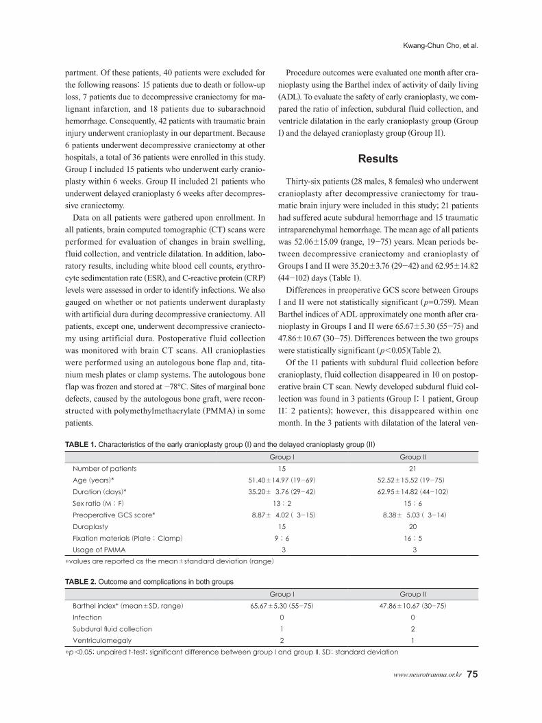
TABLE 1. Characteristics of the early cranioplasty group (I) and the delayed cranioplasty group (II)
Group I Group IINumber of patients 15 21Age (years)* 51.40±14.97 (19-69) 52.52±15.52 (19-75)0Duration (days)* 35.20±03.76 (29-42) 62.95±14.82 (44-102)
Sex ratio (M : F) 13 : 2 15 : 6Preoperative GCS score* 08.87±04.02 (03-15) 08.38±05.03 (03-14)
Duraplasty 15 20Fixation materials (Plate : Clamp) 9 : 6 16 : 5Usage of PMMA 03 03
*values are reported as the mean±standard deviation (range)
TABLE 2. Outcome and complications in both groups
Group I Group IIBarthel index* (mean±SD, range) 65.67±5.30 (55-75) 47.86±10.67 (30-75)
Infection 0 0Subdural fluid collection 1 2Ventriculomegaly 2 1
*p<0.05; unpaired t-test; significant difference between group I and group II. SD: standard deviation
76 J Korean Neurotraumatol Soc 2011;7:74-77
Safety and Efficacy of Early Cranioplasty
tricle underlying the cranial defect after decompressive craniectomy, ventricular dilatation was not aggravated in the follow-up period.
None of our patients presented with symptoms or signs of infection, such as fever and elevated level of laboratory findings, including leukocyte, ESR, and CRP during the postoperative one month follow-up period.
Discussion
Decompressive craniectomy is one of the most impor-tant methods for management of refractory intracranial hypertension after severe traumatic brain injury. However, the value of decompressive craniectomy in achieving a better outcome remains controversial and certain compli-cations may follow the operation.9,14)
In 1945, Gardner reported on a syndrome characterized by severe headache, dizziness, undue fatigability, poor memory, irritability, epilepsy, discomfort, and psychiatric symptoms observed in patients with large cranial defects, which he called “syndrome of the trephined”. Occurrence of the syndrome of the trephined is frequent after a large craniectomy and is a well-known indication for cranio-plasty. It is believed to be related to atmospheric pressure transmitted through the unsupported scalp; it was also called “syndrome of the sinking skin flap” by Yamaura and Makino in 1977.13) After cranioplasty, symptoms related to this syndrome can be relieved to different degrees in some patients; however, the procedure is not always reli-ably effective.
In addition, the large craniectomy would also lead to sig-nificant changes in the dynamics of local cerebral blood flow. In the early phases after decompressive craniectomy, perfusion of brain tissue underlying the cranial defect in-creases with reduced ICP; however, soon after the process, the sinking skin flap transmits atmospheric pressure to the underlying brain tissue and lead to a low cortical per-fusion, compared with the contralateral brain, as well as a disturbance of venous drainage, which would be partially rectified after the cranioplasty.7,14)
The hydrodynamic information about cerebrospinal flu-id (CSF) before and after cranioplasty have been studied.6) Changes in CSF hydrodynamics after large craniectomy lead to dilatation and shift of the ipsilateral lateral ventri-cle, hydrocephalus, and subdural collections. Cranioplasty is helpful for the correction of these disturbances as well as partial relief of symptoms.2)
Winkler et al.11) demonstrated that chronic decompressive craniectomy impairs not only postural blood flow regula-
tion in the ipsilateral hemisphere, but also cerebrovascular reserve capacity in the brain as a whole. Cranioplasty im-proves both postural blood flow regulation and cerebro-vascular reserve capacity. Therefore, cranioplasty resulted in marked improvement of metabolic activity.
At present, cranioplasty may be performed not only for cosmetic reasons, but also for its therapeutic effects, par-ticularly for patients with huge cranial defects after de-compressive craniectomy.5,10) Commonly, performance of cranioplasty 3 months after craniectomy is recommended, and if the patient has a history of intracranial infection or open craniocerebral injury, the procedure can delayed for at least 6 months after the first surgery. However, some au-thors have advanced the idea of early cranioplasty after de-compressive craniectomy to alleviate complications from craniectomy.4,8,15) Some authors reported that early cranio-plasty provides a satisfactory securing dissection plane dur-ing operative procedures, compared with later cranioplas-ty, without causing additional complications, including infection, subdural hygroma, and brain parenchymal dam-age, in selected cases.4,12) Liang et al. reported that early cranioplasty was safe and assisted in improvement of pa-tient’s neurological function and prognosis. In addition, early cranioplasty has an advantage in dissection for cra-nioplasty.8) Early cranioplasty performed before massive scar formation reduces operative time by facilitating soft tissue dissection. Beauchamp et al. suggested that early cra-nioplasty would lower the overall cost of care by eliminat-ing the need for additional hospital admissions.1)
In this study, early cranioplasty was effective in improv-ing ADL of patients. The Barthel index of ADL was found to be significantly higher in the early cranioplasty group. In addition, early cranioplasty does not increase relative risk of complications, such as infection or fluid collection. In addition, fixation materials and usage of bone cement (PMMA) have no effect on the rate of cranioplasty infec-tion.3)
The limitations of our study included its retrospective na-ture, small sample size, and lack of long-term follow-up data. A prospective, randomized, controlled study with extend-ed follow-up duration will be needed to fully establish ef-ficacy of early cranioplasty.
Conclusion
We consider that with appropriate selection of patients, early cranioplasty for large cranial defects after decom-pressive craniectomy will be a safe and helpful strategy for improvement of the neurologic function of patients
www.neurotrauma.or.kr 77
Kwang-Chun Cho, et al.
with severe traumatic brain injury.
■ The authors have no financial conflicts of interest.
REFERENCES1) Beauchamp KM, Kashuk J, Moore EE, Bolles G, Rabb C, Seinfeld
J, et al. Cranioplasty after postinjury decompressive craniectomy: is timing of the essence? J Trauma 69:270-274, 2010
2) Carvi Y, Nievas MN, Höllerhage HG. Early combined cranio-plasty and programmable shunt in patients with skull bone de-fects and CSF-circulation disorders. Neurol Res 28:139-144, 2006
3) Cheng YK, Weng HH, Yang JT, Lee MH, Wang TC, Chang CN. Factors affecting graft infection after cranioplasty. J Clin Neu-rosci 15:1115-1119, 2008
4) Chun HJ, Yi HJ. Efficacy and safety of early cranioplasty, at least within 1 month. J Craniofac Surg 22:203-207, 2011
5) Dujovny M, Aviles A, Agner C, Fernandez P, Charbel FT. Cranio-plasty: cosmetic or therapeutic? Surg Neurol 47:238-241, 1997
6) Fodstad H, Ekstedt J, Fridén H. CSF hydrodynamic studies before and after cranioplasty. Acta Neurochir Suppl (Wien) 28:514-518, 1979
7) Isago T, Nozaki M, Kikuchi Y, Honda T, Nakazawa H. Sinking skin flap syndrome: a case of improved cerebral blood flow after cranioplasty. Ann Plast Surg 53:288-292, 2004
8) Liang W, Xiaofeng Y, Weiguo L, Gang S, Xuesheng Z, Fei C, et al. Cranioplasty of large cranial defect at an early stage after decom-pressive craniectomy performed for severe head trauma. J Cra-niofac Surg 18:526-532, 2007
9) Schiffer J, Gur R, Nisim U, Pollak L. Symptomatic patients after craniectomy. Surg Neurol 47:231-237, 1997
10)Segal DH, Oppenheim JS, Murovic JA. Neurological recovery after cranioplasty. Neurosurgery 34:729-731; discussion 731, 1994
11) Winkler PA, Stummer W, Linke R, Krishnan KG, Tatsch K. In-fluence of cranioplasty on postural blood flow regulation, cere-brovascular reserve capacity, and cerebral glucose metabolism. J Neurosurg 93:53-61, 2000
12)Yadla S, Campbell PG, Chitale R, Maltenfort MG, Jabbour P, Sharan AD. Effect of early surgery, material, and method of flap preservation on cranioplasty infections: a systematic review. Neu-rosurgery 68:1124-1129; discussion 1130, 2011
13)Yamaura A, Makino H. Neurological deficits in the presence of the sinking skin f lap following decompressive craniectomy. Neurol Med Chir (Tokyo) 17:43-53, 1977
14)Yang XJ, Hong GL, Su SB, Yang SY. Complications induced by decompressive craniectomies after traumatic brain injury. Chin J Traumatol 6:99-103, 2003
15)Zhang GL, Yang WZ, Jiang YW, Zeng T. Extensive duraplasty with autologous graft in decompressive craniectomy and subse-quent early cranioplasty for severe head trauma. Chin J Trauma-tol 13:259-264, 2010