Embed Size (px)
DESCRIPTION
surgery
Citation preview

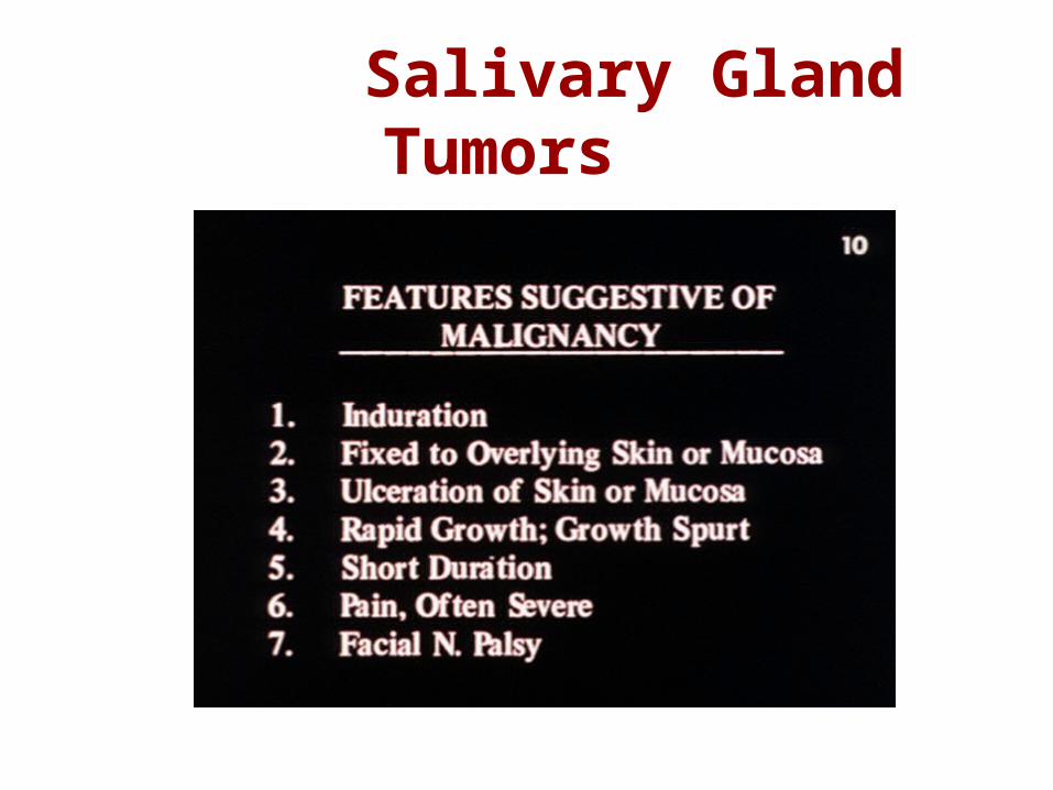
Salivary Gland Pathology
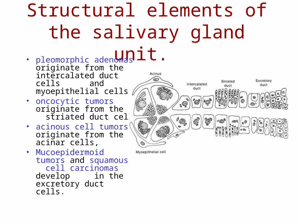
Structural elements of the salivary gland unit.
• pleomorphic adenomas originate from the intercalated duct cells and myoepithelial cells
• oncocytic tumors originate from the striated duct cells
• acinous cell tumors originate from the acinar cells,
• Mucoepidermoid tumors and squamous cell carcinomas develop in the excretory duct cells.
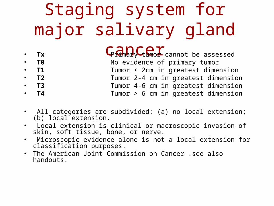
Staging system for major salivary gland cancer
• Tx Primary tumor cannot be assessed• T0 No evidence of primary tumor• T1 Tumor < 2cm in greatest dimension• T2 Tumor 2-4 cm in greatest dimension• T3 Tumor 4-6 cm in greatest dimension• T4 Tumor > 6 cm in greatest dimension
• All categories are subdivided: (a) no local extension; (b) local extension.
• Local extension is clinical or macroscopic invasion of skin, soft tissue, bone, or nerve.
• Microscopic evidence alone is not a local extension for classification purposes.
• The American Joint Commission on Cancer .see also handouts.
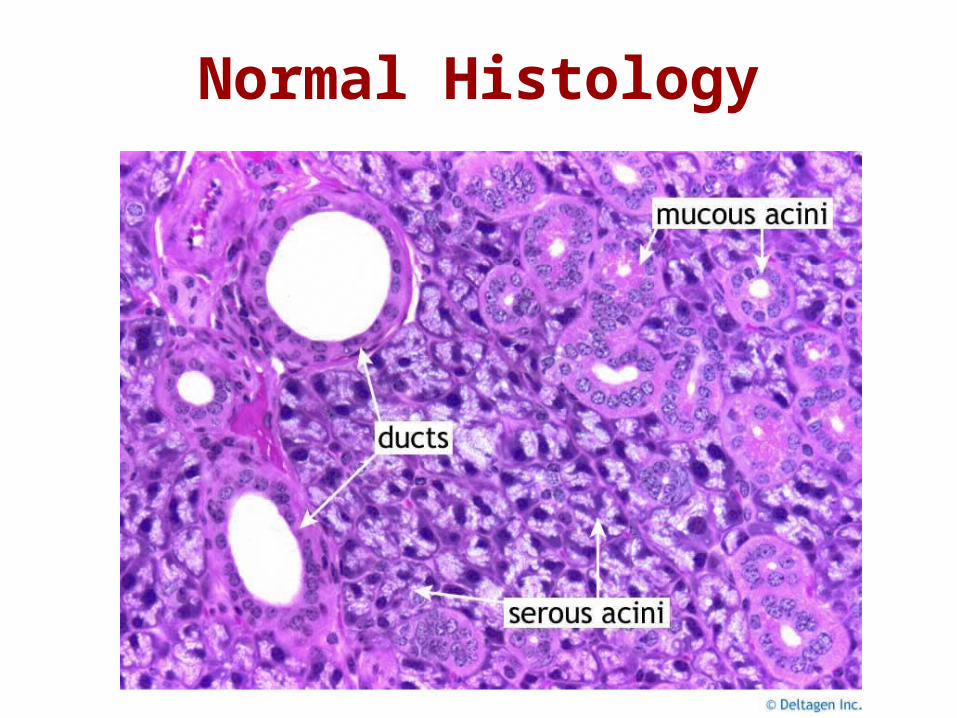
Normal Histology
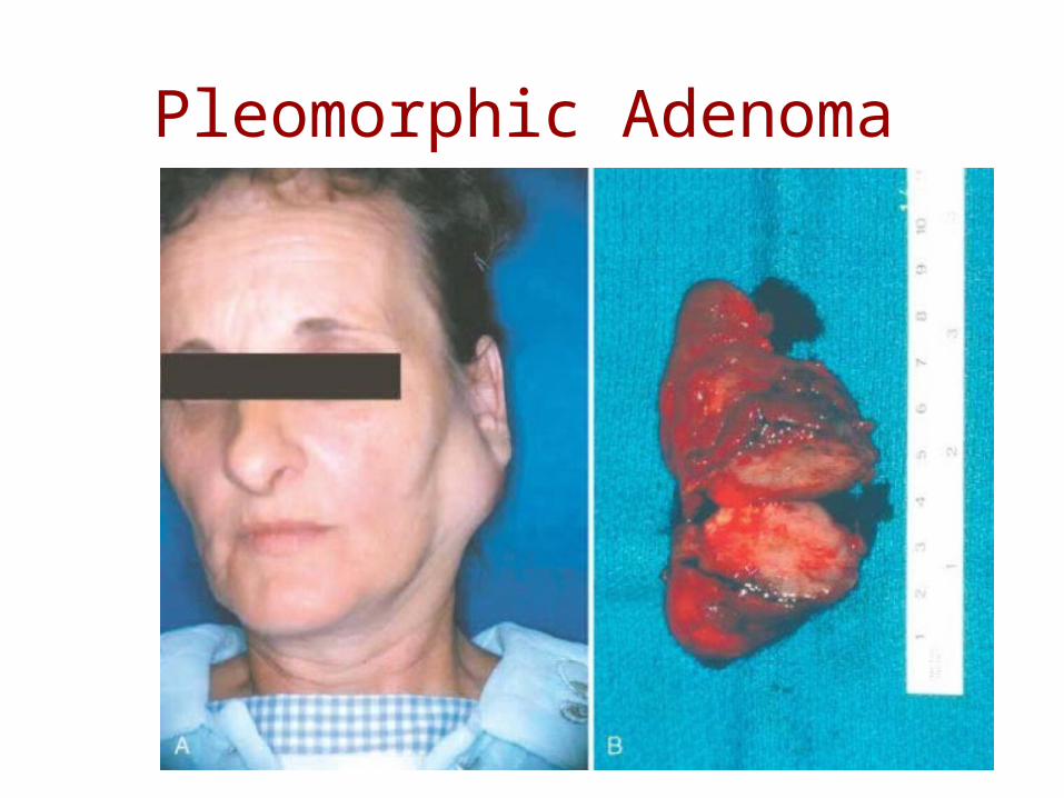
Pleomorphic Adenoma
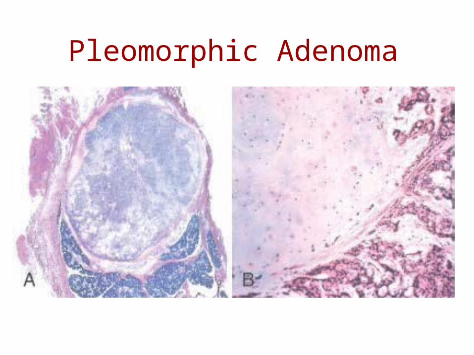
Pleomorphic Adenoma
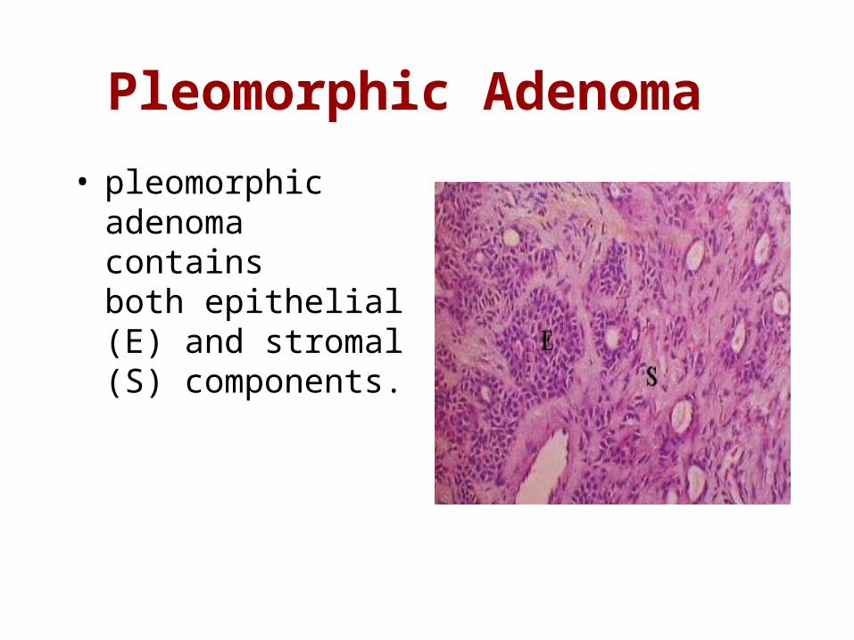
Pleomorphic Adenoma
• pleomorphic adenoma contains both epithelial (E) and stromal (S) components.
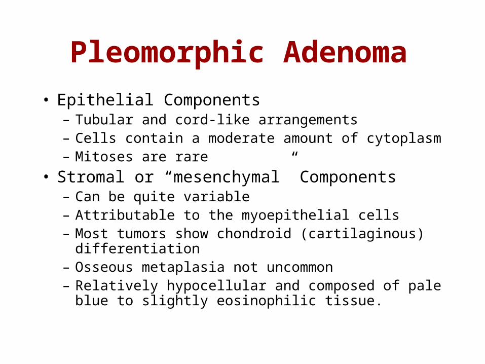
Pleomorphic Adenoma
• Epithelial Components– Tubular and cord-like arrangements– Cells contain a moderate amount of cytoplasm– Mitoses are rare
• Stromal or “mesenchymal” Components– Can be quite variable– Attributable to the myoepithelial cells– Most tumors show chondroid (cartilaginous)
differentiation– Osseous metaplasia not uncommon– Relatively hypocellular and composed of pale blue to
slightly eosinophilic tissue.
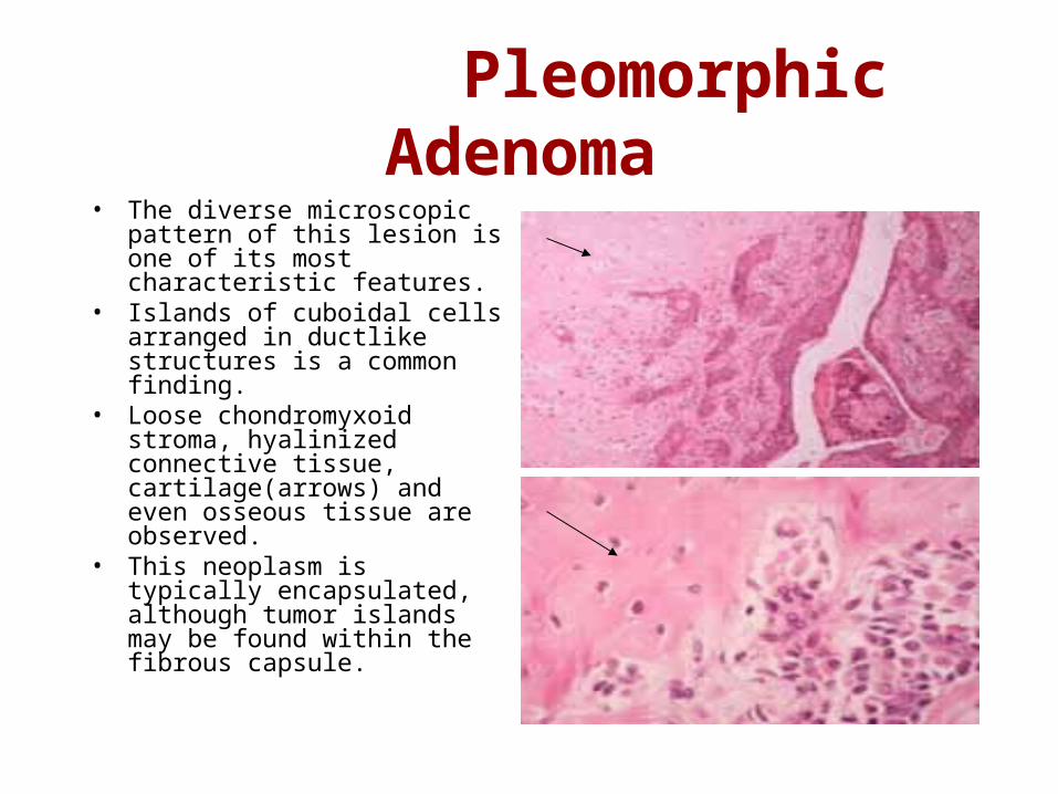
Pleomorphic Adenoma• The diverse microscopic pattern
of this lesion is one of its most characteristic features.
• Islands of cuboidal cells arranged in ductlike structures is a common finding.
• Loose chondromyxoid stroma, hyalinized connective tissue, cartilage(arrows) and even osseous tissue are observed.
• This neoplasm is typically encapsulated, although tumor islands may be found within the fibrous capsule.
Warthin's Tumor
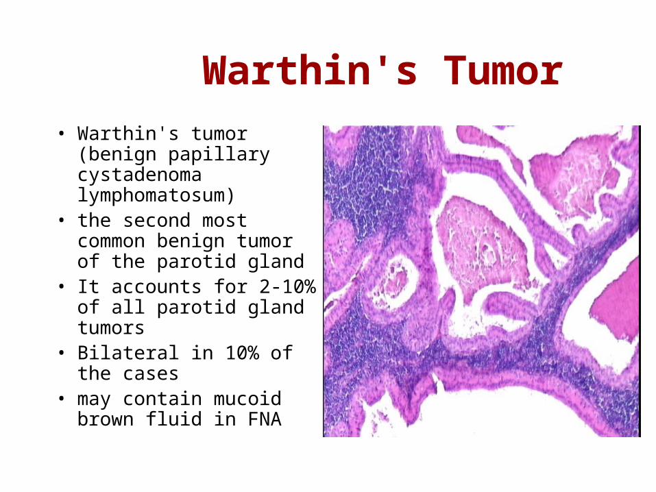
• Warthin's tumor (benign papillary cystadenoma lymphomatosum)
• the second most common benign tumor of the parotid gland
• It accounts for 2-10% of all parotid gland tumors
• Bilateral in 10% of the cases
• may contain mucoid brown fluid in FNA
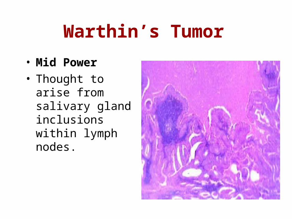
Warthin’s Tumor
• Mid Power • Thought to arise
from salivary gland inclusions within lymph nodes.
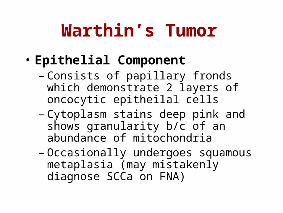
Warthin’s Tumor
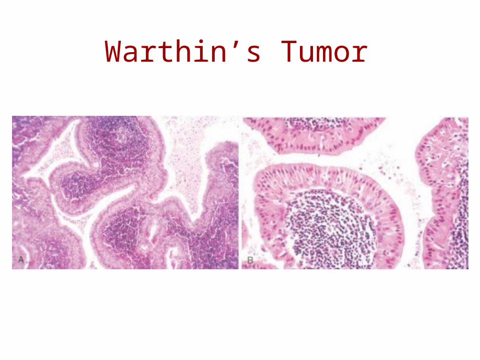
• Epithelial Component– Consists of papillary fronds which
demonstrate 2 layers of oncocytic epitheilal cells
– Cytoplasm stains deep pink and shows granularity b/c of an abundance of mitochondria
– Occasionally undergoes squamous metaplasia (may mistakenly diagnose SCCa on FNA)
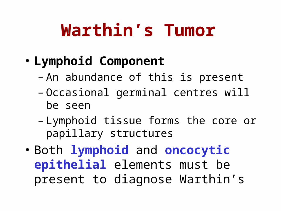
Warthin’s Tumor
• Lymphoid Component– An abundance of this is present– Occasional germinal centres will be seen– Lymphoid tissue forms the core or papillary
structures
• Both lymphoid and oncocytic epithelial elements must be present to diagnose Warthin’s
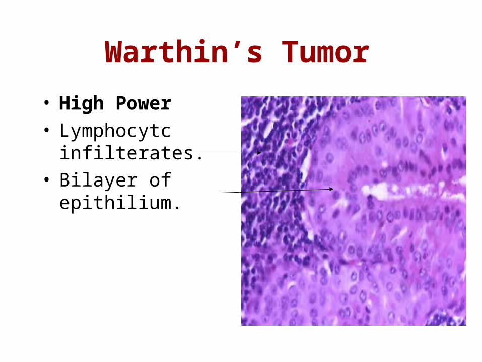
Warthin’s Tumor
• High Power • Lymphocytc
infilterates.• Bilayer of epithilium.
Warthin’s Tumor
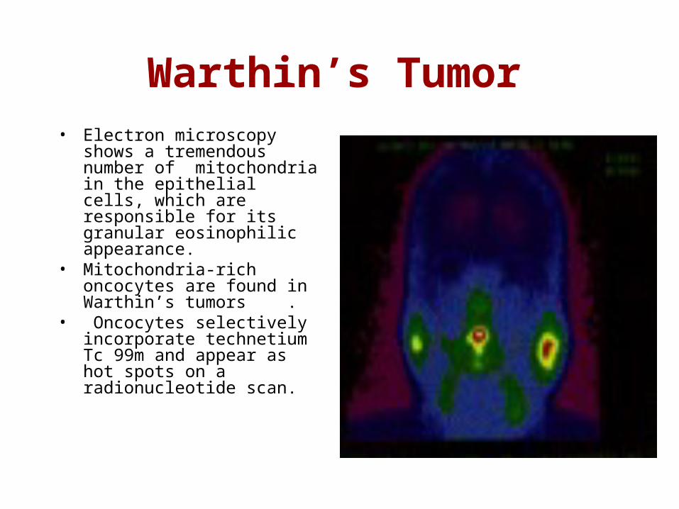
Warthin’s Tumor• Electron microscopy shows a
tremendous number of mitochondria in the epithelial cells, which are responsible for its granular eosinophilic appearance.
• Mitochondria-rich oncocytes are found in Warthin’s tumors .
• Oncocytes selectively incorporate technetium Tc 99m and appear as hot spots on a radionucleotide scan.
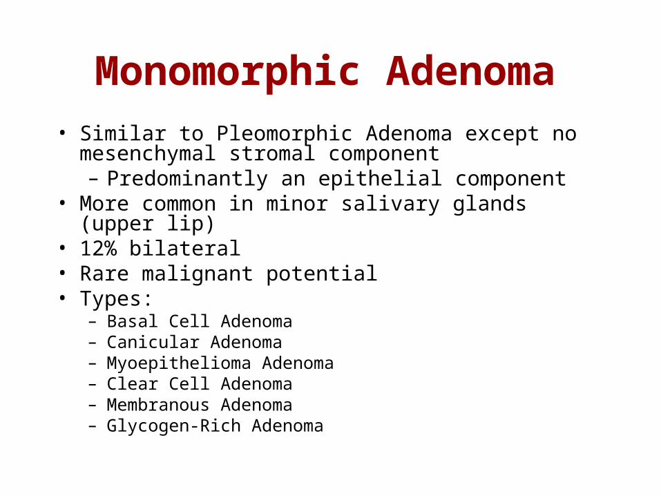
Monomorphic Adenoma• Similar to Pleomorphic Adenoma except no
mesenchymal stromal component– Predominantly an epithelial component
• More common in minor salivary glands (upper lip)• 12% bilateral• Rare malignant potential• Types:
– Basal Cell Adenoma– Canicular Adenoma– Myoepithelioma Adenoma– Clear Cell Adenoma– Membranous Adenoma– Glycogen-Rich Adenoma
Basal Cell Adenoma
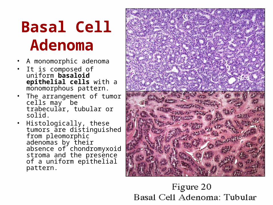
• A monomorphic adenoma• It is composed of uniform
basaloid epithelial cells with a monomorphous pattern.
• The arrangement of tumor cells may be trabecular, tubular or solid.
• Histologically, these tumors are distinguished from pleomorphic adenomas by their absence of chondromyxoid stroma and the presence of a uniform epithelial pattern.

Malignant Salivary Gland Tumors

Mucoepidermoid
Carcinoma • MECs contain two
major elements:• mucin-producing cells
and epithelial cells of the epidermoid variety
• (Epidermoid and Mucinous components).
• MEC is divided into low-grade (well differentiated).
• High-grade (poorly differentiated).
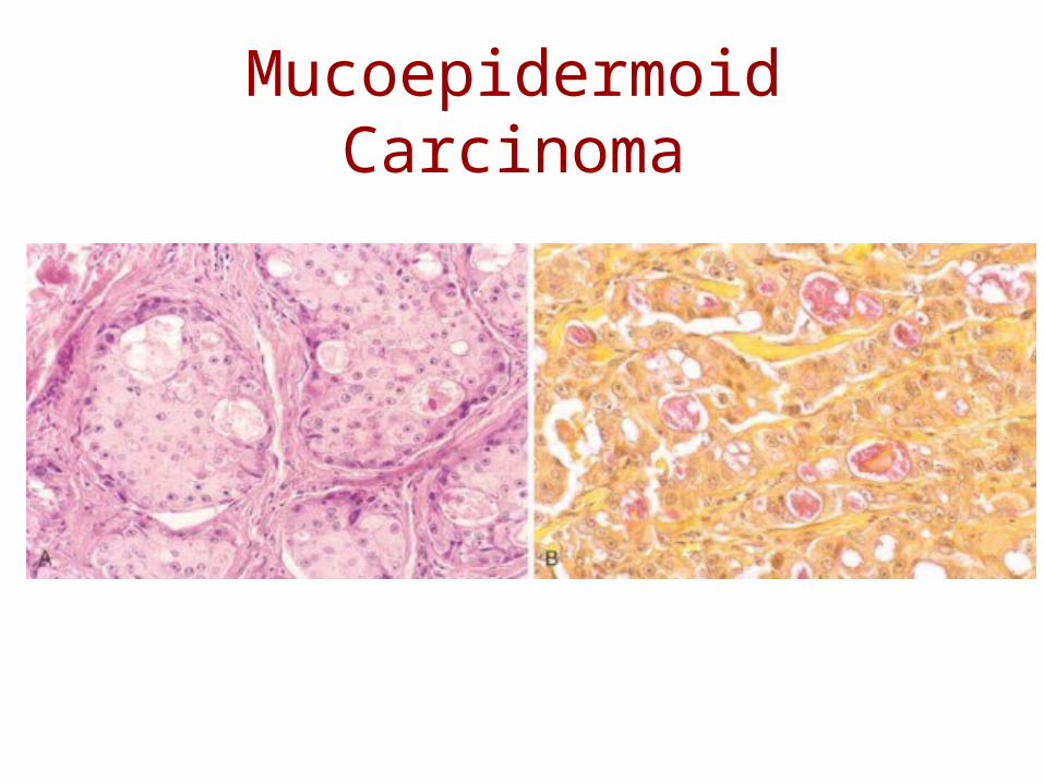
Mucoepidermoid Carcinoma
Mucoepidermoid Carcinoma
• Mucoepidermoid carcinoma (MEC) is the most common malignant tumor of the parotid gland and the second-most common malignancy (adenoid cystic carcinoma is more common) of the submandibular and minor salivary glands.
• Stained +ve by musicarmine.• MECs constitute approximately 35% of
salivary gland malignancy, and 80% to 90% of MECs occur in the parotid gland.
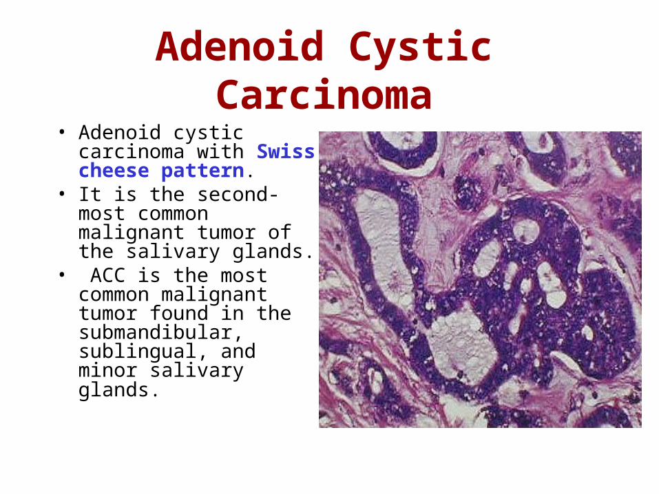
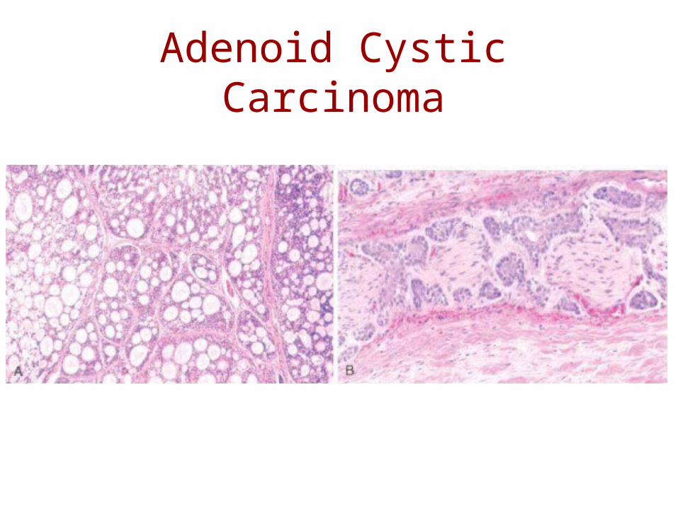
Adenoid Cystic Carcinoma• Adenoid cystic
carcinoma with Swiss cheese pattern.
• It is the second-most common malignant tumor of the salivary glands.
• ACC is the most common malignant tumor found in the submandibular, sublingual, and minor salivary glands.
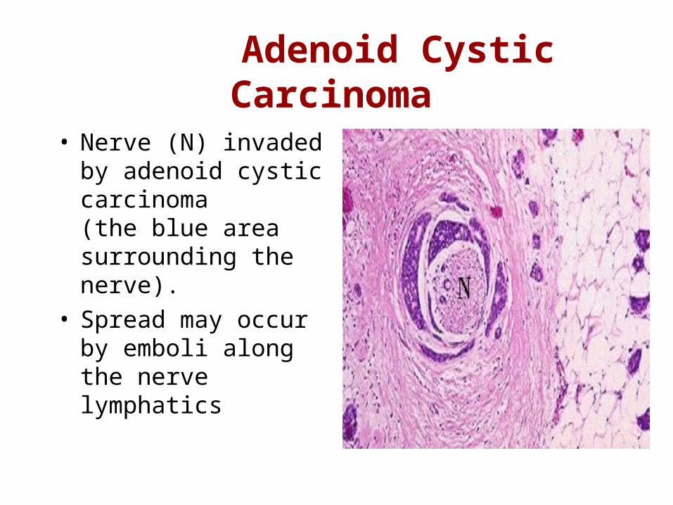
Adenoid Cystic Carcinoma
• Nerve (N) invaded by adenoid cystic carcinoma (the blue area surrounding the nerve).
• Spread may occur by emboli along the nerve lymphatics
Adenoid Cystic Carcinoma
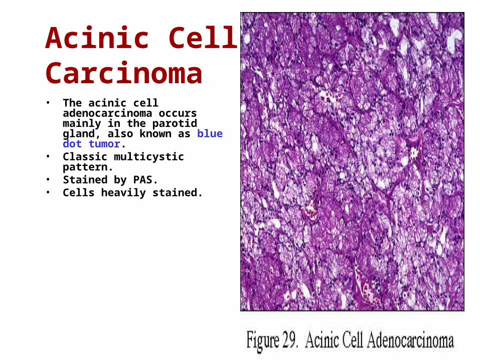
Acinic Cell Carcinoma • The acinic cell adenocarcinoma
occurs mainly in the parotid gland, also known as blue dot tumor.
• Classic multicystic pattern.• Stained by PAS.• Cells heavily stained.
Acinic Cell Carcinoma
• This lesion is characterized by a benign histomorphologic picture but by occasional malignant behavior.
• These lesions are treated by surgical excision
• Bilateral involvement occurs in 3% of patients, making acinic cell carcinoma the second-most common neoplasm, after Warthin’s tumor, to exhibit bilateral presentation.
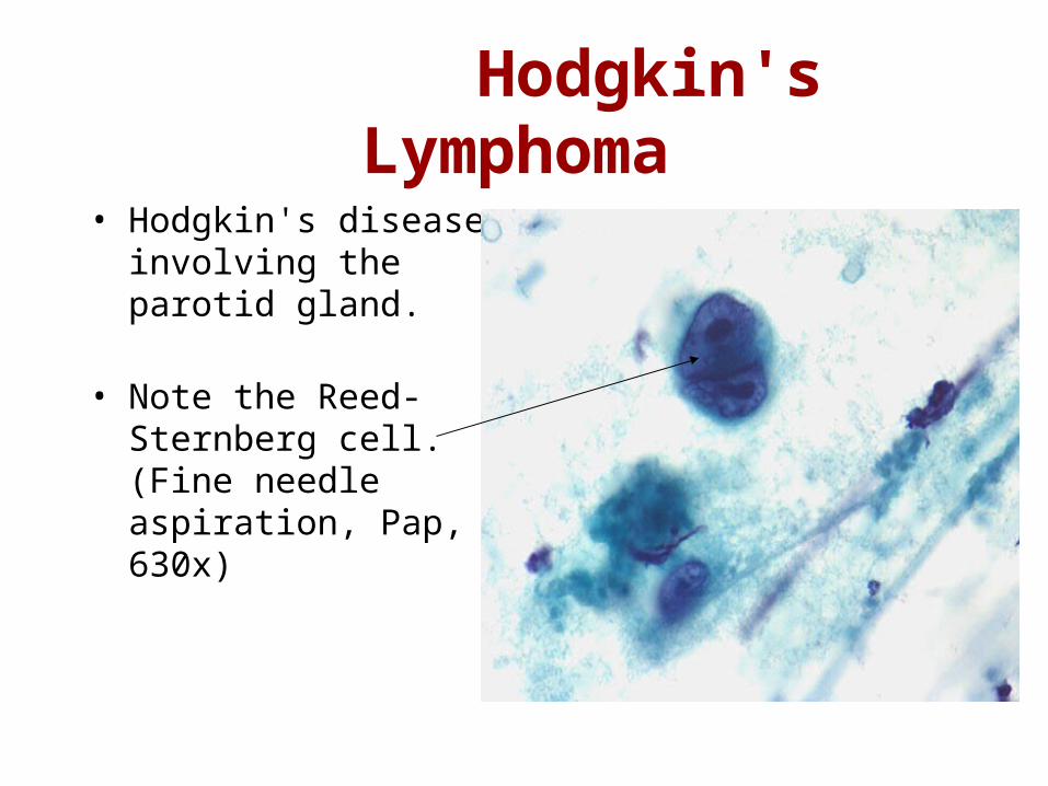
Hodgkin's Lymphoma
• Hodgkin's disease involving the parotid gland.
• Note the Reed-Sternberg cell. (Fine needle aspiration, Pap, 630x)