Embed Size (px)
Citation preview
Salivary Gland TumorsPrepared by Kurt Schaberg
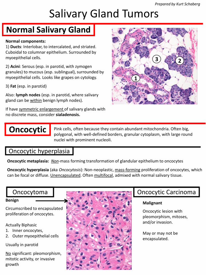
Oncocytic hyperplasia
Normal Salivary Gland
Oncocytic metaplasia: Non-mass forming transformation of glandular epithelium to oncocytes
Oncocytic hyperplasia (aka Oncocytosis): Non-neoplastic, mass-forming proliferation of oncocytes, which can be focal or diffuse. Unencapsulated. Often multifocal, admixed with normal salivary tissue.
Oncocytic
Benign
Circumscribed to encapsulated proliferation of oncocytes.
Actually Biphasic 1. Inner oncocytes, 2. Outer myoepithelial cells
Usually in parotid
No significant: pleomorphism, mitotic activity, or invasive growth
Oncocytoma Oncocytic Carcinoma
Malignant
Oncocytic lesion with pleomorphism, mitoses, and/or invasion.
May or may not be encapsulated.
Normal components:1) Ducts: Interlobar, to intercalated, and striated. Cuboidal to columnar epithelium. Surrounded by myoepithelial cells.
2) Acini: Serous (esp. in parotid, with zymogen granules) to mucous (esp. sublingual), surrounded by myoepithelial cells. Looks like grapes on cytology.
3) Fat (esp. in parotid)
Also: lymph nodes (esp. in parotid, where salivary gland can be within benign lymph nodes).
If have symmetric enlargement of salivary glands with no discrete mass, consider sialadenosis.
1
23
Pink cells, often because they contain abundant mitochondria. Often big, polygonal, with well-defined borders, granular cytoplasm, with large round nuclei with prominent nucleoli.
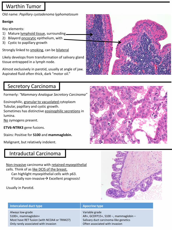
Old name: Papillary cystadenoma lyphomatosum
Benign
Key elements:1) Mature lymphoid tissue, surrounding2) Bilayerd oncocytic epithelium, with3) Cystic to papillary growth
Strongly linked to smoking, can be bilateral
Likely develops from transformation of salivary gland tissue entrapped in a lymph node.
Almost exclusively in parotid, usually at angle of jaw.Aspirated fluid often thick, dark “motor oil.”
Formerly: “Mammary Analogue Secretory Carcinoma”
Eosinophilic, granular to vacuolated cytoplasmTubular, papillary and cystic growth.Sometimes has distinctive eosinophilic secretions in lumina.No zymogens present.
ETV6-NTRK3 gene fusions.
Stains: Positive for S100 and mammaglobin.
Malignant, but relatively indolent.
Warthin Tumor
Secretory Carcinoma
Non-invasive carcinoma with retained myoepithelial cells. Think of as like DCIS of the breast.
Can highlight myoepithelial cells with p63.If totally non-invasive→ Excellent prognosis!
Usually in Parotid.
Intraductal Carcinoma
Intercalated duct type Apocrine type
Always low-grade S100+, mammaglobin+ Most have RET fusion (with NCOA4 or TRIM27) Only rarely associated with invasion
Variable grade AR+, GCDFP15+, S100 –, mammaglobin –Salivary duct carcinoma-like geneticsOften associated with invasion
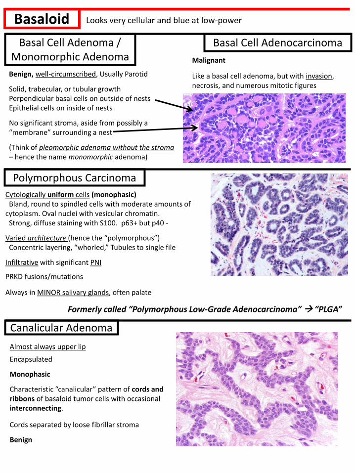
Basaloid
Benign, well-circumscribed, Usually Parotid
Solid, trabecular, or tubular growthPerpendicular basal cells on outside of nestsEpithelial cells on inside of nests
No significant stroma, aside from possibly a “membrane” surrounding a nest
(Think of pleomorphic adenoma without the stroma– hence the name monomorphic adenoma)
Cytologically uniform cells (monophasic)Bland, round to spindled cells with moderate amounts of
cytoplasm. Oval nuclei with vesicular chromatin.Strong, diffuse staining with S100. p63+ but p40 -
Varied architecture (hence the “polymorphous”)Concentric layering, “whorled,” Tubules to single file
Infiltrative with significant PNI
PRKD fusions/mutations
Always in MINOR salivary glands, often palate
Basal Cell Adenoma / Monomorphic Adenoma
Polymorphous Carcinoma
Basal Cell Adenocarcinoma
Malignant
Like a basal cell adenoma, but with invasion, necrosis, and numerous mitotic figures
Formerly called “Polymorphous Low-Grade Adenocarcinoma” → “PLGA”
Canalicular Adenoma
Almost always upper lip
Encapsulated
Monophasic
Characteristic “canalicular” pattern of cords and ribbons of basaloid tumor cells with occasional interconnecting.
Cords separated by loose fibrillar stroma
Benign
Looks very cellular and blue at low-power
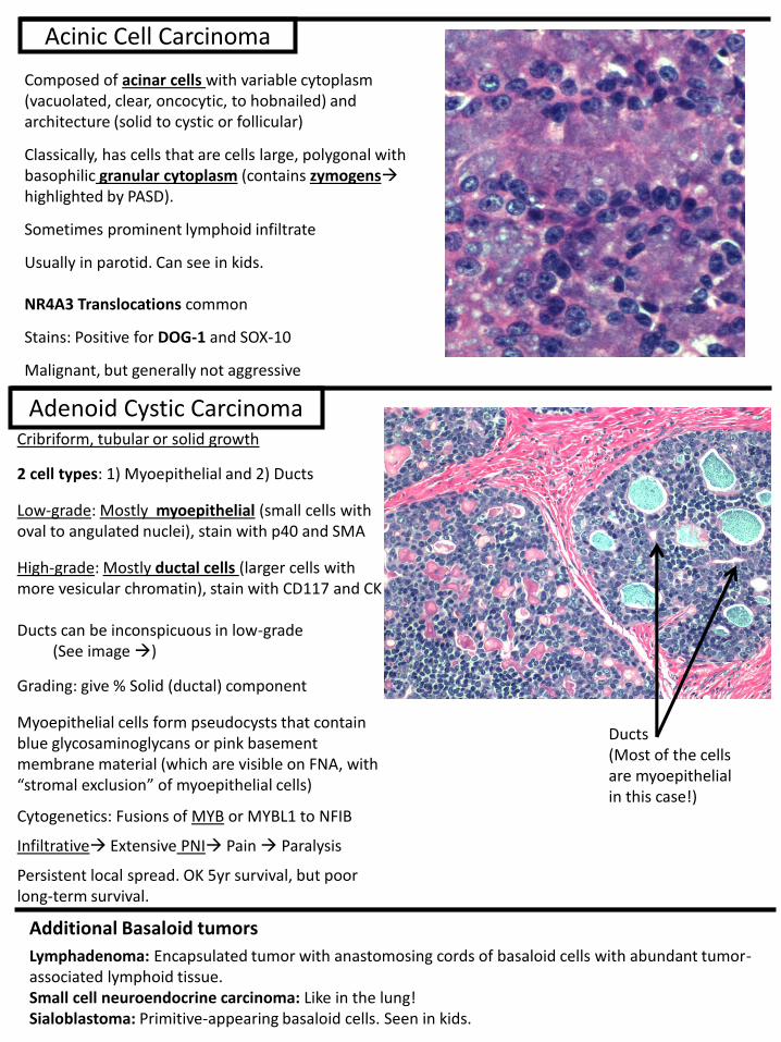
Composed of acinar cells with variable cytoplasm (vacuolated, clear, oncocytic, to hobnailed) and architecture (solid to cystic or follicular)
Classically, has cells that are cells large, polygonal with basophilic granular cytoplasm (contains zymogens→highlighted by PASD).
Sometimes prominent lymphoid infiltrate
Usually in parotid. Can see in kids.
NR4A3 Translocations common
Stains: Positive for DOG-1 and SOX-10
Malignant, but generally not aggressive
Acinic Cell Carcinoma
Adenoid Cystic CarcinomaCribriform, tubular or solid growth
2 cell types: 1) Myoepithelial and 2) Ducts
Low-grade: Mostly myoepithelial (small cells with oval to angulated nuclei), stain with p40 and SMA
High-grade: Mostly ductal cells (larger cells with more vesicular chromatin), stain with CD117 and CK
Ducts can be inconspicuous in low-grade (See image →)
Grading: give % Solid (ductal) component
Myoepithelial cells form pseudocysts that contain blue glycosaminoglycans or pink basement membrane material (which are visible on FNA, with “stromal exclusion” of myoepithelial cells)
Cytogenetics: Fusions of MYB or MYBL1 to NFIB
Infiltrative→ Extensive PNI→ Pain → Paralysis
Persistent local spread. OK 5yr survival, but poor long-term survival.
Additional Basaloid tumors
Lymphadenoma: Encapsulated tumor with anastomosing cords of basaloid cells with abundant tumor-associated lymphoid tissue.Small cell neuroendocrine carcinoma: Like in the lung!Sialoblastoma: Primitive-appearing basaloid cells. Seen in kids.
Ducts(Most of the cells are myoepithelial in this case!)
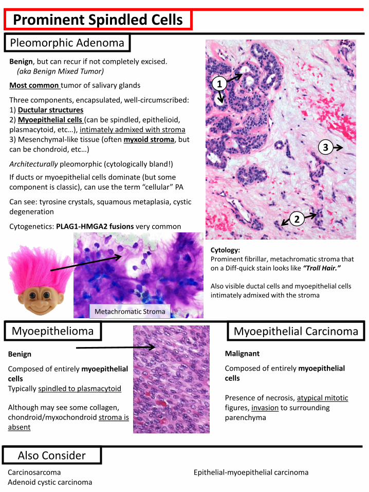
Prominent Spindled Cells
Benign, but can recur if not completely excised.(aka Benign Mixed Tumor)
Most common tumor of salivary glands
Three components, encapsulated, well-circumscribed:1) Ductular structures2) Myoepithelial cells (can be spindled, epithelioid, plasmacytoid, etc…), intimately admixed with stroma3) Mesenchymal-like tissue (often myxoid stroma, but can be chondroid, etc…)
Architecturally pleomorphic (cytologically bland!)
If ducts or myoepithelial cells dominate (but some component is classic), can use the term “cellular” PA
Can see: tyrosine crystals, squamous metaplasia, cystic degeneration
Cytogenetics: PLAG1-HMGA2 fusions very common
Benign
Composed of entirely myoepithelial cellsTypically spindled to plasmacytoid
Although may see some collagen, chondroid/myxochondroid stroma is absent
Pleomorphic Adenoma
Myoepithelioma Myoepithelial Carcinoma
2
3
1
Cytology: Prominent fibrillar, metachromatic stroma that on a Diff-quick stain looks like “Troll Hair.”
Also visible ductal cells and myoepithelial cells intimately admixed with the stroma
Malignant
Composed of entirely myoepithelial cells
Presence of necrosis, atypical mitotic figures, invasion to surrounding parenchyma
Metachromatic Stroma
CarcinosarcomaAdenoid cystic carcinoma
Epithelial-myoepithelial carcinoma
Also Consider
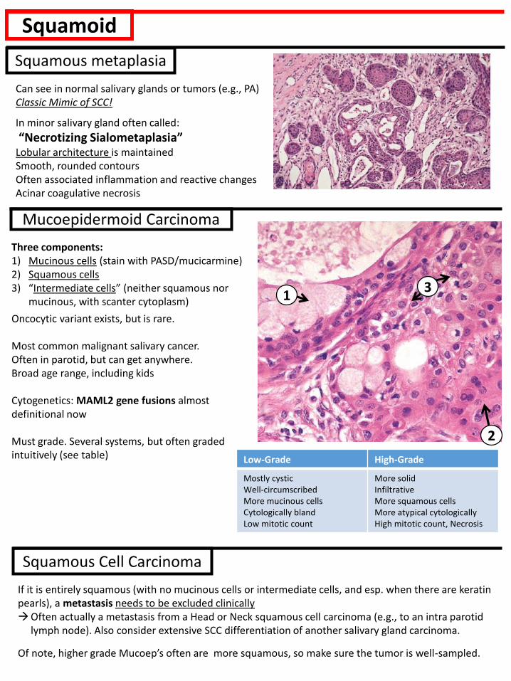
Squamoid
Can see in normal salivary glands or tumors (e.g., PA)Classic Mimic of SCC!
In minor salivary gland often called:
“Necrotizing Sialometaplasia”Lobular architecture is maintainedSmooth, rounded contoursOften associated inflammation and reactive changesAcinar coagulative necrosis
If it is entirely squamous (with no mucinous cells or intermediate cells, and esp. when there are keratin pearls), a metastasis needs to be excluded clinically→Often actually a metastasis from a Head or Neck squamous cell carcinoma (e.g., to an intra parotid
lymph node). Also consider extensive SCC differentiation of another salivary gland carcinoma.
Of note, higher grade Mucoep’s often are more squamous, so make sure the tumor is well-sampled.
Squamous metaplasia
Squamous Cell Carcinoma
Three components:1) Mucinous cells (stain with PASD/mucicarmine)2) Squamous cells3) “Intermediate cells” (neither squamous nor
mucinous, with scanter cytoplasm)
Oncocytic variant exists, but is rare.
Most common malignant salivary cancer. Often in parotid, but can get anywhere.Broad age range, including kids
Cytogenetics: MAML2 gene fusions almost definitional now
Must grade. Several systems, but often graded intuitively (see table)
Mucoepidermoid Carcinoma
13
2
Low-Grade High-Grade
Mostly cysticWell-circumscribedMore mucinous cellsCytologically blandLow mitotic count
More solidInfiltrativeMore squamous cellsMore atypical cytologicallyHigh mitotic count, Necrosis
High-Grade
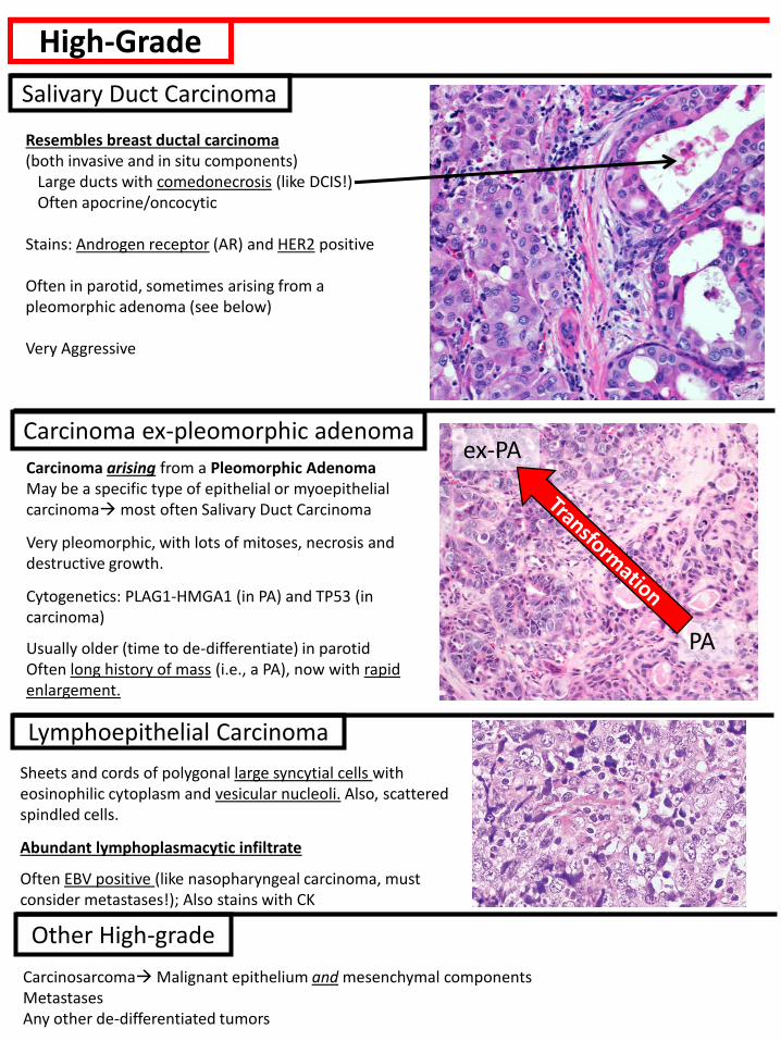
Resembles breast ductal carcinoma(both invasive and in situ components)
Large ducts with comedonecrosis (like DCIS!)Often apocrine/oncocytic
Stains: Androgen receptor (AR) and HER2 positive
Often in parotid, sometimes arising from a pleomorphic adenoma (see below)
Very Aggressive
Sheets and cords of polygonal large syncytial cells with eosinophilic cytoplasm and vesicular nucleoli. Also, scattered spindled cells.
Abundant lymphoplasmacytic infiltrate
Often EBV positive (like nasopharyngeal carcinoma, must consider metastases!); Also stains with CK
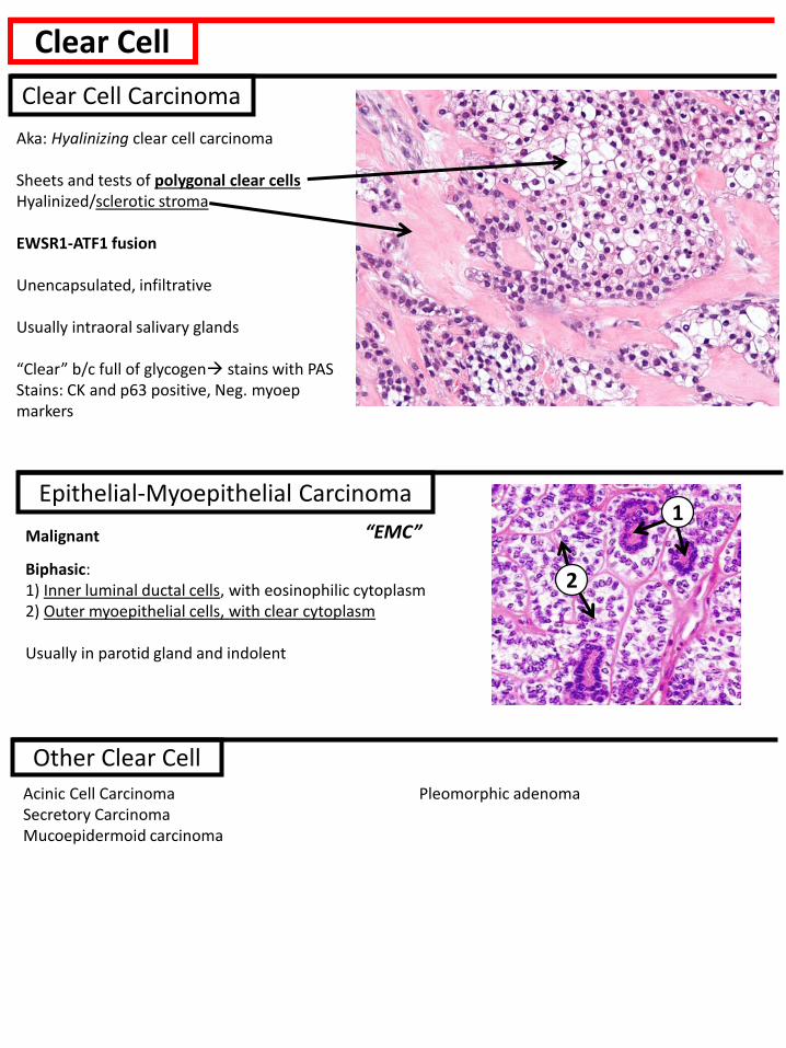
Carcinoma arising from a Pleomorphic AdenomaMay be a specific type of epithelial or myoepithelial carcinoma→most often Salivary Duct Carcinoma
Very pleomorphic, with lots of mitoses, necrosis and destructive growth.
Cytogenetics: PLAG1-HMGA1 (in PA) and TP53 (in carcinoma)
Usually older (time to de-differentiate) in parotidOften long history of mass (i.e., a PA), now with rapid enlargement.
Salivary Duct Carcinoma
Carcinoma ex-pleomorphic adenoma
Lymphoepithelial Carcinoma
Carcinosarcoma→Malignant epithelium and mesenchymal componentsMetastasesAny other de-differentiated tumors
Other High-grade
PA
ex-PA
Clear Cell
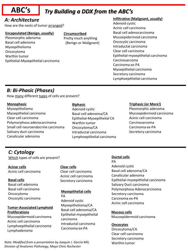
Aka: Hyalinizing clear cell carcinoma
Sheets and tests of polygonal clear cellsHyalinized/sclerotic stroma
EWSR1-ATF1 fusion
Unencapsulated, infiltrative
Usually intraoral salivary glands
“Clear” b/c full of glycogen→ stains with PASStains: CK and p63 positive, Neg. myoepmarkers
Acinic Cell CarcinomaSecretory CarcinomaMucoepidermoid carcinoma
Pleomorphic adenoma
Clear Cell Carcinoma
Other Clear Cell
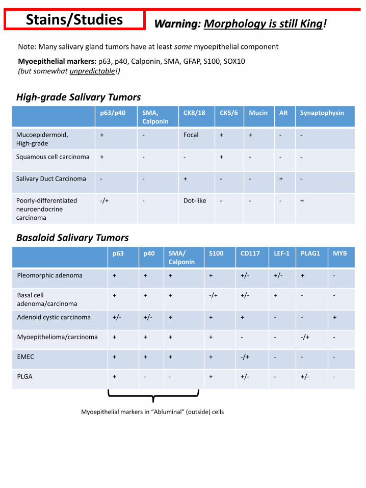
Malignant
Biphasic:1) Inner luminal ductal cells, with eosinophilic cytoplasm2) Outer myoepithelial cells, with clear cytoplasm
Usually in parotid gland and indolent
Epithelial-Myoepithelial Carcinoma1
2
“EMC”
ABC’s
Note: Modified from a presentation by Joaquín J. García MD, Division of Anatomic Pathology, Mayo Clinic Rochester
A: ArchitectureHow are the nests of tumor arranged?
B: Bi-Phasic (Phases)How many different types of cells are present?
Try Building a DDX from the ABC’s
C: CytologyWhich types of cells are present?
MonophasicMyoepitheliomaMyoepithelial carcinomaClear cell carcinomaPolymorphous adenocarcinomaSmall cell neuroendocrine carcinomaSalivary duct carcinomaCanalicular adenoma
Triphasic (or More!)Pleomorphic adenomaMucoepidermoid carcinomaAcinic cell carcinomaCarcinosarcomaCarcinoma ex-PASecretory carcinoma
BiphasicAdenoid cysticBasal cell adenoma/CAEpithelial-Myoepithelial CAWarthin tumorOncocytoma/CAIntraductal carcinomaLymphoepithelial carcinoma
Encapsulated (Benign, usually)Pleomorphic adenomaBasal cell adenomaMyoepitheliomaOncocytomaWarthin tumorEpithelial-Myoepithelial carcinoma
CircumscribedPretty much anything
(Benign or Malignant)
Infiltrative (Malignant, usually)Adenoid cysticAcinic cell carcinomaBasal cell adenocarcinomaMucoepidermoid carcinomaOncocytic carcinomaIntraductal carcinomaClear cell carcinomaEpithelial-myoepithelial carcinomaCarcinosarcomaCarcinoma ex-PAMyoepithelial carcinomaSecretory carcinomaLymphoepithelial carcinoma
Acinar cellsAcinic cell carcinoma
Basal cellsBasal cell adenomaBasal cell carcinomaOncocytomaOncocytic carcinoma
Clear cellsClear cell carcinomaAcinic cell carcinomaSecretory carcinoma
Mucous cellsMucoepidermoid carcinoma
Ductal cellsPAAdenoid cysticBasal cell adenoma/CACanalicular adenomaEpithelial-myoepithelial carcinomaSalivary Duct carcinomaPolymorphous AdenocarcinomaSecretory carcinomaCarcinoma ex-PAAcinic cell carcinoma
Myoepithelial cellsPAAdenoid cysticMyoepithelioma/CABasal cell adenoma/CAEpithelial-myoepithelial carcinomaIntraductal carcinomaCarcinoma ex-PA
Tumor-Associated Lymphoid ProliferationsMucoepidermoid carcinomaAcinic cell carcinomaLymphoepithelial carcinomaLymphadenoma
OncocytesOncocytoma/CAClear cell carcinomaSecretory carcinomaWarthin tumor
Stains/Studies
Note: Many salivary gland tumors have at least some myoepithelial component
Myoepithelial markers: p63, p40, Calponin, SMA, GFAP, S100, SOX10 (but somewhat unpredictable!)
p63/p40 SMA, Calponin
CK8/18 CK5/6 Mucin AR Synaptophysin
Mucoepidermoid, High-grade
+ - Focal + + - -
Squamous cell carcinoma + - - + - - -
Salivary Duct Carcinoma - - + - - + -
Poorly-differentiated neuroendocrine carcinoma
-/+ - Dot-like - - - +
High-grade Salivary Tumors
p63 p40 SMA/Calponin
S100 CD117 LEF-1 PLAG1 MYB
Pleomorphic adenoma + + + + +/- +/- + -
Basal cell adenoma/carcinoma
+ + + -/+ +/- + - -
Adenoid cystic carcinoma +/- +/- + + + - - +
Myoepithelioma/carcinoma + + + + - - -/+ -
EMEC + + + + -/+ - - -
PLGA + - - + +/- - +/- -
Basaloid Salivary Tumors
Myoepithelial markers in “Abluminal” (outside) cells
Warning: Morphology is still King!
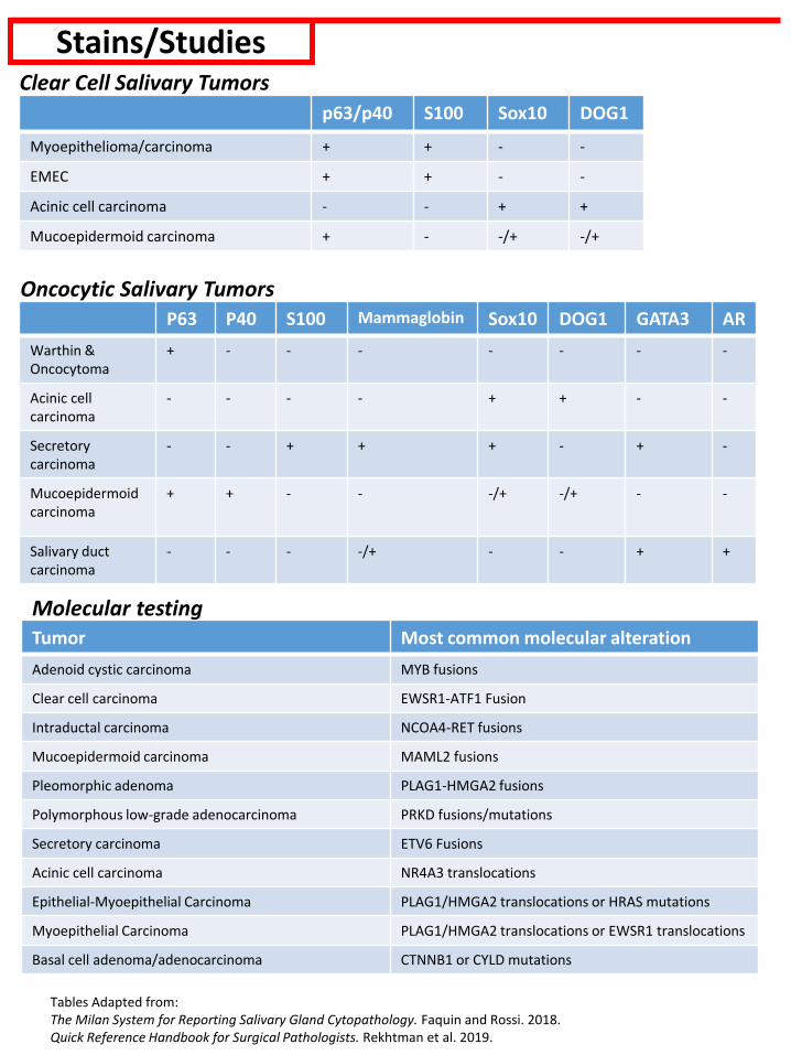
Stains/StudiesClear Cell Salivary Tumors
Oncocytic Salivary Tumors
p63/p40 S100 Sox10 DOG1
Myoepithelioma/carcinoma + + - -
EMEC + + - -
Acinic cell carcinoma - - + +
Mucoepidermoid carcinoma + - -/+ -/+
P63 P40 S100 Mammaglobin Sox10 DOG1 GATA3 AR
Warthin & Oncocytoma
+ - - - - - - -
Acinic cell carcinoma
- - - - + + - -
Secretory carcinoma
- - + + + - + -
Mucoepidermoid carcinoma
+ + - - -/+ -/+ - -
Salivary duct carcinoma
- - - -/+ - - + +
Tumor Most common molecular alteration
Adenoid cystic carcinoma MYB fusions
Clear cell carcinoma EWSR1-ATF1 Fusion
Intraductal carcinoma NCOA4-RET fusions
Mucoepidermoid carcinoma MAML2 fusions
Pleomorphic adenoma PLAG1-HMGA2 fusions
Polymorphous low-grade adenocarcinoma PRKD fusions/mutations
Secretory carcinoma ETV6 Fusions
Acinic cell carcinoma NR4A3 translocations
Epithelial-Myoepithelial Carcinoma PLAG1/HMGA2 translocations or HRAS mutations
Myoepithelial Carcinoma PLAG1/HMGA2 translocations or EWSR1 translocations
Basal cell adenoma/adenocarcinoma CTNNB1 or CYLD mutations
Molecular testing
Tables Adapted from: The Milan System for Reporting Salivary Gland Cytopathology. Faquin and Rossi. 2018.Quick Reference Handbook for Surgical Pathologists. Rekhtman et al. 2019.
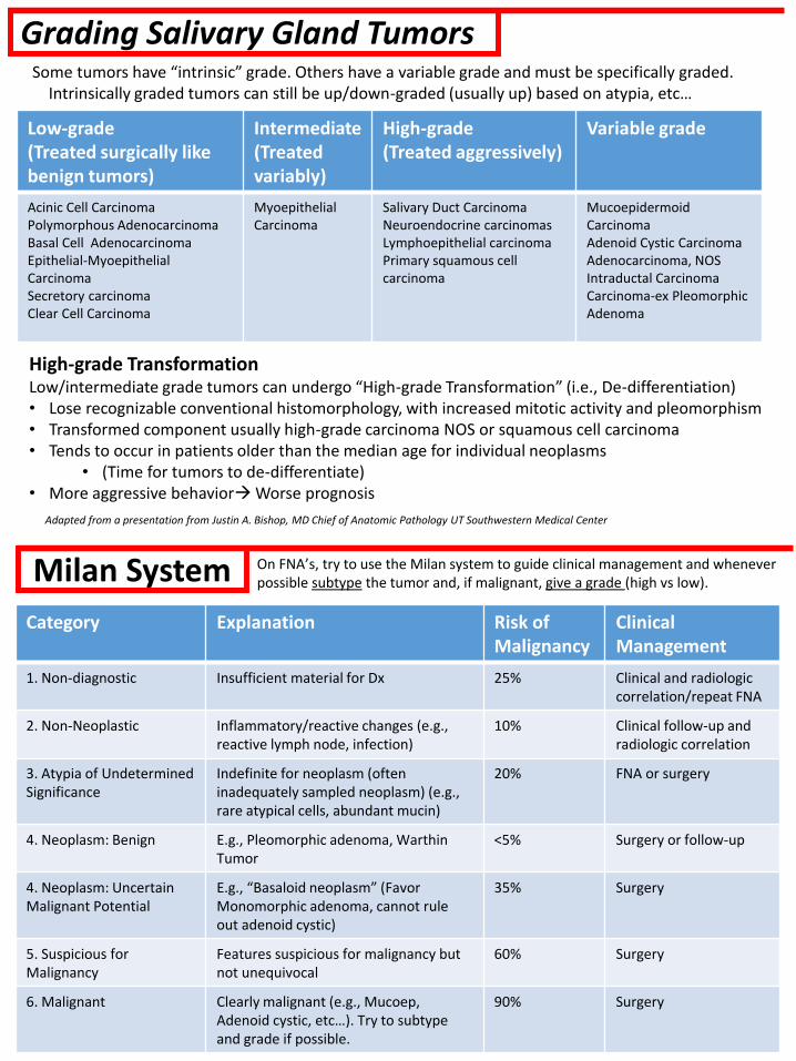
Grading Salivary Gland Tumors
Adapted from a presentation from Justin A. Bishop, MD Chief of Anatomic Pathology UT Southwestern Medical Center
Low-grade(Treated surgically like benign tumors)
Intermediate(Treated variably)
High-grade(Treated aggressively)
Variable grade
Acinic Cell Carcinoma Polymorphous Adenocarcinoma Basal Cell Adenocarcinoma Epithelial-Myoepithelial Carcinoma Secretory carcinoma Clear Cell Carcinoma
Myoepithelial Carcinoma
Salivary Duct Carcinoma Neuroendocrine carcinomas Lymphoepithelial carcinoma Primary squamous cell carcinoma
Mucoepidermoid Carcinoma Adenoid Cystic Carcinoma Adenocarcinoma, NOS Intraductal Carcinoma Carcinoma-ex Pleomorphic Adenoma
Some tumors have “intrinsic” grade. Others have a variable grade and must be specifically graded.Intrinsically graded tumors can still be up/down-graded (usually up) based on atypia, etc…
High-grade TransformationLow/intermediate grade tumors can undergo “High-grade Transformation” (i.e., De-differentiation)• Lose recognizable conventional histomorphology, with increased mitotic activity and pleomorphism• Transformed component usually high-grade carcinoma NOS or squamous cell carcinoma • Tends to occur in patients older than the median age for individual neoplasms
• (Time for tumors to de-differentiate)• More aggressive behavior→Worse prognosis
Milan System
Category Explanation Risk of Malignancy
Clinical Management
1. Non-diagnostic Insufficient material for Dx 25% Clinical and radiologic correlation/repeat FNA
2. Non-Neoplastic Inflammatory/reactive changes (e.g., reactive lymph node, infection)
10% Clinical follow-up and radiologic correlation
3. Atypia of Undetermined Significance
Indefinite for neoplasm (often inadequately sampled neoplasm) (e.g., rare atypical cells, abundant mucin)
20% FNA or surgery
4. Neoplasm: Benign E.g., Pleomorphic adenoma, Warthin Tumor
<5% Surgery or follow-up
4. Neoplasm: Uncertain Malignant Potential
E.g., “Basaloid neoplasm” (Favor Monomorphic adenoma, cannot rule out adenoid cystic)
35% Surgery
5. Suspicious for Malignancy
Features suspicious for malignancy but not unequivocal
60% Surgery
6. Malignant Clearly malignant (e.g., Mucoep, Adenoid cystic, etc…). Try to subtype and grade if possible.
90% Surgery
On FNA’s, try to use the Milan system to guide clinical management and whenever possible subtype the tumor and, if malignant, give a grade (high vs low).