Embed Size (px)
Citation preview

Salivary gland pathology non- neoplastic diseases
.

• -salivary glands -classified according to prevailing type of secretory cells:
• -serous glands, mucous glands and mixed seromucous • -salivary gland tissue consists of- acinar cells of serous
and mucous type- myoepithelial cells- epithelial cells of salivary ducts, mesenchymal cells of the connective tissue (adipose and fibrous, nerves, blood vessels), and inflammatory cells
• according to the anatomic location- salivary glands include paired major glands (parotid, submandibular, sublingual) and minor glands of upper aerodigestive tract

• -1.- major glands include parotid- the largest gland, enclosed by capsule, located anterior to the ear, composed of serous acini, secretory ducts
• -about 20 lymph nodes located in each parotid gland • -submandibular-located in the submandibular triangle, seromucous gland • -subligual gland, the smallest of major glands, in the floor of mouth,
prevailing mucous acini
• -2.- minor glands of the oral mucosa • -minor glands have mixed serous and mucous acini in varying proportions • cellular components: • -of particular interest- myoepithelial cells (MEC) - believed to have
contractile properties that assist in secretion of saliva • -microscopical examination shows that MECs are thin and spindle-shaped
cells around acini and intercalated ducts situated between basement membrane and epithelial ductal cells
• -they display features of both smooth muscle cells and epithelium
Saliva
• Saliva in composed of water 99% , • but laso electrolytes, (natrium, potassim, calcium, chloride ions,
magnesium, fluorine, iodine, phosphateshydrogencarbomnates • Mucus – mucopolysacharides, glycoproteins • antiseptic substances- (thiocyanate, hydrogen peroxide, and secretory
immunoglobulin A) • enzymes
– α-amylase - breaking down starch into simpler sugars – lysozyms – damage bacterias – lipase- Salivary lipase plays a large role in fat digestion in newborn infants as
their pancreatic lipase still needs some time to develop – Others (phosphatases, , lactoperoxidases)
• opiorphin – a newly researched pain-killing substance found in human saliva
• Epidermal growth factor or EGF • pH –neutral (pH = 7 - 8) – buffering system
Daily salivary output
• There is much debate about the amount of saliva that is produced in a healthy person per day; estimates range from 0.75 to 1.5 liters per day while it is generally accepted that during sleep the amount drops to almost zero.
• In humans, the submandibular gland contributes around 70–75% of secretion, while the parotid gland secretes about 20–25% and small amounts are secreted from the other salivary glands. During night – production is reduced to nearly zero.
• In elderly people – physiological reduction of salivation • Pathological reduction: dehydratation, stress,drugs
(antihistamines and antidepressantsspasmolytics, diuretics).

Saliva
Mechanical oral clearance Food digestion Antibacterial properties Enamel remineralization ( calcium + phosphate
minareals) Mediate selective adhesions and bacterial colonization Buffering system (activated glands content more
bicarbonates Protective function (dental and soft tissue of the oral
cavity) Source of DNA (cancer dg…)
Funcional disorders
• The production of saliva is stimulated both by the sympathetic nervous system and the parasympathetic
• The saliva stimulated by sympathetic innervation is thicker, and saliva stimulated parasympathetically is more watery.
• Sympathetic stimulation of saliva is to facilitate respiration, whereas parasympathetic stimulation is to facilitate digestion.
• Increased production - sialorea (ptyalismus) – foreifn bodies, (new dentures), ulcerations of the oral cavity, stress, tumors,
• Decreased production - xerostomia
Developmental disorders
• Very rare
• aplazia (one or more lobes, or the entire gland)
• Accesory or ectopic gland (in lymph node)
• Congenital polycystic degeneration of parotc gland- girls, fluctuating enlargement
• Anomalies of ducts (congenital atresia, ectasia may be associated with –bronchiektasias)
Inflammation
• Etilogy – Bacterial
– Viral
– Autoimmune
• Acute bacterial sialoadenitis
• Chronic interstitial sialoadenitis
• Epidemic parotitis(Mumps)
• Cytomegaloviral sialoadenitis
• Autoimmune sialoadenitis
Acute bacterial sialadenitis
• Mostly as ascending infection • Less frequentky hematogenically (sepsis, scarlet fever) lymphogenically • Risc factors: decreased salivation,
(dehydratatione, post-operative diuretics…), cachexiae, sialothiliasis Poor oral hygiene
• Staphylococcus aureus, Streptococcus viridans , Streptococcus pneumoniae
• Catarrhal, catarrhaly purulent, absceding • Complication : fistulation, flegmone, tromboflebitis of facial
venes
Acute bacterial sialadenitida
Clinical features
• Painful swelling
• Reddened skin
• Edema of the cheek, periorbital region and neck
• fever
• malaise
• raised ESR, CRP, leucocytosis
• purulent exudate from duct punctum


Viral sialadenitis
• Sialotropic viruses
• Hematogenic spread in viremia
– Clinically manifested glandular inflammation
– Persistent infection reactivation
– Only in saliva (polio, lyssa, influenza, …)
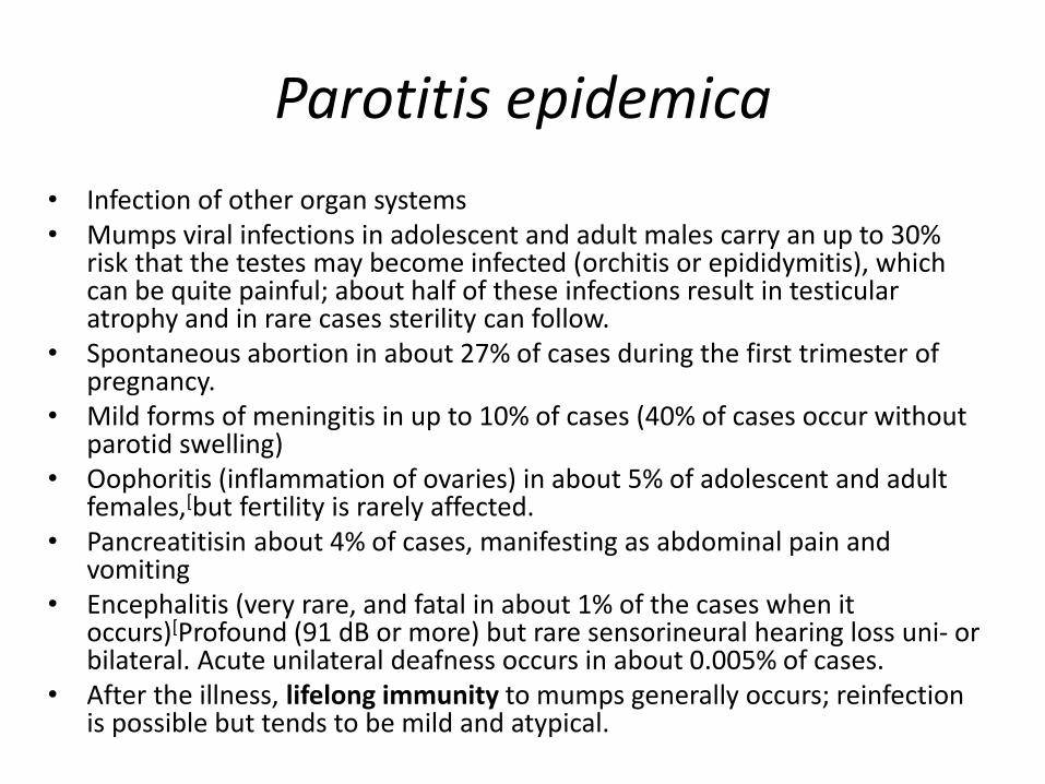
Epidemic parotitis, mumps
• RNA paramyxovirus, mostly in the childhood contagious disease through contact with respiratory
secretions, such as saliva from an infected person (droplets aerosol) Mumps can also be spread by sharing food and drinks.
mumps is contagious from approximately 6 days before the onset of symptoms until about 9 days after symptoms start.
The incubation period (time until symptoms begin) can be from 14–25 days, but is more typically 16–18 days
Symptoms lasts 7 -14 days Clinically: - (30-40% cases - inaparently!)
– headache – uni- / bi-lateral enlargement of parotis, also submandibular and ,
sublingual gl.
Parotitis epidemica
• Infection of other organ systems • Mumps viral infections in adolescent and adult males carry an up to 30%
risk that the testes may become infected (orchitis or epididymitis), which can be quite painful; about half of these infections result in testicular atrophy and in rare cases sterility can follow.
• Spontaneous abortion in about 27% of cases during the first trimester of pregnancy.
• Mild forms of meningitis in up to 10% of cases (40% of cases occur without parotid swelling)
• Oophoritis (inflammation of ovaries) in about 5% of adolescent and adult females,[but fertility is rarely affected.
• Pancreatitisin about 4% of cases, manifesting as abdominal pain and vomiting
• Encephalitis (very rare, and fatal in about 1% of the cases when it occurs)[Profound (91 dB or more) but rare sensorineural hearing loss uni- or bilateral. Acute unilateral deafness occurs in about 0.005% of cases.
• After the illness, lifelong immunity to mumps generally occurs; reinfection is possible but tends to be mild and atypical.


Cytomegaloviral sialadenitis
• kids – Transplacental transmission – per partum by cervical secret
• Adults – immunosupression – generalized infection – Infected saliva, transplantation, dialysis – + CMV hepatitis, myocarditis, polyradiculoneuritis
• Mi: intranuclear or cytoplasmatic inclusions in ductal epithelium


Chronic sialadenitis
• From previous acute infl.
• Primarily chronic infl.
• Riscs: – Sialolithiasis
– Trauma
– Thicken food
– Scarring
– Tumor
• chronic reccurent sialadenitis
• chronic sclerosing sialadenitis
• obstructive sialadenitis

chronic reccurent sialadenitis
Clinical features: • unilateral/bilateral • mild pain / swelling • common after meals • duct orifice is reddened and flow decreases • may or may not have visible/palpable stone. • Parotid gland
– Recurrent painful swellings
• Submandibular gland – Usually secondary to sialolithiasis or stricture
• microscopy – Lymphocytic infiltartion+ fibrosis + acinic atrophy
• attac (7-10 days) + remission (month ); trigger ???
• If there are attacks more than approximately 3 times per year or severe attacks, surgical excision of the affected gland should be considered

Chronic sclerosing sialadenitis
• Küttner´s „tumor“ (1896)
• Middle, old age,
• gl. submandibularis
• Clinically (dif. dg. Malignant tu) – bilateral x unilateral; focal x diffuse
– tough, non-painful
• microscopy – fibrosis, lymphoplasmocytic inflammation , vasculitis
• Belongs to „IgG4-related disseases“ !!!
• Therapy : corticoids, exstirpation

Chronic sclerosing sialadenitis

Chronic obstructive sialadenitis
• Duct obstruction
– sialolithiasis
– tumors
– Trauma
– Small salivary glands– denture pressure, smoking

HIV and salivary glands
• Children (20%), very typical for HIV!
• May be very early manifestation
• Unknown pathogenesis
• Clinically – Parotic enlargement (in 60% bilateral), xerostomy
• Microscopy – Follicular hyperplasia of intraglandulatr LN
– Lymhocytic inflammation (CD8+)
– Multiple lymhoepithelial cyst
• dif. dg.: Sjögren’s syndrome
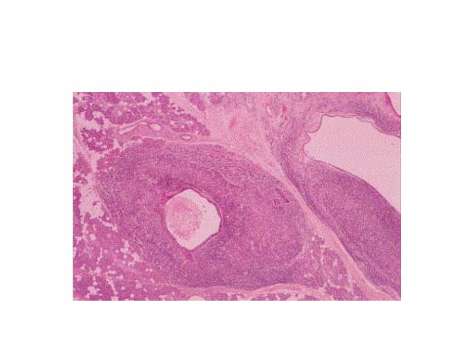

Pneumoparotitis
• air being forced through Stensen’s duct, resulting in pneumoparotitis. This may occur as a transient or recurrent phenomenon. Recurrent parotid insufflation is not entirely benign and may predispose to sialectasias, recurrent parotitis, and even subcutaneous emphysema.
• Glass-blowers, trumpeters, kids
Pneumoparotitis

Postactinic sialadenitis
• As a complication of irradiation of head and neck malignant tumor
• Serous gland are more sensitive than mucinous
• predisposing to ascending bacterial sialadenitis
• Small glands – xerostomy decay osteomyelitis
• Th: symptomatic, artificial saliva

Specific sialadenitis
• sarcoidosis
• tuberculosis
– Very rare
– Lymphogenically from oral cavity and oropharynx
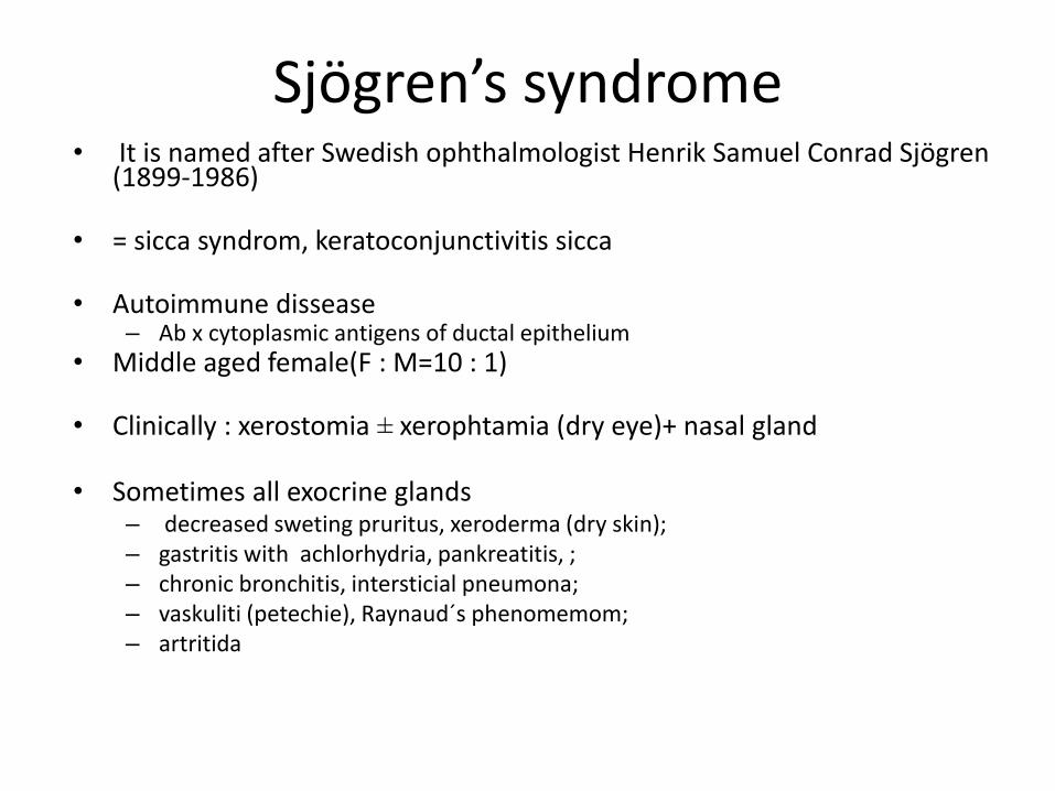
Sjögren’s syndrome • It is named after Swedish ophthalmologist Henrik Samuel Conrad Sjögren
(1899-1986) • = sicca syndrom, keratoconjunctivitis sicca • Autoimmune dissease
– Ab x cytoplasmic antigens of ductal epithelium • Middle aged female(F : M=10 : 1) • Clinically : xerostomia ± xerophtamia (dry eye)+ nasal gland • Sometimes all exocrine glands
– decreased sweting pruritus, xeroderma (dry skin); – gastritis with achlorhydria, pankreatitis, ; – chronic bronchitis, intersticial pneumona; – vaskuliti (petechie), Raynaud´s phenomemom; – artritida

Sjögren’s syndrome
• Primary (in its own right)
• Secondary – rheumatoid arthritis, systemic lupus erythematosus,
scleroderma, primary biliary cirrhosis
• microscopy – Lymphoplasmocellular inflammation (CD4) around dilated
ducts (MALT-like) basal cell proliferation lymphoepithelial islet
– Acinic atrophy xerostomia (50% pts.)
• Risc of MALToma, large cell lymphoma!!!
• Irreversible damage!!!

Sjögren’s syndrome
• Clinically:
– Difficulties with swallowing, eating, speaking,
– Dry, reddish, thin mucosa, lobulated tongue
– Frequent infection - Candida
– Dental plaque, decay
– Acute absceding parotitis

Sjögren’s syndrome

Sjögren’s syndrome angular stomatitis

The typical picture of a patient with long-standing xerostomia due to SS. The lack of appreciation of the likelihood of rapid cervical caries has
led to multiple restorations.

Swelling of the palatal minor salivary glands in Sjogren‘s syndrome – a much less common finding than that of parotid
swelling.

Parotid swelling in Sjogren’s syndrome.

A keratoconjuctivitis resulting frm the reduced tear secretion in Sjogren‘s syndrome.

Lymphoepitelial sialadenitis
• = known by a variety of names, including Mikulicz disease, benign lymphoepithelial lesion, and myoepithelial sialadenitis (MESA)
could be a form of primary Sjogren‘s syndrome.
often bilateral and in parotis
X
• Mikulicz syndrome = enlargement
– + lymphoma, leukemia, …
• Middle aged females
• parotis – 80-90% unilateral,
• Clinically:
– Non - painful, 0 systemic sy (no sicca sy)

Differencial diagnosis of xerostomia
• organic – Sjögren’s syndrome – irradiation – HIV infection – sarcoidosis – amyloidosis
• functional – Dehydratation – Psychological (depression) – Drugs – Diabetes mellitus

Sialolithiasis
• Middle aged male (M : F … 2 : 1)
• submandibularis (85%) parotis (6-10%) – Course of Wharton´s duct
– Property of saliva
• Intraductal close to orrifice (50%) intraductal intraparenchymatous
• Etiology – Inflammation, desquamation of epitelial cells, Ca ions + nidus
• Clinically – Saliváry colic x asymptomatic

Sialadenosis (sialosis)
• It is also referred to as sialosis or "nutritional mumps"
• Sialadenosis is a non-neoplastic, noninflammatory enlargement of the salivary gland
• the parotid gland, often bilateral • It results from acinar cell hypertrophy
• wide range of etiologies: • endocrine abnormalities like diabetes mellitus, thyroid gland
disease, adrenal gland dissease • nutritional deficiencies, • alcoholism, • cirrhosis, • pregnancy • bilimia • certain drugs, especially antihypertensives, β-sympatomimetics.
Cysts
• retention
– Elderly men
– Desquamation of epithelial cells, trauma, inflammation
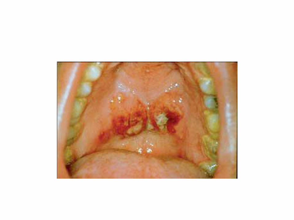
• Mucocele – mucinous glands (lip, buccal mucosa,
• Ranula – mouth floor –subliqual gland
• traumatic cysts / pseudocysts

Cyst
Dysotogenetic cysty
branchial cleft cysts (lymphoepithelial cyst)
– Embryonal epihtelial inclusions in LN or a result from a failure of obliteration of the branchial cleft, which in fish develop into gills
– Unilateral , solitary
– Most branchial cleft cysts are asymptomatic, but they may become infected
– The cyst wall will be composed of either squamous or columnar cells with lymphoid infiltrate, often with prominent germinal centers. The cyst may contain granular and keratinaceous cellular debris. Cholesterol crystals may be found in the fluid extracted from a branchial cyst.


Necrotizing sialometaplasia
• unknown etiology - spontaneous x microtraumata • Middle aged males, smokers
• Lobular infarction (ischemic necrosis) with or without extravasation of mucus.
• 2. Pseudoepitheliomatous hyperplasia at the periphery of the ulcer.
• 3. Squamous metaplasia of ducts and acini.
• 4. Inflammation secondary to extravasated mucous.
• 5. Preservation of lobular architecture.
• 12
• Because of the metaplasia, it is frequently misdiagnosed as squamous cell carcinoma
• Spontaneous healing within 1-2 month
Adenomatous hyperplasia
• Rare
• Not paiful
• Clinically mimics tumour
• Microscopy:
– Normal gland, no inflammation