Embed Size (px)
Citation preview
Salmonella
Mutagenicity Test Kit
Instruction Manual 31-100.2

2
INTRODUCTION
The materials contained in this Salmonella Mutagenicity Assay Kit include virtually all of the supplies necessary for the conduct of the “Ames Assay” as described by Maron & Ames (Maron, D. M. and B. N. Ames, Revised methods for the Salmonella mutagenicity test, Mutation Research, 113: 173 - 215, 1983) and Mortelmans & Zeiger (Mortelmans, K. and E. Zeiger, The Ames Salmonella/microsome mutagenicity assay, Mutation Research, 455: 29 - 60, 2000). This manual is a brief summary of the important aspects of testing - We strongly recommend that you carefully read one of these papers and OECD guideline 471 before you attempt to perform the assay.
All elements of the MOLTOX® kit are formulated and manufactured using the highest quality components and are consistent with the recommendations of Maron & Ames (ibid.). Certain materials supplied (e.g., STDiscs™ and lyophilized S9) have been specifically developed for inclusion in the assay kit by our laboratory. Most of the materials contained in the kit are accompanied by GLP level Quality Control and Formulation Statements - you may be assured that each element of each kit has been thoroughly tested for performance in the assay.
The MOLTOX® Salmonella Mutagenicity Assay Kit is intended for use by individuals skilled in the science and art of microbiology; the use of strict aseptic technique is essential for the successful application of the materials included in the kit. While the bacterial strains included in the kit (S. typhimurium strains TA1535, TA1537, TA98, and TA100) are attenuated, they are potentially pathogenic and must be handled accordingly. If you have any doubts about the safe handling of the strains included in this kit do not proceed until you have consulted with us at (828) 264-9099 or have obtained the advice of a skilled biochemist ormicrobiologist.
The performance of the “Ames Assay” includes several distinct experimental steps; e.g., test design; S9 mix formulation; dosing and plating; phenotype confirmation; target cell titer determination; reading (counting) and analysis. The materials contained in the kit were selected so as to provide the user with considerable flexibility as regards test design. This manual was developed to assist in the utilization of the kit contents; the information provided in the manual is intended to supplement that contained in the Maron & Ames and Mortlemans & Zeiger papers. While presented in a step-by-step manner, the instructions contained in this manual are amenable to modification - if you desire assistance in any phase of the assay, please contact our Customer Service department at (828) 264-9099. We will be happy to work with you to help solve any problems that may arise.
3
TABLE OF CONTENTS
Introduction………………………………………………………………………………………………..………2
Salmonella Mutagenicity Assay Kits Components…….……………………..…………….4
Kit Components and Additional Items Needed to Complete Assay…………….4
The Assay……………………………………………………………………………………………………………..5
A. Getting Started…………………………………………………………………………………….6
B. Cell Culture…………………………………………………………………………………………...8
C. Treatment and Plating………………………………………………………………………...9
D. The Results…………………………………………………………………………………………..13
4
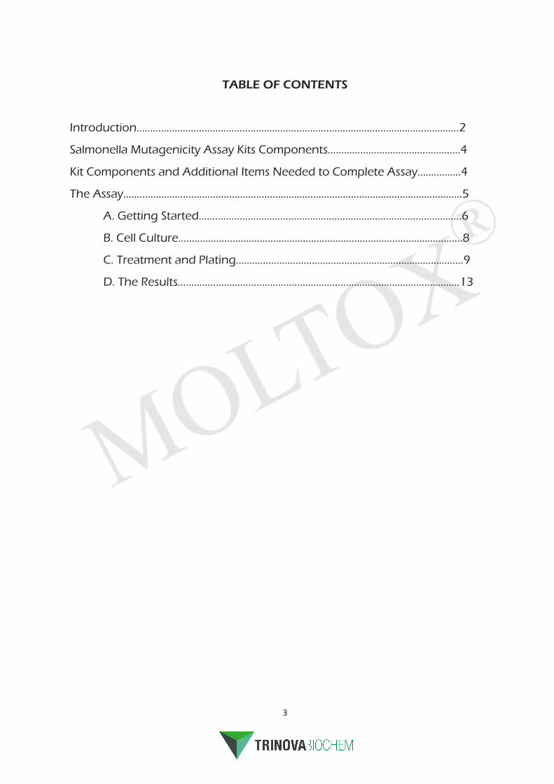
Salmonella Mutagenicity Assay Kit Components
Additional items you will need to complete the assay:
4 - Sterile Erlenmeyer flasks Sterile inoculating loops
Sharpie or wax pencil Sterile swabs (optional)
200 μL and 1000μL sterile pipette tips Ice
1 mL and 5 mL sterile pipets Ice bucket
Pipet-aid or rubber bulb Nitrile gloves
Test tube rack Sterile water
13 x 100 mm sterile disposable test tubes Vortex mixer
Dimethyl Sulfoxide (DMSO) Sterile forceps, small (optional)
Microwave or boiling water bath 37°C incubator with shaker
45°C water bath or heating block Water, deionized or distilled, sterile, ice-cold
Automatic colony counter or magnifying counter
Displacement pipette aids (e.g. Pipette-man, Eppendorf)
331-100.2 Salmonella Mutagenicity Assay Kit11-01L.2 Lyophilized Aroclor S9 2 each 21-100 Nutrient Agar Plates 1 each 21-200 ST Quad PC™ Plates 1 each 21-400.2 MGA Plates 8 each 26-505.1 Nutrient Broth, 100 mL 1 each 26-503.1 His/Bio Top Agar, 100 mL 4 each 60-101 ICR 191, 10 μg/vial 1each
60-102 Daunomycin, 60 μg/vial 1 each60-103 Sodium Azide, 15 μg/vial 1 each60-107 2-Aminoanthracene, 100 μg/vial 1 each60-200.4 Regensys™ “A”, 40 mL 1 each
60-201.4L Lyophilized Regensys™ “B”, 123 mg 1 each
71-098L TA98, 10/vial 1 each
71-100L TA100, 10/vial 1 each
71-1535L TA1535, 10/vial 1 each
71-1537L TA1537, 10/vial 1 each

5
THE ASSAY
The MOLTOX® kit contains four tester strains: TA1535, TA1537, TA98, and TA100 Each strain was constructed with a different lesion in the histidine operon (see Table 1, Mortelmans & Zeiger). This mutation renders them incapable of synthesizing histidine, i.e. they are histidine auxotrophs requiring exogenous histidine. In addition, TA1535, TA1537, TA98, and TA100 have altered cell walls (rfa) that increase the cell’s permeability to certain high molecular weight materials. These strains also share a lesion in a DNA repair-coding gene (uvrB) which results in an increase in sensitivity to a variety of mutagens. Since this lesion extends through the gene for biotin synthesis (bio), biotin is also required for growth]. Tester strains TA98 and TA100 carry a plasmid (pKM101) which acts to increase the activity of an error-prone DNA repair system and to confer resistance to the antibiotic ampicillin, tester strains TA1535 and TA1537 contain no plasmids.
Because of the characteristics of the tester strains, the “Ames Assay” is uniquely suited for the detection of mutagenic activities. The several tester strains differ in their response to DNA-damaging chemical mutagens and therefore are generally employed in combination. Each strain tends to be responsive to specific classes of mutagenic chemicals due to the specific lesion in their histidine operon. However, exposure to mutagens may result in genetic reversions in the histidine operon resulting in restoration of the wild type phenotype; mutants have their histidine operons functionally “restored” and can synthesize histidine. The assay depends on the ability to distinguish between histidine auxotrophs (the tester strains) and histidine prototrophs (the mutants). Accordingly, the target cells are plated on media containing trace quantities of histidine that allows for a few rounds of cell division necessary to “fix” a mutation event; the histidine is rapidly exhausted resulting in cessation of the growth of nonmutated cells. If a mutagenic chemical is present, (comparatively) rare reversions may occur in the altered histidine operon resulting in the continuation of growth after trace histidine exhaustion. Revertant bacterial colonies that appear on histidine-limiting media plates represent prototrophs that arose either spontaneously or due to the action of a mutagen.
The sections that follow describe the procedures for the conduct of the basic four strain assay. For the most part, the methods described are taken directly from Maron & Ames (ibid.) and, subsequently, Mortelmans & Zeiger. If you have no prior experience with the assay, we suggest that you follow these instructions closely; those experienced in the method will find useful information about the use of STDiscs™, QUAD PC™ plates, and lyophilized S9.
6
A. Getting Started
1. Before setting up the assay, you should gather as much information as possibleconcerning your test material:
a. Using the available references, structural analyses or activity data, assembleas much information as is possible concerning your test material or itsanalogues or closely related congeners. Of particular importance arequestions of bacteriostatic or bacteriocidal activities, hazardousqualities and stability.
b. Determine the solubility of your test chemical in the appropriate solvent(water and DMSO are preferred solvents - see p. 200, Maron & Ames forlisting or check the Merck Index). In many cases, you may find it necessary todose with suspensions rather that true solutions. If an organic solvent isused (e.g. DMSO, acetone), the test material may precipitate upon addition tothe aqueous top agar; e.g. 2-aminofluorene is essentially insoluble in waterand is solubilized in DMSO for dosing.
c. Decide on the doses that you wish to test. The conventional test solutiondose volumes is 100 μL per plate, but this can be increased for aqueousformulations or reduced, e.g. if a somewhat toxic organic solvent is used. Ingeneral, the upper dose should not exceed 5 mg/plate (50 mg/mL assuminga 100 μL/plate dosing volume). Select 5 to 7 doses separated by factors of 2,3 (or half logs) or 5. There are 160 minimal glucose agar plates in your kit.This is sufficient for a 5 dose triplicate plate assay (including controls)conducted with and without S9.
2. To avoid difficulties on the day of the assay, design your experiment carefullyand well in advance. Examples of questions you will need to have resolvedare as follows:
a. Dosimetry: Top dose? Dose intervals? Number of doses? Solvent? Volumeof each dosing solution dilution required for the complete assay?
b. Replicates: Are you going to dose duplicate plates per condition?Triplicates? In many cases, duplicate assays may be sufficient.
c. Metabolic activation: Are you going to perform the assay in the presence aswell as the absence of S9 mix?
d. Strains: Which strains are you going to use? Depending on your objective,you may wish to use only TA98 and TA100; e.g. if your test material is acomplex mixture, a two strain assay may be desirable.
3. Assemble the supplies and equipment needed to perform the test on the day before.
7
a. Remove the QUAD PC™ plates from the refrigerator, cut off the plastic sleeve andallow to dry upright at room temperature overnight.
b. Label the Minimal Glucose Agar plates appropriately: Strain number, testmaterial identification and dose, S9 (+/-) and date of test should be included.Writing using a wax pencil or “Sharpie” should be restricted to the dish top or sideof the plate - never write on the bottom as such will interfere with scoring. Be sureto include the diagnostic positive and negative controls.
c. Adjust the temperature of your water bath or dry block heater to approximately45°C. Make sure that your incubator is adjusted to 37°C. You will need amicrowave oven or a boiling water bath to melt the top agar. Set your shaker toapproximately 100-150 rpm. Note: Some shakers generate excessive heat andcannot be used in standard above ambient bacteriological incubators. It is best ifyou can connect your incubator shaker to a timing device - depending on thewattage of your equipment, a simple inexpensive residential lighting timer maysuffice.
8
DAY BEFORE THE ASSAY
*USE ASEPTIC TECHNIQUE *
1. Label sterile Erlenmeyer flasks with the strain number (e.g., TA1535,TA1537, TA98, and TA100) in accordance with your experimental design.
2. Using aseptic technique, carefully pipet approximately 20 - 25 mL of Oxoid#2 nutrient broth into Erlenmeyer flasks.
3. Remove the STDisc™ vials from the refrigerator and warm to roomtemperature before opening to avoid the formation of condensation onthe inner surfaces.
4. Unscrew the vial closures and remove the slotted gray butyl rubber stopperfrom one vial (this is best accomplished by use of forceps). Do notcontaminate the inner surfaces of the stopper (e.g., place the stopper in itscorrect position in the screw cap closure).
5. Using a sterile loop/needle, pick up one or more discs and drop into theappropriately labeled flask containing nutrient broth. Carefully close theSTDisc™ vial.
6. After the flasks are inoculated, transfer to a 37°C incubator and holdstationary overnight. Early the next morning incubate with shaking (125 -150 rpm, avoid foaming) at 37°C until a density of 1 - 2 X 10⁹ bacteria/mL isachieved (approximately 1.0 - 1.4, OD 650 nm). Your culture should reachan absorbance of 1.2 to 1.4 (@ 660 nm).
7. After incubation (e.g., on the morning of the assay) remove the flaskcultures and place them in the refrigerator until you begin the assay.
B. Cell Culture - Use of STDiscs™
Your kit is supplied with lyophilized strains in disc format. Each disc contains sufficient viable cells to serve as the inoculum for a 20 - 25 mL culture. STDiscs™ are accompanied by a QC sheet that describes their phenotype. The kit includes materials to confirm the phenotypes of your cultures and you may find it useful to compare your results with those described on the aforementioned QC sheets and with the strain descriptions in the Maron & Ames and Mortelmans & Zeiger papers. To prepare strain cultures for use in the assay:
9
DAY OF THE ASSAY
*USE ASEPTIC TECHNIQUE *
C. Treatments and Plating
1. Melt the histidine/biotin supplemented top agar in a boiling water bath or microwaveoven. Be sure that you have loosened the container caps - failure to do so mayresult in a violent explosion due to pressure build-up. Examine the melted agarcarefully - if any opalescence persists, continue heating until a perfectly clear solutionis obtained. After melting, place the top agar bottles into a 45°C water bath - allow atleast 45 minutes for temperature equilibration.
2. As with any enzyme assay, all materials should be placed on ice prior to use and kepton ice throughout the assay. If you are using the S9 activation system, remove thetear-off seal from one or both lyophilized S9 vials. Rehydrate each vial with 2.1 mL icecold sterile water and mix to homogeneity. Your Regensys™ system can be used at5% or 10% S9 concentration. For 5%, add 2.0 mL rehydrated S9 and 2.0 mL sterilewater to the Regensys™ “A” bottle. For 10% S9, add a total of 4 mL rehydrated S9.Keep on ice. Just before use, add the contents of the Regensys™ “B” tube (NADP), mixthoroughly, and hold on ice.
3. Open the CONTROLCHEM™ packages:
Nitrile gloves must be worn when handling these chemicals. Latex gloves do notprovide adequate protection for chemicals dissolved in organic solvents. Remove gloves carefully to avoid skin contamination. Wash hands after use.
Add 1.0 mL of the appropriate solvent to each of the CONTROLCHEM™ tubes.
4. Perform the dilutions of your test material. Remember that you will be dosing using100 μl volumes. Therefore, your dosing solutions should be made up at 10x thedesired dose. Arrange the test material dilutions so that they follow a logicalsequence - e.g., solvent control, low dose to high dose.
Mutagen Amount (μg) Strain Solvent
Sodium Azide 15 TA1535, TA100 Water
ICR 191 Acridine 10 TA1537 Water
Daunomycin 60 TA98 Water 2-Aminoanthracen(S9 activation control)
100 All DMSO
10
5. Load a test tube rack with sterile 13 x 100 mm tubes with closures equal to thenumber of minimal glucose agar plates labeled in step A 3b. Place rack in 45°Cwater bath or heating block and pipet 2 mL of molten, 45°C, top agar into each tube.Remove and replace closures carefully so as to avoid contamination.
6. Arrange your previously labeled Minimal Glucose Agar plates by strain andcondition (e.g., controls, +/- S9, etc.).
7. Decide which strain you are going to begin with. In the example below it isassumed that TA98 will be used first in a duplicate plate, + and - S9 assay.
8. Assay your Test material:
- WITHOUT S9
a. Add the test material doses to the tubes containing top agar. Begin with thesolvent control; add 100 μL of water or DMSO (or other solvent used to solubilizeyour test material) to the first two tubes. Then, in ascending sequence, add 100μL of each test material dilution to each additional pair of top agar-containingtubes.
b. Add 100 μL of the TA98 culture to the first two tubes (solvent control tubes).
c. Without delay, gently mix the tube contents using a vortex mixer and decantthe mixture onto the surface of the appropriately labeled Minimal Glucose Agarplate. Do one tube at a time. Immediately upon decantation, gently tilt the plateand rotate so as to obtain an even distribution of the plating mixture over thesurface of the bottom agar. Place onto a perfectly level surface, re-cover plateand allow to harden.
d. Repeat steps 8b. and 8c. for each dose of the test material.
- WITH S9
e. Repeat step 8a. using an additional set of top agar-containing tubes.
f. Add 500 μL of the previously prepared S9 mix to the first two tubes (solventcontrol tubes).
g. Without delay, add 100 μL of the TA98 culture as described in step 8b (above).
h. Immediately mix the tube contents as before, decant onto the Minimal GlucoseAgar plate, re-cover and set aside to harden. Repeat steps 8f. and 8g. for each ofthe test material doses.
11
REPEAT THE ABOVE PROCEDURES FOR EACH STRAIN
9. Treat the Positive Control Cultures:
a. Set up 4 tubes for each strain. 2 tubes will be used for the - S9 diagnosticcontrol and 2 will be used for the + S9 positive control.
b. Add 2 mL of molten agar to each tube as before. Add 100 μL of theCONTROLCHEM™ solutions according to the following scheme:
c. Following the methods described in Section 8, add 100 μl of the appropriatestrain and decant, spread and set aside. For 2-AA, add 500 μl S9 mix and strains aswas previously described.
10. Inoculate the QUAD PC™ Plates:
a. Using a sterile loop or swab, wet with the appropriate culture and inoculateeach of the four sectors of a QUAD PC™ plate using a “Z” inoculation pattern.
b. Repeat for each strain. After all plates are inoculated, open the vial containingthe crystal violet discs and using forceps or an inoculating loop, place single discon the agar surface in Sector II of each of the Quad PC™ plates.
11. Determine the Titer of the Strain Cultures
a. Arrange sets of 3 sterile tubes with closures for each strain. Pipet 4.95 mL sterilewater into each tube.
b. Using your positive displacement pipette, inoculate the first tube with 50 μL ofthe appropriate strain culture. Mix thoroughly using a vortex mixer at low speedor by use of a 5 mL pipette. This tube contains 1:100 dilution of the sampledculture. Add 50 μL of the 1:100 dilution to the second tube containing 4.95 mLsterile water and mix as before. The second dilution is 1:10,000. Complete thedilutions by adding 50 μL of the 1:10,000 dilution to the third 4.95 mL tube andmix. The final dilution is 1:1,000,000.
Chemical Strain Dose/Plate
Sodium Azide TA1535, TA100 1.5 μg Daunomycin TA98 6.0 μg
ICR 191 Acridine TA1537 1.0 μg 2-Aminoanthracene All (+S9) 10.0 μg
12
c. Arrange sets of 2 sterile tubes with closures for each strain and place in 45°Cwater bath. Add 2.5 or 3 mL of molten top agar to each tube.
d. Using the positive displacement pipette, inoculate the top agar-containingtubes with 50 μL of the 1:10,000 and 1:1,000,000 dilutions. Mix and pour ontothe appropriately labeled Nutrient Agar plates (provided in the kit). The platedvolumes result in final dilutions of 5 x 10⁻⁶ and 5 x 10⁻⁸ for the 1:10,000 and1:1,000,000 dilutions in water, respectively.
12. Incubate the Assay
a. Invert the plates and arrange in stacks corresponding to each experimentalcondition.
b. Place in a 37°C incubator and incubate for approximately 48 h.
13. Read the Assay
a. After the incubation period, remove the inverted plates and allow to come toroom temperature. Turn the stacks of plates over so that the tops are up. Ifexcessive condensate has formed on the lids while incubated in the invertedposition, remove the condensate by removing the lids one at a time (keepingthe plate upside down; agar should face the floor), sharply shaking the lid andreplacing before turning the plates over.
b. Colony counting can be performed manually with the aid of a magnifyingcounter (e.g., “Quebec” counter) or with an automatic colony counter (e.g.,Biotran, Synbiosis). Depending on the activity of your test material, largenumbers of colonies may develop in certain dose groups. In some cases, it maybe desirable to utilize sector-counting techniques rather than full plate counts.However, sector counting is not appropriate if the distribution of colonies isnonuniform across the surface of the agar. Be sure to examine the backgroundlawn using a microscope (40x) or similar instrument. A normal backgroundconsists of densely packed microcolonies forming a thin, somewhat granular ,film. If a plate contains many very small, just macroscopic “pinpoint” coloniesand reveals an absence or “thinning” of the background lawn, the test materialdose was toxic. Mutant colony counts from plates exhibiting toxicity should notbe considered in activity determinations.
c. After counting and recording the results for the test material treatments, thediagnostic positive control plates should be counted. The colony counts for thepositive control treatments should be compared to the values described in theSTDisc™ QC sheets. Values of the diagnostic positive control chemicals shownin the Maron & Ames paper (see p. 194) were derived from “dose responsecurves” and, in our experience may not be representative of the frequenciesexpected for the doses utilized here.

13
d. Examine the cell titer (nutrient agar) plates. The 5 x 10⁻⁶ plates should be toonumerous to count. In contrast, the 5 x 10⁻⁸ plates should contain approximately50-100 colonies. Such a result indicates that the initial population (the stockculture) was in the range of 1 - 2 x 10⁹ “viable” cells per ml. Very much lower orhigher initial titers may result in reduced frequencies and background orincreased backgrounds, respectively.
e. The QUAD PC™ plates (phenotypic confirmation media) should be examinedand the results compared to those described in the relevant strain QC sheets andthe Maron & Ames paper.
D. The Results - Evaluation and Interpretation
1. Negative (solvent) Control Counts
The colonies that grew on the Minimal Glucose Agar plates developed fromsingle cells that had regained their ability to grow in the absence of addedhistidine. The genetic reversion, from histidine auxotrophy to prototrophy, thatenabled those cells to grow in the absence of exogenous histidine might havearisen spontaneously or as the result of a mutation induced by the treatments(see Maron & Ames, p. 181). It is important to realize that some of the coloniesthat arose in the positive control plates would have grown in the absence oftreatment; they arose spontaneously. Accordingly, the negative (solvent) controlcolony counts constitute an important baseline in your evaluation of the testresults.
Unfortunately, the spontaneous reversion frequencies for the various testerstrains can be quite variable. Nevertheless, large deviations form the “normal”range of spontaneous reversion values may signal systematic problems with theassay. Taking into account the fact that there are significant lab-to-labdifferentials, the following ranges for spontaneous reversion values for the severalstrains may be representative : TA1535, 5 - 20; TA1537, 5 - 20; TA98, 20 - 50,TA100, 75 -200.
2. Diagnostic Positive Control Counts
If you used CONTROLCHEM™ chemicals as suggested, the numbers ofrevertants (mutants) should fall within the following ranges:
Strain Chemical Number of Colonies/Plate
TA1535 Sodium Azide ≥ 200 - 650 TA1537 ICR 191 ≥ 40 - 200
TA98 Daunomycin ≥ 450 - 1,850
TA100 Sodium Azide ≥ 300 - 650
14
In general, the positive control frequencies (number of colonies per plate) should be at least 2.5 times the negative control counts (spontaneous frequency). Large deviations usually indicate problems with cell husbandry; e.g., high spontaneous frequencies (due, perhaps to culture overgrowth) often are paralleled by low induced frequencies. Such eventualities reduce the resolving power of the assay and raise questions regarding the interpretation of the results of the test material treatments.
3. Phenotypic Confirmation
The QUAD PC™ plates are prepared with four different media that provide basicinformation concerning the genotypes of the strains provided in the kit (see theQC sheets for the specific strains). By sector, the results should be:
4. Test Material Results
Various investigators have applied different criteria for the analysis of the assayresults. In general, the 2 or 2.5 times over the background (spontaneous frequency) “rule-of-thumb” may serve as a useful way of distinguishing active mutagens from non-mutagenic materials. The presence of a dose response (not necessarily linear) is often used as an adjunct criterion for and interpretation of positive activity in the assay. We highly recommend that you read pages 195-197 in Maron & Ames (1983) or pages 49 – 50 in Mortelmans & Zeiger foradditional information concerning the interpretation of the assay results.
Sector Observation Genotype
I No growth (all strains) his
II Growth. Zonal inhibition around CV disc (all strains)
his, rfa
III Growth of TA98 and TA100 only
pKM101
IV No growth (all strains ) pAQ1 (present in TA102 only)
15
02/25/2014
157 Industrial Park Drive Boone, NC 28607 828.264.9099 Toll Free: 800.536.7232 Fax: 828.264.0103
www.MOLTOX.com [email protected]
Trinova Biochem GmbH is the European distributor of Moltox®, the leading manufacturer of products used in the Salmonella and E. coli WP2 mutagenicity tests: Minimal glucose agar plates, top agars, Salmonella and E. coli tester strains, frozen and lyophilized S9, MUTAZYME™, NADPH-regenerating systems and positive control chemicals. The BioReliance® Ames II™ test kit produced by Moltox® is distributed by Trinova in Europe as well.