Embed Size (px)
Citation preview
Scales and Dermal Skeletal Histology of an Early BonyFish Psarolepis romeri and Their Bearing on theEvolution of Rhombic Scales and Hard TissuesQingming Qu1,2*, Min Zhu2*, Wei Wang2
1 Subdepartment of Evolution and Development, Department of Organismal Biology, Evolutionary Biology Centre, Uppsala University, Uppsala, Sweden, 2 Key Laboratory
of Evolutionary Systematics of Vertebrates of Chinese Academy of Sciences, Institute of Vertebrate Paleontology and Paleoanthropology, Chinese Academy of Sciences,
Beijing, China
Abstract
Recent discoveries of early bony fishes from the Silurian and earliest Devonian of South China (e.g. Psarolepis, Achoania,Meemannia, Styloichthys and Guiyu) have been crucial in understanding the origin and early diversification of theosteichthyans (bony fishes and tetrapods). All these early fishes, except Guiyu, have their dermal skeletal surface puncturedby relatively large pore openings. However, among these early fishes little is known about scale morphology and dermalskeletal histology. Here we report new data about the scales and dermal skeletal histology of Psarolepis romeri, a taxon withimportant implications for studying the phylogeny of early gnathostomes and early osteichthyans. Seven subtypes ofrhombic scales with similar histological composition and surface sculpture are referred to Psarolepis romeri. They aregenerally thick and show a faint antero-dorsal process and a broad peg-and-socket structure. In contrast to previouslyreported rhombic scales of osteichthyans, these scales bear a neck between crown and base as in acanthodian scales.Histologically, the crown is composed of several generations of odontodes and an irregular canal system connectingcylindrical pore cavities. Younger odontodes are deposited on older ones both superpositionally and areally. The bonytissues forming the keel of the scale are shown to be lamellar bone with plywood-like structure, whereas the other parts ofthe base are composed of pseudo-lamellar bone with parallel collagen fibers. The unique tissue combination in the keel (i.e.,extrinsic Sharpey’s fibers orthogonal to the intrinsic orthogonal sets of collagen fibers) has rarely been reported in the keelof other rhombic scales. The new data provide insights into the early evolution of rhombic (ganoid and cosmoid) scales inosteichthyans, and add to our knowledge of hard tissues of early vertebrates.
Citation: Qu Q, Zhu M, Wang W (2013) Scales and Dermal Skeletal Histology of an Early Bony Fish Psarolepis romeri and Their Bearing on the Evolution ofRhombic Scales and Hard Tissues. PLoS ONE 8(4): e61485. doi:10.1371/journal.pone.0061485
Editor: Vincent Laudet, Ecole Normale Superieure de Lyon, France
Received July 20, 2012; Accepted March 14, 2013; Published April 9, 2013
Copyright: � 2013 Qu et al. This is an open-access article distributed under the terms of the Creative Commons Attribution License, which permits unrestricteduse, distribution, and reproduction in any medium, provided the original author and source are credited.
Funding: Funding was provided by the Major Basic Research Projects (2012CB821902) of MST of China, the National Nature Science Foundation of China(40930208), the Chinese Academy of Sciences (KZCX2-YW-156), and ERC Advanced Investigator Grant 233111. The funders had no role in study design, datacollection and analysis, decision to publish, or preparation of the manuscript.
Competing Interests: The authors have declared that no competing interests exist.
* E-mail: [email protected] (QQ); [email protected] (MZ)
Introduction
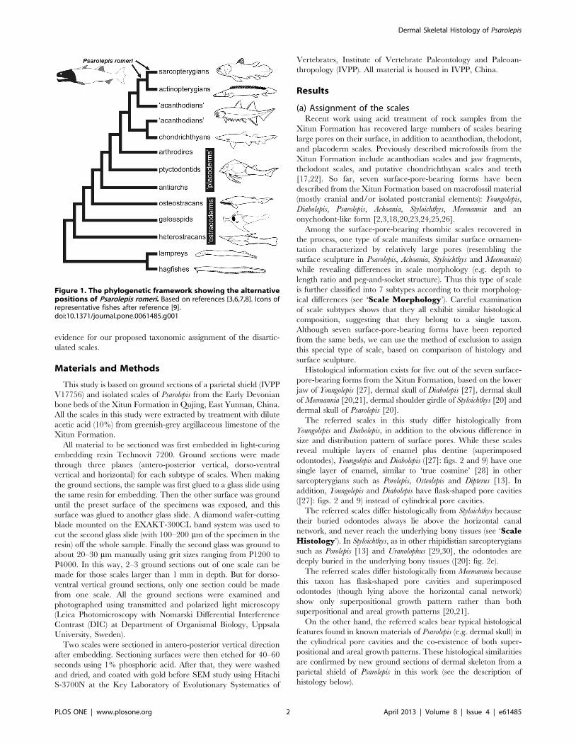
Psarolepis romeri, from the Pridoli (Silurian) and Lochkovian
(Devonian) of South China [1,2,3] and the late Silurian of
Vietnam [4], is one of the earliest known sarcopterygians (lobe-
finned fishes and tetrapods). Initially referred to crown sarcopter-
ygians (Dipnomorpha sensu Ahlberg [5])[2], Psarolepis was soon
assigned to either the osteichthyan or sarcopterygian stem based
on cladistic analysis [3] (Figure 1, based on references [3,6,7,8,9]).
Later phylogenetic studies except that of Zhu and Schultze [10]
have generally resolved Psarolepis as a stem sarcopterygian (e.g.
[7,11]). The morphological reconstruction of Psarolepis was based
on disarticulated remains [3], and has been corroborated by its
close relative Guiyu, the oldest articulated osteichthyan from the
Ludlow (Silurian), South China [11,12]. The dermal skeleton of
Guiyu lacks cosmine, a unique sarcopterygian tissue complex
[13,14,15,16]; Psarolepis thus represents the oldest known sarcop-
terygian with cosmine-like tissue complex, with the potential to
contribute to the understanding of the origin of cosmine, as well as
the dermal skeleton of early osteichthyans.
Wang [17] mentioned the abundant occurrence of the surface-
pore-bearing scales from the Xitun Formation (Lochkovian) of
Yunnan, which corresponds to the high diversity of early
sarcopterygians in this stratum [3,11], but only the trunk scales
of Styloichthys have been briefly described [18,19]. This work on the
scales of Psarolepis represents the starting point for detailed study of
squamation of the osteichthyans discovered in the Xitun
Formation.
The dermal skeletal histology of Psarolepis and Styloichthys was
illustrated briefly by Zhu et al. [20], for the purpose of the
comparison with the histology of the coeval Meemannia. Zhu et al.
[21] gave a more thorough description of the histology of
Meemannia and provided detailed information for further compar-
ative studies. The present work provides additional description of
the dermal skull histology of Psarolepis and reveals histological
differences, such as the shape of pore cavities and diverse hard
tissue resorption conditions, among the early osteichthyans from
the Xitun Formation. The dermal skull histology will also be
compared with the scale histology, thus serving as complementary
PLOS ONE | www.plosone.org 1 April 2013 | Volume 8 | Issue 4 | e61485
evidence for our proposed taxonomic assignment of the disartic-
ulated scales.
Materials and Methods
This study is based on ground sections of a parietal shield (IVPP
V17756) and isolated scales of Psarolepis from the Early Devonian
bone beds of the Xitun Formation in Qujing, East Yunnan, China.
All the scales in this study were extracted by treatment with dilute
acetic acid (10%) from greenish-grey argillaceous limestone of the
Xitun Formation.
All material to be sectioned was first embedded in light-curing
embedding resin Technovit 7200. Ground sections were made
through three planes (antero-posterior vertical, dorso-ventral
vertical and horizontal) for each subtype of scales. When making
the ground sections, the sample was first glued to a glass slide using
the same resin for embedding. Then the other surface was ground
until the preset surface of the specimens was exposed, and this
surface was glued to another glass slide. A diamond wafer-cutting
blade mounted on the EXAKT-300CL band system was used to
cut the second glass slide (with 100–200 mm of the specimen in the
resin) off the whole sample. Finally the second glass was ground to
about 20–30 mm manually using grit sizes ranging from P1200 to
P4000. In this way, 2–3 ground sections out of one scale can be
made for those scales larger than 1 mm in depth. But for dorso-
ventral vertical ground sections, only one section could be made
from one scale. All the ground sections were examined and
photographed using transmitted and polarized light microscopy
(Leica Photomicroscopy with Nomarski Differential Interference
Contrast (DIC) at Department of Organismal Biology, Uppsala
University, Sweden).
Two scales were sectioned in antero-posterior vertical direction
after embedding. Sectioning surfaces were then etched for 40–60
seconds using 1% phosphoric acid. After that, they were washed
and dried, and coated with gold before SEM study using Hitachi
S-3700N at the Key Laboratory of Evolutionary Systematics of
Vertebrates, Institute of Vertebrate Paleontology and Paleoan-
thropology (IVPP). All material is housed in IVPP, China.
Results
(a) Assignment of the scalesRecent work using acid treatment of rock samples from the
Xitun Formation has recovered large numbers of scales bearing
large pores on their surface, in addition to acanthodian, thelodont,
and placoderm scales. Previously described microfossils from the
Xitun Formation include acanthodian scales and jaw fragments,
thelodont scales, and putative chondrichthyan scales and teeth
[17,22]. So far, seven surface-pore-bearing forms have been
described from the Xitun Formation based on macrofossil material
(mostly cranial and/or isolated postcranial elements): Youngolepis,
Diabolepis, Psarolepis, Achoania, Styloichthys, Meemannia and an
onychodont-like form [2,3,18,20,23,24,25,26].
Among the surface-pore-bearing rhombic scales recovered in
the process, one type of scale manifests similar surface ornamen-
tation characterized by relatively large pores (resembling the
surface sculpture in Psarolepis, Achoania, Styloichthys and Meemannia)
while revealing differences in scale morphology (e.g. depth to
length ratio and peg-and-socket structure). Thus this type of scale
is further classified into 7 subtypes according to their morpholog-
ical differences (see ‘Scale Morphology’). Careful examination
of scale subtypes shows that they all exhibit similar histological
composition, suggesting that they belong to a single taxon.
Although seven surface-pore-bearing forms have been reported
from the same beds, we can use the method of exclusion to assign
this special type of scale, based on comparison of histology and
surface sculpture.
Histological information exists for five out of the seven surface-
pore-bearing forms from the Xitun Formation, based on the lower
jaw of Youngolepis [27], dermal skull of Diabolepis [27], dermal skull
of Meemannia [20,21], dermal shoulder girdle of Styloichthys [20] and
dermal skull of Psarolepis [20].
The referred scales in this study differ histologically from
Youngolepis and Diabolepis, in addition to the obvious difference in
size and distribution pattern of surface pores. While these scales
reveal multiple layers of enamel plus dentine (superimposed
odontodes), Youngolepis and Diabolepis ([27]: figs. 2 and 9) have one
single layer of enamel, similar to ‘true cosmine’ [28] in other
sarcopterygians such as Porolepis, Osteolepis and Dipterus [13]. In
addition, Youngolepis and Diabolepis have flask-shaped pore cavities
([27]: figs. 2 and 9) instead of cylindrical pore cavities.
The referred scales differ histologically from Styloichthys because
their buried odontodes always lie above the horizontal canal
network, and never reach the underlying bony tissues (see ‘ScaleHistology’). In Styloichthys, as in other rhipidistian sarcopterygians
such as Porolepis [13] and Uranolophus [29,30], the odontodes are
deeply buried in the underlying bony tissues ([20]: fig. 2e).
The referred scales differ histologically from Meemannia because
this taxon has flask-shaped pore cavities and superimposed
odontodes (though lying above the horizontal canal network)
show only superpositional growth pattern rather than both
superpositional and areal growth patterns [20,21].
On the other hand, the referred scales bear typical histological
features found in known materials of Psarolepis (e.g. dermal skull) in
the cylindrical pore cavities and the co-existence of both super-
positional and areal growth patterns. These histological similarities
are confirmed by new ground sections of dermal skeleton from a
parietal shield of Psarolepis in this work (see the description of
histology below).
Figure 1. The phylogenetic framework showing the alternativepositions of Psarolepis romeri. Based on references [3,6,7,8]. Icons ofrepresentative fishes after reference [9].doi:10.1371/journal.pone.0061485.g001
Dermal Skeletal Histology of Psarolepis
PLOS ONE | www.plosone.org 2 April 2013 | Volume 8 | Issue 4 | e61485
Thus far, no histological information is available for Achoania
(only based on one anterior portion of the skull [25], with five
lower jaw specimens [26] and one shoulder girdle [31] assigned to
the genus) and the onychodont-like form (only based on an
incomplete lower jaw [26]), consequently no histological compar-
ison can be made with these two poorly represented forms.
However, as the referred scales in this study make up about 50%
of all surface-pore-bearing scales in the entire sample, it is
reasonable to assign the referred scales to Psarolepis, which is
abundantly represented among macrofossils, rather than to the
poorly represented Achoania or the onychodont-like form.
While this assignment based on similarities in histology and
dermal surface sculpture, and the relative abundance of speci-
mens, must remain tentative pending the discovery of articulated
Psarolepis specimen with squamation, previous assignment of
isolated Psarolepis materials (shoulder girdles, cheek plates, median
fin spines, and most recently pelvic girdles) has received indirect
corroboration from articulated Guiyu specimens in terms of the
reconstructed body form and the restored position of isolated
elements [11,32].
Although we cannot exclude the possibility that Achoania may
have scales similar to the scales here referred to Psarolepis, the
overall significance of the scales referred to Psarolepis as described
below would not be affected, as Achoania and Psarolepis are closely
related to each other in most phylogenetic analyses (e.g. [11]).
While recognizing the tentative nature of the assignment of these
isolated scales, we believe that the morphological and histological
details revealed by these scales will add to our understanding of
Psarolepis, contribute to the ongoing discussion of the phylogenetic
position of Psarolepis (either as a stem sarcopterygian or a stem
osteichthyan), and bear on the study of ganoid and cosmoid scales
in early bony fishes (see ‘Discussion’).
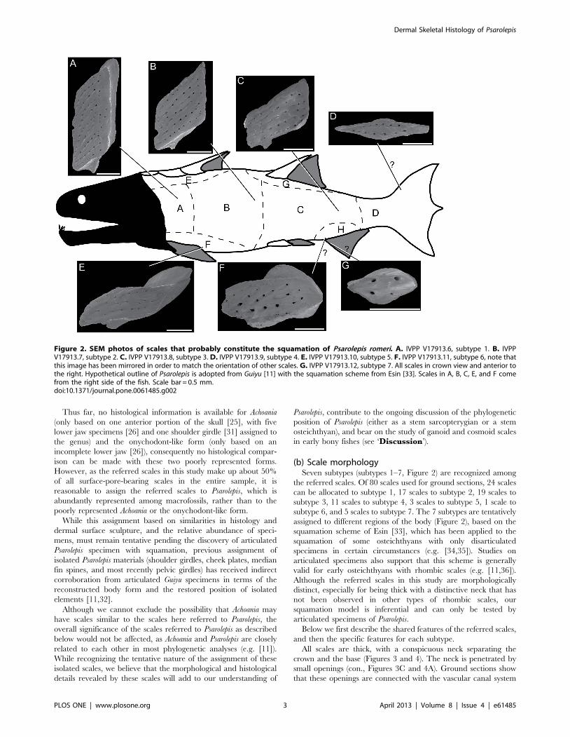
(b) Scale morphologySeven subtypes (subtypes 1–7, Figure 2) are recognized among
the referred scales. Of 80 scales used for ground sections, 24 scales
can be allocated to subtype 1, 17 scales to subtype 2, 19 scales to
subtype 3, 11 scales to subtype 4, 3 scales to subtype 5, 1 scale to
subtype 6, and 5 scales to subtype 7. The 7 subtypes are tentatively
assigned to different regions of the body (Figure 2), based on the
squamation scheme of Esin [33], which has been applied to the
squamation of some osteichthyans with only disarticulated
specimens in certain circumstances (e.g. [34,35]). Studies on
articulated specimens also support that this scheme is generally
valid for early osteichthyans with rhombic scales (e.g. [11,36]).
Although the referred scales in this study are morphologically
distinct, especially for being thick with a distinctive neck that has
not been observed in other types of rhombic scales, our
squamation model is inferential and can only be tested by
articulated specimens of Psarolepis.
Below we first describe the shared features of the referred scales,
and then the specific features for each subtype.
All scales are thick, with a conspicuous neck separating the
crown and the base (Figures 3 and 4). The neck is penetrated by
small openings (con., Figures 3C and 4A). Ground sections show
that these openings are connected with the vascular canal system
Figure 2. SEM photos of scales that probably constitute the squamation of Psarolepis romeri. A. IVPP V17913.6, subtype 1. B. IVPPV17913.7, subtype 2. C. IVPP V17913.8, subtype 3. D. IVPP V17913.9, subtype 4. E. IVPP V17913.10, subtype 5. F. IVPP V17913.11, subtype 6, note thatthis image has been mirrored in order to match the orientation of other scales. G. IVPP V17913.12, subtype 7. All scales in crown view and anterior tothe right. Hypothetical outline of Psarolepis is adopted from Guiyu [11] with the squamation scheme from Esin [33]. Scales in A, B, C, E, and F comefrom the right side of the fish. Scale bar = 0.5 mm.doi:10.1371/journal.pone.0061485.g002
Dermal Skeletal Histology of Psarolepis
PLOS ONE | www.plosone.org 3 April 2013 | Volume 8 | Issue 4 | e61485
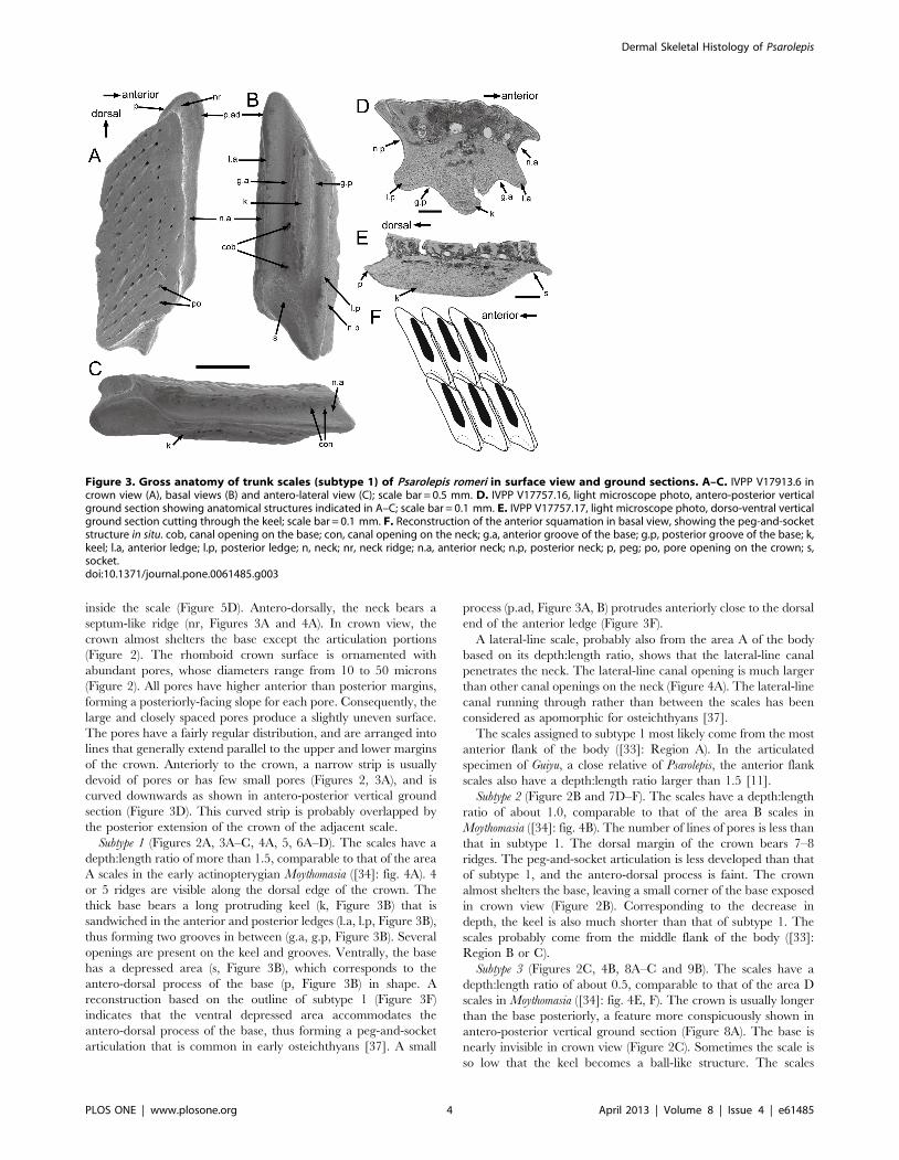
inside the scale (Figure 5D). Antero-dorsally, the neck bears a
septum-like ridge (nr, Figures 3A and 4A). In crown view, the
crown almost shelters the base except the articulation portions
(Figure 2). The rhomboid crown surface is ornamented with
abundant pores, whose diameters range from 10 to 50 microns
(Figure 2). All pores have higher anterior than posterior margins,
forming a posteriorly-facing slope for each pore. Consequently, the
large and closely spaced pores produce a slightly uneven surface.
The pores have a fairly regular distribution, and are arranged into
lines that generally extend parallel to the upper and lower margins
of the crown. Anteriorly to the crown, a narrow strip is usually
devoid of pores or has few small pores (Figures 2, 3A), and is
curved downwards as shown in antero-posterior vertical ground
section (Figure 3D). This curved strip is probably overlapped by
the posterior extension of the crown of the adjacent scale.
Subtype 1 (Figures 2A, 3A–C, 4A, 5, 6A–D). The scales have a
depth:length ratio of more than 1.5, comparable to that of the area
A scales in the early actinopterygian Moythomasia ([34]: fig. 4A). 4
or 5 ridges are visible along the dorsal edge of the crown. The
thick base bears a long protruding keel (k, Figure 3B) that is
sandwiched in the anterior and posterior ledges (l.a, l.p, Figure 3B),
thus forming two grooves in between (g.a, g.p, Figure 3B). Several
openings are present on the keel and grooves. Ventrally, the base
has a depressed area (s, Figure 3B), which corresponds to the
antero-dorsal process of the base (p, Figure 3B) in shape. A
reconstruction based on the outline of subtype 1 (Figure 3F)
indicates that the ventral depressed area accommodates the
antero-dorsal process of the base, thus forming a peg-and-socket
articulation that is common in early osteichthyans [37]. A small
process (p.ad, Figure 3A, B) protrudes anteriorly close to the dorsal
end of the anterior ledge (Figure 3F).
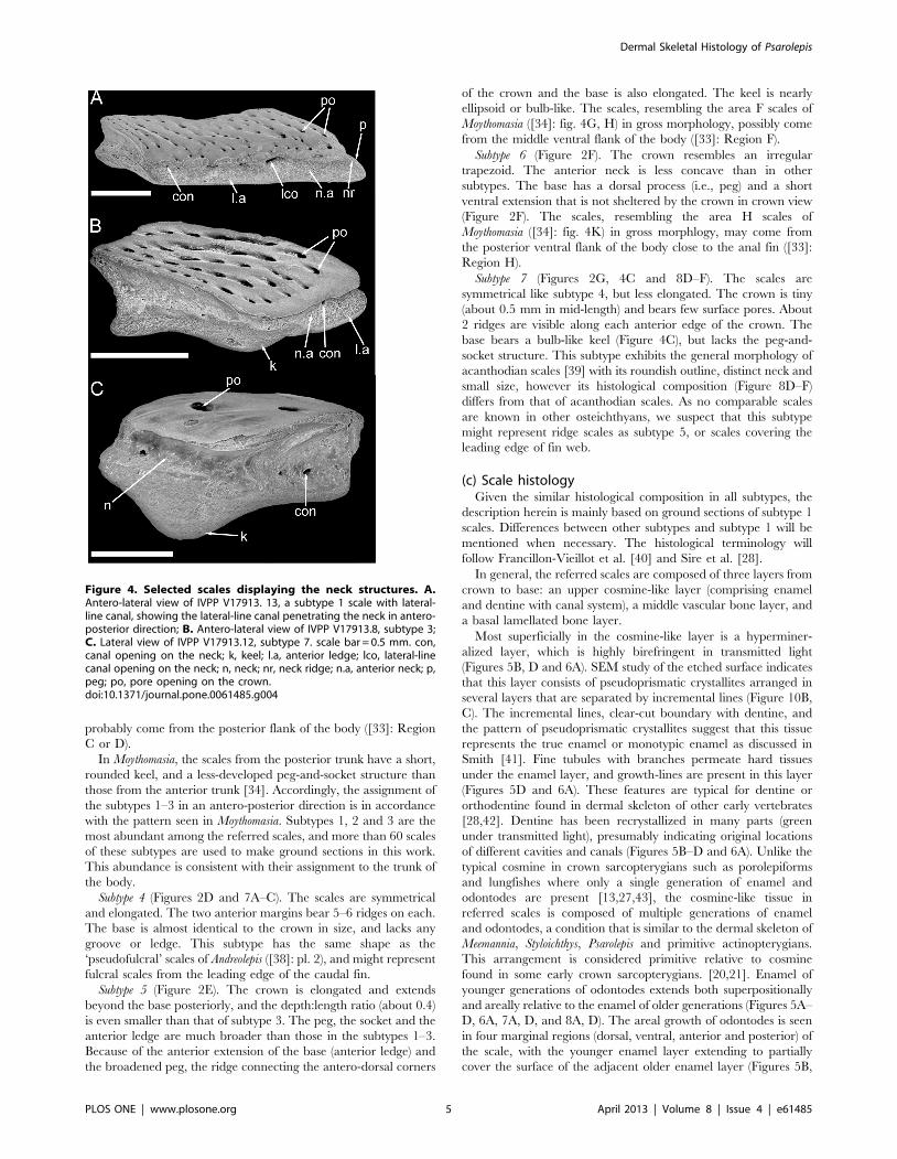
A lateral-line scale, probably also from the area A of the body
based on its depth:length ratio, shows that the lateral-line canal
penetrates the neck. The lateral-line canal opening is much larger
than other canal openings on the neck (Figure 4A). The lateral-line
canal running through rather than between the scales has been
considered as apomorphic for osteichthyans [37].
The scales assigned to subtype 1 most likely come from the most
anterior flank of the body ([33]: Region A). In the articulated
specimen of Guiyu, a close relative of Psarolepis, the anterior flank
scales also have a depth:length ratio larger than 1.5 [11].
Subtype 2 (Figure 2B and 7D–F). The scales have a depth:length
ratio of about 1.0, comparable to that of the area B scales in
Moythomasia ([34]: fig. 4B). The number of lines of pores is less than
that in subtype 1. The dorsal margin of the crown bears 7–8
ridges. The peg-and-socket articulation is less developed than that
of subtype 1, and the antero-dorsal process is faint. The crown
almost shelters the base, leaving a small corner of the base exposed
in crown view (Figure 2B). Corresponding to the decrease in
depth, the keel is also much shorter than that of subtype 1. The
scales probably come from the middle flank of the body ([33]:
Region B or C).
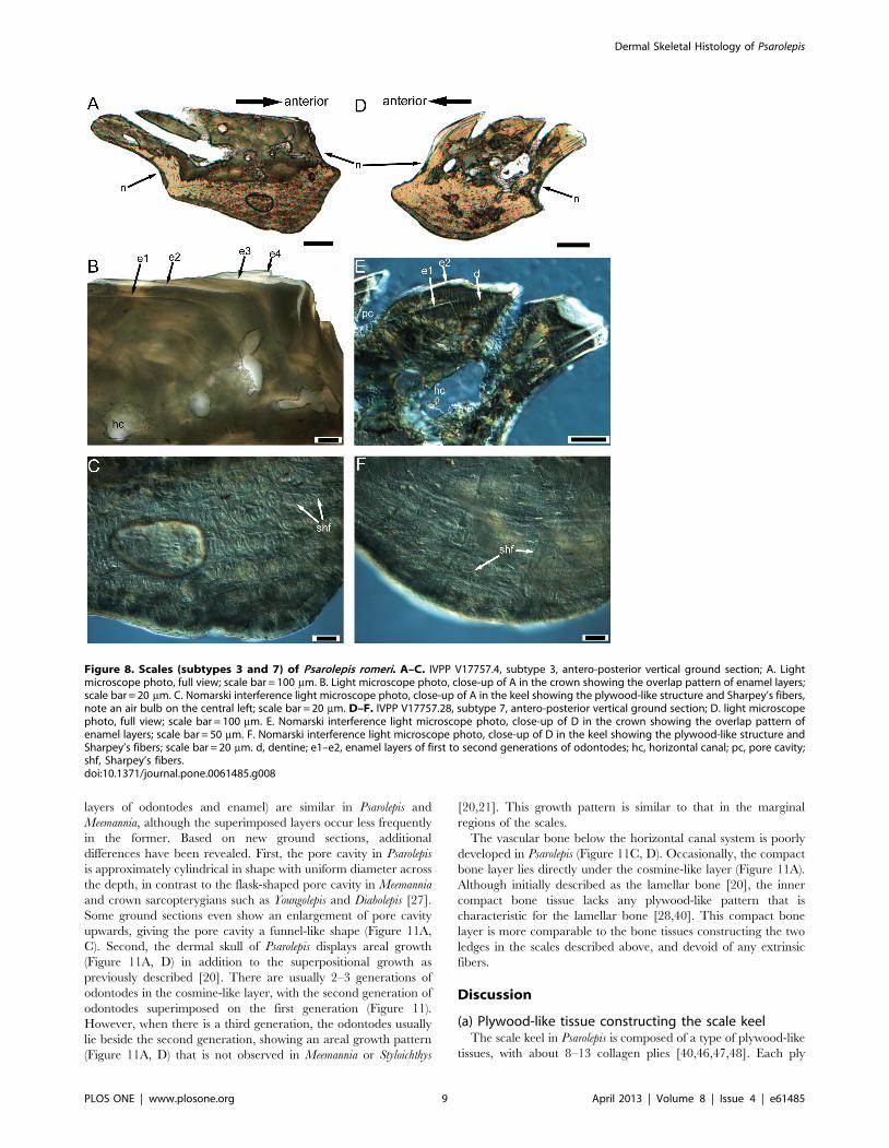
Subtype 3 (Figures 2C, 4B, 8A–C and 9B). The scales have a
depth:length ratio of about 0.5, comparable to that of the area D
scales in Moythomasia ([34]: fig. 4E, F). The crown is usually longer
than the base posteriorly, a feature more conspicuously shown in
antero-posterior vertical ground section (Figure 8A). The base is
nearly invisible in crown view (Figure 2C). Sometimes the scale is
so low that the keel becomes a ball-like structure. The scales
Figure 3. Gross anatomy of trunk scales (subtype 1) of Psarolepis romeri in surface view and ground sections. A–C. IVPP V17913.6 incrown view (A), basal views (B) and antero-lateral view (C); scale bar = 0.5 mm. D. IVPP V17757.16, light microscope photo, antero-posterior verticalground section showing anatomical structures indicated in A–C; scale bar = 0.1 mm. E. IVPP V17757.17, light microscope photo, dorso-ventral verticalground section cutting through the keel; scale bar = 0.1 mm. F. Reconstruction of the anterior squamation in basal view, showing the peg-and-socketstructure in situ. cob, canal opening on the base; con, canal opening on the neck; g.a, anterior groove of the base; g.p, posterior groove of the base; k,keel; l.a, anterior ledge; l.p, posterior ledge; n, neck; nr, neck ridge; n.a, anterior neck; n.p, posterior neck; p, peg; po, pore opening on the crown; s,socket.doi:10.1371/journal.pone.0061485.g003
Dermal Skeletal Histology of Psarolepis
PLOS ONE | www.plosone.org 4 April 2013 | Volume 8 | Issue 4 | e61485
probably come from the posterior flank of the body ([33]: Region
C or D).
In Moythomasia, the scales from the posterior trunk have a short,
rounded keel, and a less-developed peg-and-socket structure than
those from the anterior trunk [34]. Accordingly, the assignment of
the subtypes 1–3 in an antero-posterior direction is in accordance
with the pattern seen in Moythomasia. Subtypes 1, 2 and 3 are the
most abundant among the referred scales, and more than 60 scales
of these subtypes are used to make ground sections in this work.
This abundance is consistent with their assignment to the trunk of
the body.
Subtype 4 (Figures 2D and 7A–C). The scales are symmetrical
and elongated. The two anterior margins bear 5–6 ridges on each.
The base is almost identical to the crown in size, and lacks any
groove or ledge. This subtype has the same shape as the
‘pseudofulcral’ scales of Andreolepis ([38]: pl. 2), and might represent
fulcral scales from the leading edge of the caudal fin.
Subtype 5 (Figure 2E). The crown is elongated and extends
beyond the base posteriorly, and the depth:length ratio (about 0.4)
is even smaller than that of subtype 3. The peg, the socket and the
anterior ledge are much broader than those in the subtypes 1–3.
Because of the anterior extension of the base (anterior ledge) and
the broadened peg, the ridge connecting the antero-dorsal corners
of the crown and the base is also elongated. The keel is nearly
ellipsoid or bulb-like. The scales, resembling the area F scales of
Moythomasia ([34]: fig. 4G, H) in gross morphology, possibly come
from the middle ventral flank of the body ([33]: Region F).
Subtype 6 (Figure 2F). The crown resembles an irregular
trapezoid. The anterior neck is less concave than in other
subtypes. The base has a dorsal process (i.e., peg) and a short
ventral extension that is not sheltered by the crown in crown view
(Figure 2F). The scales, resembling the area H scales of
Moythomasia ([34]: fig. 4K) in gross morphlogy, may come from
the posterior ventral flank of the body close to the anal fin ([33]:
Region H).
Subtype 7 (Figures 2G, 4C and 8D–F). The scales are
symmetrical like subtype 4, but less elongated. The crown is tiny
(about 0.5 mm in mid-length) and bears few surface pores. About
2 ridges are visible along each anterior edge of the crown. The
base bears a bulb-like keel (Figure 4C), but lacks the peg-and-
socket structure. This subtype exhibits the general morphology of
acanthodian scales [39] with its roundish outline, distinct neck and
small size, however its histological composition (Figure 8D–F)
differs from that of acanthodian scales. As no comparable scales
are known in other osteichthyans, we suspect that this subtype
might represent ridge scales as subtype 5, or scales covering the
leading edge of fin web.
(c) Scale histologyGiven the similar histological composition in all subtypes, the
description herein is mainly based on ground sections of subtype 1
scales. Differences between other subtypes and subtype 1 will be
mentioned when necessary. The histological terminology will
follow Francillon-Vieillot et al. [40] and Sire et al. [28].
In general, the referred scales are composed of three layers from
crown to base: an upper cosmine-like layer (comprising enamel
and dentine with canal system), a middle vascular bone layer, and
a basal lamellated bone layer.
Most superficially in the cosmine-like layer is a hyperminer-
alized layer, which is highly birefringent in transmitted light
(Figures 5B, D and 6A). SEM study of the etched surface indicates
that this layer consists of pseudoprismatic crystallites arranged in
several layers that are separated by incremental lines (Figure 10B,
C). The incremental lines, clear-cut boundary with dentine, and
the pattern of pseudoprismatic crystallites suggest that this tissue
represents the true enamel or monotypic enamel as discussed in
Smith [41]. Fine tubules with branches permeate hard tissues
under the enamel layer, and growth-lines are present in this layer
(Figures 5D and 6A). These features are typical for dentine or
orthodentine found in dermal skeleton of other early vertebrates
[28,42]. Dentine has been recrystallized in many parts (green
under transmitted light), presumably indicating original locations
of different cavities and canals (Figures 5B–D and 6A). Unlike the
typical cosmine in crown sarcopterygians such as porolepiforms
and lungfishes where only a single generation of enamel and
odontodes are present [13,27,43], the cosmine-like tissue in
referred scales is composed of multiple generations of enamel
and odontodes, a condition that is similar to the dermal skeleton of
Meemannia, Styloichthys, Psarolepis and primitive actinopterygians.
This arrangement is considered primitive relative to cosmine
found in some early crown sarcopterygians. [20,21]. Enamel of
younger generations of odontodes extends both superpositionally
and areally relative to the enamel of older generations (Figures 5A–
D, 6A, 7A, D, and 8A, D). The areal growth of odontodes is seen
in four marginal regions (dorsal, ventral, anterior and posterior) of
the scale, with the younger enamel layer extending to partially
cover the surface of the adjacent older enamel layer (Figures 5B,
Figure 4. Selected scales displaying the neck structures. A.Antero-lateral view of IVPP V17913. 13, a subtype 1 scale with lateral-line canal, showing the lateral-line canal penetrating the neck in antero-posterior direction; B. Antero-lateral view of IVPP V17913.8, subtype 3;C. Lateral view of IVPP V17913.12, subtype 7. scale bar = 0.5 mm. con,canal opening on the neck; k, keel; l.a, anterior ledge; lco, lateral-linecanal opening on the neck; n, neck; nr, neck ridge; n.a, anterior neck; p,peg; po, pore opening on the crown.doi:10.1371/journal.pone.0061485.g004
Dermal Skeletal Histology of Psarolepis
PLOS ONE | www.plosone.org 5 April 2013 | Volume 8 | Issue 4 | e61485
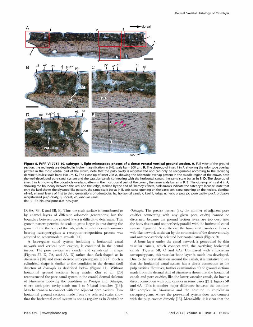
D, 6A, 7B, E and 8B, E). Thus the scale surface is contributed to
by enamel layers of different odontode generations, but the
boundary between two enamel layers is difficult to determine. This
growth pattern permits the scale to grow larger in area during the
growth of the the body of the fish, while in more derived cosmine-
bearing sarcopterygians a resorption-redeposition process was
adopted to accommodate growth [44].
A less-regular canal system, including a horizontal canal
network and vertical pore cavities, is contained in the dental
tissues. The pore cavities are slender and cylindrical in shape
(Figures 5B–D, 7A, and 8A, D) rather than flask-shaped as in
Meemannia [20] and more derived sarcopterygians [13,27]. Such a
cylindrical shape is similar to the condition in the dermal skull
skeleton of Psarolepis as described below (Figure 11). Without
horizontal ground sections being made, Zhu et al. [20]
reconstructed the pore-canal system in the cranial dermal skeleton
of Meemannia following the condition in Porolepis and Osteolepis,
where each pore cavity sends out 4 to 5 basal branches ([13]:
Maschencanals) to connect with the adjacent pore cavities. Two
horizontal ground sections made from the referred scales show
that the horizontal canal system is not as regular as in Porolepis or
Osteolepis. The precise pattern (i.e., the number of adjacent pore
cavities connecting with any given pore cavity) cannot be
discerned, because the ground section levels are too deep into
the bony tissues and not perfectly parallel with the horizontal canal
system (Figure 9). Nevertheless, the horizontal canals do form a
web-like network as shown by the connection of the dorsoventrally
and anteroposteriorly oriented horizontal canals (Figure 9).
A bone layer under the canal network is penetrated by thin
vascular canals, which connect with the overlying horizontal
canals (Figures 5B, C and 6A). Compared with rhipidistian
sarcopterygians, this vascular bone layer is much less developed.
Due to the recrystalization around the canals, it is tentative to say
that the horizontal canal system has a direct connection to the
pulp cavities. However, further examination of the ground sections
made from the dermal skull of Meemannia shows that the horizontal
canals and pore cavities, like the lower vascular canals, do have a
direct connection with pulp cavities in some cases ([21]: figures 5B
and 6A). This is another major difference between the cosmine-
like complex in Meemannia and the cosmine in rhipidistian
sarcopterygians, where the pore-canal system does not connect
with the pulp cavities directly [13]. Meanwhile, it is clear that the
Figure 5. IVPP V17757.19, subtype 1, light microscope photos of a dorso-ventral vertical ground section. A. Full view of the groundsection, the red insets are detailed in higher magnification in B–E, scale bar = 200 mm. B. The close-up of inset 1 in A, showing the odontode overlappattern in the most ventral part of the crown, note that the pulp cavity is recrystallized and can only be recognizable according to the radiatingdentine tubules; scale bar = 100 mm. C. The close-up of inset 2 in A, showing the odontode overlap pattern in the middle region of the crown, notethe well-developed pore-canal system and the vascular canals connecting with the horizontal canals, the same scale bar as in B; D. The close-up ofinset 3 in A, showing the odontode overlap pattern in the most dorsal part of the crown, the same scale bar as in B; E. The close-up of inset 4 in A,showing the boundary between the keel and the ledge, marked by the end of Sharpey’s fibers, pink arrows indicate the osteocyte lacunae, note thatonly the keel shows the plywood-like pattern, the same scale bar as in B. cob, canal opening on the base; con, canal opening on the neck; d, dentine;e1–e3, enamel layers of first to third generations of odontodes; hc, horizontal canal; k, keel; l, ledge; n, neck; p, peg; pc, pore cavity; puc?, probablerecrystallized pulp cavity; s, socket; vc, vascular canal.doi:10.1371/journal.pone.0061485.g005
Dermal Skeletal Histology of Psarolepis
PLOS ONE | www.plosone.org 6 April 2013 | Volume 8 | Issue 4 | e61485
lower vascular canal system also contributes to the pulp cavities
(Figure 5B–D). It is likely that the regular pattern of the pore-canal
system separated from the pulp canals in crown sarcopterygians
was attained in a stepwise way from the less regular pattern seen in
Psarolepis and Meemannia.
Under the vascular bone layer lies the base, which consists of
one keel flanked by two ledges (Figure 3D). Histologically the keel
and two ledges are marked by two sharp lines under polarized
light, and the two boundary lines (g.a, g.p, Figure 6C) usually lie at
the deepest points of the two grooves on the base. For those scales
without a distinct ledge region, the base is almost fully occupied by
the keel (Figures 7A and 8A). Several differences are observed
between the two types of tissues. While the tissue of the keel
exhibits a plywood-like structure (Figures 5E and 6B–D), the two
ledges show no such structure but only parallel fibers (Figures 5E
and 6B). In the keel, each ply or lamella (approximately 20 mm in
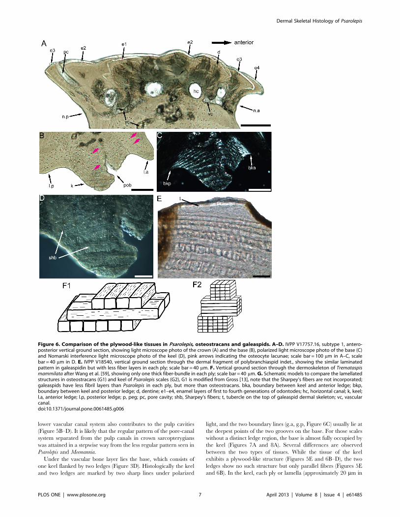
Figure 6. Comparison of the plywood-like tissues in Psarolepis, osteostracans and galeaspids. A–D. IVPP V17757.16, subtype 1, antero-posterior vertical ground section, showing light microscope photo of the crown (A) and the base (B), polarized light microscope photo of the base (C)and Nomarski interference light microscope photo of the keel (D), pink arrows indicating the osteocyte lacunae; scale bar = 100 mm in A–C, scalebar = 40 mm in D. E. IVPP V18540, vertical ground section through the dermal fragment of polybranchiaspid indet., showing the similar laminatedpattern in galeaspidin but with less fiber layers in each ply; scale bar = 40 mm. F. Vertical ground section through the dermoskeleton of Tremataspismammilata after Wang et al. [59], showing only one thick fiber-bundle in each ply; scale bar = 40 mm. G. Schematic models to compare the lamellatedstructures in osteostracans (G1) and keel of Psarolepis scales (G2), G1 is modified from Gross [13], note that the Sharpey’s fibers are not incorporated;galeaspids have less fibril layers than Psarolepis in each ply, but more than osteostracans. bka, boundary between keel and anterior ledge; bkp,boundary between keel and posterior ledge; d, dentine; e1–e4, enamel layers of first to fourth generations of odontodes; hc, horizontal canal; k, keel;l.a, anterior ledge; l.p, posterior ledge; p, peg; pc, pore cavity; shb, Sharpey’s fibers; t, tubercle on the top of galeaspid dermal skeleton; vc, vascularcanal.doi:10.1371/journal.pone.0061485.g006
Dermal Skeletal Histology of Psarolepis
PLOS ONE | www.plosone.org 7 April 2013 | Volume 8 | Issue 4 | e61485
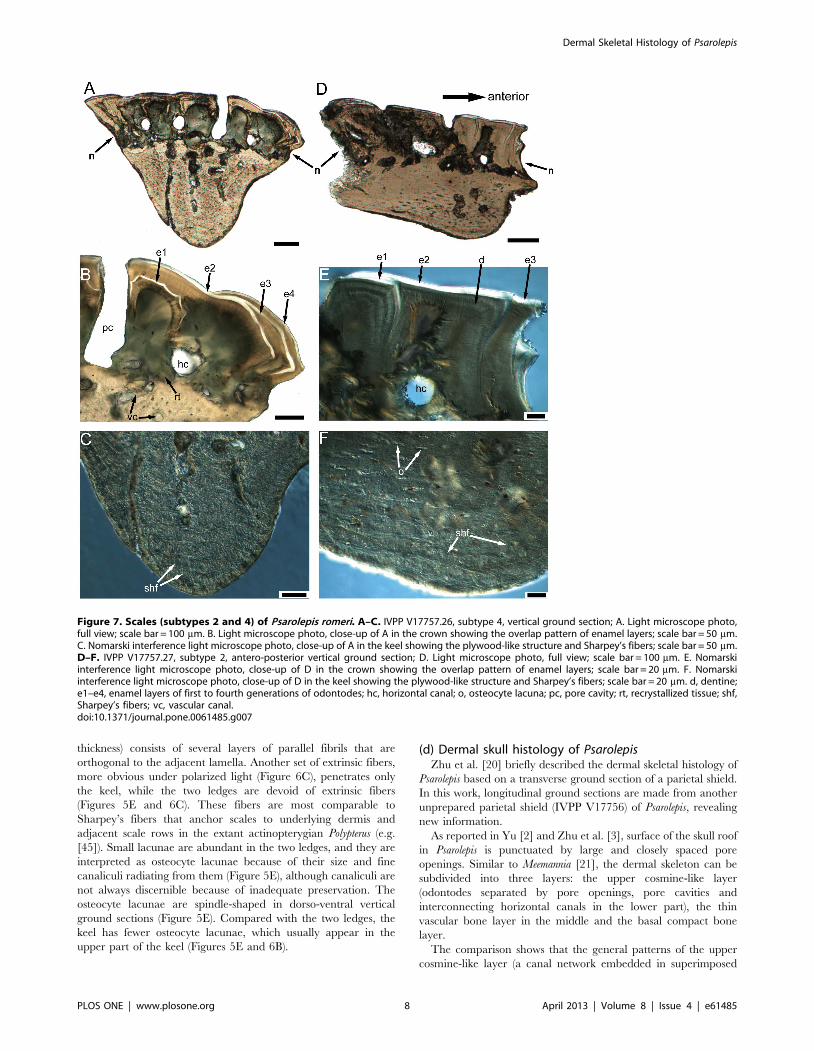
thickness) consists of several layers of parallel fibrils that are
orthogonal to the adjacent lamella. Another set of extrinsic fibers,
more obvious under polarized light (Figure 6C), penetrates only
the keel, while the two ledges are devoid of extrinsic fibers
(Figures 5E and 6C). These fibers are most comparable to
Sharpey’s fibers that anchor scales to underlying dermis and
adjacent scale rows in the extant actinopterygian Polypterus (e.g.
[45]). Small lacunae are abundant in the two ledges, and they are
interpreted as osteocyte lacunae because of their size and fine
canaliculi radiating from them (Figure 5E), although canaliculi are
not always discernible because of inadequate preservation. The
osteocyte lacunae are spindle-shaped in dorso-ventral vertical
ground sections (Figure 5E). Compared with the two ledges, the
keel has fewer osteocyte lacunae, which usually appear in the
upper part of the keel (Figures 5E and 6B).
(d) Dermal skull histology of PsarolepisZhu et al. [20] briefly described the dermal skeletal histology of
Psarolepis based on a transverse ground section of a parietal shield.
In this work, longitudinal ground sections are made from another
unprepared parietal shield (IVPP V17756) of Psarolepis, revealing
new information.
As reported in Yu [2] and Zhu et al. [3], surface of the skull roof
in Psarolepis is punctuated by large and closely spaced pore
openings. Similar to Meemannia [21], the dermal skeleton can be
subdivided into three layers: the upper cosmine-like layer
(odontodes separated by pore openings, pore cavities and
interconnecting horizontal canals in the lower part), the thin
vascular bone layer in the middle and the basal compact bone
layer.
The comparison shows that the general patterns of the upper
cosmine-like layer (a canal network embedded in superimposed
Figure 7. Scales (subtypes 2 and 4) of Psarolepis romeri. A–C. IVPP V17757.26, subtype 4, vertical ground section; A. Light microscope photo,full view; scale bar = 100 mm. B. Light microscope photo, close-up of A in the crown showing the overlap pattern of enamel layers; scale bar = 50 mm.C. Nomarski interference light microscope photo, close-up of A in the keel showing the plywood-like structure and Sharpey’s fibers; scale bar = 50 mm.D–F. IVPP V17757.27, subtype 2, antero-posterior vertical ground section; D. Light microscope photo, full view; scale bar = 100 mm. E. Nomarskiinterference light microscope photo, close-up of D in the crown showing the overlap pattern of enamel layers; scale bar = 20 mm. F. Nomarskiinterference light microscope photo, close-up of D in the keel showing the plywood-like structure and Sharpey’s fibers; scale bar = 20 mm. d, dentine;e1–e4, enamel layers of first to fourth generations of odontodes; hc, horizontal canal; o, osteocyte lacuna; pc, pore cavity; rt, recrystallized tissue; shf,Sharpey’s fibers; vc, vascular canal.doi:10.1371/journal.pone.0061485.g007
Dermal Skeletal Histology of Psarolepis
PLOS ONE | www.plosone.org 8 April 2013 | Volume 8 | Issue 4 | e61485
layers of odontodes and enamel) are similar in Psarolepis and
Meemannia, although the superimposed layers occur less frequently
in the former. Based on new ground sections, additional
differences have been revealed. First, the pore cavity in Psarolepis
is approximately cylindrical in shape with uniform diameter across
the depth, in contrast to the flask-shaped pore cavity in Meemannia
and crown sarcopterygians such as Youngolepis and Diabolepis [27].
Some ground sections even show an enlargement of pore cavity
upwards, giving the pore cavity a funnel-like shape (Figure 11A,
C). Second, the dermal skull of Psarolepis displays areal growth
(Figure 11A, D) in addition to the superpositional growth as
previously described [20]. There are usually 2–3 generations of
odontodes in the cosmine-like layer, with the second generation of
odontodes superimposed on the first generation (Figure 11).
However, when there is a third generation, the odontodes usually
lie beside the second generation, showing an areal growth pattern
(Figure 11A, D) that is not observed in Meemannia or Styloichthys
[20,21]. This growth pattern is similar to that in the marginal
regions of the scales.
The vascular bone below the horizontal canal system is poorly
developed in Psarolepis (Figure 11C, D). Occasionally, the compact
bone layer lies directly under the cosmine-like layer (Figure 11A).
Although initially described as the lamellar bone [20], the inner
compact bone tissue lacks any plywood-like pattern that is
characteristic for the lamellar bone [28,40]. This compact bone
layer is more comparable to the bone tissues constructing the two
ledges in the scales described above, and devoid of any extrinsic
fibers.
Discussion
(a) Plywood-like tissue constructing the scale keelThe scale keel in Psarolepis is composed of a type of plywood-like
tissues, with about 8–13 collagen plies [40,46,47,48]. Each ply
Figure 8. Scales (subtypes 3 and 7) of Psarolepis romeri. A–C. IVPP V17757.4, subtype 3, antero-posterior vertical ground section; A. Lightmicroscope photo, full view; scale bar = 100 mm. B. Light microscope photo, close-up of A in the crown showing the overlap pattern of enamel layers;scale bar = 20 mm. C. Nomarski interference light microscope photo, close-up of A in the keel showing the plywood-like structure and Sharpey’s fibers,note an air bulb on the central left; scale bar = 20 mm. D–F. IVPP V17757.28, subtype 7, antero-posterior vertical ground section; D. light microscopephoto, full view; scale bar = 100 mm. E. Nomarski interference light microscope photo, close-up of D in the crown showing the overlap pattern ofenamel layers; scale bar = 50 mm. F. Nomarski interference light microscope photo, close-up of D in the keel showing the plywood-like structure andSharpey’s fibers; scale bar = 20 mm. d, dentine; e1–e2, enamel layers of first to second generations of odontodes; hc, horizontal canal; pc, pore cavity;shf, Sharpey’s fibers.doi:10.1371/journal.pone.0061485.g008
Dermal Skeletal Histology of Psarolepis
PLOS ONE | www.plosone.org 9 April 2013 | Volume 8 | Issue 4 | e61485
consists of several layers of fiber-bundles that are parallel to each
other but orthogonal to the fiber-bundles of the adjacent ply
(Figure 6D, F2). Orthogonal to these intrinsic fiber-bundles is a set
of extrinsic thick fibers penetrating multiple plies (Figures 5E,
6C, D, 7C, F, and 8C, F). When viewed under polarized light, the
keel exhibits alternating stripes of black and white (Figure 6C).
This structure conforms well to the definition of the lamellar bone
[40]. By comparison, the rest of the scale base including the
flanking ledges always exhibits a homogenous pattern and does not
show any plywood-like organization. A boundary between the keel
and the rest of the base is usually evident when examined under
polarized light (Figure 6C). By definition, the collagen tissue in the
rest of the base is a type of pseudo-lamellar bone or parallel-fibered
bone [40].
It needs to be pointed out that in this paper we follow the
terminology in Francillon-Vieillot et al. [40] and use the term
‘‘lamellar bone’’ only to refer to the lamellated bone with a
plywood-like structure. Meanwhile, the term ‘isopedine’ is adopted
to describe a subtype of lamellar bone (either cellular or cellular)
with an orthogonal plywood-like structure [contra Meunier [47]
who employed isopedine to describe elasmodine in teleosts]. In this
paper, isopedine is interchangeable with lamellar bone as no
twisted plywood-like structure is involved.
Comparison with the rhombic scales of other early osteichth-
yans shows that the histological organization in the scale base of
Psarolepis is unique. In the trunk scale of Ligulalepis, the bony base is
constructed by homogenous cellular lamellated bone, with
Sharpey’s fibers restricted in the keel [49]. The scale base in
Andreolepis is composed of homogenous cellular bone without any
plywood-like organization and with Sharpey’s fibers restricted in
the keel [50]. In Moythomasia and Mimipiscis, the scale base was also
described to be composed of homogenous cellular lamellated bone
penetrated partially by Sharpey’s fibers, but the microstructure of
the lamellated bone was not illustrated [51,52]. Traditionally,
many authors employed the term ‘lamellar bone’ to describe the
lamellated bone (e.g. [20,49,51,52,53]). The ‘lamellar bone’ in
these works does not necessarily show a plywood-like pattern and
might be the pseudo-lamellar bone sensu Francillon-Vieillot et al.
[40]. For example, the ‘lamellar bone’ in the dermal skull of
Meemannia and Psarolepis [20] can be referred to the pseudo-
lamellar bone because of its homogenous nature under polarized
light. In Polypterus, the bony base of the scale has been described as
constructed by homogenous pseudo-lamellar bone, and Sharpey’s
fibers are also restricted to the keel [45,54,55].
The histological organization of the scale base in the porolepi-
form sarcopterygian Heimenia [43] is also different from that in
Psarolepis. The scale keel ([43]: ‘internal bone layer’) in Heimenia
does not show any plywood-like structure. However, the rest of the
scale base ([43]: ‘basal layer’) is composed of lamellar bone
showing a plywood-like structure. In addition, both the keel and
the rest of the base are penetrated by Sharpey’s fibers. Other early
sarcopterygians such as Porolepis and Osteolepis also have typical
cosmoid scales, whose base (excluding the keel) is composed of a
thick layer of isopedine [13]. The keel was described as
constructed by spongy bone in Gross [13].
To summarize, no plywood-like tissue has been found in the
keel of the scale among osteichthyans except Psarolepis. However,
the plywood-like tissue is present in the base of non-rhombic scales
referred to some acanthodians [56,57] and putative early
chondrichthyans ([58]), suggesting the possibility that the keel
microstructure in Psarolepis may represent a retained primitive
feature for osteichthyans. Like the scale keel of Psarolepis, the base
of non-rhombic scales of some acanthodians and putative
chondrichthyans is also constructed by intrinsic isopedine plus
extrinsic Sharpey’s fibers, although the thickness and pattern of the
Sharpey’s fibers show some variations in different taxa [56,57,58].
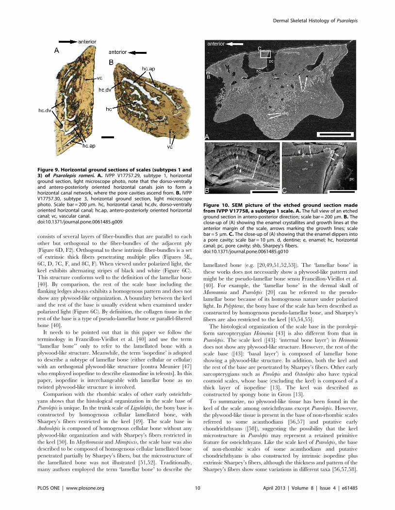
Figure 9. Horizontal ground sections of scales (subtypes 1 and3) of Psarolepis romeri. A. IVPP V17757.29, subtype 1, horizontalground section, light microscope photo, note that the dorso-ventrallyand antero-posteriorly oriented horizontal canals join to form ahorizontal canal network, where the pore cavities ascend from. B. IVPPV17757.30, subtype 3, horizontal ground section, light microscopephoto. Scale bar = 200 mm. hc, horizontal canal; hc.dv, dorso-ventrallyoriented horizontal canal; hc.ap, antero-posteriorly oriented horizontalcanal; vc, vascular canal.doi:10.1371/journal.pone.0061485.g009
Figure 10. SEM picture of the etched ground section madefrom IVPP V17758, a subtype 1 scale. A. The full view of an etchedground section in antero-posterior direction; scale bar = 200 mm. B. Theclose-up of (A) showing the enamel crystallites and growth lines at theanterior margin of the scale, arrows marking the growth lines; scalebar = 5 mm. C. The close-up of (A) showing that the enamel dippers intoa pore cavity; scale bar = 10 mm. d, dentine; e, enamel; hc, horizontalcanal; pc, pore cavity; shb, Sharpey’s fibers.doi:10.1371/journal.pone.0061485.g010
Dermal Skeletal Histology of Psarolepis
PLOS ONE | www.plosone.org 10 April 2013 | Volume 8 | Issue 4 | e61485
The keel of rhombic scales is the most interior part and functions
as a structure to connect the scales with the subdermis, usually
indicated by the presence of Sharpey’s fibers (e.g. [45]). This is also
the case for the bony base of acanthodian scales [56,57] and
putative early chondrichthyan scales [58], implying that this bony
base might be homologous to the keel of rhombic scales.
It is noteworthy that the microstructure of the plywood-like
tissue in Psarolepis resembles that of galeaspidin, an enigmatic tissue
only known from galeaspids [59], an early jawless vertebrate group
endemic to China and Vietnam [9,60,61]. The dermal skeleton of
galeaspids is composed of two types of tissues, the galeaspidin in
the inner thick layer and the microspherulitic acellular bone in the
outer capping layer [59] (Figure 6E). The intrinsic collagen fibrils
of galeaspidin form an orthogonal plywood-like tissue that is
similar to isopedine as in some osteostracans [13,62,63]. However,
for each ply, galeaspidin has several thinner layers of parallel fibrils
(as in the plywood-like tissue of Psarolepis) while the osteostracan
isopedine has only one layer of thick fibrils (Figure 6F1, F2). In
addition, galeaspidin has other thick extrinsic fibers (Sharpey’s
fibers) penetrating the entire depth. Although galeaspidin has been
explained as metaplastic ossification of the stratum compactum of
dermis [28], the tissue composition of galeaspidin prompts
comparison to the plywood-like tissue in Psarolepis, which equally
suggests its nature as a type of lamellar bone [59].
(b) Evolution of rhombic scales in early osteichthyansThe rhombic scale, a diagnostic feature of osteichthyans [37],
can be defined by its rhomboid shape with long diagonal axis, and
a long keel flanked by two grooves in the base. Usually, the peg-
and-socket articulation (either broad or narrow) exists between
adjoining rhombic scales. The rhombic scales are also known from
some placoderms and jawless fishes such as osteostracans and
anaspids, however, their base structures and histology show no
similarity to those in osteichthyans [9].
Conventionally, the rhombic scales can be classified into two
groups: ganoid and cosmoid scales [28,64]. Schultze [64]
thoroughly discussed early evolution of rhombic scales in
osteichthyans, and proposed a scenario that ganoid and cosmoid
scales evolved from a Lophosteus-like scale morphotype. Based on
this scenario, the referred scales (subtypes 1–7) can be identified as
cosmoid-like scales because of the presence of canal system and
underlying vascular bone layer. However, these scales also show
areal or lateral addition of new enamel layers and odontodes, a
feature that is characteristic of ganoid scales and absent in any
known cosmoid scales. The growth pattern of enamel is
comparable to that in the scales of Andreolepis [50], Moythomasia
[52] and other primitive actinopterygians, where the young
marginal enamel layer only partially overlaps the older layer.
Unlike the geologically younger actinopterygian taxa Palaeoniscum
and Lepisosteus [65], the enamel in Psarolepis scales never grows in
an onion-like pattern. In addition, the canal system and the
vascular bone layer are less developed than in cosmoid scales of
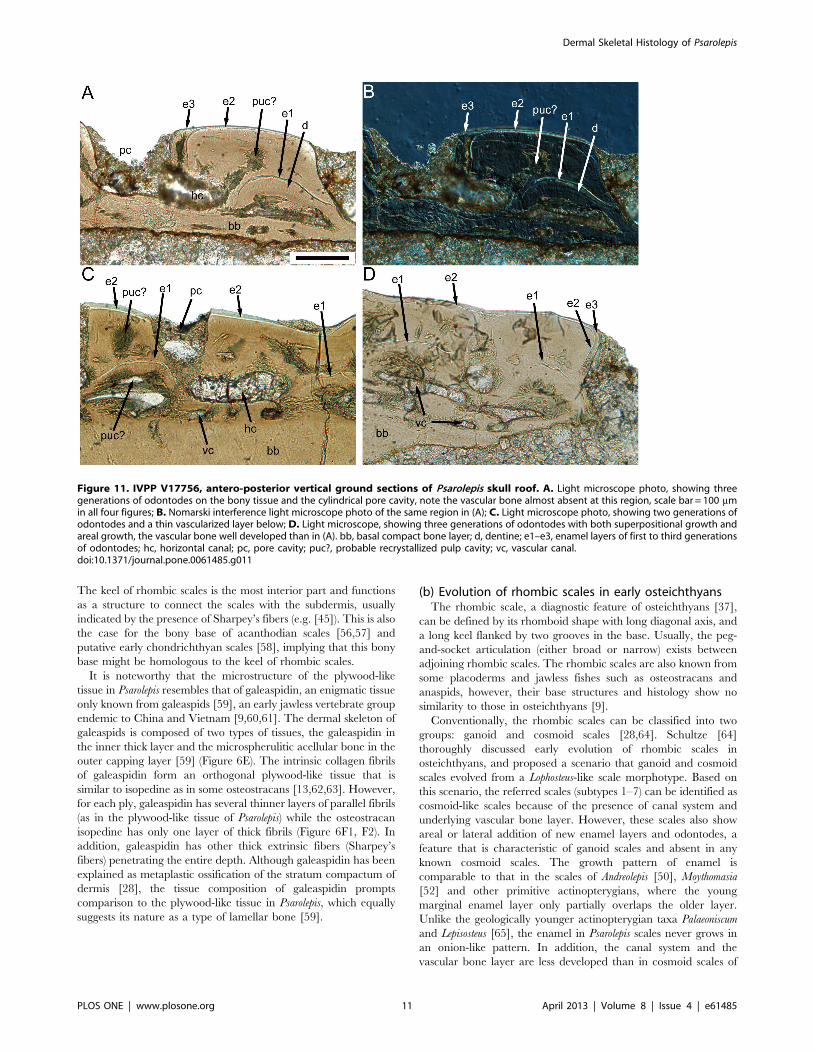
Figure 11. IVPP V17756, antero-posterior vertical ground sections of Psarolepis skull roof. A. Light microscope photo, showing threegenerations of odontodes on the bony tissue and the cylindrical pore cavity, note the vascular bone almost absent at this region, scale bar = 100 mmin all four figures; B. Nomarski interference light microscope photo of the same region in (A); C. Light microscope photo, showing two generations ofodontodes and a thin vascularized layer below; D. Light microscope, showing three generations of odontodes with both superpositional growth andareal growth, the vascular bone well developed than in (A). bb, basal compact bone layer; d, dentine; e1–e3, enamel layers of first to third generationsof odontodes; hc, horizontal canal; pc, pore cavity; puc?, probable recrystallized pulp cavity; vc, vascular canal.doi:10.1371/journal.pone.0061485.g011
Dermal Skeletal Histology of Psarolepis
PLOS ONE | www.plosone.org 11 April 2013 | Volume 8 | Issue 4 | e61485
more derived sarcopterygians (e.g. Porolepis and Osteolepis), where
one pore cavity sends out 3–5 basal branches to connect with
adjacent pore cavities, forming a regular grid-like horizontal canal
network [13]. By comparison, the horizontal canals in the referred
scales constitute an irregular web, dominated by antero-posteriorly
oriented canals (Figure 9). Under the prevailing phylogenetic
framework [7,11], the less regular horizontal canal system in
Psarolepis scales represents a plesiomorphic state of sarcopterygians.
The vascular bone layer, as in the dermal skull of Meemannia and
Psarolepis [20], is much thinner than in Porolepis and Osteolepis. To
sum up, the scales of Psarolepis combine the characters of ganoid
and cosmoid scales, and might provide a new model for discussing
the origin of ganoid and cosmoid scales.
It is noteworthy that the referred scales (subtypes 1–7) bear a
distinct neck separating the crown and base, and lack the
depressed field as seen in other rhombic scales (df, Figure 12A1,
B1, C1). Comparison with lateral-line scales of Moythomasia ([52]:
fig. 142) and Mimipiscis ([52]: fig. 141) indicates that the ventral
part of the anterior neck or the dorsal surface of the anterior ledge
corresponds topologically to the depressed field where the lateral-
line canal enters the scale anteriorly. A depressed field of the scale
is evident in Ligulalepis and Andreolepis (Figure 12A1, C1), although
the scale thickness in these two forms is comparable to that in
Psarolepis. Without a depressed field, the overlap pattern of
adjacent scale rows in Psarolepis might differ from that in other
early osteichthyans. If we follow the model in other osteichthyans
[9], we might consider that the scale in the front overlaps the scale
behind it on the dorsal surface of the anterior ledge, which is a
topological equivalent of the depressed field in other osteichthyans.
However, this overlap relationship is not functional as it will
impede the posterior growth of the crown of the scale in front.
Alternatively, we consider the scale in the front overlaps the scale
behind it along the anterior, downward-curving belt of the crown,
which functionally corresponds to the depressed field, but is not
part of the bony base as in other rhombic scales.
The posterior ledge in the referred scales corresponds to the
second keel (k9) identified by Schultze [49] on the scales of
Ligulalepis (Figure 12A). In the referred scales, only subtype 4
exhibits a prominent anterior ledge that is not sheltered by the
crown, thus forming a structure similar to the depressed field
(Figure 2D). The lack of the depressed field in other types may be
due to the growing odontode encircling the crown, thus making
the neck concaved and invisible in crown view. Given the fact that
the neck is widely present in non-osteichthyan jawed vertebrates
(e.g. placoderms and acanthodians; [39,66]), it is tempting to
explain the distinctive neck in the scales of Psarolepis, like the
dermal pelvic girdle in Psarolepis [32], as a retained primitive
gnathostome feature. However, the prevailing hypotheses of
relationships among early osteichthyans [11,37], which resolve
Psarolepis as a stem sarcopterygian, would favor the interpretation
that the scale neck in Psarolepis is an apomorphic reversal to the
plesiomorphic condition. This character discrepancy with the
prevalent phylogenies calls attention to the alternative scenario,
which places Psarolepis as a stem osteichthyan [3,10,32]. A more
comprehensive phylogenetic analysis incorporating the scale
characters revealed in this study is needed to test whether the
neck in the scales of Psarolepis is a primitive retention or an
apomorphic reversal.
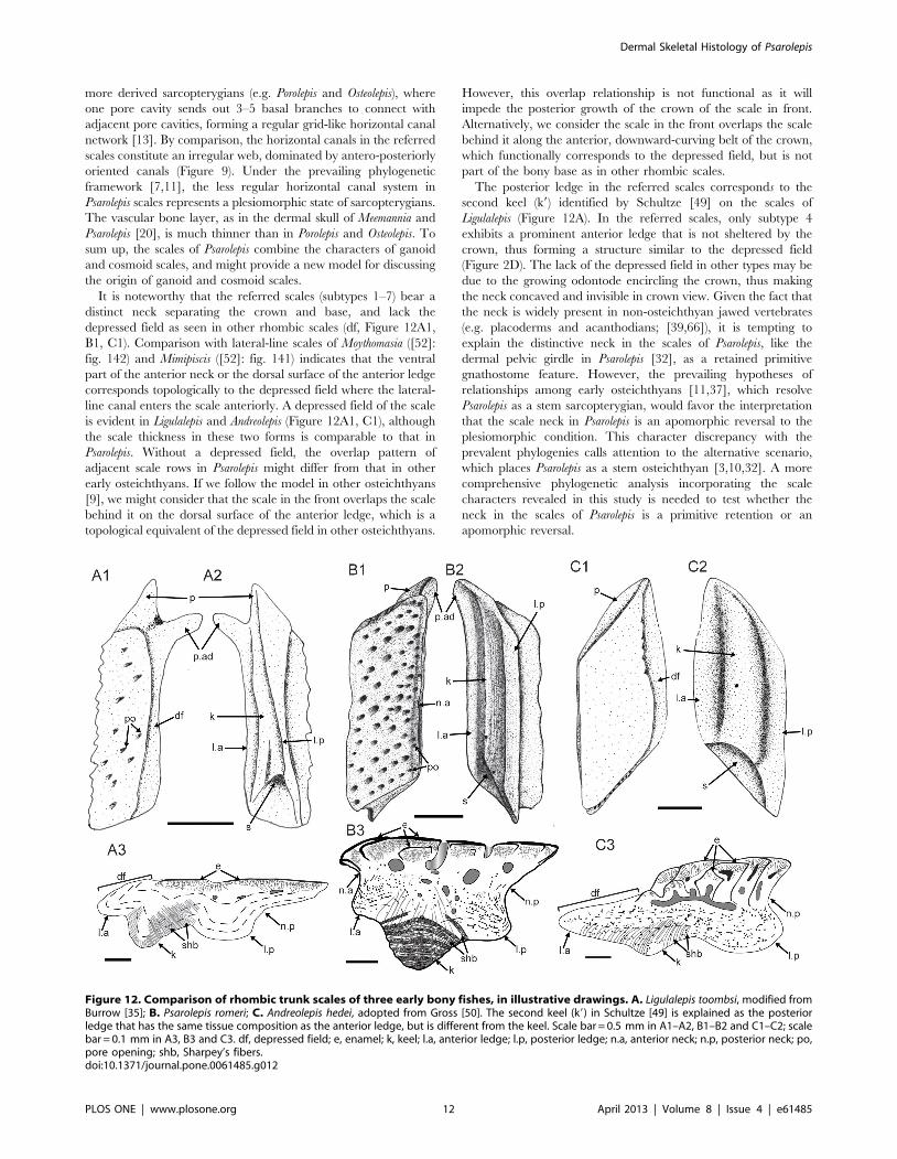
Figure 12. Comparison of rhombic trunk scales of three early bony fishes, in illustrative drawings. A. Ligulalepis toombsi, modified fromBurrow [35]; B. Psarolepis romeri; C. Andreolepis hedei, adopted from Gross [50]. The second keel (k9) in Schultze [49] is explained as the posteriorledge that has the same tissue composition as the anterior ledge, but is different from the keel. Scale bar = 0.5 mm in A1–A2, B1–B2 and C1–C2; scalebar = 0.1 mm in A3, B3 and C3. df, depressed field; e, enamel; k, keel; l.a, anterior ledge; l.p, posterior ledge; n.a, anterior neck; n.p, posterior neck; po,pore opening; shb, Sharpey’s fibers.doi:10.1371/journal.pone.0061485.g012
Dermal Skeletal Histology of Psarolepis
PLOS ONE | www.plosone.org 12 April 2013 | Volume 8 | Issue 4 | e61485
Acknowledgments
This paper is dedicated to Dr. Nianzhong Wang (1940–2010), a pioneer
researcher of early vertebrate microfossils in China. We are indebted to
X.B. Yu for commenting on the manuscript and improving it stylistically.
We thank P. Ahlberg, H. Blom, D. Chen, J. Mondejar-Fernandez, H.-P.
Schultze, Jean-Yves Sire and two anonymous reviewers for discussions and
helpful comments, W.D. Zhang, S.K. Zhang, G. Wife and B. Ryll for their
support in making ground sections, SEM photography and light
microscope photography. We thank W.J. Zhao for his help in the field
and acid preparation. Z.K. Gai kindly provided an unpublished ground
section of galeaspids for comparison.
Author Contributions
Conceived and designed the experiments: QQ MZ WW. Performed the
experiments: QQ MZ WW. Analyzed the data: QQ MZ WW.
Contributed reagents/materials/analysis tools: QQ MZ WW. Wrote the
paper: QQ MZ.
References
1. Zhu M, Schultze H-P (1997) The oldest sarcopterygian fish. Lethaia 30: 293–
304.
2. Yu XB (1998) A new porolepiform-like fish, Psarolepis romeri, gen. et sp. nov.(Sarcopterygii, Osteichthyes) from the Lower Devonian of Yunnan, China.
Journal of Vertebrate Paleontology 18: 261–274.
3. Zhu M, Yu XB, Janvier P (1999) A primitive fossil fish sheds light on the origin
of bony fishes. Nature 397: 607–610.
4. Tong-Dzuy T, Phuong TH, Boucot AJ, Goujet D, Janvier P (1997) Silurianvertebrates from Central Vietnam (Vertebres siluriens du Vietnam central).
Comptes Rendus de l’Academie des Sciences–Series IIA–Earth and PlanetaryScience 324: 1023–1030.
5. Ahlberg PE (1991) A re-examination of sarcopterygian interrelationships, with
special reference to the Porolepiformes. Zoological Journal of the LinneanSociety 103: 241–287.
6. Donoghue PCJ, Smith MP (2001) The anatomy of Turinia pagei (Powrie), and the
phylogenetic status of the Thelodonti. Transactions of the Royal Society ofEdinburgh: Earth Sciences 92: 15–37.
7. Brazeau MD (2009) The braincase and jaws of a Devonian ‘acanthodian’ andmodern gnathostome origins. Nature 457: 305–308.
8. Janvier P (2010) microRNAs revive old views about jawless vertebrate
divergence and evolution. Proceedings of the National Academy of Sciences107: 19137–19138.
9. Janvier P (1996) Early Vertebrates. Oxford: Clarendon Press. 393 p.
10. Zhu M, Schultze H-P (2001) Interrelationships of basal osteichthyans. In:Ahlberg P, editor. Major Events in Early Vertebrate Evolution: Palaeontology,
Phylogeny, Genetics and Development. London: Taylor & Francis. pp 289–314.
11. Zhu M, Zhao WJ, Jia LT, Lu J, Qiao T, et al. (2009) The oldest articulated
osteichthyan reveals mosaic gnathostome characters. Nature 458: 469–474.
12. Qiao T, Zhu M (2010) Cranial morphology of the Silurian sarcopterygian Guiyu
oneiros (Gnathostomata: Osteichthyes). Science China Earth Sciences 53: 1836–
1848.
13. Gross W (1956) Uber Crossopterygier und Dipnoer aus dem baltischenOberdevon im Zusammenhang einer vergleichenden Untersuchung des
Porenkanalsystems palaozoischer Agnathen und Fische. Kungliga SvenskaVetenskapsakademiens Handlingar 5: 1–140.
14. Thomson KS (1975) On the biology of cosmine. Bulletin of Peabody Museum of
Natural History, Yale University 40: 1–59.
15. Meinke DK (1984) A review of cosmine: its structure, development, and
relationship to other forms of the dermal skeleton in osteichthyans. Journal ofVertebrate Paleontology 4: 457–470.
16. Borgen UJ (1989) Cosmine resorption structures on three osteolepid jaws and
their biological significance. Lethaia 22: 413–424.
17. Wang NZ (1984) Thelodont, acanthodian and chondrichthyan fossils from the
Lower Devonian of Southwest China. Proceedings of the Linnean Society of
New South Wales 107: 419–441.
18. Zhu M, Yu XB (2002) A primitive fish close to the common ancestor of
tetrapods and lungfish. Nature 418: 767–770.
19. Lu J, Zhu M (2008) An Early Devonian (Pragian) sarcopterygian from
Zhaotong, Yunnan, China. Vertebrata PalAsiatica 46: 161–170.
20. Zhu M, Yu XB, Wang W, Zhao WJ, Jia LT (2006) A primitive fish provides keycharacters bearing on deep osteichthyan phylogeny. Nature 441: 77–80.
21. Zhu M, Wang W, Yu XB (2010) Meemannia eos, a basal sarcopterygian fish from
the Lower Devonian of China–expanded description and significance. In: ElliottDK, Maisey JG, Yu X, Miao D, editors. Morphology, Phylogeny and
Paleobiogeography of Fossil Fishes. Munchen: Verlag Dr. Friedrich Pfeil. pp199–214.
22. Wang NZ (1997) Restudy of thelodont microfossils from the lower part of the
Cuifengshan Group of Qujing, eastern Yunnan, China. Vertebrata PalAsiatica35: 1–17.
23. Chang MM, Yu XB (1981) A new crossopterygian, Youngolepis praecursor, gen. etsp. nov., from Lower Devonian of E. Yunnan, China. Scientia Sinica 24: 89–97.
24. Chang MM, Yu XB (1984) Structure and phylogenetic significance of
Diabolichthys speratus gen. et sp. nov., a new dipnoan-like form from the LowerDevonian of eastern Yunnan, China. Proceedings of the Linnean Society of New
South Wales 107: 171–184.
25. Zhu M, Yu XB, Ahlberg PE (2001) A primitive sarcopterygian fish with aneyestalk. Nature 410: 81–84.
26. Zhu M, Yu XB (2004) Lower jaw character transitions among majorsarcopterygian groups–a survey based on new materials from Yunnan, China.
In: Arratia G, Wilson MVH, Cloutier R, editors. Recent Advances in the Origin
and Early Radiation of Vertebrates. Munchen: Verlag Dr. Friedrich Pfeil. pp
271–286.
27. Chang MM, Smith MM (1992) Is Youngolepis a porolepiform? Journal of
Vertebrate Paleontology 12: 294–312.
28. Sire J-Y, Donoghue PCJ, Vickaryous MK (2009) Origin and evolution of the
integumentary skeleton in non-tetrapod vertebrates. Journal of Anatomy 214:
409–440.
29. Denison RH (1968) Early Devonian lungfishes from Wyoming, Utah, and
Idaho. Fieldiana Geology 17: 353–413.
30. Denison RH (1968) The evolutionary significance of the earliest known lungfishUranolophus. In: Ørvig T, editor. Current Problems of Lower Vertebrate
Phylogeney Nobel Symposium 4. Stockholm: Almqvist and Wiksell. pp 247–257.
31. Zhu M, Yu XB (2009) Stem sarcopterygians have primitive polybasal fin
articulation. Biology Letters 5: 372–375.
32. Zhu M, Yu XB, Choo B, Qu QM, Jia LT, et al. (2012) Fossil fishes from Chinaprovide first evidence of dermal pelvic girdles in osteichthyans. PLos ONE 7:
e35103.
33. Esin DN (1990) The scale cover of Amblypterina costata (Eichwald) and the
palaeoniscid taxonomy based on isolated scales. Paleontological Journal 2: 90–
98.
34. Trinajstic K (1999) Scale morphology of the Late Devonian palaeoniscoid
Moythomasia durgaringa Gardiner and Bartram, 1997. Alcheringa 23: 9–19.
35. Burrow C (1994) Form and function in scales of Ligulalepis toombsi Schultze, apalaeoniscoid from the Early Devonian of Australia. Records of the Australian
Museum 27: 175–185.
36. Choo B (2011) Revision of the actinopterygian genus Mimipiscis ( = Mimia) from
the Upper Devonian Gogo Formation of western Australia and the
interrelationships of the early Actinopterygii. Earth and Environmental ScienceTransactions of the Royal Society of Edinburgh 102: 1–28.
37. Friedman M, Brazeau MD (2010) A reappraisal of the origin and basal radiationof the Osteichthyes. Journal of Vertebrate Paleontology 30: 36–56.
38. Janvier P (1978) On the oldest known teleostome fish Andreolepis hedei Gross
(Ludlow of Gotland), and the systematic position of the lophosteids. Eesti NSVTeaduste Akadeemia Toimetised, Geoloogia 27: 88–95.
39. Denison RH (1979) Acanthodii; Schultze H-P, editor. Stuttgart: Gustav Fischer
Verlag. 62 p.
40. Francillon-Vieillot H, de Buffrenil V, Castanet J, Gerauldie J, Meunier FJ, et al.
(1990) Microstructure and mineralization of vertebrate skeletal tissues. In: CarterJG, editor. Skeletal biomineralization: patterns, processes and evolutionary
trends. New York: Van Nostrand Reinhold. pp 471–530.
41. Smith MM (1989) Distribution and variation in enamel structure in the oralteeth of sarcopterygians: its significance for the evolution of a protoprismatic
enamel. Historical Biology 3: 97–126.
42. Ørvig T (1967) Phylogeny of tooth tissues: evolution of some calcified tissues inearly vertebrates. In: Miles A, editor. Structural and chemical organization of
teeth. New York: Academic Press. pp 45–110.
43. Mondejar-Fernandez J, Clement G (2012) Squamation and scale microstructure
evolution in the Porolepiformes (Sarcopterygii, Dipnomorpha) based on Heimenia
ensis from the Devonian of Spitsbergen. Journal of Vertebrate Paleontology 32:267–284.
44. Ørvig T (1969) Cosmine and cosmine growth. Lethaia 2: 241–260.
45. Kerr T (1952) The scales of primitive living actinopterygians. Proceedings of the
Zoological Society, London 122: 55–78.
46. Meunier FJ, Gerauldie J (1980) Les structures en contre-plaque du derme et desecailles des vertebres inferieurs. L’Annee biologique 19: 1–18.
47. Meunier FJ (1984) Spatial organization and mineralization of the basal plate of
elasmoid scales in osteichthyans. American Zoologist 24: 953–964.
48. Meunier FJ (2011) The Osteichtyes, from the Paleozoic to the extant time,
through histology and palaeohistology of bony tissues. Comptes Rendus Palevol10: 347–355.
49. Schultze H-P (1968) Palaeoniscoidea-schuppen aus dem Unterdevon Australiens
und Kansas und aus dem Mitteldevon Spitzbergens. Bulletin of the BritishMuseum (Natural History), Geology 16: 343–368.
50. Gross W (1968) Fragliche Actinopterygier-schuppen aus dem Silur Gotlands.
Lethaia 1: 184–218.
51. Jessen HL (1968) Moythomasia nitida Gross und M. cf. striata Gross, Devonische
Palaeonisciden aus dem oberen Plattenkalk der Bergisch-Gladbach-PaffratherMudle (Rheinisches Schiefergebirge). Palaeontographica Abt A 128: 87–114.
Dermal Skeletal Histology of Psarolepis
PLOS ONE | www.plosone.org 13 April 2013 | Volume 8 | Issue 4 | e61485
52. Gardiner BG (1984) The relationships of the palaeoniscid fishes, a review based
on new specimens of Mimia and Moythomasia from the Upper Devonian ofWestern Australia. Bulletin of the British Museum (Natural History), Geology
37: 173–428.
53. Burrow CJ (1995) A new palaeoniscoid from the Lower Devonian trundle bedsof Australia. Geobios M S 19: 319–325.
54. Meunier FJ (1980) Recherches histologiques sur le squelette dermique desPolypteridae. Archives de Zoologie Experimentale et Generale 121: 279–295.
55. Daget J, Gayet M, Meunier FJ, Sire J-Y (2001) Major discoveries on the dermal
skeleton of fossil and recent polypteriforms: a review. Fish and Fisheries 2001:113–124.
56. Gross W (1947) Die Agnathen und Acanthodier des ObersilurischenBeyrichienkalks. Palaeontographica Abt A 96: 91–158.
57. Gross W (1971) Downtonische und dittonische Acanthodier-Reste desOstseegebietes (Downtonian and Dittonian acanthodian remains from the
Baltic Sea area). Palaeontographica Abt A 136: 1–82.
58. Ørvig T (1966) Histologic studies of ostracoderms, placoderms and fossilelasmobranchs. 2: On the dermal skeleton of two late Palaeozoic elasmobranchs.
Arkiv for Zoologi 19: 1–39.59. Wang N, Donoghue PCJ, Smith MM, Sansom IJ (2005) Histology of the
galeaspid dermoskeleton and endoskeleton, and the origin and early evolution of
the vertebrate cranial endoskeleton. Journal of Vertebrate Paleontology 25: 745–
756.
60. Zhu M, Gai ZK (2007) Phylogenetic relationships of galeaspids (Agnatha).
Frontiers of Biology in China 2: 1–19.
61. Gai ZK, Donoghue PCJ, Zhu M, Janvier P, Stampanoni M (2011) Fossil jawless
fish from China foreshadows early jawed vertebrate anatomy. Nature 476: 324–
327.
62. Pander CH (1856) Monographie der fossilen Fische des silurischen Systems der
russischbaltischen Gouvernements. St Petersburg: Akademie der Wissenschaf-
ten. 91 p.
63. Gross W (1968) Beobachtungen mit dem Elektronenraster-Auflichtmikroskop an
den Siebplatten und dem Isopedin von Dartmuthia (Osteostraci). Palaontologische
Zeitschrift 42: 73–82.
64. Schultze H-P (1977) Ausgangsform und Entwicklung der rhombischen
Schuppen der Osteichthyes (Pisces). Palaontologische Zeitschrift 51: 152–168.
65. Richter M, Smith MM (1995) A microstructural study of the ganoine tissue of
selected lower vertebrates. Zoological Journal of the Linnean Society 114: 173–
212.
66. Burrow CJ, Turner S (1999) A review of placoderm scales, and their significance
in placoderm phylogeny. Journal of Vertebrate Paleontology 19: 204–219.
Dermal Skeletal Histology of Psarolepis
PLOS ONE | www.plosone.org 14 April 2013 | Volume 8 | Issue 4 | e61485