Embed Size (px)
Citation preview

7/29/2019 Screening CA Servix
http://slidepdf.com/reader/full/screening-ca-servix 1/11
Screening and cervical cancer cure: population basedcohort study
OPEN ACCESS
Bengt Andrae senior consultant 1 2, Therese M-L Andersson doctoral student
2, Paul C Lambertreader
2 3, Levent Kemetli statistician 4, Lena Silfverdal senior consultant
5, Björn Strander senior
consultant 6, Walter Ryd associate professor
7, Joakim Dillner professor 2 8, Sven Törnberg associate
professor 4, Pär Sparén professor
2
1Centre for Research and Development, Uppsala University/County Council of Gävleborg S-80188 Gävle, Sweden; 2Department of Medical
Epidemiology and Biostatistics, Karolinska Institutet, Stockholm, Sweden; 3Department of Health Sciences, Centre for Biostatistics and Genetic
Epidemiology, University of Leicester, Leicester, UK; 4Department of Cancer Screening, Karolinska University Hospital, Stockholm, Sweden;5Department of Obstetrics and Gynaecology, Umeå University Hospital, Umeå, Sweden; 6Department of Obstetrics and Gynaecology, Institute of
Clinical Sciences, Sahlgrenska Academy, Gothenburg University, Sweden; 7Department of Pathology and Clinical Cytology, Sahlgrenska University
Hospital, Gothenburg, Sweden; 8Department of Laboratory Medicine, Karolinska Institutet, Stockholm, Sweden
Abstract
Objective To determine whether detection of invasive cervical cancer
by screening results in better prognosis or merely increases the lead
time until death.
Design Nationwide population based cohort study.
Setting Sweden.
Participants All 1230 women with cervical cancer diagnosed during
1999-2001 in Sweden prospectively followed up for an average of 8.5
years.
Main outcome measures Cureproportionsand five year relative survival
ratios, stratified by screening history, mode of detection, age,
histopathological type, and FIGO (International Federation of Gynecology
and Obstetrics) stage.
Results In the screening ages, the cure proportion for women with
screen detected invasive cancerwas 92%(95% confidence interval 75%
to 98%) and for symptomatic women was 66% (62% to 70%), a
statistically significant difference in cure of 26% (16% to 36%). Among
symptomatic women, the cure proportion was significantly higher for
those who had been screened according to recommendations (interval
cancers) than among those overdue for screening: difference in cure
14% (95% confidence interval 6% to 23%). Cure proportions were similar
for all histopathological types except small cell carcinomas and were
closely related to FIGO stage. A significantly higher cure proportion for
screen detectedcancers remainedafter adjustment for stage at diagnosis
(difference 15%, 7% to 22%).
Conclusions Screening is associated with improved cure of cervical
cancer. Confounding cannot be ruled out, but the effect was not
attributable to lead time bias and was larger than what is reflected by
down-staging. Evaluations of screening programmes should consider
the assessment of cure proportions.
Introduction
The rationale of cervical screening is to reduce the incidence
of cancer by the detection and treatment of precursors. 1 2 A
secondary aim is the early detection of invasive disease, which
might improve the prognosis thereby also reducing mortality
from the disease. Prognosis may depend on age, FIGO
(International Federation of Gynecology and Obstetrics) stage,
histopathological type, screening history, and mode of detection.3
Thus cancers may be detected on thebasis of either an abnormal
screening test result or symptoms, and the women may also
have been screened previously according to recommendations
or not. The Swedish cervical screening programme carried out
a nationwide audit of the screening history of all cases in the
country and found that in addition to preventing cervical cancer,
regular screening also detected invasive cervical cancers at
earlier stages. In the nationwide Swedish audit1 around 50% of
women who were not screened according to recommendations
were detected at FIGO stage II or higher, whereas among women
participating in the screening programme most were at stages
IA or IB (30% and 52%, respectively). For screen detected
cancers the drift towards detection at early stages was even more
apparent (47% of cancers were detected at stage IA and 46% at
stage IB).
However, the early detection of asymptomatic cancers isintuitively but not necessarily beneficial,4-6 as lead time and
length biases can distort the apparent benefit of screening
Correspondence to: B Andrae [email protected]
No commercial reuse: See rights and reprints http://www.bmj.com/permissions Subscribe: http://www.bmj.com/subscribe
BMJ 2012;344:e900 doi: 10.1136/bmj.e900 (Published 1 March 2012) Page 1 of 11
Research
RESEARCH
7/29/2019 Screening CA Servix
http://slidepdf.com/reader/full/screening-ca-servix 2/11
programmes.7 8 Randomised controlled trials are not feasible
for the evaluation of established cervical screening programmes,
which is why the only alternative is well designed observational
studies.9 In an extension of the nationwide Swedish audit, we
investigated if, and to what extent, participation in cervical
screening according to guidelines and/or screen detection of invasive cancer is reflected in improved cure of disease by
utilising recently developed statistical methods for estimating
the cured proportion of women with cervical cancer. 10 11
Methods
The organised Swedish cervical screening programme issues
invitations three years after the latest smear test for women aged
23-50 and every five years for women aged 51-60. Invitations
to screening are issued by regional offices to all women in the
population register who have not been screened according to
recommendations. 12-14 The cytology databases used to assess
screening history include all smear tests taken, not just the
organised ones. The design of the nationwide audit of cervicalscreening in Sweden has been described previously.1 In the
present prospective cohort study we linked all women with
cervical cancer in Sweden diagnosed during 1999-2001 to the
national Swedish causes of death register, with follow-up to 31
December 2006. Death from cervical cancer or unspecified
uterine cancer was considered cause specific mortality.15 To
ascertain the date of death from any cause until 31 December
2008, we linked all cases to the Swedish population register.
We analysed separately those women with cancer diagnosed at
screening ages 23-65 (including one woman with a diagnosis
at age 21) and those with a diagnosis more than five years
beyond the last invitation to screening (≥66 years). Screen
detected cancers were defined as those in women with anabnormal smeartest result recorded 1-6 months before diagnosis.
We classified the remaining women as symptomatic.1 Smear
tests taken less than one month before diagnosis were not
considered as they might be part of the diagnostic process of a
symptomatic invasive cancer (fig 1⇓).
We divided the women with symptomatic cancer into
symptomatic interval cases if the cancer was diagnosed more
than six months after the smear test but within the recommended
screening interval of 3.5 years in women under the age of 54,
or within 5.5 years before diagnosis in women over that age.
Symptomatic overdue, or not screened, comprised womenwhose
screening test was more than half a year overdue according to
screening guidelines, and included women without any previous
smear test. We divided the women with screen detected cancer,
having a smear test done 1-6 months before diagnosis, into
women who also had a smear test taken within the preceding
recommended interval (screen detected interval cancers) and
those whose screening was overdue or who had no recorded
smear test (screen detected, overdue or not screened).
FIGO stage is considered a good predictor of survival.16-18 The
classification used in this study—IA, microinvasive; IB,
localised; and II or higher, advanced—reflect distinct levels of
treatment, consequences for fertility, complications, and costs.
Statistical analysis
We calculated relative survival ratios as the overall (all cause)
survival in the cohort over the expected survival in the general
female population, comparable with the women diagnosed as
having cancer according to age and calendar year. Relative
survival estimates mortality, associated with a diagnosis of a
particular disease, without the need for information on cause of
death.19
Statistical cure is defined as the point where the relative survival
curve reaches a plateau, and this occurs when the women who
are still alive no longer experience any excess mortality
compared with the general female population. These women
are considered statistically cured as they experience the same
mortality as women of the same age without cancer. The levelat which the relative survival curve reaches a plateau is named
the cure proportion.10 The concept of “statistical cure” applies
at a grouped level and is distinct from “medical cure” at an
individual level, as it is difficult to determinewith anycertainty
that someone has been medically cured.
The statistical model for cure used in this paper—the mixture
cure model10—assumes that a proportion of patients will be
cured (experience the same mortality as the general population)
whereas the remaining (1−proportion) of patients will continue
to experience excess mortality compared with the general
population. The group who continue to experience excess
mortality are considered to be uncured or those bound to die of
the disease under study. For the models in this article we assume
that the survival times of those who are uncured have a Weibull
distribution. We used a logit link to model the cure proportion,
which gives parameter estimates that can be interpreted as odds
ratios of cure, with values greater than 1 indicating a higher
odds of cure and values less than 1 indicating a lower odds of
cure. In addition we calculated the difference, with 95%
confidence intervals, in the cure proportions.
For all cure models we applied either included terms for mode
of detection (screen detected versus symptomatic) or attendance
at screening (screened within recommended interval versus
screening overdue or no smear test). We then fitted separate
models, with the following covariates also included: FIGO
stages (IA, IB, II, or III or higher) and histopathology (squamous
cell versus adenocarcinoma). From the model we excluded thosewomen with adenosquamous, small cell, neuroendocrine, or
undifferentiated carcinomas owing to small numbers. We
restricted the analysis to the younger age group when adding
stage and histopathology, because the models did not give a
good fit for the older age group—that is, the relative survival
curves did not appear to reach a plateau. To formally assess
whether a difference existed in cure proportion between women
with screen detected cancer and those with symptomatic cancer,
we estimated odds ratios of cure with 95% confidence intervals,
where the women with screen detected cancer served as the
reference group, with an odds ratio of 1. We then introduced
FIGO stage into the model to test the hypothesis that clinical
stage at detection explains any difference in cure proportionbetween women with screen detected cancer and those with
symptomatic cancers. For univariate models we report the
difference in the cure proportion with standard errors, calculated
using the delta method. For the models incorporating FIGO
stage, we computed stage standardised differences in the cure
proportion by assuming that the stage distribution in the each
of the two groups was the same as that of the whole study
population. We used the same approach to estimate the
difference in cure proportions between women screened within
the recommended interval compared with thosewhose screening
was overdue or who had no smear test, and the hypothesis that
clinical stage at detection explains any difference in cure.
When presenting estimates of the cure proportion, we categorise
FIGO stage in three levels (IA, IB, and II or higher), because
the estimates for further subdivisions into stages II and III or
higher were unreliable, and consequently did not add any further
information.
Estimation of the model variables was obtained with maximum
likelihood using individual level data.11 20 We modelled both
No commercial reuse: See rights and reprints http://www.bmj.com/permissions Subscribe: http://www.bmj.com/subscribe
BMJ 2012;344:e900 doi: 10.1136/bmj.e900 (Published 1 March 2012) Page 2 of 11
RESEARCH

7/29/2019 Screening CA Servix
http://slidepdf.com/reader/full/screening-ca-servix 3/11
Weibull parameters (λ and γ). (See Lambert et al 11 for further
discussion of the interpretation of estimates from cure models.)
The strsmix command in Stata was used to fit models. 21
Results
At least seven years of potential follow-up were available from
diagnosis of cervical cancer for all the women. Five years after
diagnosis, 440 out of the 1230 women had died. Among them
373 had a recorded death from cervical cancer. Thirty one
women died from other cancers, and 36 died of diagnoses not
related to cancer.
The five year relative survival for women at screening ages with
screen detected cancers was 95% (95% confidence interval 92%
to 97%), whereas for women with symptomatic cancers it was
69% (65% to 73%; table 1⇓). The corresponding cure
proportions were 92% (75% to 98%) and 66% (62% to 70%),
with an estimated difference in cure of 26% (15% to 36%).
However, the cure proportion for women with symptomatic
cancers presenting within the recommended screening interval
was 74% (68% to 79%), whereas for symptomatic cancers in
women overdue for screening it was 60% (53% to 66%), with
a difference in cure of 14% (6% to 23%). Differences in cure
proportions between different FIGO stages were large both for
women with screen detected cancers and for women with
symptomatic cancers (table 1). In FIGO stage IA the difference
between screen detected and symptomatic cancers was 4% (3%
to 7%), whereas in stages IB and II or higher the difference
increased to 16% (8% to 23%) and 29% (13% to 45%),
respectively (table 1). For screen detected cancers evidence of
any substantial difference in cure was lacking between squamous
cell carcinoma and adenocarcinoma (cure proportions 93%) and
this was also the case for symptomatic cancers (cure proportions67% and 66%, respectively; table 1). For both of the major
histological subtypes, however, differences in cure were apparent
between women with screen detected cancers and women with
symptomatic cancers (table 1). Deaths related to
adenosquamous, small cell, neuroendocrine, or undifferentiated
carcinomas were too few to reliably calculate cure rates, and
no deaths occurred among women with these histological
subtypes for screen detected cancers.
Table 2⇓ presents odds ratios and differences of cure by mode
of detection and screening history. For odds ratios, the women
with interval cancers served as the reference group, with an
odds ratio of 1. The difference in cure between the women with
symptomatic cancers and those with screen detected cancerswas26% (95% confidence interval 15%to 36%; table 2).When
FIGO stage was introduced into the model, the difference in
cure between the groups decreased to 15% (7% to 22%).
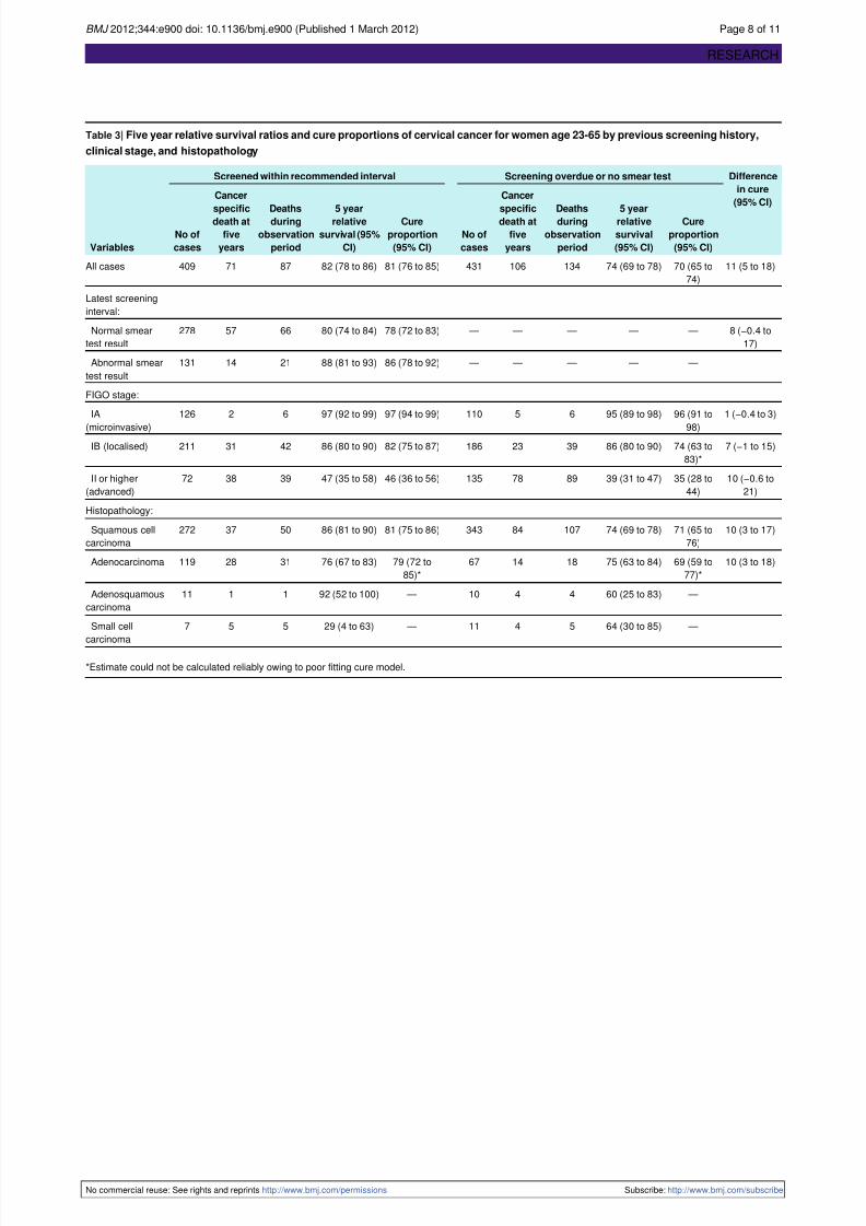
The cure proportion for women who had a smear test within the
recommended screening interval at ages 23 to 65 was 11% (95%
confidence interval 5% to 18%) higher than for women who
were overdue or who had never had a smear test (table 3⇓).
Women with a normal smear test result had a non-significantly
lower cure proportion (difference 8%, 95% confidence interval
−0.4% to 17%) than women with an abnormal smear test result.
The patterns of lower cure proportions with increasing FIGO
stage was seen for both interval and overdue cases, although
the 95% confidence intervals for the stage specific differences
all included zero (table 3). The evidence for an essential
difference in relative survival or cure proportion between
squamous cell carcinomas and adenocarcinoma was insufficient
for interval as well as for overdue cases. Deaths in women with
the other histological subtypes were too few to make any
meaningful comparisons.
As previously done for screen detected and symptomatic
cancers, odds ratios and differences of cure were calculated for
interval and overdue cases. Compared with the interval cases,
the difference in cure for the overdue cases was 11% (95%
confidence interval 5% to 18%; table 2). In a model also
including FIGO stage, the difference in cure for overduecompared with interval cases was statistically non-significant
(5%, −2% to 11%).
A similar model was constructed for histopathological subtypes,
where no difference in cure could be discerned between women
with adenocarcinomas and squamous cell carcinomas (0.1%,
−8% to 8%). When adjusting for FIGO stage (IA, IB, II, and
III or higher), this result did not change notably (difference in
cure 2%, −5% to 9%).
Since age is known to be an important modifier of cervical
cancer risk and survival, all analyses were repeated, adjusting
for linear effect of age. Differences were negligible (data not
shown).
Table 4⇓ displays five year relative survival ratios, cureproportions, and differences in cure for women over the age of
organised screening (≥66 years). In this age group the cure
proportion of women with screen detected cancers was higher
than for those with symptomatic cancers (difference in cure
36%, 95% confidence interval 11% to 80%), whereas this was
not the case for interval cases compared with overdue cases
(14%, −7% to 35%).
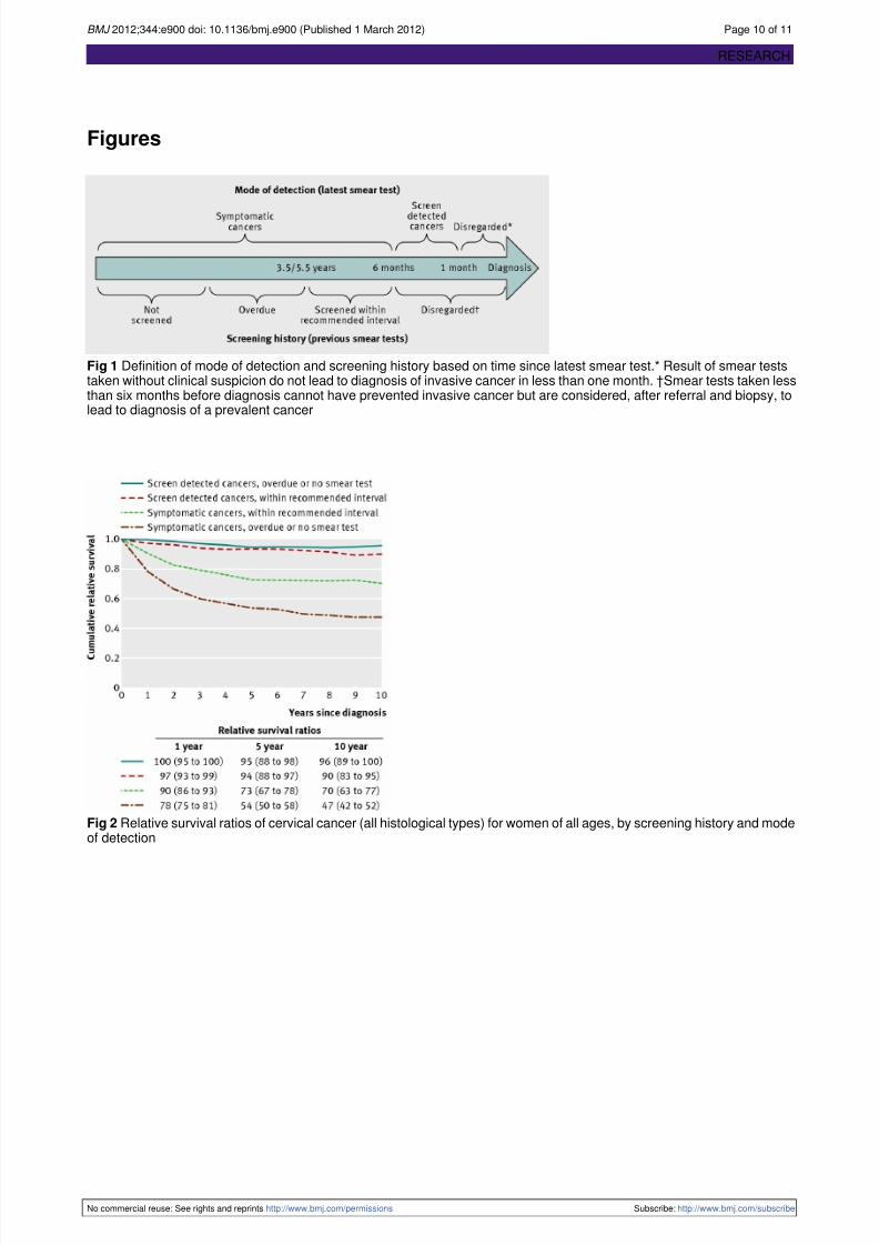
Figures 2-4⇓⇓⇓ present the relative survival curves, by screening
history and mode of detection, histological type, and FIGO
stage. Women with screen detected cancer had excellent relative
survival independent of previous screening history. Women
with symptomatic interval cancer had a better relative survival
at all times during follow-up than women who weresymptomatic with an overdue or absent screening test (fig 2).
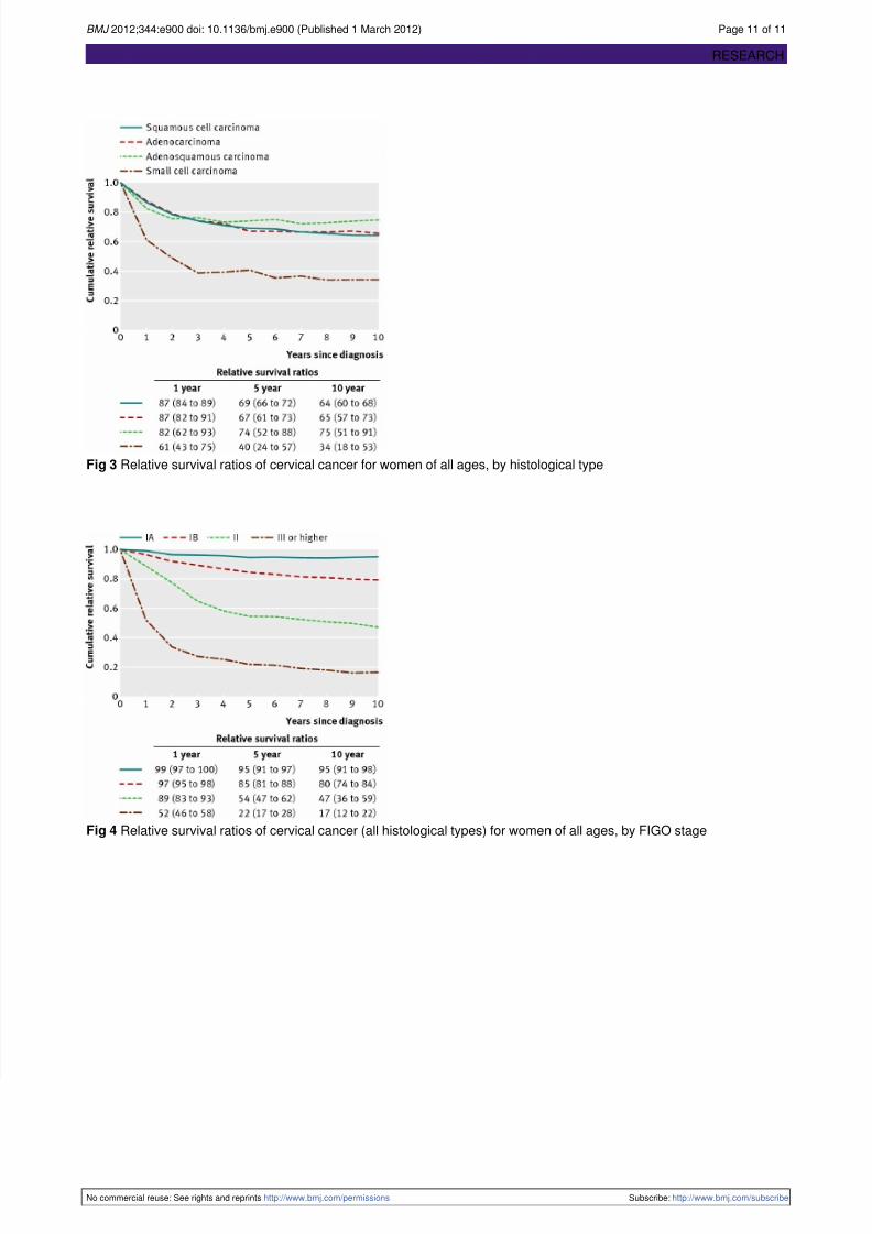
Relative survival did not differ between histological types,
except for small cell neuroendocrine and undifferentiated cancers
(fig 3). FIGO stage was a good predictor of prognosis (fig 4).
Discussion
Women with cervical cancer diagnosed as a result of a smear
test (screen detected cancers) have a better prognosis than
women whose cancer is detected on the basis of symptoms, and
this improvement was not attributable to lead time bias. To a
large extent theimprovedcure wasattributedto screendetected
cancers being generally found at earlier clinical FIGO stages
than symptomatic cancers.
Women with symptomatic cancer who present within the
recommended screening interval—that is, symptomatic interval
cancers—have better chances of cure than women with
symptomatic cancer with an overdue or absent smear test result.
If the screening effect on cure proportions had been due to
selective detection of harmless cancers, the more aggressive
cases would have appeared as symptomatic interval cancers
with higher mortality. On the contrary, we found that women
with symptomatic cancers have a better prognosis when cervical
cancer is diagnosed between screening intervals than when
diagnosed in women who are overdue for screening or not
screened.
If cancers were screen detected, relative survival and cureproportion was high irrespective of whether women had
previously participated in screening, suggesting that
determinants of screening attendance have not confounded the
effect.
No commercial reuse: See rights and reprints http://www.bmj.com/permissions Subscribe: http://www.bmj.com/subscribe
BMJ 2012;344:e900 doi: 10.1136/bmj.e900 (Published 1 March 2012) Page 3 of 11
RESEARCH
7/29/2019 Screening CA Servix
http://slidepdf.com/reader/full/screening-ca-servix 4/11
Feeling healthy22 and a lack of confidence in the benefits of
screening23 24 are barriers to screening attendance for some
women, but our data imply that all women (regardless of
previous participation) can be advised that screening will
increase the likelihood of cure in case an invasive cancer is
detected.More than three quarters of women who died from cervical
cancer had no smear test taken within the recommended
screening intervals, implying that the recommended intervals
are adequate and that further reduction of incidence and
mortality in cervical cancer should focus on reaching women
who have not attended screening.
Down-staging
In women with screen detected cancer down-staging explained
a large portion of the improved cure but not the whole
difference, compared with women who had symptomatic cancer.
A plausible reason could be that our categorisation of FIGO
clinical staging of the advanced cancers was too crude toeffectively capture the whole effect of early detection by
screening. Down-staging does explain the difference in cure
between women with interval cancers compared with those who
are overdue for screening or have not been screened.
Age
Half of the women who died from cervical cancer within five
years of diagnosis were over the recommended age for
screening. As few older women who continued screening died
from cervical cancer, half of the cervical cancer mortality was
in women with cancers diagnosed beyond screening ages and
who had not had a screening test for more than five years. Lethal
cervical cancers in young women before screening ages arerare.25 No such cases occurred in Sweden during the three years
studied. The mortality in patients younger than 30 years was so
low we were not able to study the cure proportion separately.
Histopathological type
Our results showed that the cure proportion for women with
adenocarcinomas was similar to that of women with squamous
carcinoma, also when controlling for FIGO stage, and that the
relative survival of women with adenosquamous carcinoma did
not differ from these subtypes. Only women with the poorly
differentiated, small cell or neuroendocrine carcinomas had a
worse relative survival than women with the other
histopathological types.
Abnormal smears
Women with interval cancers and an abnormal smear test result
during the past six years had a non-significantly higher cure
proportion than women with only a normal smear test result. If
this difference is real, the reason might be that women with
abnormal smear test results are followed up to a greater extent
than women with normal smear test results.26
Other studies
Survival studies have been carried out in other organised
programmes but most have either not distinguished between
microinvasive and higher stages3 or not considered the influenceof opportunistic smear tests in the same population.27-29 A recent
Italian study30 included opportunistic screening. However, no
previous study has considered the possible influence of lead
time bias or the use of cure proportions to avoid this bias.
Strengths and limitations of the study
Our audit was designed to eliminate several biases such as those
related to selection, testing, and recall1 31 through prospective
follow up of all the cases in the nationwide Swedish audit,
utilising screening histories from comprehensive cytology
registers.1 32
We have shown that cervical screening not only reduces the
risk for invasive cervical cancer1 but is also associated with
improved relative survival and cure. When evaluating screening
for cancer it has to be excluded if apparent improvements are
due to lead time bias, length bias, overdiagnosis, or
confounding.8 29 A possible explanation for a seemingly better
relative survival is lead time bias—that is, when detection at an
early stage adds time to follow-up but does not alter the course
of the disease and does not prolong life. This can be a major
confounder in the evaluation of cancer screening.8 The cure
proportion is a measure independent of lead time bias11 and we
found significantly higher cure proportions for women with
screen detected cancers than for those with symptomatic cancersas well as higher cure proportions for women with symptomatic
interval cancers compared with symptomatic overdue cancers.
The possibility of length time bias also has to be considered. If
screening picks up small indolent carcinomas but misses a
significant proportion of the rapidly growing aggressive tumours,
the aggressive tumours would appear in women as symptomatic
interval cancers with higher mortality.28 29 33 34 However, the
relative survival for women with symptomatic interval cancers
was better at all times during follow-up than for women with
symptomatic cancers and an overdue or absent screening test,
and they also had a significantly higher cure proportion.
One possible explanation for the better cure of women with
screen detected cancers compared with symptomatic cancers isconfounding by “healthy volunteer bias”—that people who
participate in screening generally could be more healthy than
non-participants. Since most of the female population participate
according to recommendations,35 non-participants could be
considered as “unhealthy abstainers” who theoretically could
have a poor prognosis, irrespective of screening. Studies have
shown socioeconomic, cultural, and educational differences
between participants and non-participants to cervical screening,
but less so in countries with nationwide population based
programmes.36-38 In our study the women with cancers detected
by screening had an equally excellent relative survival
irrespective of whether or not they had a previous screening test
taken within the recommended time, suggesting that
determinants of screening attendance were not confounders of
the effect. In addition, all Swedish residents are covered by a
common health insurance and have access to the same cancer
treatment centres. Therefore, a healthy volunteer bias or health
selection does not seem to explain our findings, although an
effect of confounding cannot be ruled out.
Our cohort comprised all women with cervical cancer in Sweden
during three years, classified by age, clinically relevant FIGO
stage, and histopathological type, factors individually related
to screening history and to the mode of detection based on
nationwide databases. All the women in this audit have been
followed prospectively in population based registries and all
smear tests, also outside the screening programme, are included
in the database. To our knowledge the present study is the firstto estimate cure proportion after cervical cancer.
One limitation of cure models is that they estimate a cure
proportion even when statistical cure is not reached. We have
graphically assessed the appropriateness of assuming statistical
cure and compared estimated survival from cure models with
No commercial reuse: See rights and reprints http://www.bmj.com/permissions Subscribe: http://www.bmj.com/subscribe
BMJ 2012;344:e900 doi: 10.1136/bmj.e900 (Published 1 March 2012) Page 4 of 11
RESEARCH
7/29/2019 Screening CA Servix
http://slidepdf.com/reader/full/screening-ca-servix 5/11
standard relative survival estimates. In the cases where cure was
questionable we have chosen not to present results from the
cure models. In addition we investigated relative survival by
age group using Finnish and Swedish cancer registry data to
assess whether a plateau in the relative survival function was
observed. If excess mortality occurred after 10 years then themodels would be estimating something close to 10 year relative
survival rather than the cure proportion. The definition of screen
detected and symptomatic cancer is based on interpretation of
timing of smear tests in relation to diagnosis of cancer and it is
possible that a small number of women with symptomatic cancer
and slow investigations could have been classified as having
screen detected cancers.
Conclusions
Detection of invasive cancer by cervical screening implies a
favourable prognosis compared with cancer being detected on
the basis of symptoms. The effect was stronger than what is
reflected in down-staging and was not attributable to lead timebias. Also, women with symptomatic interval cancers had a
better prognosis than women with symptomatic cancers who
did not have a smear test within the recommended screening
interval. The effect on cervical cancer cure should be included
when evaluating cervical screening programmes.
Contributors: BA, PS, ST, JD, and BS conceived the audit. BA, PS,
TM-LA, PCL, and JD conceived this analysis. BA, LS, LK, ST, and PS
collected and standardised the audit database. WR reviewed the
diagnostic histopathology specimens of all cancer cases. TM-LA, PCL,
and PS did the statistical analyses and drafted the statistical methods
section. BA drafted the manuscript with PS and JD. All authors actively
participated in the discussion and approved the final version. PS is
guarantor.
Funding: This work was supported by grants from the Swedish Cancer
Society (02-6988 and 2010/900), the Swedish Foundation for Strategic
Research (KF 10-0046), Gävle Cancer Fund (2009-09-17), and the
Centre for Research and Development, Uppsala University/County
Council of Gävleborg, Sweden (CFUG-82261). The corresponding
author had full access to all the data in the study and had the full
responsibility to submit the report for publication. The study was
conducted and analysed independently from its funders.
Competing interests: All authors have completed the ICMJE uniform
disclosure form at www.icmje.org/coi_disclosure.pdf(available on
request from the corresponding author) and declare: no support from
any organisation for the submitted work; no financial relationships with
any organisations that might have an interest in the submitted work inthe previous three years; and no other relationships or activities that
could appear to have influenced the submitted work.
Ethical approval: This study was approved by the ethical review boards
in Uppsala, Stockholm, Umeå, Gothenburg, and Lund and the joint
board of Örebro and Linköping, Sweden (FEK Ups 01-322 and EPN
2008/185). The ethical review boards determined that informed consent
from participating women was not required.
Data sharing: The analysis dataset for this study is available from the
corresponding author at [email protected].
1 Andrae B, Kemetli L, Sparen P, Silfverdal L, Strander B, Ryd W, et al.Screening-preventablecervicalcancerrisks:evidence froma nationwide audit in Sweden.J Natl Cancer Inst 2008;100:622-9.
2 Sasieni P,CastanonA, CuzickJ. Effectiveness ofcervicalscreeningwithage: populationbased case-control study of prospectively recorded data. BMJ 2009;339:b2968.
3 Van der Aa MA, Schutter EM, Looijen-Salamon M, Martens JE, Siesling S. Differencesin screening history, tumour characteristics and survival between women withscreen-detectedversusnot screen-detected cervical cancer in theeast of theNetherlands,1992-2001. Eur J Obstet Gynecol Reprod Biol 2008;139:204-9.
4 Djulbegovic M, Beyth RJ, Neuberger MM, Stoffs TL, Vieweg J, Djulbegovic B, et al.Screening for prostate cancer: systematic review and meta-analysis of randomisedcontrolled trials. BMJ 2010;341:c4543.
5 SandblomG, Varenhorst E, Rosell J, Lofman O, CarlssonP. Randomised prostatecancerscreening trial: 20 year follow-up. BMJ 2011;342:d1539.
6 Buys SS, Partridge E, Black A, Johnson CC, Lamerato L, Isaacs C, et al. Effect ofscreening on ovariancancermortality:the Prostate, Lung, Colorectal andOvarian(PLCO)Cancer Screening Randomized Controlled Trial. JAMA 2011;305:2295-303.
7 Grimes DA, Schulz KF. Uses and abuses of screening tests. Lancet 2002;359:881-4.8 Kramer BS, Croswell JM. Cancer screening: the clash of science and intuition. Annu Rev
Med 2009;60:125-37.9 Arbyn M, Rebolj M, De Kok IM, Fender M, Becker N, O’Reilly M, et al. The challenges of
organising cervical screening programmes in the 15 old member states of the EuropeanUnion. Eur J Cancer 2009;45:2671-8.
10 De Angelis R, Capocaccia R, Hakulinen T, Soderman B, Verdecchia A. Mixture modelsfor cancer survival analysis: application to population-based data with covariates. Stat
Med 1999;18:441-54.11 Lambert PC, Thompson JR, Weston CL, Dickman PW. Estimating and modeling the cure
fraction in population-based cancer survival analysis. Biostatistics 2007;8:576-94.12 Socialstyrelsen. Gynekologisk cellprovskontroll. [Cervicalcancer screening.]SoS-rapport
1998:15. National Board of Health and Welfare, 1998.13 Attförebygga cervixcancersamt vaginal-och vulvacancer.[Preventionof cervical, vaginal
and vulvar cancer—guidelines.] ARG rapport Vol 34. Swedish Society for Obstetriciansand Gynaecologists, 1997.
14 Cervix cancer prevention. [Cervical cancer prevention, guidelines for the management ofCIN.] ARG-rapport Vol 63. Swedish Society of Obstetricians and Gynaecologists, 2010.
15 Arbyn M, Raifu AO, Weiderpass E, Bray F, Anttila A. Trends of cervical cancer mortality
in the member states of the European Union. Eur J Cancer 2009;45:2640-8.16 Kosary C. FIGO stage, histology, histologic grade, age and race as prognostic factors indetermining survival of cancers. Semin Surg Oncol 2006;10:31-46.
17 Odicino F, Pecorelli S, Zigliani L, Creasman WT. History of the FIGO cancer stagingsystem. Int J Gynaecol Obstet 2008;101:205-10.
18 SparenP, GustafssonL, Friberg LG,PontenJ, BergstromR, AdamiHO. Improvedcontrolof invasive cervical cancer in Sweden over six decades by earlier clinical detection andbetter treatment. J Clin Oncol 1995;13:715-25.
19 Dickman PW, Adami HO. Interpreting trends in cancer patient survival. J Intern Med
2006;260:103-17.20 Lambert PC, Dickman PW, Osterlund P, Andersson T, Sankila R, Glimelius B. Temporal
trends in the proportion cured for cancer of the colon and rectum: a population-basedstudy using data from the Finnish Cancer Registry. Int J Cancer 2007;121:2052-9.
21 Lambert PC. Modeling of the cure fraction in survival studies. Stata J 2007;7:351-75.22 OscarssonMG, BenzeinEG, WijmaBE. Reasonsfor non-attendanceat cervical screening
as reported by non-attendees in Sweden. J Psychosom Obstet Gynaecol 2008;29:23-31.23 Blomberg K, Ternestedt BM, Tornberg S, Tishelman C. How do women who choose not
to participate in population-based cervical cancer screening reason about their decision?Psychooncology 2008;17:561-9.
24 Waller J, Bartoszek M, Marlow L, Wardle J. Barriers to cervical cancer screening
attendance in England: a population-based survey. J Med Screen 2009;16:199-204.25 Edelstein ZR, Madeleine MM, Hughes JP, Johnson LG, Schwartz SM, Galloway DA, et
al. Age of diagnosis of squamous cell cervical carcinoma and early sexual experience.Cancer Epidemiol Biomarkers Prev 2009;18:1070-6.
26 Silfverdal L, Kemetli L, Andrae B, Sparen P, Ryd W, Dillner J, et al. Risk of invasivecervical cancer in relation to management of abnormal Pap smear results. Am J Obstet
Gynecol 2009;201:188,e1-7.27 NieminenP, Kallio M,HakamaM. Theeffectof mass screening onincidenceandmortality
of squamous and adenocarcinoma of cervix uteri. Obstet Gynecol 1995;85:1017-21.28 Shingleton HM, Bell MC, Fremgen A, Chmiel JS, Russell AH, Jones WB, et al. Is there
really a difference in survival of women with squamous cell carcinoma, adenocarcinoma,and adenosquamous cell carcinoma of the cervix? Cancer 1995;76(Suppl 10):1948-55.
29 Welch HG, Black WC. Overdiagnosis in cancer. J Natl Cancer Inst 102:605-13.30 Zucchetto A, Franceschi S, Clagnan E, Serraino D, Zanier L, Franzo A. Screening history
of women with invasive cervical cancer in north-east Italy. Eur J Obstet Gynecol Reprod
Biol 2010;152:200-4.31 Andrae B, Strander B, Silfverdal L, Ryd W, Dillner J, Tornberg S, et al. Benefit of cervical
cancer screening inyoungwomen—amatterof adherence to therecommended screeninginterval. Response to Sasieni et al. BMJ 2009;339:b2968.
32 Cuzick J. Routine audit of large-scale cervical cancer screening programs. J Natl Cancer Inst 2008;100:605-6.
33 Hildesheim A, Hadjimichael O, Schwartz PE, Wheeler CM, Barnes W, Lowell DM, et al.Risk factors for rapid-onset cervical cancer. Am J Obstet Gynecol 1999;180:571-7.
34 KitchenerHC.Survivalfromcancer ofthe uterine cervix inEnglandandWales upto 2001.Br J Cancer 2008;99(Suppl 1):S63-4.
35 Swedish cervical cancer screening register. Report on data to 2006. [In Swedish.] 2007.http://ki.se/content/1/c6/05/05/04/Rapport_2007.pdf.
36 Rodvall Y,KemetliL, Tishelman C, TornbergS. Factorsrelatedto participation ina cervicalcancer screening programme in urban Sweden. Eur J Cancer Prev 2005;14:459-66.
37 Azerkan F, Sparen P, Sandin S, Tillgren P, Faxelid E, Zendehdel K. Cervical screeningparticipation andrisk among Swedish-bornand immigrant women in Sweden. IntJ Cancer
2012;130:927-47.38 Palencia L, Espelt A, Rodriguez-Sanz M, Puigpinos R, Pons-Vigues M, Pasarin MI, et al.
Socio-economic inequalities in breast and cervical cancer screening practices in Europe:influence of the type of screening program. Int J Epidemiol 39:757-65.
Accepted: 8 December 2011
Cite this as: BMJ 2012;344:e900This is an open-access article distributed under the terms of the Creative Commons
Attribution Non-commercial License, which permits use, distribution, and reproduction in
any medium, provided the original work is properly cited, the use is non commercial and
isotherwisein compliance with thelicense.See:http://creativecommons.org/licenses/by-
nc/2.0/ and http://creativecommons.org/licenses/by-nc/2.0/legalcode.
No commercial reuse: See rights and reprints http://www.bmj.com/permissions Subscribe: http://www.bmj.com/subscribe
BMJ 2012;344:e900 doi: 10.1136/bmj.e900 (Published 1 March 2012) Page 5 of 11
RESEARCH

7/29/2019 Screening CA Servix
http://slidepdf.com/reader/full/screening-ca-servix 6/11
What is already known on this topic
Women with cervical cancers detected by screening have an improved survival
However, evaluations of the benefit of screening programmes can be distorted by lead time bias
What this study adds
Cervical cancer screening is associated with improved cure
The effect is not attributable to lead time bias and is larger than what is reflected in earlier stage diagnosis (down-staging)
Evaluations of screening programmes should consider the assessment of cure proportions
Tables
Table 1| Five year relative survival ratios and cure proportions of cervical cancer for women age 23-65, by mode of detection, clinical stage,
and histopathology
Difference
in cure
(95% CI)
Symptomatic cancersScreen detected cancers
Variables
Cure
proportion
(95% CI)
5 yearrelative
survival
(95% CI)
Deathsduring
observation
period
Cancer
specificdeath at
five
years
No of
cases
Cure
proportion
(95% CI)
5 yearrelative
survival
(95% CI)
Deathsduring
observation
period
Cancer
specificdeath at
five
years
No of
cases
26 (15 to36)
66(62to 70)69 (65 to 73)19916656792 (75 to98)
95 (92 to 97)2211273All cases
Latest screeninginterval:
14 (6 to23)†
74(68to 79)75 (69 to 80)7265256 —*95 (90 to 98)156153Interval cases
60(53to 66)65 (60 to 70)12710131195 (87 to98)
96 (90 to 98)75120Overdue cases
FIGO stage:
4 (3 to 7)94(88to 97)94 (86 to 97)759998 (96 to
99)
98 (93 to
100)
52137IA
(microinvasive)
16 (8 to 23)75(68to 81)81 (76 to 85)715128091 (83 to95)
97 (92 to100)
103117IB (localised)
29 (13 to45)
37(30to 44)40 (32 to 47)12111018865 (48 to80)
64 (38 to 81)7619II or higher(advanced)
Histopathology:
26 (20 to33)
67(61to 72)71 (66 to 75)14311541293 (86 to96)
97 (93 to 99)146203Squamous cellcarcinoma
27 (18 to35)
66(57to 74)68 (59 to 76)413712193 (86 to96)
90 (79 to 96)8565Adenocarcinoma
— —73 (46 to 88)5518 —>100003Adenosquamouscarcinoma
— —44 (20 to 66)10916 —>100002Small cell
carcinoma
*Estimate could not be calculated reliably owing to poor fitting cure model.
†Interval and overdue symptomatic cancer cases compared.
No commercial reuse: See rights and reprints http://www.bmj.com/permissions Subscribe: http://www.bmj.com/subscribe
BMJ 2012;344:e900 doi: 10.1136/bmj.e900 (Published 1 March 2012) Page 6 of 11
RESEARCH

7/29/2019 Screening CA Servix
http://slidepdf.com/reader/full/screening-ca-servix 7/11
Table 2| Odds ratios and differences in cure from cervical cancer on women aged 23-65, by mode of detection and previous screening
history
Difference in cure (95% CI)Odds ratio (95% CI)
Variables Adjusted for FIGO stage*UnadjustedAdjusted for FIGO stageUnadjusted
Mode of detection:15 (7 to 22)26 (15 to 36)1.001.00Screen detected (reference)
0.31 (0.16 to 0.62)0.17 (0.05 to 0.65)Symptomatic
Previous screening history:
5 (−2 to 11)11 (5 to 18)1·001·00Screened (reference)
0.72 (0.46 to 1.12)0.55 (0.38 to 0.79)Screening overdue or no smear
*Stage standardised difference.
No commercial reuse: See rights and reprints http://www.bmj.com/permissions Subscribe: http://www.bmj.com/subscribe
BMJ 2012;344:e900 doi: 10.1136/bmj.e900 (Published 1 March 2012) Page 7 of 11
RESEARCH
7/29/2019 Screening CA Servix
http://slidepdf.com/reader/full/screening-ca-servix 8/11
Table 3| Five year relative survival ratios and cure proportions of cervical cancer for women age 23-65 by previous screening history,
clinical stage, and histopathology
Difference
in cure
(95% CI)
Screening overdue or no smear testScreened within recommended interval
Variables
Cure
proportion
(95% CI)
5 year
relative
survival
(95% CI)
Deaths
during
observation
period
Cancer
specific
death at
five
years
No of
cases
Cure
proportion
(95% CI)
5 year
relative
survival (95%
CI)
Deaths
during
observation
period
Cancer
specific
death at
five
years
No of
cases
11 (5 to 18)70 (65 to74)
74 (69 to 78)13410643181 (76 to 85)82 (78 to 86)8771409All cases
Latest screeninginterval:
8 (−0.4 to17)
— — — — —78 (72 to 83)80 (74 to 84)6657278Normal smeartest result
— — — — —86 (78 to 92)88 (81 to 93)2114131Abnormal smeartest result
FIGO stage:
1 (−0.4 to 3)96 (91 to
98)
95 (89 to 98)6511097 (94 to 99)97 (92 to 99)62126IA
(microinvasive)7 (−1 to 15)74 (63 to
83)*86 (80 to 90)392318682 (75 to 87)86 (80 to 90)4231211IB (localised)
10 (−0.6 to21)
35 (28 to44)
39 (31 to 47)897813546 (36 to 56)47 (35 to 58)393872II or higher(advanced)
Histopathology:
10 (3 to 17)71 (65 to76)
74 (69 to 78)1078434381 (75 to 86)86 (81 to 90)5037272Squamous cellcarcinoma
10 (3 to 18)69 (59 to77)*
75 (63 to 84)18146779 (72 to85)*
76 (67 to 83)3128119Adenocarcinoma
—60 (25 to 83)4410 —92 (52 to 100)1111Adenosquamouscarcinoma
—64 (30 to 85)5411 —29 (4 to 63)557Small cell
carcinoma
*Estimate could not be calculated reliably owing to poor fitting cure model.
No commercial reuse: See rights and reprints http://www.bmj.com/permissions Subscribe: http://www.bmj.com/subscribe
BMJ 2012;344:e900 doi: 10.1136/bmj.e900 (Published 1 March 2012) Page 8 of 11
RESEARCH

7/29/2019 Screening CA Servix
http://slidepdf.com/reader/full/screening-ca-servix 9/11
Table 4| Five year relative survival ratios and cure proportions of cervical cancer for women age 66 or older by mode of detection and
previous screening history
Difference in cure
(95% CI)
Cure proportion (95%
CI)
5 year relative survival
(95% CI)
Deaths during
observation period
Cancer specific
death at five yearsNo of casesVariables
Mode of detection:
36 (11 to 80)76 (46 to 92)77 (48 to 97)11523Screen detected cancers
40 (33 to 47)44 (38 to 50)278191367Symptomatic cancers
Screening history:
14 (−7 to 35)54 (35 to 72)56 (35 to 74)191232Screened within recommendedinterval
40 (32 to 48)45 (38 to 51)270184358Screening overdue or nosmear test
No commercial reuse: See rights and reprints http://www.bmj.com/permissions Subscribe: http://www.bmj.com/subscribe
BMJ 2012;344:e900 doi: 10.1136/bmj.e900 (Published 1 March 2012) Page 9 of 11
RESEARCH
7/29/2019 Screening CA Servix
http://slidepdf.com/reader/full/screening-ca-servix 10/11
Figures
Fig 1 Definition of mode of detection and screening history based on time since latest smear test.* Result of smear teststaken without clinical suspicion do not lead to diagnosis of invasive cancer in less than one month. †Smear tests taken lessthan six months before diagnosis cannot have prevented invasive cancer but are considered, after referral and biopsy, tolead to diagnosis of a prevalent cancer
Fig 2 Relative survival ratios of cervical cancer (all histological types) for women of all ages, by screening history and modeof detection
No commercial reuse: See rights and reprints http://www.bmj.com/permissions Subscribe: http://www.bmj.com/subscribe
BMJ 2012;344:e900 doi: 10.1136/bmj.e900 (Published 1 March 2012) Page 10 of 11
RESEARCH
7/29/2019 Screening CA Servix
http://slidepdf.com/reader/full/screening-ca-servix 11/11
Fig 3 Relative survival ratios of cervical cancer for women of all ages, by histological type
Fig 4 Relative survival ratios of cervical cancer (all histological types) for women of all ages, by FIGO stage
BMJ 2012;344:e900 doi: 10.1136/bmj.e900 (Published 1 March 2012) Page 11 of 11
RESEARCH