Embed Size (px)
Citation preview
Research Article
Serine catabolism is essential to maintain mitochondrialrespiration in mammalian cellsStephanie Lucas1, Guohua Chen1, Siddhesh Aras2, Jian Wang1,3
Breakdown of serine by the enzyme serine hydroxymethyl-transferase (SHMT) produces glycine and one-carbon (1C) units.These serine catabolites provide important metabolic intermedi-ates for the synthesis of nucleotides, as well as methyl groups forbiosynthetic and regulatory methylation reactions. Recently, it hasbeen shown that serine catabolism is required for efficient cellularrespiration. Using CRISPR-Cas9 gene editing, we demonstrate thatthe mitochondrial SHMT enzyme, SHMT2, is essential to maintaincellular respiration, the main process through which mammaliancells acquire energy. We show that SHMT2 is required for the as-sembly of Complex I of the respiratory chain. Furthermore, sup-plementation of formate, a bona fide 1C donor, restores Complex Iassembly in the absence of SHMT2. Thus, provision of 1C units bymitochondrial serine catabolism is critical for cellular respiration,at least in part by influencing the assembly of the respiratoryapparatus.
DOI 10.26508/lsa.201800036 | Received 22 February 2018 | Revised 7 May2018 | Accepted 8 May 2018 | Published online 21 May 2018
Introduction
Apart from its role in protein synthesis, serine is a major metabolicsource for generating one-carbon (1C) units in mammalian cells (deKoning et al, 2003). Two serine hydroxymethyltransferase (SHMT)enzymes, SHMT1 and 2, break down serine into glycine andmethylene-tetrahydrofolate (THF) in the cytosol and mitochondria,respectively (Stover & Schirch, 1990; Stover et al, 1997). The latterserine catabolite feeds into cellular 1C pool, and either directlyparticipates in thymidine synthesis or indirectly in purine or me-thionine synthesis after its oxidative or reductive conversion toformyl- or methyl-THF (Tibbetts & Appling, 2010). Because the 1C-derived products are key anabolic building blocks, sustaining the 1Cpool is vital for cellular proliferation and is required for a number ofphysiological and pathophysiological processes ranging from stemcell renewal to cancer progression (Wang et al, 2009; Locasale,2013). Consistent with their critical roles in supporting cell pro-liferation, SHMTs are highly active in many rapidly growing cancer
cells and are important molecular targets for cancer intervention(Snell et al, 1988; Nikiforov et al, 2002; Ducker et al, 2017).
Interestingly, 1C metabolism also functionally interacts withmitochondrial oxidative phosphorylation (OXPHOS) system, themain process through which mammalian cells generate ATP. TheOXPHOS system comprises an electron transport chain of fourrespiratory enzyme complexes (Complex I–IV) that use nutrient-derived redox potentials to drive Complex V (CV), the ATP synthase(Alberts et al, 2002). The protein components of the OXPHOS systemare encoded by both nuclear and mitochondrial genes (Ott et al,2016). It was recently shown that electron transport chain dys-function owing to mitochondrial DNA (mtDNA) depletion dramat-ically alters the expression of SHMT2 as well as the production of 1Cunits from serine catabolism (Bao et al, 2016; Nikkanen et al, 2016).In addition, system-wide metabolic modeling indicates that oxi-dation of the serine-derived 1C units provides a significant fractionof the redox potential to drive ATP synthesis via OXPHOS (Vazquezet al, 2011; Tedeschi et al, 2013). These observations strongly suggestthat the 1C metabolic cycle and the OXPHOS system are functionallycoupled. Recent studies further demonstrated that serine catab-olism by SHMT2 is required tomaintainmitochondrial respiration inhuman cell lines (Minton et al, 2018; Morscher et al, 2018) andmouse tissues (Tani et al, 2018). Interestingly, these works revealeddistinct mechanisms underlying a crucial role of SHMT2 in sus-taining mitochondrial translation in different cell types (Mintonet al, 2018; Morscher et al, 2018), indicating that complex mecha-nisms exist, linking serine catabolism to the modulation of theOXPHOS system.
In the present study, we independently investigated the meta-bolic adaptions in response to targeted deletion of SHMT enzymesin mammalian cells. Consistent with the previous reports (Mintonet al, 2018; Morscher et al, 2018), we found that the cells lackingSHMT2, but not SHMT1, preferentially metabolized glucose to lactateand were unable to survive in the presence of galactose media,suggestive of mitochondrial dysfunction in the absence of SHMT2.Mechanistically, we found that SHMT2 is dispensable for mtDNAmaintenance and OXPHOS gene expression. However, our resultsstrongly suggest that SHMT2 plays a critical role in supporting theassembly of Complex I by supplying the 1C intermediate derived
1Department of Pathology, Wayne State University, Detroit, MI, USA 2Center for Molecular Medicine and Genetics, Wayne State University, Detroit, MI, USA3Cardiovascular Research Institute, Wayne State University, Detroit, MI, USA
Correspondence: [email protected]
© 2018 Lucus et al. https://doi.org/10.26508/lsa.201800036 vol 1 | no 2 | e201800036 1 of 10
on 12 August, 2019life-science-alliance.org Downloaded from http://doi.org/10.26508/lsa.201800036Published Online: 21 May, 2018 | Supp Info:
from serine catabolism. Together, our findings revealed a novel reg-ulatory link between SHMT2-mediated 1C metabolism and the main-tenance of the mitochondrial respiratory chain in mammalian cells.
Results
Loss of SHMT2 stimulates aerobic glycolysis
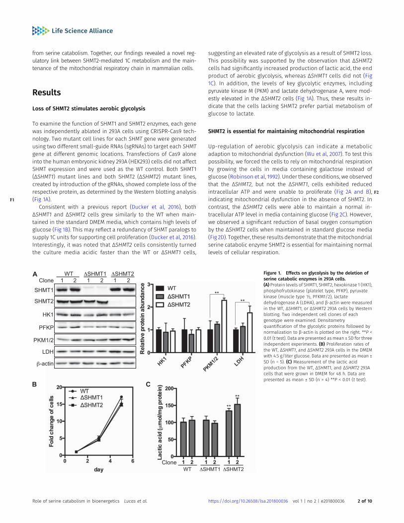
To examine the function of SHMT1 and SHMT2 enzymes, each genewas independently ablated in 293A cells using CRISPR-Cas9 tech-nology. Two mutant cell lines for each SHMT gene were generatedusing two different small-guide RNAs (sgRNAs) to target each SHMTgene at different genomic locations. Transfections of Cas9 aloneinto the human embryonic kidney 293A (HEK293) cells did not affectSHMT expression and were used as the WT control. Both SHMT1(ΔSHMT1) mutant lines and both SHMT2 (ΔSHMT2) mutant lines,created by introduction of the gRNAs, showed complete loss of therespective protein, as determined by the Western blotting analysis(F1 Fig 1A).
Consistent with a previous report (Ducker et al, 2016), bothΔSHMT1 and ΔSHMT2 cells grew similarly to the WT when main-tained in the standard DMEM media, which contains high levels ofglucose (Fig 1B). This may reflect a redundancy of SHMT paralogs tosupply 1C units for supporting cell proliferation (Ducker et al, 2016).Interestingly, it was noted that ΔSHMT2 cells consistently turnedthe culture media acidic faster than the WT or ΔSHMT1 cells,
suggesting an elevated rate of glycolysis as a result of SHMT2 loss.This possibility was supported by the observation that ΔSHMT2cells had significantly increased production of lactic acid, the endproduct of aerobic glycolysis, whereas ΔSHMT1 cells did not (Fig1C). In addition, the levels of key glycolytic enzymes, includingpyruvate kinase M (PKM) and lactate dehydrogenase A, were mod-estly elevated in the ΔSHMT2 cells (Fig 1A). Thus, these results in-dicate that the cells lacking SHMT2 prefer partial metabolism ofglucose to lactate.
SHMT2 is essential for maintaining mitochondrial respiration
Up-regulation of aerobic glycolysis can indicate a metabolicadaption to mitochondrial dysfunction (Wu et al, 2007). To test thispossibility, we forced the cells to rely on mitochondrial respirationby growing the cells in media containing galactose instead ofglucose (Robinson et al, 1992). Under these conditions, we observedthat the ΔSHMT2, but not the ΔSHMT1, cells exhibited reducedintracellular ATP and were unable to proliferate ( F2Fig 2A and B),indicating mitochondrial dysfunction in the absence of SHMT2. Incontrast, the ΔSHMT2 cells were able to maintain a normal in-tracellular ATP level in media containing glucose (Fig 2C). However,we observed a significant reduction of basal oxygen consumptionby the ΔSHMT2 cells when maintained in standard glucose media(Fig 2D). Together, these results demonstrate that themitochondrialserine catabolic enzyme SHMT2 is essential for maintaining normallevels of cellular respiration.
Figure 1. Effects on glycolysis by the deletion ofserine catabolic enzymes in 293A cells.(A) Protein levels of SHMT1, SHMT2, hexokinase 1 (HK1),phosphofrutokinase (platelet type, PFKP), pyruvatekinase (muscle type ½, PFKM1/2), lactatedehydrogenase A (LDHA), and β-actin were measuredin the WT, ΔSHMT1, or ΔSHMT2 293A cells by Westernblotting. Two independent cell clones of eachgenotype were examined. Densitometryquantification of the glycolytic proteins followed bynormalization to β-actin is plotted on the right. **P <0.01 (t test). Data are presented as mean ± SD for threeindependent experiments. (B) Proliferation rates ofthe WT, ΔSHMT1, and ΔSHMT2 293A cells in the DMEMwith 4.5 g/liter glucose. Data are presented as mean ±SD (n = 5). (C) Measurement of the lactic acidproduction from the WT, ΔSHMT1, and ΔSHMT2 293Acells that were grown in DMEM for 48 h. Data arepresented as mean ± SD (n = 4) **P < 0.01 (t test).
Role of serine catabolism in bioenergetics Lucas et al. https://doi.org/10.26508/lsa.201800036 vol 1 | no 2 | e201800036 2 of 10
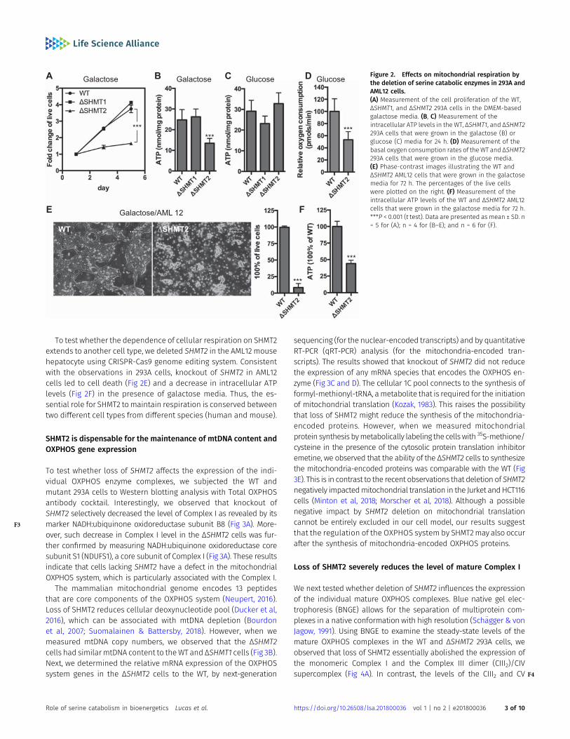
To test whether the dependence of cellular respiration on SHMT2extends to another cell type, we deleted SHMT2 in the AML12 mousehepatocyte using CRISPR-Cas9 genome editing system. Consistentwith the observations in 293A cells, knockout of SHMT2 in AML12cells led to cell death (Fig 2E) and a decrease in intracellular ATPlevels (Fig 2F) in the presence of galactose media. Thus, the es-sential role for SHMT2 tomaintain respiration is conserved betweentwo different cell types from different species (human and mouse).
SHMT2 is dispensable for the maintenance of mtDNA content andOXPHOS gene expression
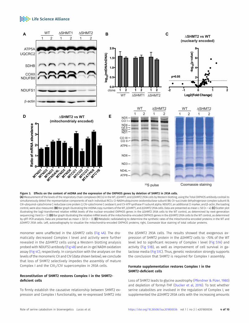
To test whether loss of SHMT2 affects the expression of the indi-vidual OXPHOS enzyme complexes, we subjected the WT andmutant 293A cells to Western blotting analysis with Total OXPHOSantibody cocktail. Interestingly, we observed that knockout ofSHMT2 selectively decreased the level of Complex I as revealed by itsmarker NADH:ubiquinone oxidoreductase subunit B8 (F3 Fig 3A). More-over, such decrease in Complex I level in the ΔSHMT2 cells was fur-ther confirmed by measuring NADH:ubiquinone oxidoreductase coresubunit S1 (NDUFS1), a core subunit of Complex I (Fig 3A). These resultsindicate that cells lacking SHMT2 have a defect in the mitochondrialOXPHOS system, which is particularly associated with the Complex I.
The mammalian mitochondrial genome encodes 13 peptidesthat are core components of the OXPHOS system (Neupert, 2016).Loss of SHMT2 reduces cellular deoxynucleotide pool (Ducker et al,2016), which can be associated with mtDNA depletion (Bourdonet al, 2007; Suomalainen & Battersby, 2018). However, when wemeasured mtDNA copy numbers, we observed that the ΔSHMT2cells had similar mtDNA content to theWT and ΔSHMT1 cells (Fig 3B).Next, we determined the relative mRNA expression of the OXPHOSsystem genes in the ΔSHMT2 cells to the WT, by next-generation
sequencing (for the nuclear-encoded transcripts) and by quantitativeRT-PCR (qRT-PCR) analysis (for the mitochondria-encoded tran-scripts). The results showed that knockout of SHMT2 did not reducethe expression of any mRNA species that encodes the OXPHOS en-zyme (Fig 3C and D). The cellular 1C pool connects to the synthesis offormyl-methionyl-tRNA, ametabolite that is required for the initiationof mitochondrial translation (Kozak, 1983). This raises the possibilitythat loss of SHMT2 might reduce the synthesis of the mitochondria-encoded proteins. However, when we measured mitochondrialprotein synthesis bymetabolically labeling the cellswith 35S-methione/cysteine in the presence of the cytosolic protein translation inhibitoremetine, we observed that the ability of the ΔSHMT2 cells to synthesizethe mitochondria-encoded proteins was comparable with the WT (Fig3E). This is in contrast to the recent observations that deletion of SHMT2negatively impactedmitochondrial translation in the Jurket andHCT116cells (Minton et al, 2018; Morscher et al, 2018). Although a possiblenegative impact by SHMT2 deletion on mitochondrial translationcannot be entirely excluded in our cell model, our results suggestthat the regulation of the OXPHOS system by SHMT2may also occurafter the synthesis of mitochondria-encoded OXPHOS proteins.
Loss of SHMT2 severely reduces the level of mature Complex I
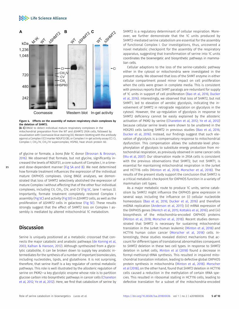
We next tested whether deletion of SHMT2 influences the expressionof the individual mature OXPHOS complexes. Blue native gel elec-trophoresis (BNGE) allows for the separation of multiprotein com-plexes in a native conformation with high resolution (Schagger & vonJagow, 1991). Using BNGE to examine the steady-state levels of themature OXPHOS complexes in the WT and ΔSHMT2 293A cells, weobserved that loss of SHMT2 essentially abolished the expression ofthe monomeric Complex I and the Complex III dimer (CIII2)/CIVsupercomplex ( F4Fig 4A). In contrast, the levels of the CIII2 and CV
Figure 2. Effects on mitochondrial respiration bythe deletion of serine catabolic enzymes in 293A andAML12 cells.(A) Measurement of the cell proliferation of the WT,ΔSHMT1, and ΔSHMT2 293A cells in the DMEM-basedgalactose media. (B, C) Measurement of theintracellular ATP levels in the WT, ΔSHMT1, and ΔSHMT2293A cells that were grown in the galactose (B) orglucose (C) media for 24 h. (D) Measurement of thebasal oxygen consumption rates of theWT and ΔSHMT2293A cells that were grown in the glucose media.(E) Phase-contrast images illustrating the WT andΔSHMT2 AML12 cells that were grown in the galactosemedia for 72 h. The percentages of the live cellswere plotted on the right. (F) Measurement of theintracellular ATP levels of the WT and ΔSHMT2 AML12cells that were grown in the galactose media for 72 h.***P < 0.001 (t test). Data are presented as mean ± SD. n= 5 for (A); n = 4 for (B–E); and n = 6 for (F).
Role of serine catabolism in bioenergetics Lucas et al. https://doi.org/10.26508/lsa.201800036 vol 1 | no 2 | e201800036 3 of 10
monomer were unaffected in the ΔSHMT2 cells (Fig 4A). The dra-matically decreased Complex I level and activity were furtherrevealed in the ΔSHMT2 cells using a Western blotting analysisprobed with NDUFS3 antibody (Fig 4B) and an in-gel NADH oxidationassay (Fig 4C), respectively. In conjunction with the analyses on thelevels of themonomeric CII and CIV (data shown below), we concludethat loss of SHMT2 selectively impedes the assembly of matureComplex I and the CIII2/CIV supercomplex in 293A cells.
Reconstitution of SHMT2 restores Complex I in the SHMT2-deficient cells
To firmly establish the causative relationship between SHMT2 ex-pression and Complex I functionality, we re-expressed SHMT2 into
the ΔSHMT2 293A cells. The results showed that exogenous ex-pression of SHMT2 protein in the ΔSHMT2 cells to ~70% of the WTlevel led to significant recovery of Complex I level (Fig S1A) andactivity (Fig S1B), as well as improvement of cell survival in ga-lactose media (Fig S1C). Thus, genetic restoration strongly supportsthe conclusion that SHMT2 is required for Complex I assembly.
Formate supplementation restores Complex I in theSHMT2-deficient cells
Loss of SHMT2 leads to glycine auxotrophy (Pfendner & Pizer, 1980)and depletion of formyl-THF (Ducker et al, 2016). To test whetherserine catabolites are involved in the regulation of Complex I, wesupplemented the ΔSHMT2 293A cells with the increasing amounts
Figure 3. Effects on the content of mtDNA and the expression of the OXPHOS genes by deletion of SHMT2 in 293A cells.(A)Measurement of the levels of the respiratory chain complexes (RCCs) in theWT, ΔSHMT1, andΔSHMT2 293A cells byWestern blotting, using the Total OXPHOSantibody cocktail tosimultaneously detect the representative components of each individual RCCs: CI-NADH:ubiquinone oxidoreductase subunit B8; CII-succinate dehydrogenase complex subunit B;CIII-ubiquinol-cytochromeC reductase core protein 2; CIV-cytochrome Coxidase II; and CV-ATP synthase F1 subunit alpha. NDUFS1, an additional CImarker, andβ-actin, the loadingcontrol, were alsomeasured. (B)Bar graph illustrating themtDNA copy numbers of theWT,ΔSHMT1, and ΔSHMT2 293A cells. Data are presented asmean ± SD (n = 4). (C) Scatter plotillustrating the log2-transformed relative mRNA levels of the nuclear-encoded OXPHOS genes in the ΔSHMT2 293A cells to the WT control, as determined by next-generationsequencing; t test (n = 3). (D)Bar graph illustrating the relativemRNA levels of themitochondria-encodedOXPHOS genes in the ΔSHMT2 293A cells to theWT control, as determinedby qRT-PCR analysis. Data are presented as mean ± SD (n = 3). (E) Metabolic radiolabeling to determine the synthetic rates of the mitochondria-encoded proteins in the WT andΔSHMT2 293A cells. Left, autoradiography to visualize the mitochondria-encoded OXPHOS proteins; right, Coomassie blue staining of total cellular proteins.
Role of serine catabolism in bioenergetics Lucas et al. https://doi.org/10.26508/lsa.201800036 vol 1 | no 2 | e201800036 4 of 10
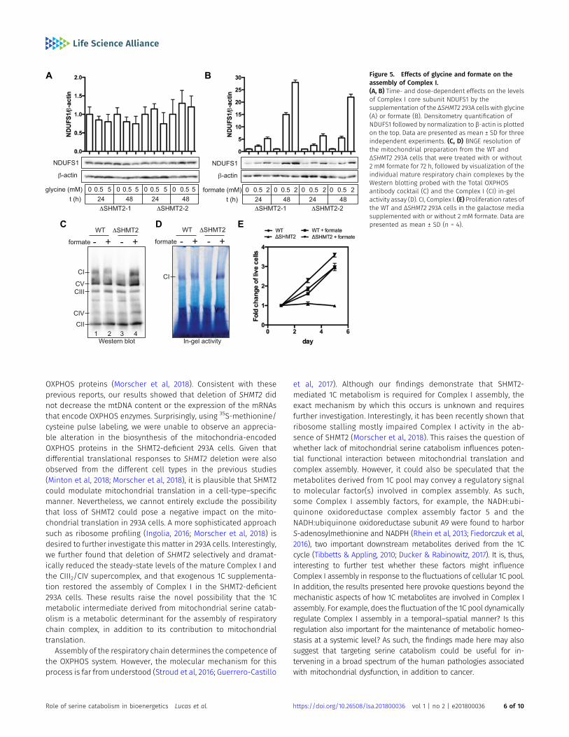
of glycine or formate, a bona fide 1C donor (Brosnan & Brosnan,2016). We observed that formate, but not glycine, significantly in-creased the levels of NDUFS1, a core subunit of Complex I, in a time-and dose-dependent manner (F5 Fig 5A and B). We next determinedhow formate treatment influences the expression of the individualmature OXPHOS complexes. Using BNGE analyses, we demon-strated that loss of SHMT2 selectively abolished the expression ofmature Complex I without affecting that of the other four individualcomplexes, including CII, CIII2, CIV, and CV (Fig 5C, lane 1 versus 3).Importantly, formate treatment completely restored Complex Iassembly (Fig 5C) and activity (Fig 5D) in ΔSHMT2 cells, as well as theproliferation of ΔSHMT2 cells in galactose (Fig 5E). These resultsstrongly suggest that the effect of SHMT2 loss on Complex I as-sembly is mediated by altered mitochondrial 1C metabolism.
Discussion
Serine is uniquely positioned at a metabolic crossroad that con-nects the major catabolic and anabolic pathways (de Koning et al,2003; Kalhan & Hanson, 2012). Although synthesized from a glyco-lytic catabolite, it can be broken down to supply key anabolic in-termediates for the synthesis of a number of important biomolecules,including nucleotides, lipids, and glutathione. It is not surprising,therefore, that serine itself is a key regulator of central metabolicpathways. This role is well illustrated by the allosteric regulation ofserine on PKM2—a key glycolytic enzyme whose role is to partitionglucose carbon into biosynthetic pathways in cancer cells (Chanetonet al, 2012; Ye et al, 2012). Here, we find that catabolism of serine by
SHMT2 is a regulatory determinant of cellular respiration. More-over, we further demonstrate that the 1C units produced bySHMT2-mediated serine catabolism are essential for the assemblyof functional Complex I. Our investigations, thus, uncovered anovel metabolic checkpoint for the assembly of the respiratoryapparatus, suggesting that transformation of serine into 1C unitscoordinates the bioenergetic and biosynthetic pathways in mamma-lian cells.
Cellular adaptions to the loss of the serine-catabolic pathwayeither in the cytosol or mitochondria were investigated in thepresent study. We observed that loss of the SHMT enzyme in eithercellular compartment posed minor impact on cell proliferationwhen the cells were grown in complete media. This is consistentwith previous reports that SHMT paralogs are redundant for supplyof 1C units in support of cell proliferation (Bao et al, 2016; Duckeret al, 2016). Interestingly, we observed that loss of SHMT2, but notSHMT1, led to elevation of aerobic glycolysis, indicating the in-volvement of SHMT2 in retrograde regulation on glycolysis in thecytosol. However, the up-regulation of glycolysis in response toSHMT2 deficiency cannot be easily explained by the allostericactivation of PKM2 by serine (Chaneton et al, 2012; Ye et al, 2012)because cellular serine levels were shown to be unaltered in theHEK293 cells lacking SHMT2 in previous studies (Bao et al, 2016;Ducker et al, 2016). Instead, our findings suggest that such ele-vation of glycolysis is a compensatory response to mitochondrialdysfunction. This compensation allows the substrate-level phos-phorylation of glycolysis to substitute energy production from mi-tochondrial respiration, as previously observed in some cancer cells(Wu et al, 2007). Our observation made in 293A cells is consistentwith the previous observations that SHMT2, but not SHMT1, isessential for maintaining mitochondrial respiration in the Jurketand HCT116 cells (Minton et al, 2018; Morscher et al, 2018). Theresults of the present study support the conclusion that SHMT2 isa critical metabolic checkpoint for OXPHOS function in a variety ofmammalian cell types.
As a major metabolic route to produce 1C units, serine catab-olism by SHMT2 might influence the OXPHOS gene expression inseveral ways including the influence on (i) cellular nucleotidehomeostasis (Bao et al, 2016; Ducker et al, 2016) and thereforemtDNA replication (Anderson et al, 2011); (ii) mRNA expression ofthe OXPHOS genes (Mentch et al, 2015; Kottakis et al, 2016); and (iii)biosynthesis of the mitochondria-encoded OXPHOS proteins(Minton et al, 2018; Morscher et al, 2018). Recent studies demon-strated that SHMT2 is necessary for sustaining mitochondrialtranslation in the Jurket human leukemic (Minton et al, 2018) andHCT116 human colon cancer (Morscher et al, 2018) cells. In-terestingly, these studies revealed distinct mechanisms that ac-count for different types of translational abnormalities consequentto SHMT2 deletion in these two cell types. In response to SHMT2deletion in Jurket cells, Minton et al (2018) found a decrease informyl-methionyl-tRNA synthesis. This resulted in impaired mito-chondrial translation initiation, leading to defective global OXPHOSprotein synthesis in mitochondria (Minton et al, 2018). Morscheret al (2018), on the other hand, found that SHMT2 deletion in HCT116cells caused a reduction in the methylation of certain tRNA spe-cies. This resulted in ribosomal stalling in HCT116 cells, leading todefective translation for a subset of the mitochondria-encoded
Figure 4. Effects on the assembly of mature respiratory chain complexes bythe deletion of SHMT2.(A–C) BNGE to detect individual mature respiratory complexes in themitochondrial preparation from the WT and ΔSHMT2 293A cells, followed byvisualization with Coomassie blue staining (A), Western blotting with the antibodyagainst a Complex I (CI) marker NDUFS3 (B), or Complex I in-gel activity assay (C). CI,Complex I; CIII2/IV, CIII2/IV supercomplex; HSP60, heat shock protein 60.
Role of serine catabolism in bioenergetics Lucas et al. https://doi.org/10.26508/lsa.201800036 vol 1 | no 2 | e201800036 5 of 10
OXPHOS proteins (Morscher et al, 2018). Consistent with theseprevious reports, our results showed that deletion of SHMT2 didnot decrease the mtDNA content or the expression of the mRNAsthat encode OXPHOS enzymes. Surprisingly, using 35S-methionine/cysteine pulse labeling, we were unable to observe an apprecia-ble alteration in the biosynthesis of the mitochondria-encodedOXPHOS proteins in the SHMT2-deficient 293A cells. Given thatdifferential translational responses to SHMT2 deletion were alsoobserved from the different cell types in the previous studies(Minton et al, 2018; Morscher et al, 2018), it is plausible that SHMT2could modulate mitochondrial translation in a cell-type–specificmanner. Nevertheless, we cannot entirely exclude the possibilitythat loss of SHMT2 could pose a negative impact on the mito-chondrial translation in 293A cells. A more sophisticated approachsuch as ribosome profiling (Ingolia, 2016; Morscher et al, 2018) isdesired to further investigate this matter in 293A cells. Interestingly,we further found that deletion of SHMT2 selectively and dramat-ically reduced the steady-state levels of the mature Complex I andthe CIII2/CIV supercomplex, and that exogenous 1C supplementa-tion restored the assembly of Complex I in the SHMT2-deficient293A cells. These results raise the novel possibility that the 1Cmetabolic intermediate derived from mitochondrial serine catab-olism is a metabolic determinant for the assembly of respiratorychain complex, in addition to its contribution to mitochondrialtranslation.
Assembly of the respiratory chain determines the competence ofthe OXPHOS system. However, the molecular mechanism for thisprocess is far from understood (Stroud et al, 2016; Guerrero-Castillo
et al, 2017). Although our findings demonstrate that SHMT2-mediated 1C metabolism is required for Complex I assembly, theexact mechanism by which this occurs is unknown and requiresfurther investigation. Interestingly, it has been recently shown thatribosome stalling mostly impaired Complex I activity in the ab-sence of SHMT2 (Morscher et al, 2018). This raises the question ofwhether lack of mitochondrial serine catabolism influences poten-tial functional interaction between mitochondrial translation andcomplex assembly. However, it could also be speculated that themetabolites derived from 1C pool may convey a regulatory signalto molecular factor(s) involved in complex assembly. As such,some Complex I assembly factors, for example, the NADH:ubi-quinone oxidoreductase complex assembly factor 5 and theNADH:ubiquinone oxidoreductase subunit A9 were found to harborS-adenosylmethionine and NADPH (Rhein et al, 2013; Fiedorczuk et al,2016), two important downstream metabolites derived from the 1Ccycle (Tibbetts & Appling, 2010; Ducker & Rabinowitz, 2017). It is, thus,interesting to further test whether these factors might influenceComplex I assembly in response to the fluctuations of cellular 1C pool.In addition, the results presented here provoke questions beyond themechanistic aspects of how 1C metabolites are involved in Complex Iassembly. For example, does the fluctuation of the 1C pool dynamicallyregulate Complex I assembly in a temporal–spatial manner? Is thisregulation also important for the maintenance of metabolic homeo-stasis at a systemic level? As such, the findings made here may alsosuggest that targeting serine catabolism could be useful for in-tervening in a broad spectrum of the human pathologies associatedwith mitochondrial dysfunction, in addition to cancer.
Figure 5. Effects of glycine and formate on theassembly of Complex I.(A, B) Time- and dose-dependent effects on the levelsof Complex I core subunit NDUFS1 by thesupplementation of the ΔSHMT2 293A cells with glycine(A) or formate (B). Densitometry quantification ofNDUFS1 followed by normalization to β-actin is plottedon the top. Data are presented as mean ± SD for threeindependent experiments. (C, D) BNGE resolution ofthe mitochondrial preparation from the WT andΔSHMT2 293A cells that were treated with or without2 mM formate for 72 h, followed by visualization of theindividual mature respiratory chain complexes by theWestern blotting probed with the Total OXPHOSantibody cocktail (C) and the Complex I (CI) in-gelactivity assay (D). CI, Complex I. (E) Proliferation rates ofthe WT and ΔSHMT2 293A cells in the galactose mediasupplemented with or without 2 mM formate. Data arepresented as mean ± SD (n = 4).
Role of serine catabolism in bioenergetics Lucas et al. https://doi.org/10.26508/lsa.201800036 vol 1 | no 2 | e201800036 6 of 10
Materials and Methods
Cell culture, plasmid construction, and mutant cell lineestablishment
HEK293A (Invitrogen) and 293T (American Type Culture Collection)cells were maintained in DMEM medium supplemented with 10%FBS, 100 U of penicillin/ml, and 0.1 ng of streptomycin/ml. MurineAML12 hepatocytes (American Type Culture Collection) weremaintained in a 1:1 mixture of DMEM and Ham’s F12 medium,supplemented with 10% FBS, 1:100 insulin-transferrin-selenium(Invitrogen), 100 U of penicillin/ml, and 0.1 ng of streptomycin/ml.
For construction of the targeting vector against human SHMT, thesgRNA sequences were determined using a web bioinformatics tool(http://crispr.mit.edu) (Ran et al, 2013). The oligo DNAs that encodesgRNA were annealed and cloned into pSpCas9(BB)-2A-Bsd, a mod-ified vector from pSpCas9(BB)-2A-Puro (PX459; Addgene) made byreplacing the puromycin-resistant to a blasticidin-resistant gene.For construction of the targeting vector against murine SHMT2, thesgRNA sequences were determined using a web bioinformaticstool (https://benchling.com/academic). The oligo DNAs that en-code the sgRNA were annealed and cloned into an AAV targetingvector (PX602; Addgene). The sgRNA sequences used are listed inTable S1.
For establishment of the 293A cell line with targeted deletion ofSHMT, the targeting vector was transfected into cells using lip-ofectamine 2000 (Invitrogen). The drug-resistant cell clones wereobtained by selecting the transfectants with 5 μg/ml of blasticidinand then were screened for loss of SHMT protein expression byWestern blotting. For establishment of the AML12 cell line withtargeted deletion of SHMT2, the adeno-assocaited virus serotypeDJ/8 viral particles that harbor the CRISPR-Cas9 system wereproduced using the AAV Helper Free Packaging System (Cell Biol-abs) and were then applied to the cells. The infectants were seededinto 96-well plates at the single-cell level using a flow cytometer.The cell clones were expanded and then screened for loss of SHMT2protein expression by Western blotting. The genomic modificationswere confirmed by automatic DNA sequencing.
For rescue of SHMT2 expression in the 293A mutant cell line, thefull-length human SHMT2 cDNA was cloned into pcDNA3.1.puro,a modified vector of pcDNA3.1 (Invitrogen) made by replacing theG418-resistant with a puromycin-resistant gene. The synonymousmutations at the sgRNA binding site were introduced into theexpression vector using a site-directed mutagenesis kit (Agilent) toevade gene targeting. The resultant SHMT2 expression vector wastransfected into the SHMT2-knockout cells, and the drug-resistantclones were obtained by selection with 2 μg/ml of puromycin andwere then screened for gain of SHMT2 protein expression byWestern blotting.
Western blotting
Cells were washed twice in PBS and lysed in cytoplasmic lysis buffer(25 mM Tris–HCl, pH 7.5, 40 mM NaCl, and 1% Triton X-100). Proteinconcentrations were determined with the Bradford reagent (Bio-Rad). Cell lysates (40 μg) were resolved by SDS-PAGE, and proteins
were transferred onto nitrocellulose filters. The blots were saturatedwith 5% nonfat milk and probed with antibodies against SHMT1(#HPA023314; Sigma-Aldrich, 1:1000), SHMT2 (#HPA020543; Sigma-Aldrich, 1:1000), HK1 (#2024; Cell Signaling Technology, 1:1000), lac-tate dehydrogenase A (#3582; Cell Signaling Technology, 1:1000), PFKP(#8164; Cell Signaling Technology, 1:1000), PKM1/2 (#3190; Cell SignalingTechnology, 1:1000), NDUFS1 (sc-271510; Santa Cruz, 1:1000), NDUFS3 (sc-374282; Santa Cruz, 1:1000), β-actin (A2066; Sigma-Aldrich, 1:1000), orTotal OXPHOS Human Antibody Cocktail (ab110411; Abcam, 1:1000).Following a wash with phosphate buffered saline with 0.1% Tween 20,the blots were incubated with peroxidase-coupled goat anti-rabbitimmunoglobulin G (Sigma-Aldrich, 1:5000). The immunolabeled pro-tein bands were detected by enhanced chemiluminescence (ECL)method (Perkin Elmer). Densitometric analysis of the blots was per-formed using Image Quant TL software (GE Healthcare).
Determination of cell proliferation and cell survival
Cells were seeded on 6-cm plates and grown for the indicated timeintervals in the glucose or galactose media. The dead cells wereexcluded with trypan blue staining (Invitrogen) and the number of livecells wasmeasured using a hemocytometer under a light microscope.
Measurements of lactic acid, ATP, and basal oxygen consumption
Measurement of lactic acid was adapted from Brandt et al (1980). Inbrief, appropriate amount of culture media was incubated at RT for30 min in a final 100 μl of reaction mix containing 160 mM Tris–hydrazine, pH 9.0, 2.5 mM NAD+, 0.01% BSA, and 8 U lactate de-hydrogenase. The amount of lactic acid was extrapolated froma standard curve based on the Ab340 reading recorded ona microplate reader (BMG LABTECH). Cellular ATP level was de-termined using ENLITEN ATP assay kit (Promega) according to themanufacturer’s specification. Intact cellular oxygen consumptionwas measured in the WT and ΔSHMT2 cells on an XFe24 seahorsebioanalyzer (Agilent) on plating 5 × 10⁴ cells per well. Data havebeen represented as oxygen consumption relative to the WT cells.
Determination of mRNA expression by next-generationsequencing and qRT-PCR
Total cellular RNA was isolated using the Trizol reagent (Invitrogen).To determine the mRNA expression of the nuclear-encodedOXPHOS genes, cDNA libraries compatible for Illumina sequenc-ing were prepared by using the QuantSeq 39 mRNA-seq Reverse(REV) Library Prep Kit (Lexogen) according to the manufacturer’sinstruction. The resultant cDNA libraries were assessed usinga TapeStation (Agilent) and subjected to 100-bp single-end sequencingusing the Illumina HiSeq 2500 system at the Wayne State UniversityApplied Genomics Technology Center. Raw sequencing reads in FASTQformatwere processedwith Trimmomatic (Bolger et al, 2014) to removelow-quality and unknown sequences. To quantify transcript abun-dance, the processed reads in FASTA format were mapped to the hg19human reference genome using bowtie2 (Langmead & Salzberg, 2012).Transcript abundance in count per million was determined usingeXpress (Roberts&Pachter, 2013), anddifferential gene expressionwasdetermined using edgeR (Robinson et al, 2010).
Role of serine catabolism in bioenergetics Lucas et al. https://doi.org/10.26508/lsa.201800036 vol 1 | no 2 | e201800036 7 of 10
To determine the mRNA expression of the mitochondria-encoded OXPHOS genes, cDNA libraries were constructed by ran-dom priming using the SuperScript III First-Strand Synthesis System(Invitrogen). The synthesized cDNA was used as a template for qRT-PCR with SYBR Green qPCR Master Mixes (Thermo Scientific) on anMx3000P cycler (Stratagene). All mRNA levels were determined asthe delta–delta threshold cycle (ΔΔCT) and normalized to pepti-dylprolyl isomerase A mRNA level. The PCR primers used are listedin Table S2.
Determination of mtDNA copy number
mtDNA copy number was determined by calculating the ratio of themitochondria-encoded ND1 gene levels to the nuclear-encoded28S rRNA gene levels using qPCR analysis. To quantify each genelevel, serial dilutions of the cloned gene-specific PCR fragmentswere used to create a standard curve. The exact gene copy numbersof the ND2 and 28S rRNA genes were obtained by plotting the log-transformed ΔΔCT values against the standard curves. The PCRprimers used are listed in Table S2.
Measurement of biosynthesis of the mitochondria-encodedproteins
Cells growing on 6-cm plates to 90% confluency were metabolicallylabeled with 400 μCi EastTag EXPRESS35S Protein Labeling Mix(PerkinElmer) in the presence of 100 μg/ml emetine for 1 h. Cellpellets were suspended in 1× Laemmli sample buffer and lysed byboiling for 10 min. Equal amounts of the extracts were resolved ona 16.5% tricine gel and dried on a GelAir gel dryer (Bio-Rad). Theradiolabeled proteins were visualized by a phosphorimager (Mo-lecular Dynamics).
Mitochondria isolation and BNGE
To isolate mitochondria, cell suspensions were incubated in ice-cold homogenization buffer (10 mM Tris–HCl, pH 7.5, 250 mM su-crose, and 1 mM EDTA) for 15 min and then homogenized witha glass douncer by 15 strokes. After centrifugation at 600 g, 4°C for10 min, the supernatants were further centrifuged at 11,000 g, 4°Cfor 10 min, to precipitate the mitochondrial fraction. Mitochondrialpellets were stored at –80°C until use.
The BNGE was carried out as described by Schagger and vonJagow (1991) withminormodifications. Briefly, mitochondrial lysateswere prepared by incubating mitochondrial suspension in the ice-cold lysis buffer (50 mM Tris–HCl, pH 7.0, 750 mM aminocapoic acid,and 1.7% n-dodecyl-β-D-moltoside) for 10 min. The lysates werethen clarified by centrifugation at 10,000 g, 4°C for 30 min. Theprotein concentrations were determined using the bicinchoninicacid assay reagents (Pierce). The gel loading mixtures were pre-pared by adding 10× loading buffer (750 mM aminocapoic acid and3% Coomassie blue brilliant G-250) to the 60 μg of mitochondriallysates. The BNGE was carried out by running the samples at 80 V/150 V on a 3–12 or 3%–16% gradient gel prepared from 41.6%, 100:1acrylamide/bis-acrylamide stock solution and 3× gel buffer (150mMBis–Tris, pH 7.0, and 1.5 M aminocaproic acid) and using separateanode (50 mM Bis–Tris, pH 7.0) and cathode (15 mM Bis–Tris and
50 mM tricine with or without 0.02% Coomassie blue brilliantG-250) buffers. For Coomassie blue staining, the gels were fixed in a50%methanol/10% acetic acid solution, stained with 0.1% Coomassieblue R-250 in the fixing solution, and destained with a 40%methanol/10% acetic acid solution. For Western blotting, the resolved proteincomplexes were transferred onto polyvinylidene fluoride membraneand then probed with either NDUFS3 antibody or the Total OXPHOSantibody cocktail. For the in-gel activity assay, Complex I activity wasdeveloped by incubating the gel in a solution containing 50 mMpotassium phosphate buffer, pH 7.0, 0.1 mg/ml NADH, and 0.2 mg/mlnitrotetrazolium blue.
Supplemental Information
Supplementary Information is available at https://doi.org/10.26508/lsa.201800036.
Acknowledgements
We sincerely thank Drs. Todd Leff, James Granneman, and Shijie Sheng forcritical comments on the manuscript.
Author Contributions
S Lucas: investigation.G Chen: investigation.S Aras: investigation and methodology.J Wang: conceptualization, formal analysis, funding acquisition,investigation, methodology, and writing—original draft, review, andediting.
Conflict of Interest Statement
The authors declare that they have no conflict of interest.
References
Alberts B, Johnson A, Lewis J, Raff M, Roberts K, Walter P (2002) MolecularBiology of the Cell, pp 767–829. New York, NY: Garland Science.
Anderson DD, Quintero CM, Stover PJ (2011) Identification of a de novothymidylate biosynthesis pathway in mammalian mitochondria. ProcNatl Acad Sci USA 108: 15163–15168. doi:10.1073/pnas.1103623108
Bao XR, Ong SE, Goldberger O, Peng J, Sharma R, Thompson DA, Vafai SB, CoxAG, Marutani E, Ichinose F (2016) Mitochondrial dysfunction remodelsone-carbon metabolism in human cells. Elife 5: e10575. doi:10.7554/elife.10575
Bolger AM, Lohse M, Usadel B (2014) Trimmomatic: A flexible trimmer forIllumina sequence data. Bioinformatics 30: 2114–2120. doi:10.1093/bioinformatics/btu170
Bourdon A, Minai L, Serre V, Jais JP, Sarzi E, Aubert S, Chretien D, de Lonlay P,Paquis-Flucklinger V, Arakawa H (2007) Mutation of RRM2B,encoding p53-controlled ribonucleotide reductase (p53R2), causessevere mitochondrial DNA depletion. Nat Genet 39: 776–780.doi:10.1038/ng2040
Role of serine catabolism in bioenergetics Lucas et al. https://doi.org/10.26508/lsa.201800036 vol 1 | no 2 | e201800036 8 of 10
Brandt RB, Siegel SA, Waters MG, Bloch MH (1980) Spectrophotometric assayfor D-(−)-lactate in plasma. Anal Biochem 102: 39–46. doi:10.1016/0003-2697(80)90314-0
Brosnan ME, Brosnan JT (2016) Formate: The neglected member of one-carbon metabolism. Annu Rev Nutr 36: 369–388. doi:10.1146/annurev-nutr-071715-050738
Chaneton B, Hillmann P, Zheng L, Martin AC, Maddocks OD, ChokkathukalamA, Coyle JE, Jankevics A, Holding FP, Vousden KH (2012) Serine isa natural ligand and allosteric activator of pyruvate kinase M2. Nature491: 458–462. doi:10.1038/nature11540
de Koning TJ, Snell K, Duran M, Berger R, Surtees R (2003) L-serine in diseaseand development. Biochem J 371: 653–661. doi:10.1042/bj20021785
Ducker GS, Chen L, Morscher RJ, Ghergurovich JM, Esposito M, Teng X, Kang Y,Rabinowitz JD (2016) Reversal of cytosolic one-carbon fluxcompensates for loss of the mitochondrial folate pathway. Cell Metab23: 1140–1153. doi:10.1016/j.cmet.2016.04.016
Ducker GS, Ghergurovich JM, Mainolfi N, Suri V, Jeong SK, Hsin-Jung Li S,Friedman A, Manfredi MG, Gitai Z, Kim H (2017) Human SHMT inhibitorsreveal defective glycine import as a targetablemetabolic vulnerabilityof diffuse large B-cell lymphoma. Proc Natl Acad Sci USA 114:11404–11409. doi:10.1073/pnas.1706617114
Ducker GS, Rabinowitz JD (2017) One-carbon metabolism in health anddisease. Cell Metab 25: 27–42. doi:10.1016/j.cmet.2016.08.009
Fiedorczuk K, Letts JA, Degliesposti G, Kaszuba K, Skehel M, Sazanov LA (2016)Atomic structure of the entire mammalian mitochondrial complex I.Nature 538: 406–410. doi:10.1038/nature19794
Guerrero-Castillo S, Baertling F, Kownatzki D, Wessels HJ, Arnold S, Brandt U,Nijtmans L (2017) The assembly pathway of mitochondrial Respiratorychain complex I. Cell Metab 25: 128–139. doi:10.1016/j.cmet.2016.09.002
Ingolia NT (2016) Ribosome footprint profiling of translation throughout thegenome. Cell 165: 22–33. doi:10.1016/j.cell.2016.02.066
Kalhan SC, Hanson RW (2012) Resurgence of serine: An often neglected butindispensable amino acid. J Biol Chem 287: 19786–19791. doi:10.1074/jbc.r112.357194
Kottakis F, Nicolay BN, Roumane A, Karnik R, Gu H, Nagle JM, Boukhali M,Hayward MC, Li YY, Chen T, et al (2016) LKB1 loss links serinemetabolism to DNA methylation and tumorigenesis. Nature 539:390–395. doi:10.1038/nature20132
Kozak M (1983) Comparison of initiation of protein synthesis in procaryotes,eucaryotes, and organelles. Microbiol Rev 47: 1–45.
Langmead B, Salzberg SL (2012) Fast gapped-read alignment with Bowtie 2.Nat Methods 9: 357–359. doi:10.1038/nmeth.1923
Locasale JW (2013) Serine, glycine and one-carbon units: Cancer metabolismin full circle. Nat Rev Cancer 13: 572–583. doi:10.1038/nrc3557
Mentch SJ, Mehrmohamadi M, Huang L, Liu X, Gupta D, Mattocks D, GómezPadilla P, Ables G, Bamman MM, Thalacker-Mercer AE, et al (2015)Histonemethylation dynamics and gene regulation occur through thesensing of one-carbon metabolism. Cell Metab 22: 861–873.doi:10.1016/j.cmet.2015.08.024
Minton DR, Nam M, McLaughlin DJ, Shin J, Bayraktar EC, Alvarez SW, SviderskiyVO, Papagiannakopoulos T, Sabatini DM, Birsoy K (2018) Serinecatabolism by SHMT2 is required for proper mitochondrial translationinitiation and maintenance of formylmethionyl-tRNAs. Mol Cell 69:610–621. doi:10.1016/j.molcel.2018.01.024
Morscher RJ, Ducker GS, Li SH, Mayer JA, Gitai Z, Sperl W, Rabinowitz JD (2018)Mitochondrial translation requires folate-dependent tRNAmethylation. Nature 554: 128. doi:10.1038/nature25460
Neupert W (2016) Mitochondrial gene expression: A playground ofevolutionary tinkering. Annu Rev Biochem 85: 65–76. doi:10.1146/annurev-biochem-011116-110824
Nikiforov MA, Chandriani S, O’Connell B, Petrenko O, Kotenko I, Beavis A,Sedivy JM, Cole MD (2002) A functional screen for Myc-responsivegenes reveals serine hydroxymethyltransferase: A major source of theone-carbon unit for cell metabolism. Mol cell Biol 22: 5793–5800.doi:10.1128/mcb.22.16.5793-5800.2002
Nikkanen J, Forsstrom S, Euro L, Paetau I, Kohnz Rebecca A, Wang L, Chilov D,Viinamaki J, Roivainen A, Marjamaki P, et al (2016) Mitochondrial DNAreplication defects disturb cellular dNTP pools and remodel one-carbon metabolism. Cell Metab 23: 635–648. doi:10.1016/j.cmet.2016.01.019
Ott M, Amunts A, Brown A (2016) Organization and regulation ofmitochondrial protein synthesis. Annu Rev Biochem 85: 77–101.doi:10.1146/annurev-biochem-060815-014334
Pfendner W, Pizer LI (1980) The metabolism of serine and glycine in mutantlines of Chinese hamster ovary cells. Arch Biochem Biophys 200:503–512. doi:10.1016/0003-9861(80)90382-3
Ran FA, Hsu PD, Wright J, Agarwala V, Scott DA, Zhang F (2013) Genomeengineering using the CRISPR-Cas9 system. Nat Protoc 8: 2281–2308.doi:10.1038/nprot.2013.143
Rhein VF, Carroll J, Ding S, Fearnley IM, Walker JE (2013) NDUFAF7 methylatesarginine 85 in the NDUFS2 subunit of human complex I. J Biol Chem288: 33016–33026. doi:10.1074/jbc.m113.518803
Roberts A, Pachter L (2013) Streaming fragment assignment for real-timeanalysis of sequencing experiments. Nat Methods 10: 71–73.doi:10.1038/nmeth.2251
Robinson BH, Petrova-Benedict R, Buncic JR, Wallace DC (1992) Nonviability ofcells with oxidative defects in galactose medium: A screening test foraffected patient fibroblasts. Biochem Med Metab Biol 48: 122–126.doi:10.1016/0885-4505(92)90056-5
Robinson MD, McCarthy DJ, Smyth GK (2010) edgeR: a Bioconductor packagefor differential expression analysis of digital gene expression data.Bioinformatics 26: 139–140. doi:10.1093/bioinformatics/btp616
Schagger H, von Jagow G (1991) Blue native electrophoresis for isolation ofmembrane protein complexes in enzymatically active form. AnalBiochem 199: 223–231. doi:10.1016/0003-2697(91)90094-a
Snell K, Natsumeda Y, Eble J, Glover J, Weber G (1988) Enzymic imbalance inserine metabolism in human colon carcinoma and rat sarcoma. Br JCancer 57: 87. doi:10.1038/bjc.1988.15
Stover P, Schirch V (1990) Serine hydroxymethyltransferase catalyzes thehydrolysis of 5, 10-methenyltetrahydrofolate to 5-formyltetrahydrofolate.J Biol Chem 265: 14227–14233
Stover PJ, Chen LH, Suh JR, Stover DM, Keyomarsi K, Shane B (1997) Molecularcloning, characterization, and regulation of the human mitochondrialserine hydroxymethyltransferase gene. J Biol Chem 272: 1842–1848.doi:10.1074/jbc.272.3.1842
Stroud DA, Surgenor EE, Formosa LE, Reljic B, Frazier AE, Dibley MG, OsellameLD, Stait T, Beilharz TH, Thorburn DR (2016) Accessory subunits areintegral for assembly and function of human mitochondrial complexI. Nature 538: 123–126. doi:10.1038/nature19754
Suomalainen A, Battersby BJ (2018) Mitochondrial diseases: The contributionof organelle stress responses to pathology. Nat Rev Mol Cell Biol 19:77–92. doi:10.1038/nrm.2017.66
Tani H, Ohnishi S, Shitara H, Mito T, Yamaguchi M, Yonekawa H, Hashizume O,Ishikawa K, Nakada K, Hayashi J-I (2018) Mice deficient in the Shmt2gene have mitochondrial respiration defects and are embryoniclethal. Sci Rep 8: 425. doi:10.1038/s41598-017-18828-3
Tedeschi PM, Markert EK, Gounder M, Lin H, Dvorzhinski D, Dolfi S, Chan LL,Qiu J, DiPaola R, Hirshfield K (2013) Contribution of serine, folateand glycine metabolism to the ATP, NADPH and purinerequirements of cancer cells. Cell Death Dis 4: e877. doi:10.1038/cddis.2013.393
Role of serine catabolism in bioenergetics Lucas et al. https://doi.org/10.26508/lsa.201800036 vol 1 | no 2 | e201800036 9 of 10

Tibbetts AS, Appling DR (2010) Compartmentalization of Mammalian folate-mediated one-carbon metabolism. Annu Rev Nutr 30: 57–81.doi:10.1146/annurev.nutr.012809.104810
Vazquez A, Markert EK, Oltvai ZN (2011) Serine biosynthesis with one carboncatabolism and the glycine cleavage system represents a novelpathway for ATP generation. PLoS One 6: e25881. doi:10.1371/journal.pone.0025881
Wang J, Alexander P, Wu L, Hammer R, Cleaver O, McKnight SL (2009)Dependence of mouse embryonic stem cells on threoninecatabolism. Science 325: 435–439. doi:10.1126/science.1173288
Wu M, Neilson A, Swift AL, Moran R, Tamagnine J, Parslow D, Armistead S,Lemire K, Orrell J, Teich J (2007) Multiparameter metabolic analysisreveals a close link between attenuated mitochondrial bioenergetic
function and enhanced glycolysis dependency in human tumorcells. Am J Physiol Cell Physiol 292: C125–C136. doi:10.1152/ajpcell.00247.2006
Ye J, Mancuso A, Tong X, Ward PS, Fan J, Rabinowitz JD, Thompson CB (2012)Pyruvate kinase M2 promotes de novo serine synthesis to sustainmTORC1 activity and cell proliferation. Proc Natl Acad Sci USA 109:6904–6909. doi:10.1073/pnas.1204176109
License: This article is available under a CreativeCommons License (Attribution 4.0 International, asdescribed at https://creativecommons.org/licenses/by/4.0/).
Role of serine catabolism in bioenergetics Lucas et al. https://doi.org/10.26508/lsa.201800036 vol 1 | no 2 | e201800036 10 of 10