Embed Size (px)
Citation preview

Review
Page 1 of 15
Licensee OA Publishing London 2013. Creative Commons Attribution Licence (CC-BY)
F : Merolla G. Shoulder replacement in advanced glenohumeral osteoarthritis: current concepts review. OA Orthopaedics 2013 Jun 19;1(1):7. Com
pe n
g in
tere
sts:
non
e de
clar
ed. C
onfl i
ct o
f int
eres
ts: n
one
decl
ared
.A
ll au
thor
s co
ntrib
uted
to th
e co
ncep
t on,
des
ign,
and
pre
para
on
of th
e m
anus
crip
t, a
s w
ell a
s re
ad a
nd a
ppro
ved
the fi n
al m
anus
crip
t.A
ll au
thor
s ab
ide
by th
e A
ssoc
ia o
n fo
r Med
ical
Eth
ics
(AM
E) e
thic
al ru
les
of d
iscl
osur
e.
Sur
gica
l Pr
oced
ures
AbstractIntroduction
Osteoarthritis of the glenohumeral joint is a source of severe pain and disability. Furthermore, shoulder osteoarthritis is frequently associated with tear or atrophy of the rotator cuff that only gets worse over time. Shoulder arthroplasty gives satisfac-tory results, restoring shoulder func-tion as well as improving patient’s quality of life. In this article, we have reviewed the biomechanics, surgical technique and results of anatomical and reverse shoulder arthroplasty.Conclusion
Shoulder arthroplasty remains the gold standard treatment for advanced shoulder osteoarthritis of the gle-nohumeral joint. However, surgeons who intend to approach this type of surgery should be aware of the need for an accurate preoperative selection of both the patient and the type of implant and a thorough understand-ing of potential complications that may arise during implantation and postoperatively.
IntroductionDegenerative osteoarthritis (OA) of glenohumeral joint is less common than that seen in weight-bearing joints, such as the hip and knee, but the incidence of OA increases with age and remains a source of severe pain and disability1,2. Furthermore, shoulder OA is frequently associ-ated with tear or atrophy of the rota-tor cuff 3,4. In patients with severe glenohumeral arthritis, shoulder
arthroplasty gives satisfactory results, restoring shoulder function and improving patient’s quality of life. Charles Neer5 irst described the results after carrying out a humeral replacement, but a long-term evalua-tion showed that a cohort of patients continued to complain of pain and weakness after going through hemi-arthroplasty. These complications were attributed to implant mobiliza-tion6, glenoid erosion7 and rotator cuff de iciency8–10. Consequently, a polyethylene glenoid component was introduced to reduce the risk of pros-theses failure and related decline in patient’s quality of life6. In order to tackle the unsatisfactory outcomes of anatomical arthroplasty carried out for treating shoulder osteoarthritis with rotator cuff insuf iciency, a new type of prosthesis was developed at the end of 1980, which is called the ‘reverse prostheses,’ the development of which was based on the assump-tion that the new design can increase the deltoid lever arm and improve shoulder function11.
In this article, we have reviewed the biomechanics, surgical technique and results of anatomical and reverse shoulder arthroplasty (RSA).
Prosthetic design
Anatomical implants
Anatomical total shoulder arthro-plasty (TSA) makes use of uncon-strained monoblock (Figure 1) or modular (Figure 2) humeral compo-nents. Instead of the standard stem, the more recently developed modern implants come with a hydroxyapa-tite-coated ‘corolla’ impacted without cement in the humeral methaphysis (TESS®; Figure 3). An example of the last-generation humeral component is the ‘short stem’ with a prevalent
metaphyseal grip (Figure 4). Head prostheses are available in several sizes, standard or with eccentric off-set (Figure 1).
Glenoid prostheses include the fol-lowing components:
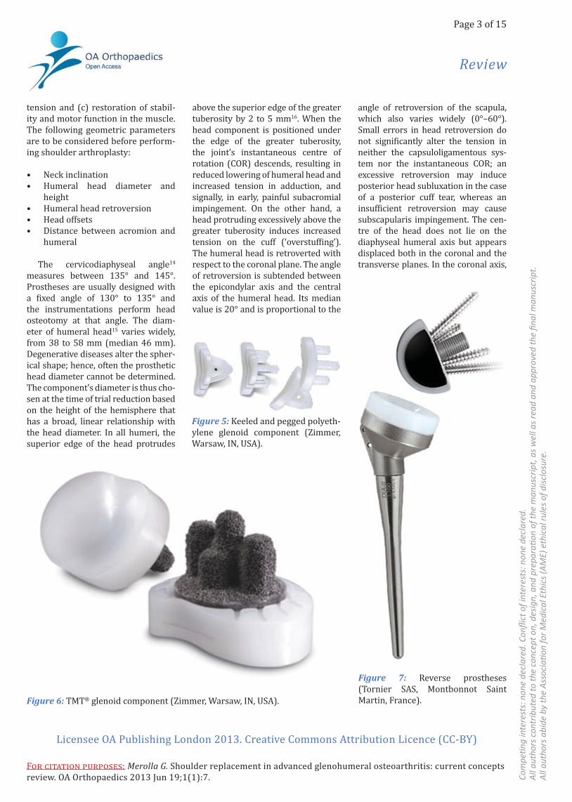
• Polyethylene components with keel or pegs (Figure 5) ixed on the cancellous bone with cement; pegged glenoids are also available with a langed unce-mented central peg to promote osseointegration
• Standard metal-backed glenoid (Figure 2) ixed with screws and covered with a polyethylene liner
• Trabecular tantalium-backed gle-noid (TMT®; Figure 6) ixed on the bone under pressure12.
As for treating glenoid, a TMT® humeral component enabling the healing of humeral fractures is available13.
A resurfacing design has been developed in several sizes with a
Shoulder replacement in advanced glenohumeral osteoarthritis: current concepts review
G Merolla*
*Corresponding authorEmails: [email protected]; [email protected]
Unit of Shoulder and Elbow Surgery, D. Cervesi Hospital, Via L. V. Beethowen 46, Cattolica, Italy
Figure 1: Monoblock humeral stem with humeral head prostheses (Zimmer, Warsaw, IN, USA).

Page 2 of 15
Review
Com
pe n
g in
tere
sts:
non
e de
clar
ed. C
onfl i
ct o
f int
eres
ts: n
one
decl
ared
.A
ll au
thor
s co
ntrib
uted
to th
e co
ncep
t on,
des
ign,
and
pre
para
on
of th
e m
anus
crip
t, a
s w
ell a
s re
ad a
nd a
ppro
ved
the fi n
al m
anus
crip
t.A
ll au
thor
s ab
ide
by th
e A
ssoc
ia o
n fo
r Med
ical
Eth
ics
(AM
E) e
thic
al ru
les
of d
iscl
osur
e.
Licensee OA Publishing London 2013. Creative Commons Attribution Licence (CC-BY)
F : Merolla G. Shoulder replacement in advanced glenohumeral osteoarthritis: current concepts review. OA Orthopaedics 2013 Jun 19;1(1):7.
short central head stem (cylindri-cal, luted, tri- in or threaded) and a metal-backed hydroxyapatite coating.
Reverse implants
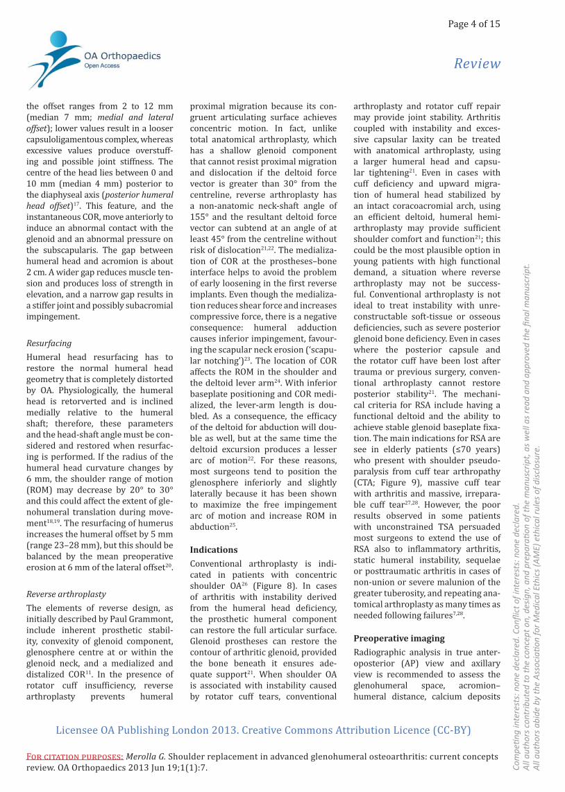
Reverse prosthesis is a semi-con-strained, totally modular device (Figure 7). The glenoid component consists of a baseplate (metaglene), provided with a large central peg and secured to the native glenoid by cor-tical screws (2 or 4), which may be straight or angled. The glenosphere (a round metal ball approximately two-third of sphere) is ixed on the baseplate with a screw. It can be com-pletely medialized or slightly lateral-ized, in order to prevent scapular neck erosion. The humeral component consists of a proximal cup-shaped portion and a metal stem press- itted or cemented on the medullary canal. A radiolucent polyethylene insert sits on this cup portion and articulates with glenosphere. As for anatomi-cal implants, reverse prostheses are available with a short stem having a predominantly metaphyseal grip.
Biomechanic rationale for shoulder prostheses
Anatomical prostheses
In order to obtain satisfactory results from shoulder replacement the fol-lowing are required: (a) prosthetic reproduction of a normal bone mor-phology, (b) restoration of capsular
Figure 2: Humeral stem (A), humeral body (B), metal-backed glenoid compo-nent (C) and polyethylene liner of a modular humeral component (LIMA, San Daniele del Friuli, Italy).
Figure 3: Stemless shoulder pros-theses: ‘corolla’ with hydroxyapatite coating and the polyethylene gle-noid component for total shoulder replacement (TESS® Biomet, Warsaw, IN, USA).
Figure 4: Short stem with offset humeral head prostheses (Tornier SAS, Montbonnot Saint Martin, France).
Aepualis Ascend™standard Press-Fit
AepualisAscend™ PTCPure Titanium Coating
Page 3 of 15
Review
Com
pe n
g in
tere
sts:
non
e de
clar
ed. C
onfl i
ct o
f int
eres
ts: n
one
decl
ared
.A
ll au
thor
s co
ntrib
uted
to th
e co
ncep
t on,
des
ign,
and
pre
para
on
of th
e m
anus
crip
t, a
s w
ell a
s re
ad a
nd a
ppro
ved
the fi n
al m
anus
crip
t.A
ll au
thor
s ab
ide
by th
e A
ssoc
ia o
n fo
r Med
ical
Eth
ics
(AM
E) e
thic
al ru
les
of d
iscl
osur
e.
Licensee OA Publishing London 2013. Creative Commons Attribution Licence (CC-BY)
F : Merolla G. Shoulder replacement in advanced glenohumeral osteoarthritis: current concepts review. OA Orthopaedics 2013 Jun 19;1(1):7.
tension and (c) restoration of stabil-ity and motor function in the muscle. The following geometric parameters are to be considered before perform-ing shoulder arthroplasty:
• Neck inclination• Humeral head diameter and
height• Humeral head retroversion• Head offsets• Distance between acromion and
humeral
The cervicodiaphyseal angle14 measures between 135° and 145°. Prostheses are usually designed with a ixed angle of 130° to 135° and the instrumentations perform head osteotomy at that angle. The diam-eter of humeral head15 varies widely, from 38 to 58 mm (median 46 mm). Degenerative diseases alter the spher-ical shape; hence, often the prosthetic head diameter cannot be determined. The component’s diameter is thus cho-sen at the time of trial reduction based on the height of the hemisphere that has a broad, linear relationship with the head diameter. In all humeri, the superior edge of the head protrudes
above the superior edge of the greater tuberosity by 2 to 5 mm16. When the head component is positioned under the edge of the greater tuberosity, the joint’s instantaneous centre of rotation (COR) descends, resulting in reduced lowering of humeral head and increased tension in adduction, and signally, in early, painful subacromial impingement. On the other hand, a head protruding excessively above the greater tuberosity induces increased tension on the cuff (‘overstuf ing’). The humeral head is retroverted with respect to the coronal plane. The angle of retroversion is subtended between the epicondylar axis and the central axis of the humeral head. Its median value is 20° and is proportional to the
angle of retroversion of the scapula, which also varies widely (0°–60°). Small errors in head retroversion do not signi icantly alter the tension in neither the capsuloligamentous sys-tem nor the instantaneous COR; an excessive retroversion may induce posterior head subluxation in the case of a posterior cuff tear, whereas an insuf icient retroversion may cause subscapularis impingement. The cen-tre of the head does not lie on the diaphyseal humeral axis but appears displaced both in the coronal and the transverse planes. In the coronal axis,
Figure 7: Reverse prostheses (Tornier SAS, Montbonnot Saint Martin, France).
Figure 5: Keeled and pegged polyeth-ylene glenoid component (Zimmer, Warsaw, IN, USA).
Figure 6: TMT® glenoid component (Zimmer, Warsaw, IN, USA).
Page 4 of 15
Review
Com
pe n
g in
tere
sts:
non
e de
clar
ed. C
onfl i
ct o
f int
eres
ts: n
one
decl
ared
.A
ll au
thor
s co
ntrib
uted
to th
e co
ncep
t on,
des
ign,
and
pre
para
on
of th
e m
anus
crip
t, a
s w
ell a
s re
ad a
nd a
ppro
ved
the fi n
al m
anus
crip
t.A
ll au
thor
s ab
ide
by th
e A
ssoc
ia o
n fo
r Med
ical
Eth
ics
(AM
E) e
thic
al ru
les
of d
iscl
osur
e.
Licensee OA Publishing London 2013. Creative Commons Attribution Licence (CC-BY)
F : Merolla G. Shoulder replacement in advanced glenohumeral osteoarthritis: current concepts review. OA Orthopaedics 2013 Jun 19;1(1):7.
the offset ranges from 2 to 12 mm (median 7 mm; medial and lateral offset); lower values result in a looser capsuloligamentous complex, whereas excessive values produce overstuff-ing and possible joint stiffness. The centre of the head lies between 0 and 10 mm (median 4 mm) posterior to the diaphyseal axis (posterior humeral head offset)17. This feature, and the instantaneous COR, move anteriorly to induce an abnormal contact with the glenoid and an abnormal pressure on the subscapularis. The gap between humeral head and acromion is about 2 cm. A wider gap reduces muscle ten-sion and produces loss of strength in elevation, and a narrow gap results in a stiffer joint and possibly subacromial impingement.
Resurfacing
Humeral head resurfacing has to restore the normal humeral head geometry that is completely distorted by OA. Physiologically, the humeral head is retorverted and is inclined medially relative to the humeral shaft; therefore, these parameters and the head-shaft angle must be con-sidered and restored when resurfac-ing is performed. If the radius of the humeral head curvature changes by 6 mm, the shoulder range of motion (ROM) may decrease by 20° to 30° and this could affect the extent of gle-nohumeral translation during move-ment18,19. The resurfacing of humerus increases the humeral offset by 5 mm (range 23–28 mm), but this should be balanced by the mean preoperative erosion at 6 mm of the lateral offset20.
Reverse arthroplasty
The elements of reverse design, as initially described by Paul Grammont, include inherent prosthetic stabil-ity, convexity of glenoid component, glenosphere centre at or within the glenoid neck, and a medialized and distalized COR11. In the presence of rotator cuff insuf iciency, reverse arthroplasty prevents humeral
proximal migration because its con-gruent articulating surface achieves concentric motion. In fact, unlike total anatomical arthroplasty, which has a shallow glenoid component that cannot resist proximal migration and dislocation if the deltoid force vector is greater than 30° from the centreline, reverse arthroplasty has a non-anatomic neck-shaft angle of 155° and the resultant deltoid force vector can subtend at an angle of at least 45° from the centreline without risk of dislocation21,22. The medializa-tion of COR at the prostheses–bone interface helps to avoid the problem of early loosening in the irst reverse implants. Even though the medializa-tion reduces shear force and increases compressive force, there is a negative consequence: humeral adduction causes inferior impingement, favour-ing the scapular neck erosion (‘scapu-lar notching’)23. The location of COR affects the ROM in the shoulder and the deltoid lever arm24. With inferior baseplate positioning and COR medi-alized, the lever-arm length is dou-bled. As a consequence, the ef icacy of the deltoid for abduction will dou-ble as well, but at the same time the deltoid excursion produces a lesser arc of motion22. For these reasons, most surgeons tend to position the glenosphere inferiorly and slightly laterally because it has been shown to maximize the free impingement arc of motion and increase ROM in abduction25.
Indications
Conventional arthroplasty is indi-cated in patients with concentric shoulder OA26 (Figure 8). In cases of arthritis with instability derived from the humeral head de iciency, the prosthetic humeral component can restore the full articular surface. Glenoid prostheses can restore the contour of arthritic glenoid, provided the bone beneath it ensures ade-quate support21. When shoulder OA is associated with instability caused by rotator cuff tears, conventional
arthroplasty and rotator cuff repair may provide joint stability. Arthritis coupled with instability and exces-sive capsular laxity can be treated with anatomical arthroplasty, using a larger humeral head and capsu-lar tightening21. Even in cases with cuff de iciency and upward migra-tion of humeral head stabilized by an intact coracoacromial arch, using an ef icient deltoid, humeral hemi-arthroplasty may provide suf icient shoulder comfort and function21; this could be the most plausible option in young patients with high functional demand, a situation where reverse arthroplasty may not be success-ful. Conventional arthroplasty is not ideal to treat instability with unre-constructable soft-tissue or osseous de iciencies, such as severe posterior glenoid bone de iciency. Even in cases where the posterior capsule and the rotator cuff have been lost after trauma or previous surgery, conven-tional arthroplasty cannot restore posterior stability21. The mechani-cal criteria for RSA include having a functional deltoid and the ability to achieve stable glenoid baseplate ixa-tion. The main indications for RSA are see in elderly patients (≤70 years) who present with shoulder pseudo-paralysis from cuff tear arthropathy (CTA; Figure 9), massive cuff tear with arthritis and massive, irrepara-ble cuff tear27,28. However, the poor results observed in some patients with unconstrained TSA persuaded most surgeons to extend the use of RSA also to in lammatory arthritis, static humeral instability, sequelae or posttraumatic arthritis in cases of non-union or severe malunion of the greater tuberosity, and repeating ana-tomical arthroplasty as many times as needed following failures7,28.
Preoperative imaging
Radiographic analysis in true anter-oposterior (AP) view and axillary view is recommended to assess the glenohumeral space, acromion–humeral distance, calcium deposits

Page 5 of 15
Review
Com
pe n
g in
tere
sts:
non
e de
clar
ed. C
onfl i
ct o
f int
eres
ts: n
one
decl
ared
.A
ll au
thor
s co
ntrib
uted
to th
e co
ncep
t on,
des
ign,
and
pre
para
on
of th
e m
anus
crip
t, a
s w
ell a
s re
ad a
nd a
ppro
ved
the fi n
al m
anus
crip
t.A
ll au
thor
s ab
ide
by th
e A
ssoc
ia o
n fo
r Med
ical
Eth
ics
(AM
E) e
thic
al ru
les
of d
iscl
osur
e.
Licensee OA Publishing London 2013. Creative Commons Attribution Licence (CC-BY)
F : Merolla G. Shoulder replacement in advanced glenohumeral osteoarthritis: current concepts review. OA Orthopaedics 2013 Jun 19;1(1):7.
or ossi ications, the proximal humeral epiphysis and the size of diaphysis. The gold standard in proeperative bone evaluation is computed tomog-raphy (CT), which is especially useful in determining glenoid bone stock and morphology according to Walch et al.7,26 (Figure 10). Magnetic resonance imaging (MRI) can more accurately assess the soft tissue and the rota-tor cuff when a decision needs to be
made whether to opt for conventional implants or reverse arthroplasty.
Surgical procedures
Anatomical arthroplasty
Operations are performed on the patient seated in the beach-chair position, using a deltopectoral or anterosuperior shoulder approach. We describe the common steps fol-lowed in deltopectoral approach, which is routinely used in our hospi-tal’s Shoulder and Elbow Unit.
The skin is marked and a cut29 is performed from the clavicle, down to and across the coracoid tip and con-tinued in a straight line to the anterior border of deltoid insertion (Figure 11). The interval between the deltoid and pectoralis major muscle with the cephalic vein that is retracted laterally with the deltoid is identi ied. The long head of biceps in the bicipital groove is tenotomized and the lesser tuberosity with subscapularis tendon is osteoto-mized. The dissection proceeds supe-riorly, from the base of the coracoid to the subacromial space, anteriorly and inferiorly, and thereafter the degener-ate capsule is carefully removed. Then
the subacromial space is explored, in order to save the coraco-acromial liga-ment. A suture is passed on the medial margin of the supraspinatus tendon, in order to have a tendon mark in case the rotator interval needs to be closed, with the subscapularis muscle medi-ally retracted to expose the joint. A manoeuvre is made to dislocate the humeral head, by a movement of the arm in adduction, extension and exter-nal rotation. At this stage, it is neces-sary to completely remove the inferior ‘goat beard’ osteophyte to obtain the complete exposure of humeral head. During humeral exposure, it is ideal to use a large retractor in the gleno-humeral joint, a blunt Hohmann under the deltoid in the subacromial space and a small Hohmann at the inferior humeral neck, with the retractor kept in contact with the bone to maintain a safe distance from the axillary nerve.
All osteophytes present along the anatomical neck are removed and the humeral head is perforated at its highest point, 1 cm superior medial to bicipital groove (‘hinge point’). The medullary canal is accessed through a graduated driving, which is mounted on the mask for cutting. Osteotomy of
Figure 8: Concentric glenohumeral osteoarthritis.
Figure 9: Cuff tear arthropathy.Figure 10: Axial CT scan in severe glenohumeral osteoarthritis. Note the glenoid erosion and the posterior subluxation of humeral head.
Page 6 of 15
Review
Com
pe n
g in
tere
sts:
non
e de
clar
ed. C
onfl i
ct o
f int
eres
ts: n
one
decl
ared
.A
ll au
thor
s co
ntrib
uted
to th
e co
ncep
t on,
des
ign,
and
pre
para
on
of th
e m
anus
crip
t, a
s w
ell a
s re
ad a
nd a
ppro
ved
the fi n
al m
anus
crip
t.A
ll au
thor
s ab
ide
by th
e A
ssoc
ia o
n fo
r Med
ical
Eth
ics
(AM
E) e
thic
al ru
les
of d
iscl
osur
e.
Licensee OA Publishing London 2013. Creative Commons Attribution Licence (CC-BY)
F : Merolla G. Shoulder replacement in advanced glenohumeral osteoarthritis: current concepts review. OA Orthopaedics 2013 Jun 19;1(1):7.
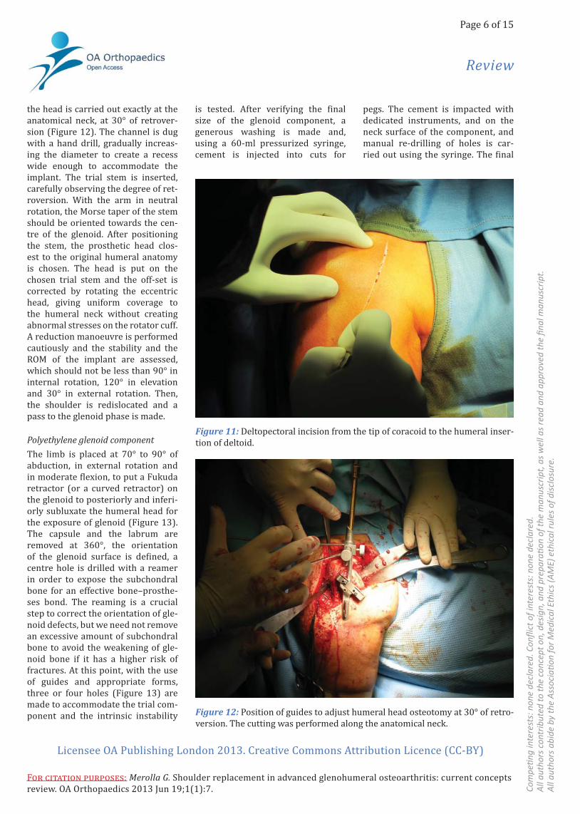
the head is carried out exactly at the anatomical neck, at 30° of retrover-sion (Figure 12). The channel is dug with a hand drill, gradually increas-ing the diameter to create a recess wide enough to accommodate the implant. The trial stem is inserted, carefully observing the degree of ret-roversion. With the arm in neutral rotation, the Morse taper of the stem should be oriented towards the cen-tre of the glenoid. After positioning the stem, the prosthetic head clos-est to the original humeral anatomy is chosen. The head is put on the chosen trial stem and the off-set is corrected by rotating the eccentric head, giving uniform coverage to the humeral neck without creating abnormal stresses on the rotator cuff. A reduction manoeuvre is performed cautiously and the stability and the ROM of the implant are assessed, which should not be less than 90° in internal rotation, 120° in elevation and 30° in external rotation. Then, the shoulder is redislocated and a pass to the glenoid phase is made.
Polyethylene glenoid component
The limb is placed at 70° to 90° of abduction, in external rotation and in moderate lexion, to put a Fukuda retractor (or a curved retractor) on the glenoid to posteriorly and inferi-orly subluxate the humeral head for the exposure of glenoid (Figure 13). The capsule and the labrum are removed at 360°, the orientation of the glenoid surface is de ined, a centre hole is drilled with a reamer in order to expose the subchondral bone for an effective bone–prosthe-ses bond. The reaming is a crucial step to correct the orientation of gle-noid defects, but we need not remove an excessive amount of subchondral bone to avoid the weakening of gle-noid bone if it has a higher risk of fractures. At this point, with the use of guides and appropriate forms, three or four holes (Figure 13) are made to accommodate the trial com-ponent and the intrinsic instability
is tested. After verifying the inal size of the glenoid component, a generous washing is made and, using a 60-ml pressurized syringe, cement is injected into cuts for
pegs. The cement is impacted with dedicated instruments, and on the neck surface of the component, and manual re-drilling of holes is car-ried out using the syringe. The inal
Figure 11: Deltopectoral incision from the tip of coracoid to the humeral inser-tion of deltoid.
Figure 12: Position of guides to adjust humeral head osteotomy at 30° of retro-version. The cutting was performed along the anatomical neck.
Page 7 of 15
Review
Com
pe n
g in
tere
sts:
non
e de
clar
ed. C
onfl i
ct o
f int
eres
ts: n
one
decl
ared
.A
ll au
thor
s co
ntrib
uted
to th
e co
ncep
t on,
des
ign,
and
pre
para
on
of th
e m
anus
crip
t, a
s w
ell a
s re
ad a
nd a
ppro
ved
the fi n
al m
anus
crip
t.A
ll au
thor
s ab
ide
by th
e A
ssoc
ia o
n fo
r Med
ical
Eth
ics
(AM
E) e
thic
al ru
les
of d
iscl
osur
e.
Licensee OA Publishing London 2013. Creative Commons Attribution Licence (CC-BY)
F : Merolla G. Shoulder replacement in advanced glenohumeral osteoarthritis: current concepts review. OA Orthopaedics 2013 Jun 19;1(1):7.
glenoid prosthesis is then impacted (Figure 14). When polyethylene29 component is used with cementless central peg, the langed luted peg is embedded within the morselized bone in the glenoid central hole.
Metal-backed component
The centre of the glenoid tracing two orthogonal lines along the lon-gitudinal and transversal axes is identi ied with an electric cautery, and then a K wire is inserted (15-cm long, 2.5-mm diameter) at least 25 mm into the bone, orthogonal to the glenoid surface and slightly off the centre. The glenoid reamer is used to remove the glenoid cartilage, exposing the subchondral bone. The glenoid drilling is continued until the peg reaches the end; in case a larger peg is used, the glenoid drill may be used to widen the hole. After choosing the correct size of the M-B cementless component, it is pushed into the central hole with a handle
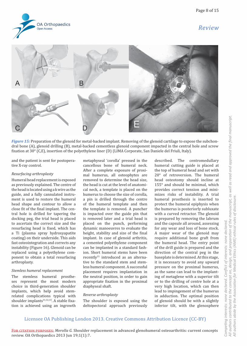
positioner, ensuring that the major axis of the implant coincides with the largest axis of glenoid. Screws are then inserted and itted within 30°. Finally, using the thumb, the pol-yethylene liner pushing is inserted (Figure 15 A–E).
TMT® glenoid without screw ixa-tion can be used to optimize the bone ingrowth and reduce the risk of gle-noid failure29.
Final assembly of the prosthetic components
Before the implantation of inal humeral component, the trial head is put again and the shoulder is lowered. The tension of soft parts, size, offset of head and the new articular rela-tionship between the glenoid pros-theses implanted and the ROM are all checked; subscapularis is returned to its bone insertion on the lesser tuber-osity to assess the degree of tension. After assessing these parameters, the humeral trial is removed and four or ive bone sutures are passed ( lexidene #4) on the neck of the humerus to ix the subscapularis. In
case cemented humeral prostheses are chosen, the plug is inserted into the canal and dried in order to per-form an accurate lavage. The cement is injected under pressure and the inal stem is introduced with the cor-
rect version as previously measured. Some time must be allowed for the cement to consolidate. The trial head is inserted again to check the offset and the tension of the subscapularis, the rotator cuff and the ROM. The trial is then removed and the inal head prostheses are implanted, ensuring that the offset previously assessed is accurately reproduced. Shoulder is reduced, close the rotator interval to its base with reabsorbable suture (ethibond #2) and the subscapula-ris is ixed using a modi ied Mason–Allen stitches. Anterior and posterior drawer manoeuvres29 are repeated to assess the stability of prostheses and evaluate the mobility achieved; the area is washed, and the status of axil-lary nerve is checked in order to place a subdeltoid drainage. Afterwards, both deep and surface layers are placed, the arm is placed in a sling
Figure 14: Cemented all-polyethylene glenoid prostheses after the impaction with cement (Zimmer, Warsaw, IN, USA).
Figure 13: Complete exposure of the glenoid using curved retractors on the humeral head and scapular neck and two Hohmann retractors on the supe-rior and inferior aspect of the glenoid. The capsule has been excised circum-ferentially and three holes have been performed to accommodate the trial.
Page 8 of 15
Review
Com
pe n
g in
tere
sts:
non
e de
clar
ed. C
onfl i
ct o
f int
eres
ts: n
one
decl
ared
.A
ll au
thor
s co
ntrib
uted
to th
e co
ncep
t on,
des
ign,
and
pre
para
on
of th
e m
anus
crip
t, a
s w
ell a
s re
ad a
nd a
ppro
ved
the fi n
al m
anus
crip
t.A
ll au
thor
s ab
ide
by th
e A
ssoc
ia o
n fo
r Med
ical
Eth
ics
(AM
E) e
thic
al ru
les
of d
iscl
osur
e.
Licensee OA Publishing London 2013. Creative Commons Attribution Licence (CC-BY)
F : Merolla G. Shoulder replacement in advanced glenohumeral osteoarthritis: current concepts review. OA Orthopaedics 2013 Jun 19;1(1):7.
and the patient is sent for postopera-tive X-ray control.
Resurfacing arthroplasty
Humeral head replacement is exposed as previously explained. The centre of the head is located using a k wire as the guide, and a fully cannulated instru-ment is used to restore the humeral head shape and contour to allow a close it of the inal implant. The cen-tral hole is drilled for tapering the docking peg, the trial head is placed to ascertain the correct size and the resurfacing head is ixed, which has a Ti (plasma spray hydroxyapatite coating) on their underside. This aids fast osteointegration and corrects any instability (Figure 16). Glenoid can be replaced using a polyethylene com-ponent to obtain a total resurfacing arthroplasty.
Stemless humeral replacement
The stemless humeral prosthe-ses represent the most modern choice in third-generation shoulder implants, which help avoid stem-related complications typical with shoulder implants31,32,33. A stable ixa-tion is achieved using an ingrowth
metaphyseal ‘corolla’ pressed in the cancellous bone of humeral neck. After a complete exposure of proxi-mal humerus, all osteophytes are removed to determine the head size, the head is cut at the level of anatomi-cal neck, a template is placed on the humerus to choose the size of corolla, a pin is drilled through the centre of the humeral template and then the template is removed. A puncher is impacted over the guide pin that is removed later and a trial head is placed on the punch, performing dynamic manoeuvres to evaluate the height, stability and size of the inal implant. In case of glenoid arthritis, a cemented polyethylene component can be implanted in a standard fash-ion. Short humeral stems have been recently31 introduced as an alterna-tive to the standard stem and stem-less humeral component. A successful placement requires implantation in the neutral position, in order to gain appropriate ixation in the proximal diaphyseal shaft.
Reverse arthroplasty
The shoulder is exposed using the deltopectoral approach previously
described. The centromedullary humeral cutting guide is placed at the top of humeral head and set with 20° of retroversion. The humeral head osteotomy should incline at 155° and should be minimal, which provides correct tension and mini-mizes risks of instability. A trial humeral prosthesis is inserted to protect the humeral epiphysis when the humerus is posteriorly subluxate with a curved retractor. The glenoid is prepared by removing the labrum and the capsule all around, assessing for any wear and loss of bone stock. A major wear of the glenoid may require additional bone graft from the humeral head. The entry point of the drill guide is prepared and the direction of the central peg in the baseplate is determined. At this stage, it is necessary to avoid any upward pressure on the proximal humerus, as the same can lead to the implant-ing of metaglene with a superior tilt or to the drilling of centre hole at a very high location, which can then lead to impingement of the humerus in adduction. The optimal position of glenoid should be with a slightly inferior tilt, with the glenosphere
Figure 15: Preparation of the glenoid for metal-backed implant. Removing of the glenoid cartilage to expose the subchon-dral bone (A), glenoid drilling (B), metal-backed cementless glenoid component impacted in the central hole and screw ixation at 30° (C,E), insertion of the polyethylene liner (D) (LIMA Corporate, San Daniele del Friuli, Italy).
Page 9 of 15
Review
Com
pe n
g in
tere
sts:
non
e de
clar
ed. C
onfl i
ct o
f int
eres
ts: n
one
decl
ared
.A
ll au
thor
s co
ntrib
uted
to th
e co
ncep
t on,
des
ign,
and
pre
para
on
of th
e m
anus
crip
t, a
s w
ell a
s re
ad a
nd a
ppro
ved
the fi n
al m
anus
crip
t.A
ll au
thor
s ab
ide
by th
e A
ssoc
ia o
n fo
r Med
ical
Eth
ics
(AM
E) e
thic
al ru
les
of d
iscl
osur
e.
Licensee OA Publishing London 2013. Creative Commons Attribution Licence (CC-BY)
F : Merolla G. Shoulder replacement in advanced glenohumeral osteoarthritis: current concepts review. OA Orthopaedics 2013 Jun 19;1(1):7.
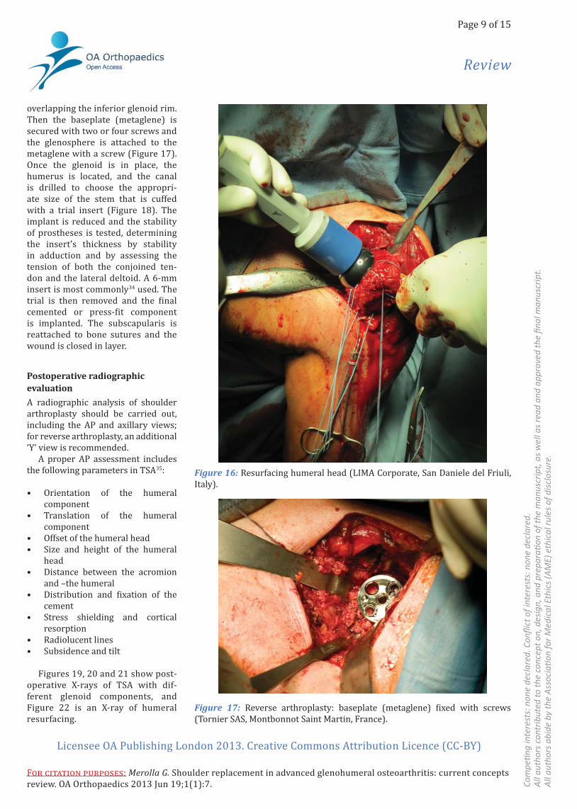
overlapping the inferior glenoid rim. Then the baseplate (metaglene) is secured with two or four screws and the glenosphere is attached to the metaglene with a screw (Figure 17). Once the glenoid is in place, the humerus is located, and the canal is drilled to choose the appropri-ate size of the stem that is cuffed with a trial insert (Figure 18). The implant is reduced and the stability of prostheses is tested, determining the insert’s thickness by stability in adduction and by assessing the tension of both the conjoined ten-don and the lateral deltoid. A 6-mm insert is most commonly34 used. The trial is then removed and the inal cemented or press- it component is implanted. The subscapularis is reattached to bone sutures and the wound is closed in layer.
Postoperative radiographic evaluation
A radiographic analysis of shoulder arthroplasty should be carried out, including the AP and axillary views; for reverse arthroplasty, an additional ‘Y’ view is recommended.
A proper AP assessment includes the following parameters in TSA35:
• Orientation of the humeral component
• Translation of the humeral component
• Offset of the humeral head• Size and height of the humeral
head• Distance between the acromion
and –the humeral• Distribution and ixation of the
cement• Stress shielding and cortical
resorption• Radiolucent lines• Subsidence and tilt
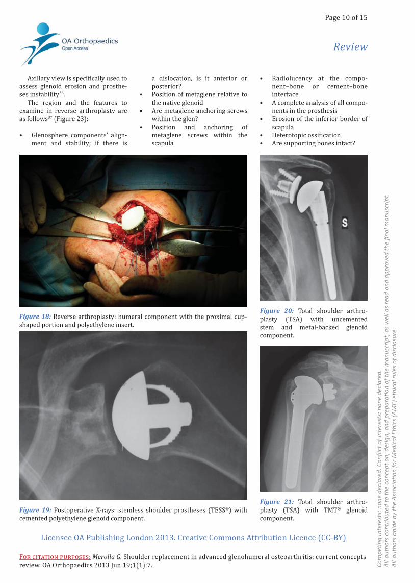
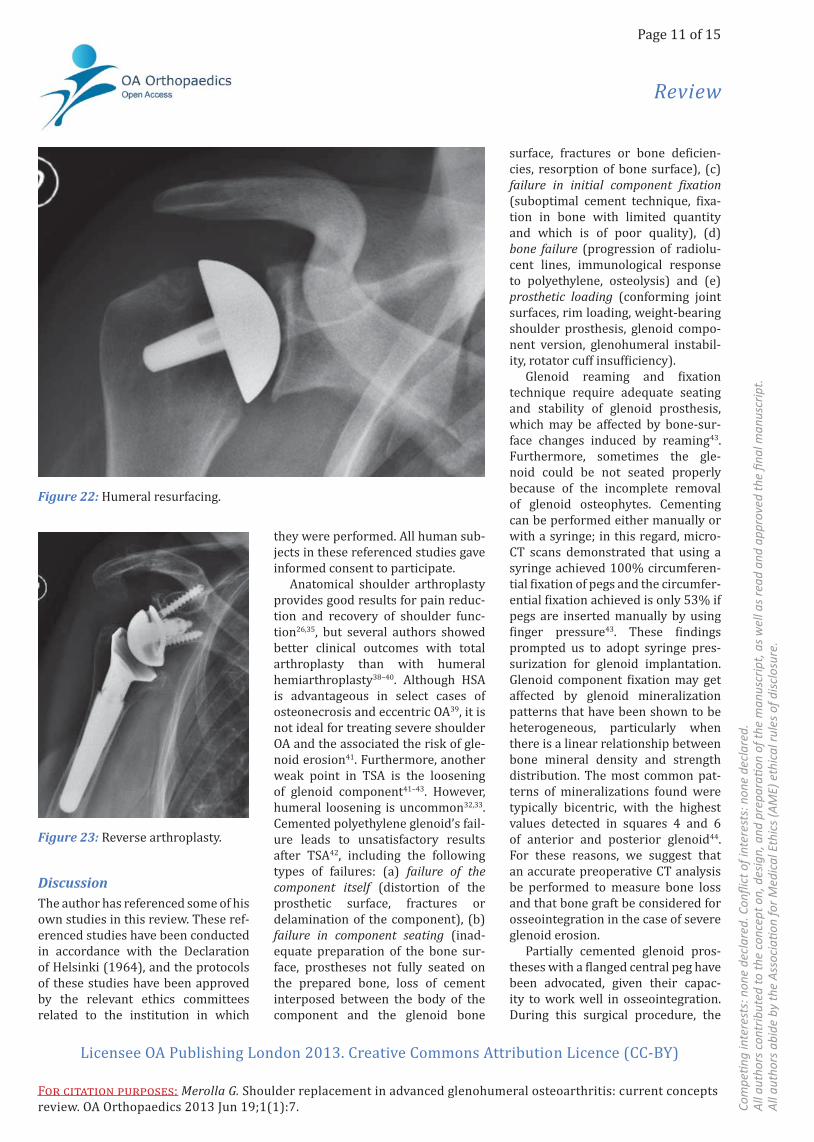
Figures 19, 20 and 21 show post-operative X-rays of TSA with dif-ferent glenoid components, and Figure 22 is an X-ray of humeral resurfacing.
Figure 16: Resurfacing humeral head (LIMA Corporate, San Daniele del Friuli, Italy).
Figure 17: Reverse arthroplasty: baseplate (metaglene) ixed with screws (Tornier SAS, Montbonnot Saint Martin, France).
Page 10 of 15
Review
Com
pe n
g in
tere
sts:
non
e de
clar
ed. C
onfl i
ct o
f int
eres
ts: n
one
decl
ared
.A
ll au
thor
s co
ntrib
uted
to th
e co
ncep
t on,
des
ign,
and
pre
para
on
of th
e m
anus
crip
t, a
s w
ell a
s re
ad a
nd a
ppro
ved
the fi n
al m
anus
crip
t.A
ll au
thor
s ab
ide
by th
e A
ssoc
ia o
n fo
r Med
ical
Eth
ics
(AM
E) e
thic
al ru
les
of d
iscl
osur
e.
Licensee OA Publishing London 2013. Creative Commons Attribution Licence (CC-BY)
F : Merolla G. Shoulder replacement in advanced glenohumeral osteoarthritis: current concepts review. OA Orthopaedics 2013 Jun 19;1(1):7.
Axillary view is speci ically used to assess glenoid erosion and prosthe-ses instability36.
The region and the features to examine in reverse arthroplasty are as follows37 (Figure 23):
• Glenosphere components’ align-ment and stability; if there is
a dislocation, is it anterior or posterior?
• Position of metaglene relative to the native glenoid
• Are metaglene anchoring screws within the glen?
• Position and anchoring of metaglene screws within the scapula
• Radiolucency at the compo-nent–bone or cement–bone interface
• A complete analysis of all compo-nents in the prosthesis
• Erosion of the inferior border of scapula
• Heterotopic ossi ication• Are supporting bones intact?
Figure 18: Reverse arthroplasty: humeral component with the proximal cup-shaped portion and polyethylene insert.
Figure 19: Postoperative X-rays: stemless shoulder prostheses (TESS®) with cemented polyethylene glenoid component.
Figure 20: Total shoulder arthro-plasty (TSA) with uncemented stem and metal-backed glenoid component.
Figure 21: Total shoulder arthro-plasty (TSA) with TMT® glenoid component.
Page 11 of 15
Review
Com
pe n
g in
tere
sts:
non
e de
clar
ed. C
onfl i
ct o
f int
eres
ts: n
one
decl
ared
.A
ll au
thor
s co
ntrib
uted
to th
e co
ncep
t on,
des
ign,
and
pre
para
on
of th
e m
anus
crip
t, a
s w
ell a
s re
ad a
nd a
ppro
ved
the fi n
al m
anus
crip
t.A
ll au
thor
s ab
ide
by th
e A
ssoc
ia o
n fo
r Med
ical
Eth
ics
(AM
E) e
thic
al ru
les
of d
iscl
osur
e.
Licensee OA Publishing London 2013. Creative Commons Attribution Licence (CC-BY)
F : Merolla G. Shoulder replacement in advanced glenohumeral osteoarthritis: current concepts review. OA Orthopaedics 2013 Jun 19;1(1):7.
DiscussionThe author has referenced some of his own studies in this review. These ref-erenced studies have been conducted in accordance with the Declaration of Helsinki (1964), and the protocols of these studies have been approved by the relevant ethics committees related to the institution in which
they were performed. All human sub-jects in these referenced studies gave informed consent to participate.
Anatomical shoulder arthroplasty provides good results for pain reduc-tion and recovery of shoulder func-tion26,35, but several authors showed better clinical outcomes with total arthroplasty than with humeral hemiarthroplasty38–40. Although HSA is advantageous in select cases of osteonecrosis and eccentric OA39, it is not ideal for treating severe shoulder OA and the associated the risk of gle-noid erosion41. Furthermore, another weak point in TSA is the loosening of glenoid component41–43. However, humeral loosening is uncommon32,33. Cemented polyethylene glenoid’s fail-ure leads to unsatisfactory results after TSA42, including the following types of failures: (a) failure of the component itself (distortion of the prosthetic surface, fractures or delamination of the component), (b) failure in component seating (inad-equate preparation of the bone sur-face, prostheses not fully seated on the prepared bone, loss of cement interposed between the body of the component and the glenoid bone
surface, fractures or bone de icien-cies, resorption of bone surface), (c) failure in initial component fixation (suboptimal cement technique, ixa-tion in bone with limited quantity and which is of poor quality), (d) bone failure (progression of radiolu-cent lines, immunological response to polyethylene, osteolysis) and (e) prosthetic loading (conforming joint surfaces, rim loading, weight-bearing shoulder prosthesis, glenoid compo-nent version, glenohumeral instabil-ity, rotator cuff insuf iciency).
Glenoid reaming and ixation technique require adequate seating and stability of glenoid prosthesis, which may be affected by bone-sur-face changes induced by reaming43. Furthermore, sometimes the gle-noid could be not seated properly because of the incomplete removal of glenoid osteophytes. Cementing can be performed either manually or with a syringe; in this regard, micro-CT scans demonstrated that using a syringe achieved 100% circumferen-tial ixation of pegs and the circumfer-ential ixation achieved is only 53% if pegs are inserted manually by using inger pressure43. These indings
prompted us to adopt syringe pres-surization for glenoid implantation. Glenoid component ixation may get affected by glenoid mineralization patterns that have been shown to be heterogeneous, particularly when there is a linear relationship between bone mineral density and strength distribution. The most common pat-terns of mineralizations found were typically bicentric, with the highest values detected in squares 4 and 6 of anterior and posterior glenoid44. For these reasons, we suggest that an accurate preoperative CT analysis be performed to measure bone loss and that bone graft be considered for osseointegration in the case of severe glenoid erosion.
Partially cemented glenoid pros-theses with a langed central peg have been advocated, given their capac-ity to work well in osseointegration. During this surgical procedure, the
Figure 22: Humeral resurfacing.
Figure 23: Reverse arthroplasty.
Page 12 of 15
Review
Com
pe n
g in
tere
sts:
non
e de
clar
ed. C
onfl i
ct o
f int
eres
ts: n
one
decl
ared
.A
ll au
thor
s co
ntrib
uted
to th
e co
ncep
t on,
des
ign,
and
pre
para
on
of th
e m
anus
crip
t, a
s w
ell a
s re
ad a
nd a
ppro
ved
the fi n
al m
anus
crip
t.A
ll au
thor
s ab
ide
by th
e A
ssoc
ia o
n fo
r Med
ical
Eth
ics
(AM
E) e
thic
al ru
les
of d
iscl
osur
e.
Licensee OA Publishing London 2013. Creative Commons Attribution Licence (CC-BY)
F : Merolla G. Shoulder replacement in advanced glenohumeral osteoarthritis: current concepts review. OA Orthopaedics 2013 Jun 19;1(1):7.
central peg remains uncemented and the langs are completely embed-ded into bleeding cancellous bone (‘morselized bone graft’)45. Although recent studies45,46 and our CT indings (unpublished data) showed a good bone mantle around the central unce-mented peg, the follow-up period proved too short to con irm that bone osseointegration is indeed complete.
Surgical procedure for metal-back glenoid requires a central press- it and ixation with two screws that provides for a rigid system with pol-yethylene liner around the surface. A lat metal-back lash with glenoid ensures prostheses stability but involves the risk of bone resorption around the metallic baseplates and screws47. Furthermore, polyethylene wear can induce metal-on-metal con-tact with associated synovitis.
Boileau et al.47 in a prospective, dou-ble-blind, randomized study showed that the survival rate of cementless, metal-backed glenoid components is inferior to that observed in those with cemented all-polyethylene com-ponents and the incidence of radiolu-cency at the glenoid–cement interface with all-polyethylene components was high. Taunton et al.48 reported a 5-year survival estimate, free of revi-sion, or radiographic failure of 79.9%, and a 10-year survival estimate of 51.9% if a lat metal-backed bone ingrowth glenoid component is used. Biomechanical laboratory studies have described high stresses on the polyethylene surface of metal-backed glenoid components. The implication then is that these components will wear out at a much faster rate49,50. These biomechanical indings, com-bined with clinical data48, indicate that increased stresses arising from metal backing increases polyethyl-ene wear rate and leads to clinical failure in some cases. Conversely, Castagna et al.51 reported good mid-term outcomes for the use of a dual radius metal-backed glenoid, sug-gesting that the design and the shape of metal back could affect the results. These authors emphasize the effects
of highly stiff and thick metal backing that provide better implant rigidity with reduced stress on the polyeth-ylene component and the underlying bone. Nevertheless, they have also highlighted that thicker metal-back-ing results in higher metal–bone and polyethylene–metal interface stresses and may lead to an interface disrup-tion due to the separation of com-ponent from bone or polyethylene from metal-backing. As an alternative to the stemmed implants, metallic humeral resurfacing or total shoulder resurfacing carried out using poly-ethylene glenoid component have become popular since they offer bet-ter ef icacy in treatment and, thus, more bene its to patients. In fact, retaining the humeral head makes it easy to maintain the correct ver-sion, offset and neck inclination52,53. However, the glenoid could be dif-icult to expose and replace because
the humeral head is not resecated54. Long-term results reported patient satisfaction was 95% and the rate of survival in cases treated with implan-tation of humeral prostheses was 96%55. We can consider humeral resurfacing as a viable option in young active patients, particularly those aged below 55 years and expect favourable results for pain relief and restoring desired level of function-ality56. As for stemmed prostheses, glenoid erosion remains the main fac-tor affecting humeral head replace-ment30, and recent research indings reported unsatisfactory outcomes with the use of meniscus allograft for glenoid arthroplasty30.
In order to reduce the risk of gle-noid erosion, Merolla et al.30 sup-ported two speculative hypotheses. First, the size should be reduced, in order to use a smaller prosthesis that covers about 80% of the head surface and has a head height not exceed-ing 1.5 mm; second, in those cases reported with preoperative glenoid arthritis, it is ideal to place the pros-theses more valgus to limit the con-centric loading of head prostheses on the glenoid surface, which helps with
reducing the risk of central glenoid erosion. An additional option to con-ventional arthroplasty is represented by stemless prostheses, which allow for anatomic reconstruction of the proximal humerus through an auto-matic centring on the metaphyseal, both for normal bone and bone with poor quality or soft bone structure57. However, when we choose this kind of prostheses, the humeral head cut-ting must be as accurate as possible to obtain a lat and stable bone surface that allows for suf icient osseointe-gration of the implant. Although early results observed with the use of mini-stem humeral component are encour-aging58, suggesting that it could be an effective option for TSA, long-term follow-up studies are still necessary to assess its ef icacy and the rate of survival of those treated with it.
RSA guarantees good results in CTA and massive irreparable rota-tor cuff tears59–64 and higher patient satisfaction when painful pseudopa-ralysis is the principle indication27. In patients with CTA, over a follow-up period ranging from 8 to 24 months, the mean active external rotation was between 7° and 14°65–67, which shows a large variation, from −44° to +60°66; pain was also signi icantly reduced65–67. Final median internal rotation reached L366,67 but again showed large variation from the greater trochanter to T1265. These clinical results con irm that most patients had functional reach but rotation still remains a concern.
The survivorship of RSA at 10 years was 89% (95% con idence interval: 83–96), but it was found that there was a gradual decline in Constant–Murley score (CS); when the CS was <30 points, the rate of survival at 10 years fell to 72%68. The age is another risk factor when performing RSA; therefore, most surgeons prefer RSA as a treatment option only for patients aged above 65 years and have low demands21. For patients aged between 70 and 73 years who present with irrepa-rable massive rotator cuff tears, at a

Page 13 of 15
Review
Com
pe n
g in
tere
sts:
non
e de
clar
ed. C
onfl i
ct o
f int
eres
ts: n
one
decl
ared
.A
ll au
thor
s co
ntrib
uted
to th
e co
ncep
t on,
des
ign,
and
pre
para
on
of th
e m
anus
crip
t, a
s w
ell a
s re
ad a
nd a
ppro
ved
the fi n
al m
anus
crip
t.A
ll au
thor
s ab
ide
by th
e A
ssoc
ia o
n fo
r Med
ical
Eth
ics
(AM
E) e
thic
al ru
les
of d
iscl
osur
e.
Licensee OA Publishing London 2013. Creative Commons Attribution Licence (CC-BY)
F : Merolla G. Shoulder replacement in advanced glenohumeral osteoarthritis: current concepts review. OA Orthopaedics 2013 Jun 19;1(1):7.
mean follow-up of 24 months, out-come scores improved and ROM was similar compared to that observed in patients with cuff-tear arthropathy69.
Furthermore, there was no sig-ni icant difference between the out-come related to patients who had undergone previous rotator cuff sur-gery and those who had not69. It was interesting to note that patients who had <90° of active forward lexion prior to surgery had a signi icantly better ROM and functional outcome and higher patient satisfaction than those who had >90° forward lexion prior to surgery69,70. The outcome of the treatment of fracture sequelae with RSA is equivalent to that of CTA. The most frequent complication of RSA is the scapular notching60,64,72–75, followed by the loosening of glenoid component, infections, instability and other complications associated with humeral component71. Notching is identi ied on X-ray as a resorption or wear of the lateral pillar of the scapula, medially and progressively superior to the inferior aspect of gle-noid baseplate as described in the Nerot classi ication76. Scapular notch-ing can induce partial destruction of the inferior aspect of glenoid, but its clinical relevance is a point of debate; in fact, some authors77 reported poor clinical outcomes associated with notching and some78 considered the phenomenon altogether clinically irrelevant. Nyffeler et al.79 showed that the superior baseplate remained solidly attached to the bone in cases where the inferior half of glenoid had been resorbed. Inferior positioning of metaglene80, lateralized COR81 and a shallow concave component82 are considered the most important fac-tors in preventing scapular notching.
ConclusionShoulder arthroplasty remains the gold standard treatment for advanced shoulder OA of the glenohumeral joint, but surgeons who intend to approach this type of surgery should be aware of the need for an accurate
preoperative selection of the patient and the type of implant as well as potential complications that may arise over time.
References1. Jurmain RD. The pattern of involve-ment of appendicular degenerative joint disease. Am J Phys Anthropol. 1980 July;53(1):143–50.2. Kerr R, Resnick D, Pineda C, Haghighi P. Osteoarthritis of the glenohumeral joint: a radiologic-pathologic study. AJR Am J Roentgenol. 1985 May;144(5):967–72.3. Olsson O. Degenerative changes of the shoulder joint and their connection with shoulder pain. Acta Chir Scand Suppl. 1953;181:1–130.4. Petersson CJ. Degeneration of the gleno-humeral joint: an anatomical study. Acta Orthop Scand. 1983 Apr;54(2):277–83.5. Neer CS II. Replacement arthroplasty for glenohumeral osteoarthritis. J Bone Joint Surg Am. 1974 Jan;56(1):1–13.6. Carroll RM, Izquierdo IR, Vazquez M, Blaine Ta, Levine WN, Bigliani LU. Conversion of painful hemiarthroplasty to total shoulder arthroplasty: long-term results. J Shoulder Elbow Surg. 2004 Nov–Dec;13(6):599–603.7. Merolla G, Campi F, Paladini P, Cavagna E, Porcellini G. Multichannel computed tomography (MCTT) analysis of glenoid erosion in shoulder hemiar-throplasty: preliminary clinical applica-tions. Musculoskelet Surg. 2010 May;94 Suppl 1:S71–7.8. Warner JJ, Shah A. Shoulder arthoplasty for the treatment of rotator cuff insuf i-ciency. Instr Course Lect. 2011;60:113–21.9. Ecklund KJ, Lee TQ, Tibone J, Gupta R. Rotator cuff tear arthropathy. J Am Acad Orthop Surg. 2007 Jun;15(6):340–9.10. Feeley BT, Gallo RA, Craig EV. Cuff tear arthropathy: current trends in diagnosis and surgical manage-ment. J Shoulder Elbow Surg. 2009 May–Jun;18(3):484–94.11. Grammont PM, Trouilloud P, Laffay JP, Deries X. Etude et realisation d’une novelle prothese d’epaule. Rhumatologie 1987;39:17–22. French12. Mroczkowski ML. Performance evalu-ation of the trabecular metal glenoid 2009. Available at http://www.zimmer.com/web/enUS/pdf/Performance_Evaluation_of_Kinectiv_Technology_Rev1.pdf13. Levine B. A new era in porous met-als: applications in orthopaedics. Adv Eng Mater. 2008;10(9):788–92.
14. Walch G, Boileau P. Morphological study of the humeral proximal epiphysis. J Bone Joint Surg Br. 1992;74B:S14.15. Kelkar R, Wang VM, Flatow EL, Newton PM, Ateshian GA, Bigliani LU, et al. Glenohumeral mechanics: a study of articular geometry, contact, and kin-ematics. J Shoulder Elbow Surg. 2001 Jan–Feb;10(1):73–84.16. Campi F, Dalla Pria P, Paladini P, Porcellini G. Concepts of anatomical, bio-mechanical, and articular physiology in shoulder arthroplasty. In: Porcellini G, Campi F, Paladini P, editors. Shoulder replacement in osteoarthritis. Bologna, Italy: Timeoeditore; 2005.p13–34.17. Boileau P, Walch G. The three-dimen-sional geometry of the proximal humerus. Implications for surgical technique and prosthetic design. J Bone Joint Surg Br. 1997 Sep;79(5):857–65.18. Harryman DT, Sidles JA, Harris SL, Lippitt SB, Matsen FA III. The effect of articular conformity and the size of the humeral head component on laxity and motion after glenohumeral arthroplasty. A study in cadavera. J Bone Joint Surg Am. 1995 Apr;77(4):555–63.19. Jobe CM, Iannotti JP. Limits imposed on glenohumeral motion by joint geom-etry. J Shoulder Elbow Surg. 1995 Jul–Aug;4(4):281–5.20. Thomas SR, Sforza G, Levy O, Copeland SA. Geometrical analysis of Copeland surface replacement shoul-der arthroplasty in relation to normal anatomy. J Shoulder Elbow Surg. 2005 Mar–Apr;14(2):186–92.21. Matsen FA, Boileau P, Walch G, Gerber C, Bicknell RT. The reverse total shoulder arthroplasty. J Bone Joint Surg Am. 2007 Mar;89(3):660–7.22. Gerber C, Pennington SD, Nyffeler RW. Reverse total shoulder arthro-plasty. J Am Acad Orthop Surg. 2009 May;17(5):284–95.23. Sirveaux F, Favard L, Oudet D, Huquet D, Walch G, Molé D. Grammont inverted total shoulder arthroplasty in the treatment of glenohumeral osteoarthritis with massive rupture of the cuff. Results of a multicen-tre study of 80 shoulders. J Bone Joint Surg Br. 2004 Apr;86(3):388–95.24. Hsu SH, Greiwe RM, Sai i C, Ahmad CS. Reverse total shoulder arthroplasty—biomechanics and rationale. Oper Tech Orthop. 2011;21:52–9.25. Walker M, Jordan Brooks J, Willis M, Frankle M. How reverse shoulder arthro-plasty works. Clin Orthop Relat Res. 2011 Sep;469(9):2440–51.

Page 14 of 15
Review
Com
pe n
g in
tere
sts:
non
e de
clar
ed. C
onfl i
ct o
f int
eres
ts: n
one
decl
ared
.A
ll au
thor
s co
ntrib
uted
to th
e co
ncep
t on,
des
ign,
and
pre
para
on
of th
e m
anus
crip
t, a
s w
ell a
s re
ad a
nd a
ppro
ved
the fi n
al m
anus
crip
t.A
ll au
thor
s ab
ide
by th
e A
ssoc
ia o
n fo
r Med
ical
Eth
ics
(AM
E) e
thic
al ru
les
of d
iscl
osur
e.
Licensee OA Publishing London 2013. Creative Commons Attribution Licence (CC-BY)
F : Merolla G. Shoulder replacement in advanced glenohumeral osteoarthritis: current concepts review. OA Orthopaedics 2013 Jun 19;1(1):7.
26. Merolla G, Paladini P, Campi F, Porcellini G. Ef icacy of anatomical prostheses in primary glenohumeral osteoarthritis. Chir Organi Mov. 2008 Feb;91(2):109–15.27. Smith CD, Guyver P, Bunker TD. Indications for reverse shoulder replace-ment: a systematic review. J Bone Joint Surg Br. 2012 May;94(5):577–83.28. Smithers CJ, Young AA, Walch G. Reverse shoulder arthroplasty. Curr Rev Musculoskelet Med. 2011 Dec;4(4):183–90.29. Merolla G, Campi F, Porcellini G. Shoulder replacement in osteoarthritis: design, biomechanics and surgical tech-nique. In: Colombo DF, Rossi GS, editors. Prostheses: design, types and complica-tions. New York: Nova Science Publisher; 2012.p123–140.30. Merolla G, Bianchi P, Lollino N, Rossi R, Paladini P, Porcellini G. Clinical and radio-graphic mid-term outcomes after shoul-der resurfacing in patients aged ifty years old or younger. Musculoskelet Surg. 2013 Jun;97(Suppl 1):23–9.31. Merolla G, Nastrucci G, Porcellini G. Shoulder arthroplasty in osteoarthritis: current concepts in biomechanics and surgical techniques. Transl Med Unis. 2013; 6:16–28.32. Verborgt O, El-Abiad R, Gazielly DF. Long-term results of uncemented humeral component in shoulder arthro-plasty. J Shoulder Elbow Surg. 2007 May-Jun;16(Suppl 3):S13–8.33. Cil A, Veilette CJH, Sanchez-Sotelo J, Sperling JW, Schleck C, Co ield RH. Revision of the humeral component for aseptic loosening in arthroplasty of the shoulder. J Bone Joint Surg Br. 2009 Jan;91(1):75–81.34. Molé D, Wein F, Dézaly C, Valenti P, Sirveaux F. Surgical techniques: the anterosuperior approach for reverse shoulder arthroplasty. Clin Orthop Relat Res. 2011 Sep;469(9):2461–8.35. Merolla G, Di Pietto F, Romano S, Paladini P, Campi F, Porcellini G. Radiographic analysis of shoulder ana-tomical arthroplasty. Eur J Radiol. 2008 Oct;68(1):159–69.36. Sperling JW, Co ield RH, Rowland CM. Minimum ifteen-year follow-up of Neer hemiarthroplasty and total shoulder arthroplasty in patients aged ifty years or younger. J Shoulder Elbow Surg. 2004 Nov–Dec;13(6):604–13.37. Roberts CC, Ekelund AL, Renfree KF, Liu PT, Chew FS. Radiological assessment of reverse shoulder
arthroplasty. Radiographics. 2007 Jan–Feb;27(1):223–35.38. Singh JA, Sperling JW, Buchbinder R, McMaken K. Surgery for shoulder osteo-arthritis: a Cochrane systematic review. J Rheumatol. 2011 Apr;38(4):598–605.39. Phaler M, Jena F, Neyton L, Sirveaux F, Molè D. Hemiarthroplasty versus total shoulder arthroplasty: results of cemented glenoid component. J Shoulder Elbow Surg. 2006 Mar-Apr;15(2):154–63.40. Gartsman GM, Roddey TS, Hammerman SM. Shoulder arthroplasty with or without resurfacing of the glenoid in patients who have osteoarthritis. J Bone Joint Surg Am. 2000 Jan;82(1):26–34.41. Merolla G, Campi F, Paladini P, Lollino N, Fauci F, Porcellini G. Correlation between radiographic risk for glenoid loosening and clinical scores in shoul-der arthroplasty. Chir Organi Mov. 2009 Apr;93(Suppl 1):S29–34.42. Matsen FA 3rd, Clinton J, Lynch J, Bertelsen A, Richardson ML. Glenoid component failure in total shoulder arthroplasty. J Bone Joint Surg Am. 2008 Apr;90(4):885–96.43. Nyffeler RW, Meyer D, Sheikh R, Koller BJ, Gerber C. The effect of cement-ing technique on structural ixation of pegged glenoid components in total shoulder arthroplasty. J Shoulder Elbow Surg. 2006 Jan–Feb;15(1):106–11.44. Kraljević M, Zumstein V, Wirz D, Hügli R, Müller-Gerbl M. Mineralisation and mechanical strength of the gle-noid cavity subchondral bone plate. Int Orthop. 2011 Dec;35(12):1813–9.45. Wirth MA, Loredo R, Garcia G, Rockwood CA Jr, Southworth C, Iannotti JP. Total shoulder arthroplasty with an all-polyethylene pegged bone-ingrowth glenoid component: a clinical and radio-graphic outcome study. J Bone Joint Surg Am. 2012 Feb 1;94(3):260–7.46. Vidil A, Valenti P, Guichoux F, Barthas JH. CT scan evaluation of gle-noid component ixation: a prospective study of 27 minimally cemented shoul-der arthroplasties. Eur J Orthop Surg Traumatol. 2013 Jul;23(5):521–5.47. Boileau P, Avidor C, Krishnan SG, Walch G, Kempf JF, Molè D. Cemented polyethylene versus uncemented metal-backed glenoid components in total shoul-der arthroplasty: a prospective, double-blind, randomized study. J Shoulder Elbow Surg. 2002 Jul-Aug;11(4):351–9.48. Taunton MJ, McIntosh AL, Sperling JW, Co ield RH. Total shoulder arthroplasty with a metal-backed bone-ingrowth
glenoid component: medium to long term results. J Bone Joint Surg Am. 2008 Oct;90(10):2180–8.49. Stone KD, Grabowski JJ, Co ield RH, Morrey BF, An KN. Stress analyses of glenoid components in total shoulder arthroplasty. J Shoulder Elbow Surg. 1999 Mar-Apr;8(2):151–8.50. Swieszkowski W, Bednarz P, Prendergast PJ. Contact stresses in the glenoid component in total shoulder arthroplasty. Proc Inst Mech Eng H. 2003;217(1):49–57.51. Castagna A, Randelli M, Garofalo R, Maradei L, Giardella A, Borroni M. Mid-term results of a metal-backed glenoid component in total shoulder replacement. J Bone Joint Surg Br. 2010 Oct;92(10):1410–5.52. Burgess D, McGrath M, Bonutti P, Marker D, Delanois R, Mont M. Current concepts review: shoulder resur-facing. J Bone Joint Surg Am. 2009 May;91(5):1228–38.53. Copeland S. The continuing develop-ment of shoulder replacement: ‘reaching the surface’. J Bone Joint Surg Am. 2006 Apr;88(4):900–5.54. Levy O, Copeland SA. Cementless surface replacement arthroplasty of the shoulder. 5- to 10-year results with the Copeland Mark-2 prosthesis. J Bone Joint Surg Br. 2001 Mar;83(2):213–21.55. Pritchett JW. Long term results and patients satisfaction after shoulder resurfacing. J Shoulder Elbow Surg. 2011 Jul;20(5):771–7.56. Bailie DS, Llinas PJ, Ellenbecker TS. Cementless humeral resurfacing arthro-plasty in active patients less than ifty- ive years of age. J Bone Joint Surg Am. 2008 Jan;90(1):110–7.57. Berth A, Pap G. Stemless shoulder prosthesis versus conventional anatomic shoulder prosthesis in patients with oste-oarthritis. A comparison of the functional outcomes after a minimum of two years follow-up. J Orthop Traumatol. 2013 Mar;14(1):31–7.58. Jost PW, Dines JS, Grif ith MH, Angel M, Altchek DW, Dines DM. Total shoulder arthroplasty utilizing mini-stem humeral components: technique and short-term results. HSS J. 2011 Oct;7(3):213–7.59. Vanhove B, Beugnies A. Grammont’s reverse shoulder prosthesis for rotator cuff arthropathy: a retrospective study of 32 cases. Acta Orthop Belg. 2004 Jun;70(3):219–25.60. Werner CM, Steinmann PA, Gilbart M, Gerber C. Treatment of painful
Page 15 of 15
Review
Com
pe n
g in
tere
sts:
non
e de
clar
ed. C
onfl i
ct o
f int
eres
ts: n
one
decl
ared
.A
ll au
thor
s co
ntrib
uted
to th
e co
ncep
t on,
des
ign,
and
pre
para
on
of th
e m
anus
crip
t, a
s w
ell a
s re
ad a
nd a
ppro
ved
the fi n
al m
anus
crip
t.A
ll au
thor
s ab
ide
by th
e A
ssoc
ia o
n fo
r Med
ical
Eth
ics
(AM
E) e
thic
al ru
les
of d
iscl
osur
e.
Licensee OA Publishing London 2013. Creative Commons Attribution Licence (CC-BY)
F : Merolla G. Shoulder replacement in advanced glenohumeral osteoarthritis: current concepts review. OA Orthopaedics 2013 Jun 19;1(1):7.
pseudoparesis due to irreparable rota-tor cuff dysfunction with the Delta III reverse-ball-and-socket total shoulder prosthesis. J Bone Joint Surg Am. 2005 Jul;87(7):1476–86.61. Seebauer L, Walter W, Keyl W. Reverse total shoulder arthroplasty for the treat-ment of defect arthropathy. Oper Orthop Traumatol. 2005 Feb;17(1):1–24.62. Boileau P, Watkinson D, Hatzidakis AM, Hovorka I. Neer Award 2005: the Grammont reverse shoulder prosthesis: results in cuff tear arthritis, fracture sequelae, and revision arthro-plasty. J Shoulder Elbow Surg. 2006 Sep-Oct;15(5):527–40.63. Guery J, Favard L, Sirveaux F, Oudet D, Mole D, Walch G. Reverse total shoul-der arthroplasty. Survivorship analysis of eighty replacements followed for ive to ten years. J Bone Joint Surg Am. 2006 Aug;88(8):1742–7.64. Sirveaux F, Favard L, Oudet D, Huquet D, Walch G, Molé D. Grammont inverted total shoulder arthroplasty in the treatment of glenohumeral osteoarthritis with massive rupture of the cuff. Results of a multicen-tre study of 80 shoulders. J Bone Joint Surg Br. 2004 Apr;86(3):388–95.65. Naveed MA, Kitson J, Bunker TD. The Delta III reverse shoulder replacement for cuff tear arthropathy: a single-centre study of 50 consecutive procedures. J Bone Joint Surg Br. 2011 Jan;93(1):57–61.66. Nolan BM, Ankerson E, Wiater JM. Reverse total shoulder arthroplasty improves function in cuff tear arthrop-athy. Clin Orthop Relat Res. 2011 Sep;469(9):2476–82.67. Wall B, Nové-Josserand L, O’Connor DP, Edwards TB, Walch G. Reverse total shoul-der arthroplasty: a review of results
according to etiology. J Bone Joint Surg Am. 2007 Jul;89(7):1476–85.68. Favard L, Levigne C, Nerot C, Gerber C, De Wilde L, Mole D. Reverse prosthe-ses in arthropathies with cuff tear: are survivorship and function maintained over time? Clin Orthop Relat Res. 2011 Sep;469(9):2469–75.69. Mulieri P, Dunning P, Klein S, Pupello D, Frankle M. Reverse shoulder arthro-plasty for the treatment of irreparable rotator cuff tear without glenohumeral arthritis. J Bone Joint Surg Am. 2010 Nov 3;92(15):2544–56.70. Boileau P, Gonzalez JF, Chuinard C, Bicknell R, Walch G. Reverse total shoul-der arthroplasty after failed rotator cuff surgery. J Shoulder Elbow Surg. 2009 Jul-Aug;18(4):600–6.71. Farshad M, Gerber C. Reverse total shoulder arthroplasty-from the most to the least common complication. Int Orthop. 2010 Dec;34(8):1075–82.72. Boileau P, Watkinson DJ, Hatzidakis AM, Balg F. Grammont reverse prosthesis: design, rationale, and biome-chanics. J Shoulder Elbow Surg. 2005 Jan–Feb;14(Suppl 1):S147–61.73. Grassi FA, Murena L, Valli F, Alberio R. Six-year experience with the Delta III reverse shoulder prosthesis. J Orthop Surg (Hong Kong). 2009 Aug;17(2):151–6.74. Rittmeister M, Kerschbaumer F. Grammont reverse total shoulder arthro-plasty in patients with rheumatoid arthri-tis and nonreconstructible rotator cuff lesions. J Shoulder Elbow Surg. 2001 Jan–Feb;10(1):17–22.75. John M, Pap G, Angst F, Flury MP, Lieske S, Schwyzer HK, et al. Short-term results after reversed shoulder arthroplasty (Delta III) in patients with rheumatoid
arthritis and irreparable rotator cuff tear. Int Orthop. 2010 Feb;34(1):71–7.76. Valenti P, Boutens D, Nerot C. Delta 3 reversed prosthesis for osteoarthritis with massive rotator cuff tear: long term results (>5 years). In: Walch G, Boileau P, Molé D, editors. Shoulder prosthesis. Montpellier, France: Sauramps Medical; 2001.p253–9.77. Simovitch RW, Zumstein MA, Lohri E, Helmy N, Gerber C. Predictors of scapu-lar notching in patients managed with the Delta III reverse total shoulder replacement. J Bone Joint Surg Am. 2007 Mar;89(3):588–600.78. Levigne C, Boileau P, Favard L. Scapular notching. In: Walch G, Boileau P, Molé D, editors. Reverse shoulder arthroplasty: clinical results, complications, revision. Montpellier, France: Sauramps Medical; 2006.p353–72.79. Nyffeler RW, Werner CM, Simmen BR, Gerber C. Analysis of a retrieved Delta III total shoulder prosthesis. J Bone Joint Surg Br. 2004 Nov;86(8):1187–91.80. Nyffeler RW, Werner CM, Gerber C. Biomechanical relevance of gle-noid component positioning in the reverse Delta III total shoulder pros-thesis. J Shoulder Elbow Surg. 2005 Sep–Oct;14(5):524–8.81. Gutierrez S, Levy JC, Frankle MA, Cuff D, Keller TS, Pupello DR, et al. Evaluation of abduction range of motion and avoidance of inferior scapular impingement in a reverse shoulder model. J Shoulder Elbow Surg. 2008 Jul–Aug;17(4):608–15.82. Gutierrez S, Luo ZP, Levy J, Frankle MA. Arc of motion and socket depth in reverse shoulder implants. Clin Biomech (Bristol, Avon). 2009 Jul;24(6):473–9.