Embed Size (px)
Citation preview
RESEARCH ARTICLE
Shroom3 functions downstream of planar cell polarity to regulatemyosin II distribution and cellular organization during neural tubeclosure
Erica M. McGreevy1, Deepthi Vijayraghavan2, Lance A. Davidson2 and Jeffrey D. Hildebrand1,*
ABSTRACT
Neural tube closure is a critical developmental event that relies on
actomyosin contractility to facilitate specific processes such as apical
constriction, tissue bending, and directional cell rearrangements.
These complicated processes require the coordinated activities of
Rho-Kinase (Rock), to regulate cytoskeletal dynamics and actomyosin
contractility, and the Planar Cell Polarity (PCP) pathway, to direct the
polarized cellular behaviors that drive convergent extension (CE)
movements. Here we investigate the role of Shroom3 as a direct linker
between PCP and actomyosin contractility during mouse neural tube
morphogenesis. In embryos, simultaneous depletion of Shroom3 and
the PCP components Vangl2 or Wnt5a results in an increased liability
to NTDs and CE failure.We further show that these pathways intersect
at Dishevelled, as Shroom3 andDishevelled 2 co-distribute and form a
physical complex in cells. We observed that multiple components of
the Shroom3 pathway are planar polarized along mediolateral cell
junctions in the neural plate of E8.5 embryos in a Shroom3 and PCP-
dependent manner. Finally, we demonstrate that Shroom3 mutant
embryos exhibit defects in planar cell arrangement during neural tube
closure, suggesting a role for Shroom3 activity in CE. These findings
support a model in which the Shroom3 and PCP pathways interact to
control CE and polarized bending of the neural plate and provide a
clear illustration of the complex genetic basis of NTDs.
KEY WORDS: Shroom3, Planar polarity, Neural tube, Myosin II,
Apical constriction
INTRODUCTIONFormation of the central nervous system is critical for vertebrate
embryogenesis. The brain and spinal cord develop from the
neural tube, which forms through a series of morphogenetic
events that transform the flat neural plate into a closed, hollow
tube (Schoenwolf, 1991). Errors in this developmental program
result in neural tube defects (NTDs), a class of highly prevalent
congenital malformations. NTDs are classified based on the
region of the neural tube affected and include spina bifida
(posterior), exencephaly (cranial), and craniorachischisis (trunk)
(Greene and Copp, 2009). There is evidence for a genetic basis of
inheritance of NTDs, however, there are no known mutations in
single genes that predispose a person to a NTD. Instead, their
etiology is thought to result from a combination of risk alleles and
environmental factors (Detrait et al., 2005). Despite their clinical
importance, their causes are poorly understood.
During neural tube morphogenesis, cells within the neural plate
intercalate towards the midline, extending the tissue along the
anterior-posterior (AP) axis in a process known as convergent
extension (CE) (Schoenwolf and Alvarez, 1989; Keller et al.,
1992; Keller, 2002). CE requires cells to be polarized within the
plane of the epithelium, a phenomenon known as Planar Cell
Polarity (PCP). PCP was first described in Drosophila, in which
PCP mutants exhibit disorganization of normally highly ordered
epithelial structures (Gubb and Garcıa-Bellido, 1982; Wong and
Adler, 1993). Disruption of PCP in vertebrates results in failed
CE, giving rise to embryos with shortened AP axes and NTDs
(Wallingford and Harland, 2002; Ciruna et al., 2006; Ybot-
Gonzalez et al., 2007). PCP mutations in vertebrates also perturb
ordered epithelial structures such as hair cells of the inner ear
(Dabdoub and Kelley, 2005; Davies et al., 2005).
The propagation of PCP is controlled by a set of conserved
proteins including the transmembrane proteins Frizzled (Fzd),
and Van Gogh (Vang), the atypical cadherin Celsr (Flamingo in
flies), and the cytoplasmic proteins Dishevelled (Dvl), Prickle
(Pk), and Inversin (Diego in flies) (Seifert and Mlodzik, 2007;
Zallen, 2007; Goodrich and Strutt, 2011; Gray et al., 2011). In
Drosophila, PCP relies on the formation of membrane-localized
protein complexes arranged asymmetrically at proximal and
distal adherens junctions (Axelrod, 2001; Strutt, 2001; Bastock
et al., 2003; Das et al., 2004; Chen et al., 2008). Asymmetric
protein localization ensures that signaling is restricted to one pole
of each cell, establishing polarity within the epithelium. Signaling
proceeds via Dvl, which activates the small GTPase Rho to elicit
polarized cytoskeletal rearrangements through the activation of
Rho-Kinase (Rock) and non-muscle-Myosin II (Myosin II)
(Habas et al., 2001; Winter et al., 2001; Marlow et al., 2002).
The PCP pathway is initiated by Wnt ligands, which bind to Fzd
receptors and transduce the signal to Dvl (Heisenberg et al., 2000;
Tada and Smith, 2000; Kilian et al., 2003; Qian et al., 2007; Wu
et al., 2013). PCP-dependent CE is critical for neural tube closure,
as loss of function alleles of Vangl2, Dvl and Dvl2, Celsr1, and
Fzd3 and Fzd6 cause craniorachischisis (Greene et al., 1998;
Curtin et al., 2003; Wang, Y. et al., 2006; Etheridge et al., 2008).
Until recently, evidence for asymmetric localization of PCP
components in the neural plate was lacking. Studies in chicken
embryos have identified planar polarized Celsr1 and Dvl2, which
localize to adherens junctions oriented along the mediolateral axis
1Department of Biological Sciences, University of Pittsburgh, Pittsburgh, PA15260, USA. 2Department of Bioengineering, University of Pittsburgh, Pittsburgh,PA 15260, USA.
*Author for correspondence ([email protected])
This is an Open Access article distributed under the terms of the Creative Commons AttributionLicense (http://creativecommons.org/licenses/by/3.0), which permits unrestricted use, distributionand reproduction in any medium provided that the original work is properly attributed.
Received 21 July 2014; Accepted 25 November 2014
� 2015. Published by The Company of Biologists Ltd | Biology Open (2015) 000, 1–11 doi:10.1242/bio.20149589
1
BiologyOpen
by guest on August 26, 2018http://bio.biologists.org/Downloaded from
of the embryo (Nishimura and Takeichi, 2008; Nishimura et al.,2012). This results in planar polarization of Rock and
phosphorylated myosin regulatory light chain (pMRLC). Cellintercalation during CE of the neural plate is thought to occurthrough a rosette-based mechanism, in which cells align locally toform multicellular units that form and resolve in a directed
fashion (Williams et al., 2014). Accumulation of Rock andpMRLC at mediolateral cell junctions drives the formation ofrosettes along this axis to promote directed cell intercalation and
subsequent CE of the tissue.In conjunction with CE, the neural plate also undergoes
folding. In the neuroepithelium, prior to closure, the actin-binding
protein Shroom3 colocalizes with F-actin at the zonula adherens(Hildebrand and Soriano, 1999; Haigo et al., 2003; Hildebrand,2005). Through direct interaction, Shroom3 binds and recruits
Rock, which activates Myosin II (Mohan et al., 2012; Mohanet al., 2013). Activation of Myosin II results in contraction of theF-actin network and subsequent apical constriction, a cell shapechange that guides neural plate bending (Haigo et al., 2003;
Hildebrand, 2005). Neural plate bending only occurs along themediolateral axes of the embryo, indicating that the molecularmachinery that drives plate bending is likely to exhibit planar
polarity. Inhibition of Shroom3-Rock interactions within theneural plate of chicken embryos disrupts planar cell arrangement,including the formation of rosettes, indicating that Shroom3 may
participate in both CE and apical constriction (Nishimura andTakeichi, 2008).
We employed a genetic approach in mice to address the
hypothesis that Shroom3 and PCP function together to coordinateCE with polarized bending of the neural plate. Shroom3 interactsgenetically with the PCP components Vangl2 and Wnt5a andthese pathways appear to be linked by a complex of Dvl2,
Shroom3, and Rock. We provide evidence that this complex isplanar polarized along the mediolateral axis of theneuroepithelium and may regulate cellular organization and
tissue bending to promote neural tube closure.
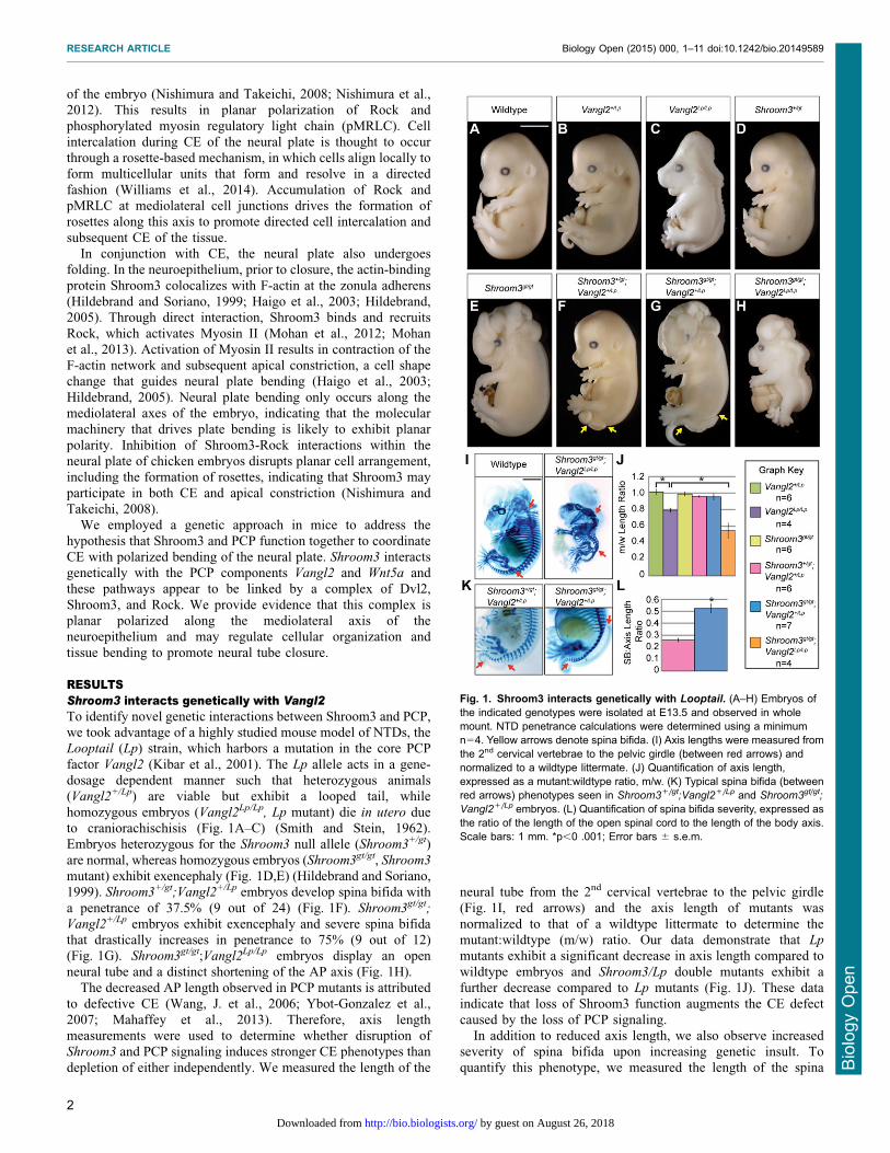
RESULTSShroom3 interacts genetically with Vangl2To identify novel genetic interactions between Shroom3 and PCP,we took advantage of a highly studied mouse model of NTDs, theLooptail (Lp) strain, which harbors a mutation in the core PCP
factor Vangl2 (Kibar et al., 2001). The Lp allele acts in a gene-dosage dependent manner such that heterozygous animals(Vangl2+/Lp) are viable but exhibit a looped tail, while
homozygous embryos (Vangl2Lp/Lp, Lp mutant) die in utero dueto craniorachischisis (Fig. 1A–C) (Smith and Stein, 1962).Embryos heterozygous for the Shroom3 null allele (Shroom3+/gt)
are normal, whereas homozygous embryos (Shroom3gt/gt, Shroom3
mutant) exhibit exencephaly (Fig. 1D,E) (Hildebrand and Soriano,1999). Shroom3+/gt;Vangl2+/Lp embryos develop spina bifida with
a penetrance of 37.5% (9 out of 24) (Fig. 1F). Shroom3gt/gt;
Vangl2+/Lp embryos exhibit exencephaly and severe spina bifidathat drastically increases in penetrance to 75% (9 out of 12)(Fig. 1G). Shroom3gt/gt;Vangl2Lp/Lp embryos display an open
neural tube and a distinct shortening of the AP axis (Fig. 1H).The decreased AP length observed in PCP mutants is attributed
to defective CE (Wang, J. et al., 2006; Ybot-Gonzalez et al.,
2007; Mahaffey et al., 2013). Therefore, axis lengthmeasurements were used to determine whether disruption ofShroom3 and PCP signaling induces stronger CE phenotypes than
depletion of either independently. We measured the length of the
neural tube from the 2nd cervical vertebrae to the pelvic girdle(Fig. 1I, red arrows) and the axis length of mutants was
normalized to that of a wildtype littermate to determine themutant:wildtype (m/w) ratio. Our data demonstrate that Lp
mutants exhibit a significant decrease in axis length compared to
wildtype embryos and Shroom3/Lp double mutants exhibit afurther decrease compared to Lp mutants (Fig. 1J). These dataindicate that loss of Shroom3 function augments the CE defect
caused by the loss of PCP signaling.In addition to reduced axis length, we also observe increased
severity of spina bifida upon increasing genetic insult. Toquantify this phenotype, we measured the length of the spina
Fig. 1. Shroom3 interacts genetically with Looptail. (A–H) Embryos ofthe indicated genotypes were isolated at E13.5 and observed in wholemount. NTD penetrance calculations were determined using a minimumn54. Yellow arrows denote spina bifida. (I) Axis lengths were measured fromthe 2nd cervical vertebrae to the pelvic girdle (between red arrows) andnormalized to a wildtype littermate. (J) Quantification of axis length,expressed as a mutant:wildtype ratio, m/w. (K) Typical spina bifida (betweenred arrows) phenotypes seen in Shroom3+/gt;Vangl2+/Lp and Shroom3gt/gt;
Vangl2+/Lp embryos. (L) Quantification of spina bifida severity, expressed asthe ratio of the length of the open spinal cord to the length of the body axis.Scale bars: 1 mm. *p,0 .001; Error bars 6 s.e.m.
RESEARCH ARTICLE Biology Open (2015) 000, 1–11 doi:10.1242/bio.20149589
2
BiologyOpen
by guest on August 26, 2018http://bio.biologists.org/Downloaded from
bifida in Shroom3+/gt;Vangl2+/Lp or Shroom3gt/gt;Vangl2+/Lp
embryos and these were normalized to the AP axis length of
each embryo (Fig. 1K, red arrows). Shroom3gt/gt;Vangl2+/Lp
embryos exhibit a significant increase in the length of the openneural tube as compared to Shroom3+/gt;Vangl2+/Lp embryos(Fig. 1L), suggesting that simultaneous disruption of Shroom3
and PCP increases both the frequency and severity of NTDs.
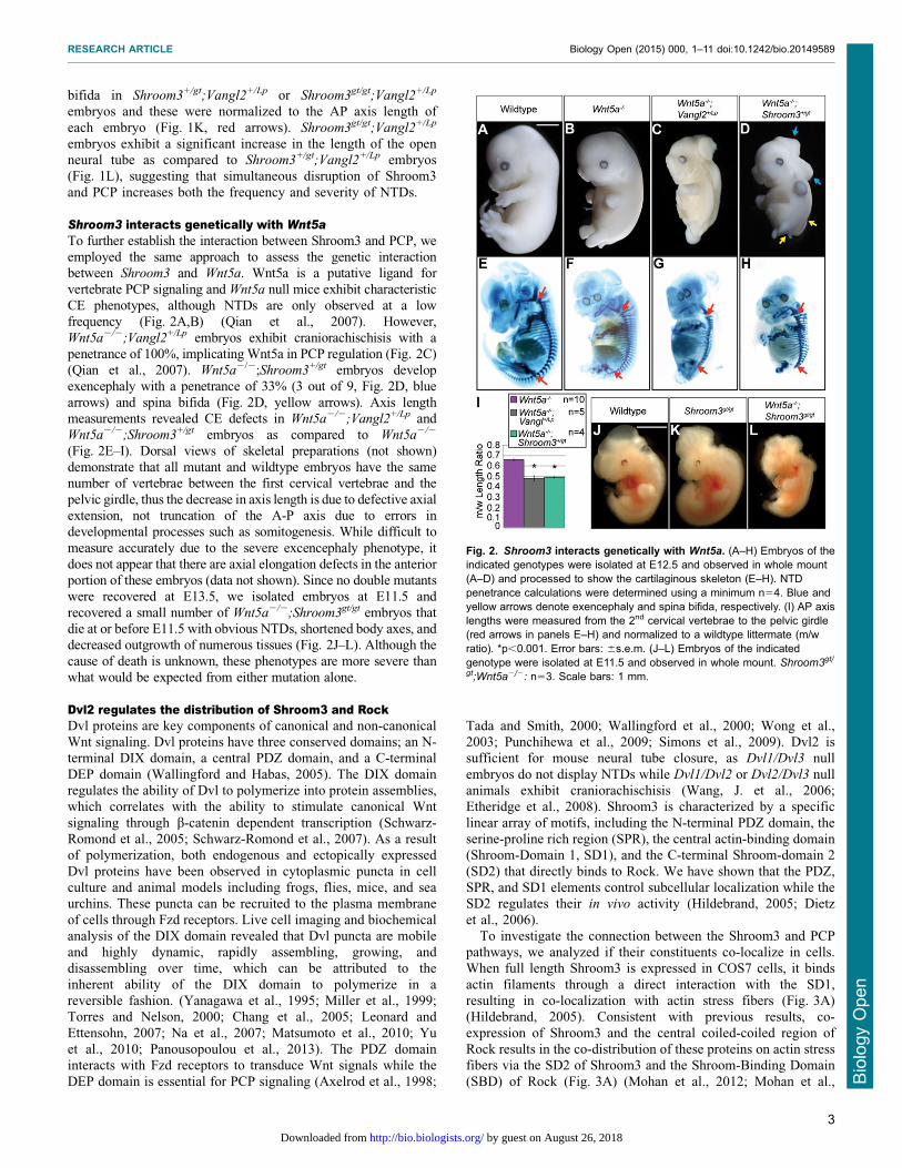
Shroom3 interacts genetically with Wnt5aTo further establish the interaction between Shroom3 and PCP, weemployed the same approach to assess the genetic interactionbetween Shroom3 and Wnt5a. Wnt5a is a putative ligand for
vertebrate PCP signaling and Wnt5a null mice exhibit characteristicCE phenotypes, although NTDs are only observed at a lowfrequency (Fig. 2A,B) (Qian et al., 2007). However,
Wnt5a2/2;Vangl2+/Lp embryos exhibit craniorachischisis with apenetrance of 100%, implicating Wnt5a in PCP regulation (Fig. 2C)(Qian et al., 2007). Wnt5a2/2;Shroom3+/gt embryos developexencephaly with a penetrance of 33% (3 out of 9, Fig. 2D, blue
arrows) and spina bifida (Fig. 2D, yellow arrows). Axis lengthmeasurements revealed CE defects in Wnt5a2/2;Vangl2+/Lp andWnt5a2/2;Shroom3+/gt embryos as compared to Wnt5a2/2
(Fig. 2E–I). Dorsal views of skeletal preparations (not shown)demonstrate that all mutant and wildtype embryos have the samenumber of vertebrae between the first cervical vertebrae and the
pelvic girdle, thus the decrease in axis length is due to defective axialextension, not truncation of the A-P axis due to errors indevelopmental processes such as somitogenesis. While difficult to
measure accurately due to the severe excencephaly phenotype, itdoes not appear that there are axial elongation defects in the anteriorportion of these embryos (data not shown). Since no double mutantswere recovered at E13.5, we isolated embryos at E11.5 and
recovered a small number of Wnt5a2/2;Shroom3gt/gt embryos thatdie at or before E11.5 with obvious NTDs, shortened body axes, anddecreased outgrowth of numerous tissues (Fig. 2J–L). Although the
cause of death is unknown, these phenotypes are more severe thanwhat would be expected from either mutation alone.
Dvl2 regulates the distribution of Shroom3 and RockDvl proteins are key components of canonical and non-canonicalWnt signaling. Dvl proteins have three conserved domains; an N-terminal DIX domain, a central PDZ domain, and a C-terminal
DEP domain (Wallingford and Habas, 2005). The DIX domainregulates the ability of Dvl to polymerize into protein assemblies,which correlates with the ability to stimulate canonical Wnt
signaling through b-catenin dependent transcription (Schwarz-Romond et al., 2005; Schwarz-Romond et al., 2007). As a resultof polymerization, both endogenous and ectopically expressed
Dvl proteins have been observed in cytoplasmic puncta in cellculture and animal models including frogs, flies, mice, and seaurchins. These puncta can be recruited to the plasma membrane
of cells through Fzd receptors. Live cell imaging and biochemicalanalysis of the DIX domain revealed that Dvl puncta are mobileand highly dynamic, rapidly assembling, growing, anddisassembling over time, which can be attributed to the
inherent ability of the DIX domain to polymerize in areversible fashion. (Yanagawa et al., 1995; Miller et al., 1999;Torres and Nelson, 2000; Chang et al., 2005; Leonard and
Ettensohn, 2007; Na et al., 2007; Matsumoto et al., 2010; Yuet al., 2010; Panousopoulou et al., 2013). The PDZ domaininteracts with Fzd receptors to transduce Wnt signals while the
DEP domain is essential for PCP signaling (Axelrod et al., 1998;
Tada and Smith, 2000; Wallingford et al., 2000; Wong et al.,2003; Punchihewa et al., 2009; Simons et al., 2009). Dvl2 issufficient for mouse neural tube closure, as Dvl1/Dvl3 null
embryos do not display NTDs while Dvl1/Dvl2 or Dvl2/Dvl3 nullanimals exhibit craniorachischisis (Wang, J. et al., 2006;Etheridge et al., 2008). Shroom3 is characterized by a specific
linear array of motifs, including the N-terminal PDZ domain, theserine-proline rich region (SPR), the central actin-binding domain(Shroom-Domain 1, SD1), and the C-terminal Shroom-domain 2
(SD2) that directly binds to Rock. We have shown that the PDZ,SPR, and SD1 elements control subcellular localization while theSD2 regulates their in vivo activity (Hildebrand, 2005; Dietz
et al., 2006).To investigate the connection between the Shroom3 and PCP
pathways, we analyzed if their constituents co-localize in cells.When full length Shroom3 is expressed in COS7 cells, it binds
actin filaments through a direct interaction with the SD1,resulting in co-localization with actin stress fibers (Fig. 3A)(Hildebrand, 2005). Consistent with previous results, co-
expression of Shroom3 and the central coiled-coiled region ofRock results in the co-distribution of these proteins on actin stressfibers via the SD2 of Shroom3 and the Shroom-Binding Domain
(SBD) of Rock (Fig. 3A) (Mohan et al., 2012; Mohan et al.,
Fig. 2. Shroom3 interacts genetically with Wnt5a. (A–H) Embryos of theindicated genotypes were isolated at E12.5 and observed in whole mount(A–D) and processed to show the cartilaginous skeleton (E–H). NTDpenetrance calculations were determined using a minimum n54. Blue andyellow arrows denote exencephaly and spina bifida, respectively. (I) AP axislengths were measured from the 2nd cervical vertebrae to the pelvic girdle(red arrows in panels E–H) and normalized to a wildtype littermate (m/wratio). *p,0.001. Error bars: 6s.e.m. (J–L) Embryos of the indicatedgenotype were isolated at E11.5 and observed in whole mount. Shroom3gt/
gt;Wnt5a2/2: n53. Scale bars: 1 mm.
RESEARCH ARTICLE Biology Open (2015) 000, 1–11 doi:10.1242/bio.20149589
3
BiologyOpen
by guest on August 26, 2018http://bio.biologists.org/Downloaded from
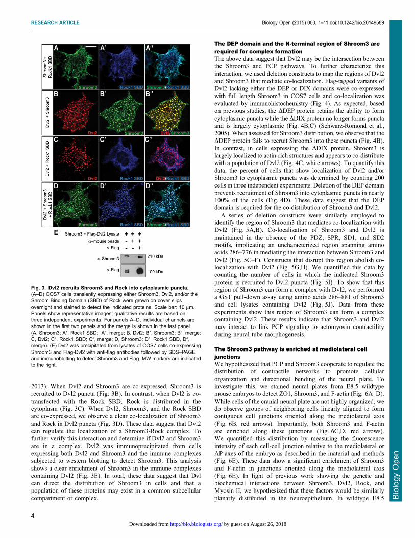
2013). When Dvl2 and Shroom3 are co-expressed, Shroom3 isrecruited to Dvl2 puncta (Fig. 3B). In contrast, when Dvl2 is co-
transfected with the Rock SBD, Rock is distributed in thecytoplasm (Fig. 3C). When Dvl2, Shroom3, and the Rock SBDare co-expressed, we observe a clear co-localization of Shroom3
and Rock in Dvl2 puncta (Fig. 3D). These data suggest that Dvl2can regulate the localization of a Shroom3-Rock complex. Tofurther verify this interaction and determine if Dvl2 and Shroom3
are in a complex, Dvl2 was immunoprecipitated from cellsexpressing both Dvl2 and Shroom3 and the immune complexessubjected to western blotting to detect Shroom3. This analysisshows a clear enrichment of Shroom3 in the immune complexes
containing Dvl2 (Fig. 3E). In total, these data suggest that Dvlcan direct the distribution of Shroom3 in cells and that apopulation of these proteins may exist in a common subcellular
compartment or complex.
The DEP domain and the N-terminal region of Shroom3 arerequired for complex formationThe above data suggest that Dvl2 may be the intersection betweenthe Shroom3 and PCP pathways. To further characterize thisinteraction, we used deletion constructs to map the regions of Dvl2and Shroom3 that mediate co-localization. Flag-tagged variants of
Dvl2 lacking either the DEP or DIX domains were co-expressedwith full length Shroom3 in COS7 cells and co-localization wasevaluated by immunohistochemistry (Fig. 4). As expected, based
on previous studies, the DDEP protein retains the ability to formcytoplasmic puncta while the DDIX protein no longer forms punctaand is largely cytoplasmic (Fig. 4B,C) (Schwarz-Romond et al.,
2005). When assessed for Shroom3 distribution, we observe that theDDEP protein fails to recruit Shroom3 into these puncta (Fig. 4B).In contrast, in cells expressing the DDIX protein, Shroom3 is
largely localized to actin-rich structures and appears to co-distributewith a population of Dvl2 (Fig. 4C, white arrows). To quantify thisdata, the percent of cells that show localization of Dvl2 and/orShroom3 to cytoplasmic puncta was determined by counting 200
cells in three independent experiments. Deletion of the DEP domainprevents recruitment of Shroom3 into cytoplasmic puncta in nearly100% of the cells (Fig. 4D). These data suggest that the DEP
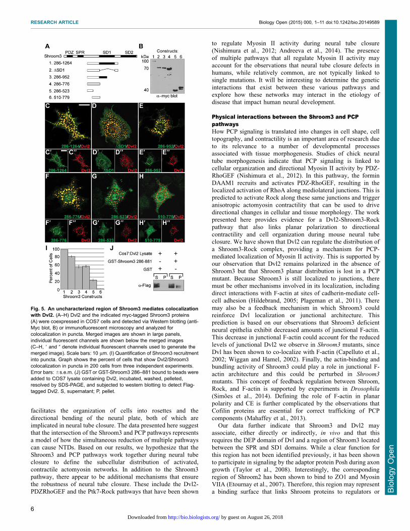
domain is required for the co-distribution of Shroom3 and Dvl2.A series of deletion constructs were similarly employed to
identify the region of Shroom3 that mediates co-localization with
Dvl2 (Fig. 5A,B). Co-localization of Shroom3 and Dvl2 ismaintained in the absence of the PDZ, SPR, SD1, and SD2motifs, implicating an uncharacterized region spanning amino
acids 286–776 in mediating the interaction between Shroom3 andDvl2 (Fig. 5C–F). Constructs that disrupt this region abolish co-localization with Dvl2 (Fig. 5G,H). We quantified this data bycounting the number of cells in which the indicated Shroom3
protein is recruited to Dvl2 puncta (Fig. 5I). To show that thisregion of Shroom3 can form a complex with Dvl2, we performeda GST pull-down assay using amino acids 286–881 of Shroom3
and cell lysates containing Dvl2 (Fig. 5J). Data from theseexperiments show this region of Shroom3 can form a complexcontaining Dvl2. These results indicate that Shroom3 and Dvl2
may interact to link PCP signaling to actomyosin contractilityduring neural tube morphogenesis.
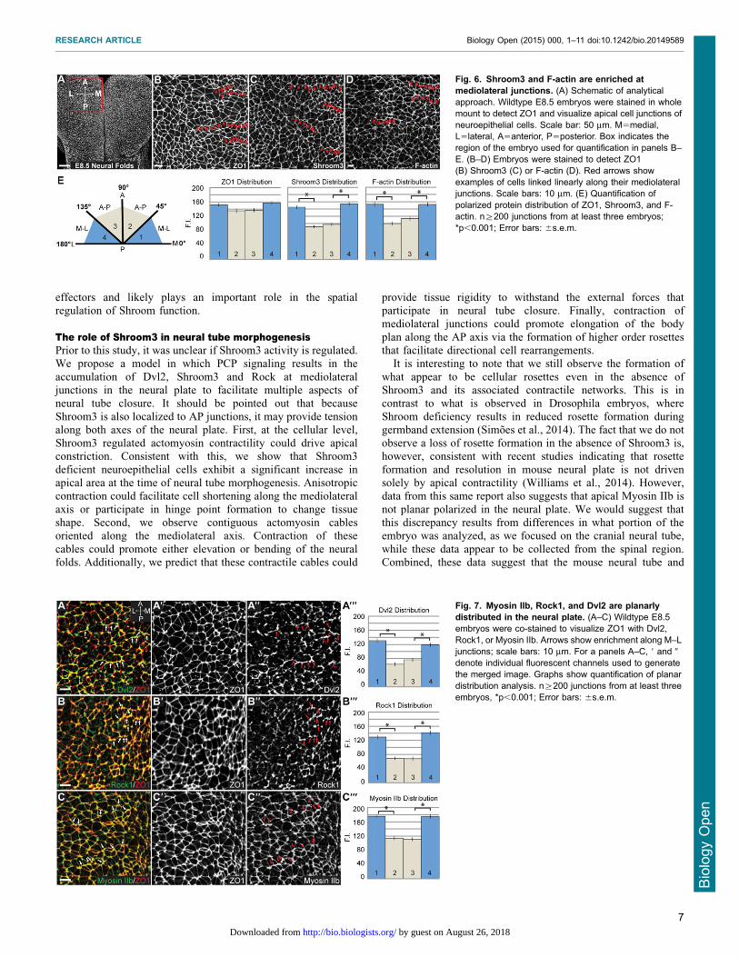
The Shroom3 pathway is enriched at mediolateral celljunctionsWe hypothesized that PCP and Shroom3 cooperate to regulate thedistribution of contractile networks to promote cellular
organization and directional bending of the neural plate. Toinvestigate this, we stained neural plates from E8.5 wildtypemouse embryos to detect ZO1, Shroom3, and F-actin (Fig. 6A–D).
While cells of the cranial neural plate are not highly organized, wedo observe groups of neighboring cells linearly aligned to formcontiguous cell junctions oriented along the mediolateral axis
(Fig. 6B, red arrows). Importantly, both Shroom3 and F-actinare enriched along these junctions (Fig. 6C,D, red arrows).We quantified this distribution by measuring the fluorescenceintensity of each cell-cell junction relative to the mediolateral or
AP axes of the embryo as described in the material and methods(Fig. 6E). These data show a significant enrichment of Shroom3and F-actin in junctions oriented along the mediolateral axis
(Fig. 6E). In light of previous work showing the genetic andbiochemical interactions between Shroom3, Dvl2, Rock, andMyosin II, we hypothesized that these factors would be similarly
planarly distributed in the neuroepithelium. In wildtype E8.5
Fig. 3. Dvl2 recruits Shroom3 and Rock into cytoplasmic puncta.(A–D) COS7 cells transiently expressing either Shroom3, Dvl2, and/or theShroom Binding Domain (SBD) of Rock were grown on cover slipsovernight and stained to detect the indicated proteins. Scale bar: 10 mm.Panels show representative images; qualitative results are based onthree independent experiments. For panels A–D, individual channels areshown in the first two panels and the merge is shown in the last panel(A, Shroom3; A9, Rock1 SBD; A0, merge; B, Dvl2; B9, Shroom3; B0, merge;C, Dvl2; C9, Rock1 SBD; C0, merge; D, Shroom3; D9, Rock1 SBD, D0,merge). (E) Dvl2 was precipitated from lysates of COS7 cells co-expressingShroom3 and Flag-Dvl2 with anti-flag antibodies followed by SDS–PAGEand immunoblotting to detect Shroom3 and Flag. MW markers are indicatedto the right.
RESEARCH ARTICLE Biology Open (2015) 000, 1–11 doi:10.1242/bio.20149589
4
BiologyOpen
by guest on August 26, 2018http://bio.biologists.org/Downloaded from

embryos, Dvl2, Rock1 and Myosin IIb are all enriched atmediolateral cell junctions (Fig. 7A–C).
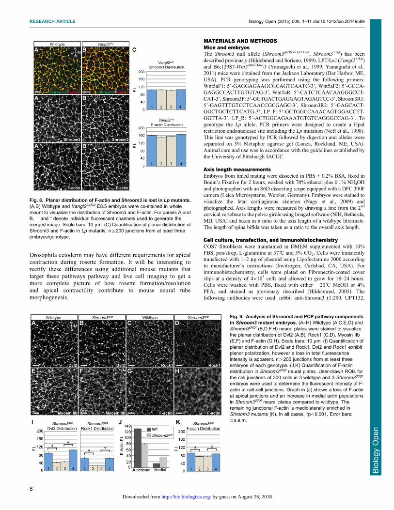
PCP is upstream of Shroom3To determine if Shroom3 is downstream of PCP signaling, we
stained E8.5 Shroom3gt/gt and Vangl2Lp/Lp embryos to detectcomponents of these pathways (Fig. 8A,B, Fig. 9A–H). In Lp
mutants, Shroom3 and F-actin are localized to cell-cell junctionsbut are no longer enriched at mediolateral junctions, indicating that
the general localization signals are intact, but mediolateralasymmetry of the adherens junctions is lost (Fig. 8A–C vsFig. 6C–E). In Shroom3gt/gt embryos, Dvl2 is recruited to
junctions and remains planarly polarized (Fig. 9A,B), althoughthe amount of Dvl2 recruited to cell junctions may be reduced(Fig. 7A vs Fig. 9I). Rock1 localization is significantly impaired in
Shroom3gt/gt embryos (Fig. 9C–F) but the remaining Rock1 ismediolaterally enriched, suggesting there are other mechanisms forlocalization (Fig. 9I). Consistent with the loss of Rock1 at
cell junctions, we see reduced junctional Myosin IIb andincreased cytoplasmic Myosin IIb (Fig. 9E,F). This increasedcytoplasmic staining prevents accurate quantification of the MyosinIIb distribution. These results provide evidence that Shroom3
recruits Rock1 and Myosin IIb to adherens junctions while the PCPpathway refines the spatial distribution of the pathway.
Based on the above results, we assessed if Shroom3 loss affects
the apical actin cytoskeleton of the neuroepithelium. Shroom3deficiency results in a diffuse, discontinuous apicojunctional F-actin network and the appearance of medial or cytoplasmic F-
actin (Fig. 9G,H). In wildtype embryos, 90% of the F-actinfluorescence is localized along cell-cell junctions. Shroom3gt/gt
embryos exhibit an approximate 40% decrease in the
fluorescence intensity of junctional F-actin and a concomitantincrease in the medial population (Fig. 9J). Consistent with whatwe observe for Rock1, the remaining junctional F-actin exhibits adistribution bias along the mediolateral axis (Fig. 9K). These data
demonstrate that Shroom3 functions downstream of PCP toregulate the distribution of Rock and Myosin II, which may feedback to regulate F-actin organization.
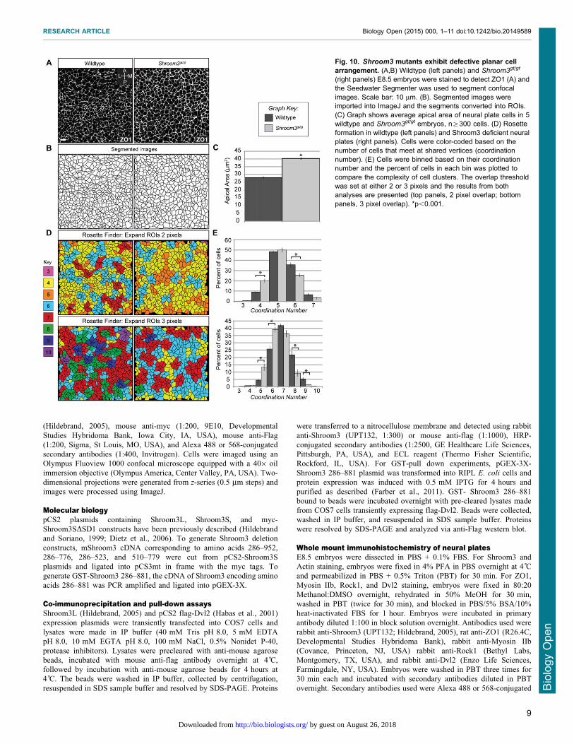
Loss of Shroom3 disrupts planar cell arrangementBased on the above results, we hypothesized that Shroom3-
induced tension at mediolateral junctions facilitates the formation
of cellular rosettes to promote CE. To test this, we stained neuralplates of E8.5 embryos to detect ZO1 to visualize the planar
arrangement of cells. Images were then segmented [SeedWaterSegmenter (Mashburn et al., 2012)] to produce separate Regionsof Interest (ROI) for each cell (Fig. 10A,B). Analysis of
segmented images indicates that Shroom3 deficient cells exhibita 48% increase in apical area (Fig. 10C), consistent with its rolein apical constriction.
ROIs were further analyzed using a custom ImageJ macro to
determine the number of cells that meet at shared vertices. This isreferred to as the coordination number and represents the cellularcomplexity of the rosette, such that cells in rosettes will have a
higher number of cells that share a common vertex. Coordinationnumbers were calculated based on cell overlap after the ROIs wereexpanded. Cell clusters were either color coded (Fig. 10D) or
binned (Fig. 10E), based on their coordination number. Cells inmultiple clusters were assigned the highest coordination number. Inpractice we found that changing the overlap threshold from 2 to 3
pixels increased the number of rosettes as well as the cellularity ofeach rosette. For comparison we present both analyses and find thatthey report qualitatively similar results. Both wildtype and Shroom3
deficient neural plates exhibit clusters containing 3–10 cells,
suggesting that Shroom3 is not required for rosettes (Fig. 10D). Thepercent of cells with each coordination number, relative to the totalnumber of cells, was graphed and used to compare the complexity
of rosettes between wildtype and Shroom3gt/gt embryos (Fig. 10E).This analysis demonstrates that Shroom3 mutants exhibit asignificant decrease in the complexity of the rosettes but not the
total number of rosettes. Live-imaging studies of mouse neuralplates and elongating kidney tubules have shown that loss of rosettecomplexity within the tissue results in a loss of cellular remodeling
and neighbor exchange and an overall decrease in tissue extension(Lienkamp et al., 2012; Williams et al., 2014). Therefore, our datasuggest that Shroom3 is likely required for the cellular remodelingthat facilitates CE and neural tube closure.
DISCUSSIONCooperation of the Shroom and PCP pathwaysWe have shown that the Shroom3 and PCP pathways interact tocontrol the planar distribution of apical contractile actomyosinnetworks in epithelial cells of the neural plate. We predict that the
asymmetric distribution of the Shroom3-Rock-Myosin II pathway
Fig. 4. The DEP domain of Dvl2 facilitatesinteraction with Shroom3. (A–C) Mapping theShroom3 binding region of Dvl2. Indicated deletionmutants of Dvl2 (A) and Shroom3 were transientlycoexpressed in COS7 cells and detected viaimmunofluorescent microcopy (B,C). Individualchannels are shown in the first two panels and themerge is shown in the last panel (B, Dvl2DDEP;B9, Shroom3; B0, merge; C, Dvl2DDIX; C9, Shroom3;C0, merge). Deletion of the DEP domain preventsrecruitment of Shroom3 into puncta. Deletion of theDIX domain prevents Dvl2 polymerization and thusno puncta are observed. Some Dvl2/Shroom3colocalization is observed at actin stress fibers(C, arrows) Scale bars: 10 mm. (D) Quantification ofShroom3 recruitment into puncta. Graph shows thepercent of cells that show Dvl2 or Shoorm3 localizedto puncta in 200 cells from three independentexperiments. Error bars: 6s.e.m.
RESEARCH ARTICLE Biology Open (2015) 000, 1–11 doi:10.1242/bio.20149589
5
BiologyOpen
by guest on August 26, 2018http://bio.biologists.org/Downloaded from
facilitates the organization of cells into rosettes and thedirectional bending of the neural plate, both of which are
implicated in neural tube closure. The data presented here suggestthat the intersection of the Shroom3 and PCP pathways representsa model of how the simultaneous reduction of multiple pathways
can cause NTDs. Based on our results, we hypothesize that theShroom3 and PCP pathways work together during neural tubeclosure to define the subcellular distribution of activated,contractile actomyosin networks. In addition to the Shroom3
pathway, there appear to be additional mechanisms that ensurethe robustness of neural tube closure. These include the Dvl2-PDZRhoGEF and the Ptk7-Rock pathways that have been shown
to regulate Myosin II activity during neural tube closure(Nishimura et al., 2012; Andreeva et al., 2014). The presence
of multiple pathways that all regulate Myosin II activity mayaccount for the observations that neural tube closure defects inhumans, while relatively common, are not typically linked tosingle mutations. It will be interesting to determine the genetic
interactions that exist between these various pathways andexplore how these networks may interact in the etiology ofdisease that impact human neural development.
Physical interactions between the Shroom3 and PCPpathwaysHow PCP signaling is translated into changes in cell shape, celltopography, and contractility is an important area of research dueto its relevance to a number of developmental processes
associated with tissue morphogenesis. Studies of chick neuraltube morphogenesis indicate that PCP signaling is linked tocellular organization and directional Myosin II activity by PDZ-RhoGEF (Nishimura et al., 2012). In this pathway, the formin
DAAM1 recruits and activates PDZ-RhoGEF, resulting in thelocalized activation of RhoA along mediolateral junctions. This ispredicted to activate Rock along these same junctions and trigger
anisotropic actomyosin contractility that can be used to drivedirectional changes in cellular and tissue morphology. The workpresented here provides evidence for a Dvl2-Shroom3-Rock
pathway that also links planar polarization to directionalcontractility and cell organization during mouse neural tubeclosure. We have shown that Dvl2 can regulate the distribution of
a Shroom3-Rock complex, providing a mechanism for PCP-mediated localization of Myosin II activity. This is supported byour observation that Dvl2 remains polarized in the absence ofShroom3 but that Shroom3 planar distribution is lost in a PCP
mutant. Because Shroom3 is still localized to junctions, theremust be other mechanisms involved in its localization, includingdirect interactions with F-actin at sites of cadherin-mediate cell-
cell adhesion (Hildebrand, 2005; Plageman et al., 2011). Theremay also be a feedback mechanism in which Shroom3 couldreinforce Dvl localization or junctional architecture. This
prediction is based on our observations that Shroom3 deficientneural epithelia exhibit decreased amounts of junctional F-actin.This decrease in junctional F-actin could account for the reducedlevels of junctional Dvl2 we observe in Shroom3 mutants, since
Dvl has been shown to co-localize with F-actin (Capelluto et al.,2002; Wiggan and Hamel, 2002). Finally, the actin-binding andbundling activity of Shroom3 could play a role in junctional F-
actin architecture and this could be perturbed in Shroom3
mutants. This concept of feedback regulation between Shroom,Rock, and F-actin is supported by experiments in Drosophila
(Simoes et al., 2014). Defining the role of F-actin in planarpolarity and CE is further complicated by the observations thatCofilin proteins are essential for correct trafficking of PCP
components (Mahaffey et al., 2013).Our data further indicate that Shroom3 and Dvl2 may
associate, either directly or indirectly, in vivo and that thisrequires the DEP domain of Dvl and a region of Shroom3 located
between the SPR and SD1 domains. While a clear function forthis region has not been identified previously, it has been shownto participate in signaling by the adaptor protein Posh during axon
growth (Taylor et al., 2008). Interestingly, the correspondingregion of Shroom2 has been shown to bind to ZO1 and MyosinVIIA (Etournay et al., 2007). Therefore, this region may represent
a binding surface that links Shroom proteins to regulators or
Fig. 5. An uncharacterized region of Shroom3 mediates colocalizationwith Dvl2. (A–H) Dvl2 and the indicated myc-tagged Shroom3 proteins(A) were coexpressed in COS7 cells and detected via Western blotting (anti-Myc blot, B) or immunofluorescent microscopy and analyzed forcolocalization in puncta. Merged images are shown in large panels,individual fluorescent channels are shown below the merged images(C–H, 9 and 0 denote individual fluorescent channels used to generate themerged image). Scale bars: 10 mm. (I) Quantification of Shroom3 recruitmentinto puncta. Graph shows the percent of cells that show Dvl2/Shroom3colocalization in puncta in 200 cells from three independent experiments.Error bars: 6s.e.m. (J) GSTor GST-Shroom3 286–881 bound to beads wereadded to COS7 lysate containing Dvl2, incubated, washed, pelleted,resolved by SDS-PAGE, and subjected to western blotting to detect Flag-tagged Dvl2. S, supernatant; P, pellet.
RESEARCH ARTICLE Biology Open (2015) 000, 1–11 doi:10.1242/bio.20149589
6
BiologyOpen
by guest on August 26, 2018http://bio.biologists.org/Downloaded from
effectors and likely plays an important role in the spatialregulation of Shroom function.
The role of Shroom3 in neural tube morphogenesisPrior to this study, it was unclear if Shroom3 activity is regulated.We propose a model in which PCP signaling results in the
accumulation of Dvl2, Shroom3 and Rock at mediolateraljunctions in the neural plate to facilitate multiple aspects ofneural tube closure. It should be pointed out that because
Shroom3 is also localized to AP junctions, it may provide tensionalong both axes of the neural plate. First, at the cellular level,Shroom3 regulated actomyosin contractility could drive apicalconstriction. Consistent with this, we show that Shroom3
deficient neuroepithelial cells exhibit a significant increase inapical area at the time of neural tube morphogenesis. Anisotropiccontraction could facilitate cell shortening along the mediolateral
axis or participate in hinge point formation to change tissueshape. Second, we observe contiguous actomyosin cablesoriented along the mediolateral axis. Contraction of these
cables could promote either elevation or bending of the neuralfolds. Additionally, we predict that these contractile cables could
provide tissue rigidity to withstand the external forces thatparticipate in neural tube closure. Finally, contraction ofmediolateral junctions could promote elongation of the body
plan along the AP axis via the formation of higher order rosettesthat facilitate directional cell rearrangements.
It is interesting to note that we still observe the formation of
what appear to be cellular rosettes even in the absence ofShroom3 and its associated contractile networks. This is incontrast to what is observed in Drosophila embryos, where
Shroom deficiency results in reduced rosette formation duringgermband extension (Simoes et al., 2014). The fact that we do notobserve a loss of rosette formation in the absence of Shroom3 is,however, consistent with recent studies indicating that rosette
formation and resolution in mouse neural plate is not drivensolely by apical contractility (Williams et al., 2014). However,data from this same report also suggests that apical Myosin IIb is
not planar polarized in the neural plate. We would suggest thatthis discrepancy results from differences in what portion of theembryo was analyzed, as we focused on the cranial neural tube,
while these data appear to be collected from the spinal region.Combined, these data suggest that the mouse neural tube and
Fig. 6. Shroom3 and F-actin are enriched atmediolateral junctions. (A) Schematic of analyticalapproach. Wildtype E8.5 embryos were stained in wholemount to detect ZO1 and visualize apical cell junctions ofneuroepithelial cells. Scale bar: 50 mm. M5medial,L5lateral, A5anterior, P5posterior. Box indicates theregion of the embryo used for quantification in panels B–E. (B–D) Embryos were stained to detect ZO1(B) Shroom3 (C) or F-actin (D). Red arrows showexamples of cells linked linearly along their mediolateraljunctions. Scale bars: 10 mm. (E) Quantification ofpolarized protein distribution of ZO1, Shroom3, and F-actin. n§200 junctions from at least three embryos;*p,0.001; Error bars: 6s.e.m.
Fig. 7. Myosin IIb, Rock1, and Dvl2 are planarlydistributed in the neural plate. (A–C) Wildtype E8.5embryos were co-stained to visualize ZO1 with Dvl2,Rock1, or Myosin IIb. Arrows show enrichment along M–Ljunctions; scale bars: 10 mm. For a panels A–C, 9 and 0
denote individual fluorescent channels used to generatethe merged image. Graphs show quantification of planardistribution analysis. n§200 junctions from at least threeembryos, *p,0.001; Error bars: 6s.e.m.
RESEARCH ARTICLE Biology Open (2015) 000, 1–11 doi:10.1242/bio.20149589
7
BiologyOpen
by guest on August 26, 2018http://bio.biologists.org/Downloaded from
Drosophila ectoderm may have different requirements for apical
contraction during rosette formation. It will be interesting torectify these differences using additional mouse mutants thattarget these pathways pathway and live cell imaging to get a
more complete picture of how rosette formation/resolutionand apical contractility contribute to mouse neural tubemorphogenesis.
MATERIALS AND METHODSMice and embryosThe Shroom3 null allele (Shroom3gt(ROSA)53sor, Shroom3+/gt) has been
described previously (Hildebrand and Soriano, 1999). LPT/LeJ (Vangl2+/Lp)
and B6;129S7-Wnt5atm1Amc/J (Yamaguchi et al., 1999; Yamaguchi et al.,
2011) mice were obtained from the Jackson Laboratory (Bar Harbor, ME,
USA). PCR genotyping was performed using the following primers:
Wnt5aF1: 59-GAGGAGAAGCGCAGTCAATC-39, Wnt5aF2: 59-GCCA-
GAGGCCACTTGTGTAG-39, Wnt5aR: 59-CATCTCAACAAGGGCCT-
CAT-39, Shroom3F: 59-GGTGACTGAGGAGTAGAGTCC-39, Shroom3R1:
59-GAGTTTGTCCTCAACCGCGAGC-39, Shroom3R2: 59-GAGCACT-
GGCTGCTCTTCATG-39, LP_F: 59-GCTGGCCAAACAGTGGACCTT-
GGTTA-39, LP_R: 59-ACTGGCAGAAATGTGTCAGGGCCAG-39. To
genotype the Lp allele, PCR primers were designed to create a HpaI
restriction endonuclease site including the Lp mutation (Neff et al., 1998).
This line was genotyped by PCR followed by digestion and alleles were
separated on 3% Metaphor agarose gel (Lonza, Rockland, ME, USA).
Animal care and use was in accordance with the guidelines established by
the University of Pittsburgh IACUC.
Axis length measurementsEmbryos from timed mating were dissected in PBS + 0.2% BSA, fixed in
Bouin’s Fixative for 2 hours, washed with 70% ethanol plus 0.1% NH4OH
and photographed with an S6D dissecting scope equipped with a DFC 300F
camera (Leica Microsystems, Wetzlar, Germany). Embryos were stained to
visualize the fetal cartilaginous skeleton (Nagy et al., 2009) and
photographed. Axis lengths were measured by drawing a line from the 2nd
cervical vertebrae to the pelvic girdle using ImageJ software (NIH, Bethesda,
MD, USA) and taken as a ratio to the axis length of a wildtype littermate.
The length of spina bifida was taken as a ratio to the overall axis length.
Cell culture, transfection, and immunohistochemistryCOS7 fibroblasts were maintained in DMEM supplemented with 10%
FBS, pen/strep, L-glutamine at 37 C and 5% CO2. Cells were transiently
transfected with 1–2 mg of plasmid using Lipofectamine 2000 according
to manufacturer’s instructions (Invitrogen, Carlsbad, CA, USA). For
immunohistochemistry, cells were plated on Fibronectin-coated cover
slips at a density of 46105 cells and allowed to grow for 18–24 hours.
Cells were washed with PBS, fixed with either 220 C MeOH or 4%
PFA, and stained as previously described (Hildebrand, 2005). The
following antibodies were used: rabbit anti-Shroom3 (1:200, UPT132,
Fig. 8. Planar distribution of F-actin and Shroom3 is lost in Lp mutants.(A,B) Wildtype and Vangl2Lp/Lp E8.5 embryos were co-stained in wholemount to visualize the distribution of Shroom3 and F-actin. For panels A andB, 9 and 0 denote individual fluorescent channels used to generate themerged image. Scale bars: 10 mm. (C) Quantification of planar distribution ofShroom3 and F-actin in Lp mutants. n§200 junctions from at least threeembryos/genotype.
Fig. 9. Analysis of Shroom3 and PCP pathway componentsin Shroom3 mutant embryos. (A–H) Wildtype (A,C,E,G) andShroom3gt/gt (B,D,F,H) neural plates were stained to visualizethe planar distribution of Dvl2 (A,B), Rock1 (C,D), Myosin IIb(E,F) and F-actin (G,H). Scale bars: 10 mm. (I) Quantification ofplanar distribution of Dvl2 and Rock1. Dvl2 and Rock1 exhibitplanar polarization, however a loss in total fluorescenceintensity is apparent. n§200 junctions from at least threeembryos of each genotype. (J,K) Quantification of F-actindistribution in Shroom3gt/gt neural plates. User-drawn ROIs forthe cell junctions of 300 cells in 3 wildtype and 3 Shroom3gt/gt
embryos were used to determine the fluorescent intensity of F-actin at cell-cell junctions. Graph in (J) shows a loss of F-actinat apical junctions and an increase in medial actin populationsin Shroom3gt/gt neural plates compared to wildtype. Theremaining junctional F-actin is mediolaterally enriched inShroom3 mutants (K). In all cases, *p,0.001, Error bars:6s.e.m.
RESEARCH ARTICLE Biology Open (2015) 000, 1–11 doi:10.1242/bio.20149589
8
BiologyOpen
by guest on August 26, 2018http://bio.biologists.org/Downloaded from
(Hildebrand, 2005), mouse anti-myc (1:200, 9E10, Developmental
Studies Hybridoma Bank, Iowa City, IA, USA), mouse anti-Flag
(1:200, Sigma, St Louis, MO, USA), and Alexa 488 or 568-conjugated
secondary antibodies (1:400, Invitrogen). Cells were imaged using an
Olympus Fluoview 1000 confocal microscope equipped with a 406 oil
immersion objective (Olympus America, Center Valley, PA, USA). Two-
dimensional projections were generated from z-series (0.5 mm steps) and
images were processed using ImageJ.
Molecular biologypCS2 plasmids containing Shroom3L, Shroom3S, and myc-
Shroom3SDSD1 constructs have been previously described (Hildebrand
and Soriano, 1999; Dietz et al., 2006). To generate Shroom3 deletion
constructs, mShroom3 cDNA corresponding to amino acids 286–952,
286–776, 286–523, and 510–779 were cut from pCS2-Shroom3S
plasmids and ligated into pCS3mt in frame with the myc tags. To
generate GST-Shroom3 286–881, the cDNA of Shroom3 encoding amino
acids 286–881 was PCR amplified and ligated into pGEX-3X.
Co-immunoprecipitation and pull-down assaysShroom3L (Hildebrand, 2005) and pCS2 flag-Dvl2 (Habas et al., 2001)
expression plasmids were transiently transfected into COS7 cells and
lysates were made in IP buffer (40 mM Tris pH 8.0, 5 mM EDTA
pH 8.0, 10 mM EGTA pH 8.0, 100 mM NaCl, 0.5% Nonidet P-40,
protease inhibitors). Lysates were precleared with anti-mouse agarose
beads, incubated with mouse anti-flag antibody overnight at 4 C,
followed by incubation with anti-mouse agarose beads for 4 hours at
4 C. The beads were washed in IP buffer, collected by centrifugation,
resuspended in SDS sample buffer and resolved by SDS-PAGE. Proteins
were transferred to a nitrocellulose membrane and detected using rabbit
anti-Shroom3 (UPT132, 1:300) or mouse anti-flag (1:1000), HRP-
conjugated secondary antibodies (1:2500, GE Healthcare Life Sciences,
Pittsburgh, PA, USA), and ECL reagent (Thermo Fisher Scientific,
Rockford, IL, USA). For GST-pull down experiments, pGEX-3X-
Shroom3 286–881 plasmid was transformed into RIPL E. coli cells and
protein expression was induced with 0.5 mM IPTG for 4 hours and
purified as described (Farber et al., 2011). GST- Shroom3 286–881
bound to beads were incubated overnight with pre-cleared lysates made
from COS7 cells transiently expressing flag-Dvl2. Beads were collected,
washed in IP buffer, and resuspended in SDS sample buffer. Proteins
were resolved by SDS-PAGE and analyzed via anti-Flag western blot.
Whole mount immunohistochemistry of neural platesE8.5 embryos were dissected in PBS + 0.1% FBS. For Shroom3 and
Actin staining, embryos were fixed in 4% PFA in PBS overnight at 4 C
and permeabilized in PBS + 0.5% Triton (PBT) for 30 min. For ZO1,
Myosin IIb, Rock1, and Dvl2 staining, embryos were fixed in 80:20
Methanol:DMSO overnight, rehydrated in 50% MeOH for 30 min,
washed in PBT (twice for 30 min), and blocked in PBS/5% BSA/10%
heat-inactivated FBS for 1 hour. Embryos were incubated in primary
antibody diluted 1:100 in block solution overnight. Antibodies used were
rabbit anti-Shroom3 (UPT132; Hildebrand, 2005), rat anti-ZO1 (R26.4C,
Developmental Studies Hybridoma Bank), rabbit anti-Myosin IIb
(Covance, Princeton, NJ, USA) rabbit anti-Rock1 (Bethyl Labs,
Montgomery, TX, USA), and rabbit anti-Dvl2 (Enzo Life Sciences,
Farmingdale, NY, USA). Embryos were washed in PBT three times for
30 min each and incubated with secondary antibodies diluted in PBT
overnight. Secondary antibodies used were Alexa 488 or 568-conjugated
Fig. 10. Shroom3 mutants exhibit defective planar cellarrangement. (A,B) Wildtype (left panels) and Shroom3gt/gt
(right panels) E8.5 embryos were stained to detect ZO1 (A) andthe Seedwater Segmenter was used to segment confocalimages. Scale bar: 10 mm. (B). Segmented images wereimported into ImageJ and the segments converted into ROIs.(C) Graph shows average apical area of neural plate cells in 5wildtype and Shroom3gt/gt embryos, n§300 cells. (D) Rosetteformation in wildtype (left panels) and Shroom3 deficient neuralplates (right panels). Cells were color-coded based on thenumber of cells that meet at shared vertices (coordinationnumber). (E) Cells were binned based on their coordinationnumber and the percent of cells in each bin was plotted tocompare the complexity of cell clusters. The overlap thresholdwas set at either 2 or 3 pixels and the results from bothanalyses are presented (top panels, 2 pixel overlap; bottompanels, 3 pixel overlap). *p,0.001.
RESEARCH ARTICLE Biology Open (2015) 000, 1–11 doi:10.1242/bio.20149589
9
BiologyOpen
by guest on August 26, 2018http://bio.biologists.org/Downloaded from

goat anti-rabbit or rat (1:400, Invitrogen). Tritc-phalloidin (1:500, Sigma)
was used to detect actin. Embryos were washed with PBT and mounted in
2% agarose for imaging. Imaging was done using a Leica TCS SP5
confocal microscope equipped with an APO L 206/1.00 water immersion
objective. Z-series (1 mm steps) were collected and projections were
generated using ImageJ.
Analysis of planar distribution and cellular arrangementUsing ImageJ, 1-pixel wide user-drawn lines across cell junctions of non-
saturated confocal projections were analyzed to determine the mean grey
value and orientation relative to the mediolateral axis of the embryo.
Those junctions between 0˚and 45˚or between 135˚and 180˚(Bins 1 and
4) were considered to be oriented along the mediolateral axis. Those
between 45˚and 135˚(Bins 2 and 3) were considered to be oriented along
the AP axis. Junctions were binned according to their angle and the
fluorescence intensities in each bin were averaged. To quantify junctional
versus medial F-actin fluorescence, the average fluorescence intensity
was measured using either ROIs drawn along the junctions (junctional) or
ROIs drawn inside the cell boundary (medial). For each experiment, at
least 200 cells were analyzed from each of three embryos.
The semi-automated image-segmenting program Seedwater Segmenter
(Mashburn et al., 2012) was used to segment images from five wildtype
and five Shroom3gt/gt embryos to produce separate ROIs for each cell.
Area measurements of each ROI were used to compare the average apical
area of neural plate cells in wildtype versus Shroom3gt/gt embryos.
n§300 cells. ROIs with an average area of 800 pixels were expanded by
2 or 3 pixels and a custom image analysis tool was used to calculate the
coordination number of each expanded ROIs. Cells were color-coded,
binned based on their coordination number, and the percent of cells in
each bin plotted.
Statistical analysesMeasures of significance were determined by two-tailed, unpaired,
Student’s t-tests with p,0.001. For all graphs, error bars represent
standard error of mean (6s.e.m.).
AcknowledgementsWe thank Dr. Raymond Habas for donating Dvl2 plasmids. Anti-myc and anti-ZO1antibodies were developed by Bishop, J.M. and Goodenough, D.A., respectively,and obtained from the Developmental Studies Hybridoma Bank, created by theNIH and maintained at The University of Iowa, Department of Biology, Iowa City,IA, USA.
Competing interestsThe authors declare no competing or financial interests.
Author contributionsE.M.M. conducted experiments, analyzed and interpreted the data, and togetherwith J.D.H. wrote the manuscript. D.V. and L.A.D. provided custom imageanalysis software, assisted with data analysis, and provided critical feedback.J.D.H. conceived and designed the study.
FundingThis work was supported by funds from the National Institute of General MedicalSciences and the Central Research Development Fund from the University ofPittsburgh (J.D.H.).
ReferencesAndreeva, A., Lee, J., Lohia, M., Wu, X., Macara, I. G. and Lu, X. (2014). PTK7-Src signaling at epithelial cell contacts mediates spatial organization ofactomyosin and planar cell polarity. Dev. Cell 29, 20-33.
Axelrod, J. D. (2001). Unipolar membrane association of Dishevelled mediatesFrizzled planar cell polarity signaling. Genes Dev. 15, 1182-1187.
Axelrod, J. D., Miller, J. R., Shulman, J. M., Moon, R. T. and Perrimon, N. (1998).Differential recruitment of Dishevelled provides signaling specificity in the planarcell polarity and Wingless signaling pathways. Genes Dev. 12, 2610-2622.
Bastock, R., Strutt, H. and Strutt, D. (2003). Strabismus is asymmetricallylocalised and binds to Prickle and Dishevelled during Drosophila planar polaritypatterning. Development 130, 3007-3014.
Capelluto, D. G., Kutateladze, T. G., Habas, R., Finkielstein, C. V., He, X. andOverduin, M. (2002). The DIX domain targets dishevelled to actin stress fibresand vesicular membranes. Nature 419, 726-729.
Chang, W., Lloyd, C. E. and Zarkower, D. (2005). DSH-2 regulates asymmetriccell division in the early C. elegans somatic gonad. Mech. Dev. 122, 781-789.
Chen, W. S., Antic, D., Matis, M., Logan, C. Y., Povelones, M., Anderson, G. A.,Nusse, R. and Axelrod, J. D. (2008). Asymmetric homotypic interactions of theatypical cadherin flamingo mediate intercellular polarity signaling. Cell 133,1093-1105.
Ciruna, B., Jenny, A., Lee, D., Mlodzik, M. and Schier, A. F. (2006). Planar cellpolarity signalling couples cell division and morphogenesis during neurulation.Nature 439, 220-224.
Curtin, J. A., Quint, E., Tsipouri, V., Arkell, R. M., Cattanach, B., Copp, A. J.,Henderson, D. J., Spurr, N., Stanier, P., Fisher, E. M. et al. (2003). Mutation ofCelsr1 disrupts planar polarity of inner ear hair cells and causes severe neuraltube defects in the mouse. Curr. Biol. 13, 1129-1133.
Dabdoub, A. and Kelley, M. W. (2005). Planar cell polarity and a potential role fora Wnt morphogen gradient in stereociliary bundle orientation in the mammalianinner ear. J. Neurobiol. 64, 446-457.
Das, G., Jenny, A., Klein, T. J., Eaton, S. and Mlodzik, M. (2004). Diego interactswith Prickle and Strabismus/Van Gogh to localize planar cell polarity complexes.Development 131, 4467-4476.
Davies, A., Formstone, C., Mason, I. and Lewis, J. (2005). Planar polarity of haircells in the chick inner ear is correlated with polarized distribution of c-flamingo-1protein. Dev. Dyn. 233, 998-1005.
Detrait, E. R., George, T. M., Etchevers, H. C., Gilbert, J. R., Vekemans, M. andSpeer, M. C. (2005). Human neural tube defects: developmental biology,epidemiology, and genetics. Neurotoxicol. Teratol. 27, 515-524.
Dietz, M. L., Bernaciak, T. M., Vendetti, F., Kielec, J. M. and Hildebrand, J. D.(2006). Differential actin-dependent localization modulates the evolutionarilyconserved activity of Shroom family proteins. J. Biol. Chem. 281, 20542-20554.
Etheridge, S. L., Ray, S., Li, S., Hamblet, N. S., Lijam, N., Tsang, M., Greer, J.,Kardos, N., Wang, J., Sussman, D. J. et al. (2008). Murine dishevelled 3functions in redundant pathways with dishevelled 1 and 2 in normal cardiacoutflow tract, cochlea, and neural tube development. PLoS Genet. 4, e1000259.
Etournay, R., Zwaenepoel, I., Perfettini, I., Legrain, P., Petit, C. and El-Amraoui, A. (2007). Shroom2, a myosin-VIIa- and actin-binding protein, directlyinteracts with ZO-1 at tight junctions. J. Cell Sci. 120, 2838-2850.
Farber, M. J., Rizaldy, R. and Hildebrand, J. D. (2011). Shroom2 regulatescontractility to control endothelial morphogenesis. Mol. Biol. Cell 22, 795-805.
Goodrich, L. V. and Strutt, D. (2011). Principles of planar polarity in animaldevelopment. Development 138, 1877-1892.
Gray, R. S., Roszko, I. and Solnica-Krezel, L. (2011). Planar cell polarity:coordinating morphogenetic cell behaviors with embryonic polarity. Dev. Cell 21,120-133.
Greene, N. D. and Copp, A. J. (2009). Development of the vertebrate centralnervous system: formation of the neural tube. Prenat. Diagn. 29, 303-311.
Greene, N. D., Gerrelli, D., Van Straaten, H. W. and Copp, A. J. (1998).Abnormalities of floor plate, notochord and somite differentiation in the loop-tail(Lp) mouse: a model of severe neural tube defects. Mech. Dev. 73, 59-72.
Gubb, D. and Garcıa-Bellido, A. (1982). A genetic analysis of the determinationof cuticular polarity during development in Drosophila melanogaster. J. Embryol.Exp. Morphol. 68, 37-57.
Habas, R., Kato, Y. and He, X. (2001). Wnt/Frizzled activation of Rho regulatesvertebrate gastrulation and requires a novel Formin homology protein Daam1.Cell 107, 843-854.
Haigo, S. L., Hildebrand, J. D., Harland, R. M. and Wallingford, J. B. (2003).Shroom induces apical constriction and is required for hingepoint formationduring neural tube closure. Curr. Biol. 13, 2125-2137.
Heisenberg, C. P., Tada, M., Rauch, G. J., Saude, L., Concha, M. L., Geisler, R.,Stemple, D. L., Smith, J. C. and Wilson, S. W. (2000). Silberblick/Wnt11mediates convergent extension movements during zebrafish gastrulation.Nature 405, 76-81.
Hildebrand, J. D. (2005). Shroom regulates epithelial cell shape via the apicalpositioning of an actomyosin network. J. Cell Sci. 118, 5191-5203.
Hildebrand, J. D. and Soriano, P. (1999). Shroom, a PDZ domain-containingactin-binding protein, is required for neural tube morphogenesis in mice. Cell 99,485-497.
Keller, R. (2002). Shaping the vertebrate body plan by polarized embryonic cellmovements. Science 298, 1950-1954.
Keller, R., Shih, J. and Sater, A. (1992). The cellular basis of the convergenceand extension of the Xenopus neural plate. Dev. Dyn. 193, 199-217.
Kibar, Z., Vogan, K. J., Groulx, N., Justice, M. J., Underhill, D. A. and Gros, P.(2001). Ltap, a mammalian homolog of Drosophila Strabismus/Van Gogh,is altered in the mouse neural tube mutant Loop-tail. Nat. Genet. 28, 251-255.
Kilian, B., Mansukoski, H., Barbosa, F. C., Ulrich, F., Tada, M. and Heisenberg,C. P. (2003). The role of Ppt/Wnt5 in regulating cell shape and movement duringzebrafish gastrulation. Mech. Dev. 120, 467-476.
Leonard, J. D. and Ettensohn, C. A. (2007). Analysis of dishevelled localizationand function in the early sea urchin embryo. Dev. Biol. 306, 50-65.
Lienkamp, S. S., Liu, K., Karner, C. M., Carroll, T. J., Ronneberger, O.,Wallingford, J. B. and Walz, G. (2012). Vertebrate kidney tubules elongateusing a planar cell polarity-dependent, rosette-based mechanism of convergentextension. Nat. Genet. 44, 1382-1387.
RESEARCH ARTICLE Biology Open (2015) 000, 1–11 doi:10.1242/bio.20149589
10
BiologyOpen
by guest on August 26, 2018http://bio.biologists.org/Downloaded from
Mahaffey, J. P., Grego-Bessa, J., Liem, K. F. and Jr and Anderson, K. V.(2013). Cofilin and Vangl2 cooperate in the initiation of planar cell polarity in themouse embryo. Development 140, 1262-1271.
Marlow, F., Topczewski, J., Sepich, D. and Solnica-Krezel, L. (2002). ZebrafishRho kinase 2 acts downstream of Wnt11 to mediate cell polarity and effectiveconvergence and extension movements. Curr. Biol. 12, 876-884.
Mashburn, D. N., Lynch, H. E., Ma, X. and Hutson, M. S. (2012). Enabling user-guided segmentation and tracking of surface-labeled cells in time-lapse imagesets of living tissues. Cytometry A 81, 409-418.
Matsumoto, S., Fumoto, K., Okamoto, T., Kaibuchi, K. and Kikuchi, A. (2010).Binding of APC and dishevelled mediates Wnt5a-regulated focal adhesiondynamics in migrating cells. EMBO J. 29, 1192-1204.
Miller, J. R., Rowning, B. A., Larabell, C. A., Yang-Snyder, J. A., Bates, R. L.and Moon, R. T. (1999). Establishment of the dorsal-ventral axis in Xenopusembryos coincides with the dorsal enrichment of dishevelled that is dependenton cortical rotation. J. Cell Biol. 146, 427-438.
Mohan, S., Rizaldy, R., Das, D., Bauer, R. J., Heroux, A., Trakselis, M. A.,Hildebrand, J. D. and VanDemark, A. P. (2012). Structure of Shroom domain 2reveals a three-segmented coiled-coil required for dimerization, Rock binding,and apical constriction. Mol. Biol. Cell 23, 2131-2142.
Mohan, S., Das, D., Bauer, R. J., Heroux, A., Zalewski, J. K., Heber, S.,Dosunmu-Ogunbi, A. M., Trakselis, M. A., Hildebrand, J. D. and Vandemark,A. P. (2013). Structure of a highly conserved domain of Rock1 required forShroom-mediated regulation of cell morphology. PLoS ONE 8, e81075.
Na, J., Lykke-Andersen, K., Torres Padilla, M. E. and Zernicka-Goetz, M.(2007). Dishevelled proteins regulate cell adhesion in mouse blastocyst andserve to monitor changes in Wnt signaling. Dev. Biol. 302, 40-49.
Nagy, A., Gertsenstein, M., Vintersten, K. and Behringer, R. (2009). Alcian bluestaining of the mouse fetal cartilaginous skeleton. Cold Spring Harb. Protoc.2009, pdb prot5169.
Neff, M. M., Neff, J. D., Chory, J. and Pepper, A. E. (1998). dCAPS, a simpletechnique for the genetic analysis of single nucleotide polymorphisms:experimental applications in Arabidopsis thaliana genetics. Plant J. 14, 387-392.
Nishimura, T. and Takeichi, M. (2008). Shroom3-mediated recruitment of Rhokinases to the apical cell junctions regulates epithelial and neuroepithelial planarremodeling. Development 135, 1493-1502.
Nishimura, T., Honda, H. and Takeichi, M. (2012). Planar cell polarity links axesof spatial dynamics in neural-tube closure. Cell 149, 1084-1097.
Panousopoulou, E., Tyson, R. A., Bretschneider, T. and Green, J. B. (2013).The distribution of Dishevelled in convergently extending mesoderm. Dev. Biol.382, 496-503.
Plageman, T. F., Jr, Zacharias, A. L., Gage, P. J. and Lang, R. A. (2011). Shroom3and a Pitx2-N-cadherin pathway function cooperatively to generate asymmetric cellshape changes during gut morphogenesis. Dev. Biol. 357, 227-234.
Punchihewa, C., Ferreira, A. M., Cassell, R., Rodrigues, P. and Fujii, N. (2009).Sequence requirement and subtype specificity in the high-affinity interactionbetween human frizzled and dishevelled proteins. Protein Sci. 18, 994-1002.
Qian, D., Jones, C., Rzadzinska, A., Mark, S., Zhang, X., Steel, K. P., Dai, X.and Chen, P. (2007). Wnt5a functions in planar cell polarity regulation in mice.Dev. Biol. 306, 121-133.
Schoenwolf, G. C. (1991). Cell movements driving neurulation in avian embryos.Development Suppl. 2, 157-168.
Schoenwolf, G. C. and Alvarez, I. S. (1989). Roles of neuroepithelial cellrearrangement and division in shaping of the avian neural plate. Development106, 427-439.
Schwarz-Romond, T., Merrifield, C., Nichols, B. J. and Bienz, M. (2005). TheWnt signalling effector Dishevelled forms dynamic protein assemblies ratherthan stable associations with cytoplasmic vesicles. J. Cell Sci. 118, 5269-5277.
Schwarz-Romond, T., Fiedler, M., Shibata, N., Butler, P. J., Kikuchi, A.,Higuchi, Y. and Bienz, M. (2007). The DIX domain of Dishevelled confers Wntsignaling by dynamic polymerization. Nat. Struct. Mol. Biol. 14, 484-492.
Seifert, J. R. and Mlodzik, M. (2007). Frizzled/PCP signalling: a conservedmechanism regulating cell polarity and directed motility. Nat. Rev. Genet. 8, 126-138.
Simoes, S. M., Mainieri, A. and Zallen, J. A. (2014). Rho GTPase and Shroomdirect planar polarized actomyosin contractility during convergent extension. J.Cell Biol. 204, 575-589.
Simons, M., Gault, W. J., Gotthardt, D., Rohatgi, R., Klein, T. J., Shao, Y., Lee,H. J., Wu, A. L., Fang, Y., Satlin, L. M. et al. (2009). Electrochemical cues
regulate assembly of the Frizzled/Dishevelled complex at the plasma membraneduring planar epithelial polarization. Nat. Cell Biol. 11, 286-294.
Smith, L. J. and Stein, K. F. (1962). Axial elongation in the mouse and itsretardation in homozygous looptail mice. J. Embryol. Exp. Morphol. 10, 73-87.
Strutt, D. I. (2001). Asymmetric localization of frizzled and the establishment ofcell polarity in the Drosophila wing. Mol. Cell 7, 367-375.
Tada, M. and Smith, J. C. (2000). Xwnt11 is a target of Xenopus Brachyury:regulation of gastrulation movements via Dishevelled, but not through thecanonical Wnt pathway. Development 127, 2227-2238.
Taylor, J., Chung, K. H., Figueroa, C., Zurawski, J., Dickson, H. M., Brace,E. J., Avery, A. W., Turner, D. L. and Vojtek, A. B. (2008). The scaffold proteinPOSH regulates axon outgrowth. Mol. Biol. Cell 19, 5181-5192.
Torres, M. A. and Nelson, W. J. (2000). Colocalization and redistribution ofdishevelled and actin during Wnt-induced mesenchymal morphogenesis. J. CellBiol. 149, 1433-1442.
Wallingford, J. B. and Habas, R. (2005). The developmental biology ofDishevelled: an enigmatic protein governing cell fate and cell polarity.Development 132, 4421-4436.
Wallingford, J. B. and Harland, R. M. (2002). Neural tube closure requiresDishevelled-dependent convergent extension of the midline. Development 129,5815-5825.
Wallingford, J. B., Rowning, B. A., Vogeli, K. M., Rothbacher, U., Fraser, S. E.and Harland, R. M. (2000). Dishevelled controls cell polarity during Xenopusgastrulation. Nature 405, 81-85.
Wang, J., Hamblet, N. S., Mark, S., Dickinson, M. E., Brinkman, B. C., Segil, N.,Fraser, S. E., Chen, P., Wallingford, J. B. and Wynshaw-Boris, A. (2006).Dishevelled genes mediate a conserved mammalian PCP pathway to regulateconvergent extension during neurulation. Development 133, 1767-1778.
Wang, Y., Guo, N. and Nathans, J. (2006). The role of Frizzled3 and Frizzled6 inneural tube closure and in the planar polarity of inner-ear sensory hair cells.J. Neurosci. 26, 2147-2156.
Wiggan, O. and Hamel, P. A. (2002). Pax3 regulates morphogenetic cell behaviorin vitro coincident with activation of a PCP/non-canonical Wnt-signalingcascade. J. Cell Sci. 115, 531-541.
Williams, M., Yen, W., Lu, X. and Sutherland, A. (2014). Distinct apical andbasolateral mechanisms drive planar cell polarity-dependent convergentextension of the mouse neural plate. Dev. Cell 29, 34-46.
Winter, C. G., Wang, B., Ballew, A., Royou, A., Karess, R., Axelrod, J. D. andLuo, L. (2001). Drosophila Rho-associated kinase (Drok) links Frizzled-mediated planar cell polarity signaling to the actin cytoskeleton. Cell 105, 81-91.
Wong, L. L. and Adler, P. N. (1993). Tissue polarity genes of Drosophila regulatethe subcellular location for prehair initiation in pupal wing cells. J. Cell Biol. 123,209-221.
Wong, H. C., Bourdelas, A., Krauss, A., Lee, H. J., Shao, Y., Wu, D., Mlodzik,M., Shi, D. L. and Zheng, J. (2003). Direct binding of the PDZ domain ofDishevelled to a conserved internal sequence in the C-terminal region ofFrizzled. Mol. Cell 12, 1251-1260.
Wu, J., Roman, A. C., Carvajal-Gonzalez, J. M. and Mlodzik, M. (2013). Wg andWnt4 provide long-range directional input to planar cell polarity orientation inDrosophila. Nat. Cell Biol. 15, 1045-1055.
Yamaguchi, T. P., Bradley, A., McMahon, A. P. and Jones, S. (1999). A Wnt5apathway underlies outgrowth of multiple structures in the vertebrate embryo.Development 126, 1211-1223.
Yamaguchi, Y., Shinotsuka, N., Nonomura, K., Takemoto, K., Kuida, K.,Yosida, H. and Miura, M. (2011). Live imaging of apoptosis in a noveltransgenic mouse highlights its role in neural tube closure. J. Cell Biol. 195,1047-1060.
Yanagawa, S., van Leeuwen, F., Wodarz, A., Klingensmith, J. and Nusse, R.(1995). The dishevelled protein is modified by wingless signaling in Drosophila.Genes Dev. 9, 1087-1097.
Ybot-Gonzalez, P., Savery, D., Gerrelli, D., Signore, M., Mitchell, C. E., Faux,C. H., Greene, N. D. and Copp, A. J. (2007). Convergent extension, planar-cell-polarity signalling and initiation of mouse neural tube closure. Development 134,789-799.
Yu, A., Xing, Y., Harrison, S. C. and Kirchhausen, T. (2010). Structural analysisof the interaction between Dishevelled2 and clathrin AP-2 adaptor, a critical stepin noncanonical Wnt signaling. Structure 18, 1311-1320.
Zallen, J. A. (2007). Planar polarity and tissue morphogenesis. Cell 129, 1051-1063.
RESEARCH ARTICLE Biology Open (2015) 000, 1–11 doi:10.1242/bio.20149589
11
BiologyOpen
by guest on August 26, 2018http://bio.biologists.org/Downloaded from