Embed Size (px)
Citation preview
ContactUs.
FORR
ES
EARCH USEO
NLY
Single Addition Luminescence ATP Detection Assay System
© 2003 - 2015 PerkinElmer, Inc.
1. Introduction 3
2. Principle 5
3. Advantages of ATPlite TM 1step 9
4. Contents and storage of ATPlite 1step 10
5. Handling 12
6. Stability 12
7. Mixing 12
8. Additional requirements 13
9. ATPlite 1step assay procedure 14
10. ATP standard 16
11. Recommendations for use 18
12. Ordering information 20
13. References 21
Contents Page
For best results, see page 18 for
product use recommendations.
ATPLite 1step booklet contents_ATPLite 1step booklet contents 23-04-15 10:37 Pagina 1

2
ATPLite 1step booklet contents_ATPLite 1step booklet contents 23-04-15 10:37 Pagina 2
1. Introduction
ATPliteTM 1step is an Adenosine TriPhosphate (ATP)
monitoring system based on firefly (Photinus pyralis)
luciferase. This luminescence assay is an alternative to
colorimetric, fluorometric and radioisotopic assays for the
quantitative evaluation of proliferation and cytotoxicity of
cultured mammalian cells. ATP monitoring can be used
to assess the cytocidal, cytostatic and proliferative effects
of a wide range of drugs, biological response modifiers
and biological compounds 1,2,3,4,5,6.
ATPlite 1step is a true homogeneous high sensitivity
ATP monitoring 1-step addition assay kit for the
quantification of viable cells. The kit can be used for
continuous process systems such as in-line systems in
high throughput environments. These in-line systems do
not require a long signal half-life since the time between
addition of the reagent and reading the resulting
luminescence is relatively short (minutes). The decrease
of the luminescent light-output is approximately 15%
after 30 minutes. This decrease is independent of cell
numbers but may differ between cell type and medium.
The maximum cell number that can be applied has been
determined to be 50,000 cells per well for 96-well and
12,500 cells per well for 384-well microplates. Because
the kit needs no stabilization of the luminescence signal,
high throughput is preserved.
3
ATPLite 1step booklet contents_ATPLite 1step booklet contents 23-04-15 10:37 Pagina 3
4
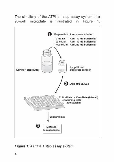
The simplicity of the ATPlite 1step assay system in a
96-well microplate is illustrated in Figure 1.
Figure 1: ATPlite 1 step assay system.
ATPlite 1step buffer
Add 100 µL/well
Seal and mix
Measureluminescence
·
CulturPlate or ViewPlate (96-well) containing cells (100 µL/well)
¸
Lyophilizedsubstrate solution
Preparation of substrate solution:
10 mL kit : Add 10 mL buffer/vial100 mL kit : Add 10 mL buffer/vial1,000 mL kit : Add 250 mL buffer/vial
ATPLite 1step booklet contents_ATPLite 1step booklet contents 23-04-15 10:37 Pagina 4
2. Principle
ATP is a marker for cell viability because it is present in
all metabolically active cells and the concentration
declines very rapidly when the cells undergo necrosis or
apoptosis. The ATPlite 1step assay system is based on
the production of light caused by the reaction of ATP with
added luciferase and d-luciferin. This is illustrated in the
following reaction scheme:
ATP + d-Luciferin + O2LUCIFERASE
Oxyluciferin +Mg2+
AMP + PPi + CO2 + Light
The emitted light is proportional to the ATP concentration
within certain limits.
The kit has been formulated in such a way that within a
measuring window of 0 to 30 minutes, a very bright
signal will be obtained, when using up to the maximum
cell number per well. Within this measuring window the
decrease of the luminescent signal in time is independent
of the cell number per well.
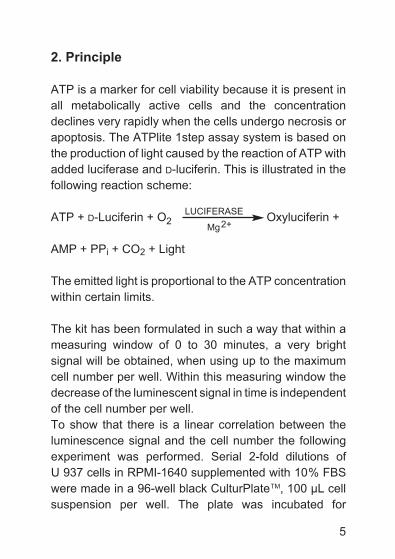
To show that there is a linear correlation between the
luminescence signal and the cell number the following
experiment was performed. Serial 2-fold dilutions of
U 937 cells in RPMI-1640 supplemented with 10% FBS
were made in a 96-well black CulturPlateTM, 100 µL cell
suspension per well. The plate was incubated for
5
ATPLite 1step booklet contents_ATPLite 1step booklet contents 23-04-15 10:37 Pagina 5
6
2 hours at 37 °C / 5% CO2. The plate was taken from the
incubator and equilibrated at room temperature for 30
minutes. Next, 100 µL of ATPlite 1step reagent was added
to the wells and the plate was shaken for 2 minutes at
700 rpm using an IKA® MTS4 plate shaker. The resulting
luminescence was monitored at 5 minutes intervals in a
temperature controlled (22 °C) TopCount® NXT and
expressed as counts per second (CPS). The results
Figure 2: Serial dilution (2-fold) U 937 cells in
RPMI-1640 10% FBS. Luminescence measured after
10 minutes incubation. Inset shows linearity at low cell
numbers per well.
0
Cells per well
Lum
ines
cen
ce in
CPS
0
62 x 10
61 x 10
41 x 10
42 x 10
43 x 10
44 x 10
45 x 10
63 x 10
64 x 10
65 x 10
00
12,000
24,000
100 200
ATPLite 1step booklet contents_ATPLite 1step booklet contents 23-04-15 10:37 Pagina 6
7
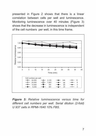
presented in Figure 2 shows that there is a linear
correlation between cells per well and luminescence.
Monitoring luminescence over 40 minutes (Figure 3)
shows that the decrease in luminescence is independent
of the cell numbers per well, in this time frame.
Figure 3: Relative luminescence versus time for
different cell numbers per well. Serial dilution (2-fold)
U 937 cells in RPMI-1640 10% FBS.
0,00
0,20
0,40
0,60
0,80
1,00
0 5 10 15 20 25 30 35 40 45
Time (min)
Relative lu
min
escence
50,000
25,000
12,500
6,250
3,125
1,563
781
391
195
98
49
24
12
6
3
0
Cell numbers per well:
ATPLite 1step booklet contents_ATPLite 1step booklet contents 23-04-15 10:37 Pagina 7
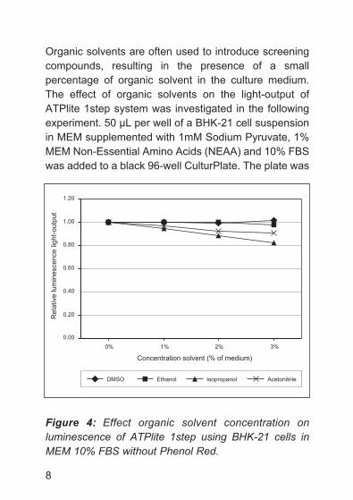
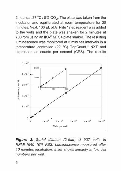
Organic solvents are often used to introduce screening
compounds, resulting in the presence of a small
percentage of organic solvent in the culture medium.
The effect of organic solvents on the light-output of
ATPlite 1step system was investigated in the following
experiment. 50 µL per well of a BHK-21 cell suspension
in MEM supplemented with 1mM Sodium Pyruvate, 1%
MEM Non-Essential Amino Acids (NEAA) and 10% FBS
was added to a black 96-well CulturPlate. The plate was
Figure 4: Effect organic solvent concentration on
luminescence of ATPlite 1step using BHK-21 cells in
MEM 10% FBS without Phenol Red.
8
0.20
0.40
0.60
0.80
1.00
1.20
0% 1% 2% 3%
Concentration solvent (% of medium)
Re
lative
lu
min
esce
nce
ligh
t-o
utp
ut
DMSO Ethanol Isopropanol Acetonitrile
0.00
ATPLite 1step booklet contents_ATPLite 1step booklet contents 23-04-15 10:37 Pagina 8
incubated at 37 °C/ 5% CO2 to let the cells adhere. The
plate was taken from the incubator and equilibrated at
room temperature. Next, 50 µL of medium with various
concentrations of organic solvents was added to the
wells and mixed gently. Hereafter 100 µL of ATPlite 1step
was added to the wells and the plate shaken for
2 minutes at 700 rpm using an IKA MTS4 orbital plate
shaker. The resulting luminescence was measured on a
TopCount NXT. The results presented in Figure 4 shows
that dMSO and Ethanol have a minimal effect. With
Isopropanol and Acetonitrile the luminescence decreases
gradually with increasing solvent concentrations.
3. Advantages of ATPlite 1step
� Simple and reproducible
- only one reagent addition step, no separation steps
� Suitable for both 96- and 384-well microplates
� Linear correlation between cell number and
luminescent signal
- up to 50,000 cells per well for 96-well microplates
and 12,500 cells per well for 384-well microplates
� Designed for continuous process systems
- luminescence should be measured between 0 and
30 minutes after reagent addition and plate shaking
9
ATPLite 1step booklet contents_ATPLite 1step booklet contents 23-04-15 10:37 Pagina 9
10
� Homogeneous
- no cell harvesting or centrifugation required
� Highly sensitive
� Reduced Phenol Red dependency
� High light-output
- kit can also be used with less sensitive luminescence
readers like multi-label readers
� Fast
- no luminescence signal stabilization time required
4. Contents and storage of ATPlite 1 step kit
6016736 - ATPlite 1step 10 mL kit
Each assay kit contains the following components:
1. 1 x 10 mL of substrate buffer solution
2. 1 vial of substrate solution (lyophilized)
3. 1 vial of ATP standard (lyophilized)
4. Instruction booklet
Using the recommended assay volumes of 100 µL for
96-well microplates and 25 µL for 384-well microplates
this kit is sufficient for 100 and 400 assays respectively.
ATPLite 1step booklet contents_ATPLite 1step booklet contents 23-04-15 10:37 Pagina 10
11
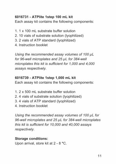
6016731 - ATPlite 1step 100 mL kit
Each assay kit contains the following components:
1. 1 x 100 mL substrate buffer solution
2. 10 vials of substrate solution (lyophilized)
3. 2 vials of ATP standard (lyophilized)
4. Instruction booklet
Using the recommended assay volumes of 100 µL
for 96-well microplates and 25 µL for 384-well
microplates this kit is sufficient for 1,000 and 4,000
assays respectively.
6016739 - ATPlite 1step 1,000 mL kit
Each assay kit contains the following components:
1. 2 x 500 mL substrate buffer solution
2. 4 vials of substrate solution (lyophilized)
3. 4 vials of ATP standard (lyophilized)
4. Instruction booklet
Using the recommended assay volumes of 100 µL for
96-well microplates and 25 µL for 384-well microplates
this kit is sufficient for 10,000 and 40,000 assays
respectively.
Storage conditions:
Upon arrival, store kit at 2 - 8 °C.
ATPLite 1step booklet contents_ATPLite 1step booklet contents 23-04-15 10:37 Pagina 11
12
5. Handling
Care should be taken during handling of the kit
components, such as opening vials and bottles, to
ensure that the contents of these are not contaminated
with ATP. Such contamination will cause high
background levels. In handling the kit, the skin of the
fingers is a very potent source of ATP-contamination,
therefore the use of clean gloves is strongly
recommended. Use also ATP-free dispensing materials.
6. Stability
Reconstituted ATPlite 1step is stable (approximately
10% loss of activity) for 8 hours at 20 °C. At 2 °C storage
the loss in activity is less than 10% after 48 hours.
Freshly prepared reagents can be aliquoted and stored
at -80 °C for one month.
7. Mixing
Mixing of the added ATPlite 1step reagent with the
contents of the wells is very important. Improper mixing
results in lower signals and deviations in the correlation
between high and low cell numbers per well. It was
found that using the IKA MTS 4 laboratory orbital
ATPLite 1step booklet contents_ATPLite 1step booklet contents 23-04-15 10:37 Pagina 12
microplate shaker (diameter of the orbit: 3 mm) set at
700 rpm for 2 minutes gave consistent results using
96-well plates. It was also found that with these settings
the contents of 384-well microplates could not be mixed
properly, even with prolonged shaking times. Increasing
to 1,100 rpm for 2 minutes resolved this.
8. Additional requirements
1. detection instrument such as the PerkinElmer
TopCount, MicroBeta, LumiCount, VICTOR3TM
Multi Label Reader, VICTOR Light, EnVisionTM or
EnSpire. CCd camera systems, such as
PerkinElmer ViewLux TM can be used for high through-
put applications.
2. Sterile, tissue culture treated, white or black 96- or
384-well microplates such as the PerkinElmer
CulturPlate and ViewPlate.
3. Pipette (multichannel) or automated pipetting device.
4. Microplate shaker suitable for efficient mixing of the
plate used.
5. ATP-free dispensing material.
13
ATPLite 1step booklet contents_ATPLite 1step booklet contents 23-04-15 10:37 Pagina 13
14
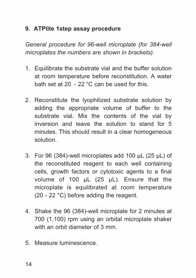
9. ATPlite 1step assay procedure
General procedure for 96-well microplate (for 384-well
microplates the numbers are shown in brackets).
1. Equilibrate the substrate vial and the buffer solution
at room temperature before reconstitution. A water
bath set at 20 - 22 °C can be used for this.
2. Reconstitute the lyophilized substrate solution by
adding the appropriate volume of buffer to the
substrate vial. Mix the contents of the vial by
inversion and leave the solution to stand for 5
minutes. This should result in a clear homogeneous
solution.
3. For 96 (384)-well microplates add 100 µL (25 µL) of
the reconstituted reagent to each well containing
cells, growth factors or cytotoxic agents to a final
volume of 100 µL (25 µL). Ensure that the
microplate is equilibrated at room temperature
(20 - 22 °C) before adding the reagent.
4. Shake the 96 (384)-well microplate for 2 minutes at
700 (1,100) rpm using an orbital microplate shaker
with an orbit diameter of 3 mm.
5. Measure luminescence.
ATPLite 1step booklet contents_ATPLite 1step booklet contents 23-04-15 10:37 Pagina 14
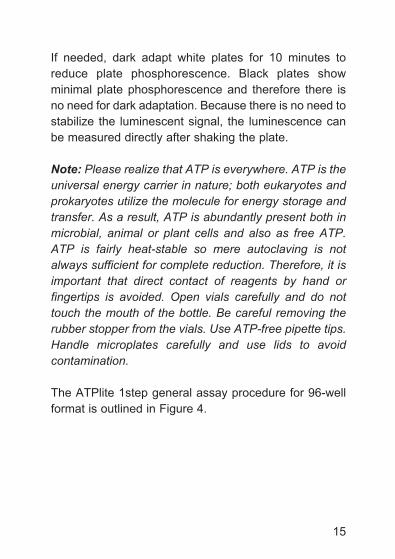
If needed, dark adapt white plates for 10 minutes to
reduce plate phosphorescence. Black plates show
minimal plate phosphorescence and therefore there is
no need for dark adaptation. Because there is no need to
stabilize the luminescent signal, the luminescence can
be measured directly after shaking the plate.
Note: Please realize that ATP is everywhere. ATP is the
universal energy carrier in nature; both eukaryotes and
prokaryotes utilize the molecule for energy storage and
transfer. As a result, ATP is abundantly present both in
microbial, animal or plant cells and also as free ATP.
ATP is fairly heat-stable so mere autoclaving is not
always sufficient for complete reduction. Therefore, it is
important that direct contact of reagents by hand or
fingertips is avoided. Open vials carefully and do not
touch the mouth of the bottle. Be careful removing the
rubber stopper from the vials. Use ATP-free pipette tips.
Handle microplates carefully and use lids to avoid
contamination.
The ATPlite 1step general assay procedure for 96-well
format is outlined in Figure 4.
15
ATPLite 1step booklet contents_ATPLite 1step booklet contents 23-04-15 10:37 Pagina 15
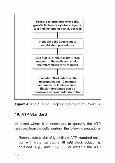
Figure 4: The ATPlite 1 step assay flow chart (96-well).
10. ATP Standard
In cases where it is necessary to quantify the ATP
released from the cells, perform the following procedure:
1. Reconstitute a vial of lyophilized ATP standard solu-
tion with water so that a 10 mM stock solution is
obtained. E.g., add 1,170 µL of water if the ATP
16
Prepare microplates with cells,growth factors or cytotoxic agents to a final volume of 100 µL per well
Incubate cells according toestablished procedures
Add 100 µL of the ATPlite 1 stepreagent to the wells and shake the microplates for 2 minutes
If needed: Dark adapt whitemicroplates for 10 minutes
and measure luminescence.Black microplates can be
measured without dark adaptation
ATPLite 1step booklet contents_ATPLite 1step booklet contents 23-04-15 10:37 Pagina 16
amount printed on the label is 11.7 µmole or add
960 µL of water if the amount is 9.6 µmole. After
addition of the water, allow the ATP to dissolve
completely by swirling the vial for one minute.
2. Set up a standard curve in the same microplate that
will be used for the experimental samples:
a. Take an aliquot of the ATP standard solution and
prepare a 1 µM ATP in culture medium.
b. Prepare a 10-fold serial dilution series of ATP in
culture medium (1 µM to 1 pM)
c. Pipette this dilution series in the wells of a
microplate (100 µL for a 96-well and 25 µL for a
384-well microplate).
3. Add ATPlite 1step reagent to these wells, 100 µL for
a 96-well and 25 µL for a 384-well microplate.
4. Mix contents of the wells for 2 minutes using a plate
shaker.
5. Measure luminescence.
Note: Endogenous ATPase’s may be present in sera
resulting in a reduction of the ATP concentration of the
ATP dilution series. Therefore the ATP dilution series
should be made just before the addition of the
ATPlite 1step reagent.
17
ATPLite 1step booklet contents_ATPLite 1step booklet contents 23-04-15 10:37 Pagina 17
11. Recommendations for use
Phenol red, as well as other colored compounds, will not
interfere with the luciferin/ luciferase reaction, but will
physically absorb some of the emitted light, resulting in
lower assay signals. For the highest light-output, the
culture medium can be substituted with medium without
phenol red or (dulbecco’s) PBS prior to the addition of
the ATPlite 1step reagent.
Whether to choose white or black microplates depends
very much on the luminescence reader used. With some
readers the detectors of the instrument may saturate
when using white plates and high ATP concentrations
due to the very high light-output of the ATPlite 1step
reaction. This can occur especially with 96-well white
plates. In this case black plates are recommended.
Optimize liquid handling procedures to attain optimal
reagent /medium mixing.
When handling the plates prior to measurement, work
under subdued light conditions and avoid direct sunlight
or bright fluorescent light. Bright light may cause plate
phosphorescence resulting in higher background levels.
Phosphorescence has a half-life of several minutes.
18
ATPLite 1step booklet contents_ATPLite 1step booklet contents 23-04-15 10:37 Pagina 18
19
If more than one vial of substrate is reconstituted for the
assay, we recommend these solutions be combined
before addition to the plates.
Optimal room and instrument temperature is 22 °C.
Allow plates to adapt at room temperature upon removal
from the incubator and prior to the addition of the
reagent. An adaptation time of 30 minutes is usually
sufficient.
ATPLite 1step booklet contents_ATPLite 1step booklet contents 23-04-15 10:37 Pagina 19
20
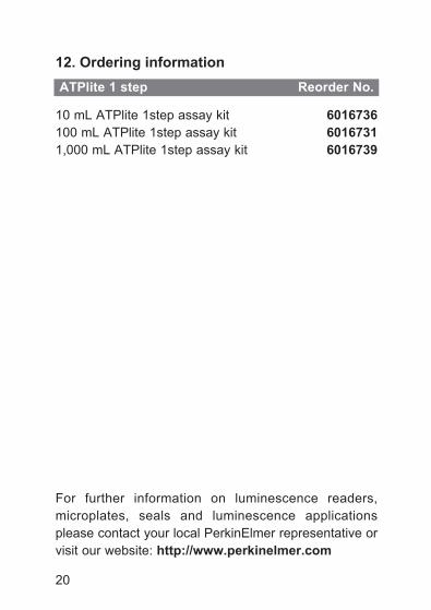
12. Ordering information
10 mL ATPlite 1step assay kit 6016736
100 mL ATPlite 1step assay kit 6016731
1,000 mL ATPlite 1step assay kit 6016739
For further information on luminescence readers,
microplates, seals and luminescence applications
please contact your local PerkinElmer representative or
visit our website: http://www.perkinelmer.com
ATPlite 1 step Reorder No.
ATPLite 1step booklet contents_ATPLite 1step booklet contents 23-04-15 10:37 Pagina 20
21
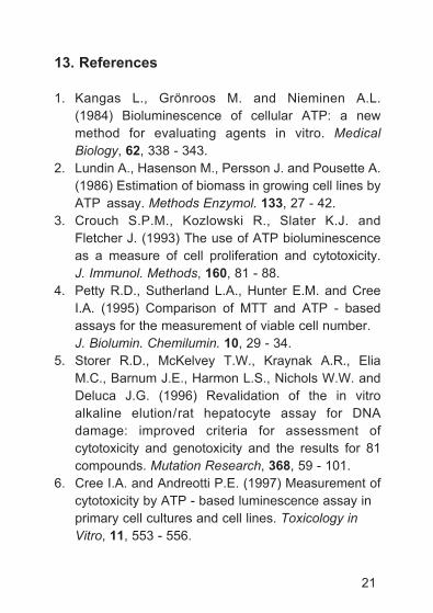
13. References
1. Kangas L., Grönroos M. and Nieminen A.L.
(1984) Bioluminescence of cellular ATP: a new
method for evaluating agents in vitro. Medical
Biology, 62, 338 - 343.
2. Lundin A., Hasenson M., Persson J. and Pousette A.
(1986) Estimation of biomass in growing cell lines by
ATP assay. Methods Enzymol. 133, 27 - 42.
3. Crouch S.P.M., Kozlowski R., Slater K.J. and
Fletcher J. (1993) The use of ATP bioluminescence
as a measure of cell proliferation and cytotoxicity.
J. Immunol. Methods, 160, 81 - 88.
4. Petty R.d., Sutherland L.A., Hunter E.M. and Cree
I.A. (1995) Comparison of MTT and ATP - based
assays for the measurement of viable cell number.
J. Biolumin. Chemilumin. 10, 29 - 34.
5. Storer R.d., McKelvey T.W., Kraynak A.R., Elia
M.C., Barnum J.E., Harmon L.S., Nichols W.W. and
deluca J.G. (1996) Revalidation of the in vitro
alkaline elution/rat hepatocyte assay for dNA
damage: improved criteria for assessment of
cytotoxicity and genotoxicity and the results for 81
compounds. Mutation Research, 368, 59 - 101.
6. Cree I.A. and Andreotti P.E. (1997) Measurement of
cytotoxicity by ATP - based luminescence assay in
primary cell cultures and cell lines. Toxicology in
Vitro, 11, 553 - 556.
ATPLite 1step booklet contents_ATPLite 1step booklet contents 23-04-15 10:37 Pagina 21

22
Limited Use License
This product is distributed and sold for life science
research and commercial applications, but not for
diagnostic use. Any use of this product other than for life
science research and commercial applications is strictly
prohibited.
Rev.
E -
April 2015
ATPLite 1step booklet contents_ATPLite 1step booklet contents 23-04-15 10:37 Pagina 22

PerkinElmer, Inc.940 Winter StreetWaltham, MA 02451 USAPhone: (800) 762-4000 or(+1) 203-925-4602www.perkinelmer.com
For a complete listing of our global offices, visit www.perkinelmer.com/ContactUs.
© 2003 - 2015 PerkinElmer, Inc. ATPLT1STP-0415 / REV. K
EX
PR
00003