Embed Size (px)
Citation preview

Single Crystal Diffraction
Arthur J. SchultzArgonne National Laboratory
National School on Neutron and X-Ray Scattering
June 13, 2011

What is a crystal?
2
• Atoms (molecules) pack
together in a regular pattern to
form a crystal.
• Periodicity: we superimpose
(mentally) on the crystal
structure a repeating lattice or
unit cell.
• A lattice is a regular array of
geometrical points each of
which has the same
environment.Unit cells of oxalic acid dihydrate
Quartz crystals

Why don’t the X-rays scatter in all directions?
3
X-ray precession photograph
(Georgia Tech, 1978).
• X-rays and neutrons have
wave properties.
• A crystal acts as a
diffraction grating producing
constructive and destructive
interference.

Bragg’s Law
4
William Henry Bragg William Lawrence Bragg
Jointly awarded the 1915
Nobel Prize in Physics

Crystallographic Planes and Miller Indices
5
c
a
b
(221)
d-spacing = spacing between origin and first plane or between
neighboring planes in the family of planes.

Laue Equations
6
Si
Ssa
a • Ss
a • (-Si)
a • Ss + a • (-Si) = a • (Ss – Si) = hλ
a • (Ss – Si) = hλ
b • (Ss – Si) = kλ
c • (Ss – Si) = lλ
Scattering from points
In three dimensions →
Max von Laue
1914 Noble Prize for Physics

Real and reciprocal Space
a* • a = b* • b = c* • c = 1
a* • b = … = 0
Laue equations:
a • (Ss – Si) = hλ, or a • s = h
b • (Ss – Si) = kλ, or b • s = k
c • (Ss – Si) = lλ, or c • s = l
where
s = (Ss – Si)/λ = ha* + kb* + lc*
7
s
Si
Ss
|S| = 1/
|s| = 1/d

θθ
θ1/λ
1/d
1/(2d)
a*
b*O
The Ewald Sphere
8

The Ewald sphere: the movie
9
Courtesy of the CSIC (Spanish National Research Council).
http://www.xtal.iqfr.csic.es/Cristalografia/index-en.html

Bragg Peak Intensity
10
0
a
b
hklhkl IF 2
Relative phase shifts
related to molecular
structure.

Two-theta
Counts
θ-2θ Step Scan
11

Omega Step Scan
Omega
Mosaic
spread
1. Detector stationary at
2θ angle.
2. Crystal is rotated
about θ by +/- ω.
3. FWHM is the mosaic
spread.
12

Something completely different - polycrystallography
What is a powder? - polycrystalline mass
All orientations of crystallites
possible
Sample: 1ml powder of 1mm crystallites -
~109 particles
Single crystal reciprocal lattice
- smeared into spherical shells
Packing efficiency – typically 50%
Spaces – air, solvent, etc.
Courtesy of R. Von Dreele

Powder Diffraction
14
Counts
2
Bragg’s Law: sin2*d
• Usually do not attempt to integrate individual
peaks.
• Instead, fit the spectrum using Rietveld profile
analysis. Requires functions that describe the
peak shape and background.

Why do single crystal diffraction (vs. powder
diffraction)?
Smaller samples – 1-10 mg vs 500-5000 mg
Larger molecules and unit cells
Hydrogen is ok – generally does not need to be deuterated
Less absorption
Fourier coefficients are more accurate – based on integrating well-resolved peaks
Uniquely characterize non-standard scattering – superlattice and satellite peaks (commensurate and incommensurate), diffuse scattering (rods, planes, etc.)
But:
Need to grow a single crystal
Data collection can be more time consuming
15

Some history of single crystal neutron diffraction
16
• 1951 – Peterson and Levy demonstrate the feasibility of single crystal
neutron diffraction using the Graphite Reactor at ORNL.
• 1950s and 1960s – Bill Busing, Henri Levy, Carroll Johnson and others wrote
a suite of programs for singe crystal diffraction including ORFLS and ORTEP.
• 1979 – Peterson and coworkers demonstrate the single crystal neutron time-
of-flight Laue technique at Argonne’s ZING-P’ spallation neutron source.

U is a rotation matrix relating the unit cell to the
instrument coordinate system.
The matrix product UB is called the orientation
matrix.
The Orientation Matrix
17

Picker 4-Circle Diffractometer
18

Kappa Diffractometer
19
Brucker AXS: KAPPA APEX II
• Full 360° rotations about ω and φ axes.
• Rotation about κ axis reproduces quarter
circle about χ axis.

Monochromatic diffractometer
20
Reactor
HFIR 4-Circle
Diffractometer
• Rotating crystal
• Vary sin in the Bragg equation:
2d sin = n
nd sin2 nd sin2

Laue diffraction
21
Polychromatic “white” spectrum
I()

Laue photo from white radiation
22
X-ray Laue photos taken
by Linus Pauling

Quasi-Laue Neutron Image Plate Diffractometer
23
Select D/ of 10-20%
2012 at HFIR: IMAGINE

Pulsed Neutron Incident Spectrum
= (h / m)•(t / L)
12.5 msec
5.0 Å
CO
UN
TS
t0
L = 10 m
1.25 msec
0.5 Å
CO
UN
TS
t0t033 1/3 msec
SOURCE
PULSED
AT 30 HZ
24

Time-of-Flight Laue Technique
25
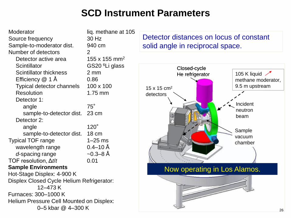
SCD Instrument Parameters
Sample Environments
Hot-Stage Displex: 4-900 K
Displex Closed Cycle Helium Refrigerator:
12–473 K
Furnaces: 300–1000 K
Helium Pressure Cell Mounted on Displex:
0–5 kbar @ 4–300 K
Incident
neutron
beam
105 K liquid
methane moderator,
9.5 m upstream15 x 15 cm2
detectors
Sample
vacuum
chamber
Closed-cycle
He refrigerator
Incident
neutron
beam
105 K liquid
methane moderator,
9.5 m upstream
105 K liquid
methane moderator,
9.5 m upstream15 x 15 cm2
detectors
Sample
vacuum
chamber
Closed-cycle
He refrigerator
Moderator liq. methane at 105
Source frequency 30 Hz
Sample-to-moderator dist. 940 cm
Number of detectors 2
Detector active area 155 x 155 mm2
Scintillator GS20 6Li glass
Scintillator thickness 2 mm
Efficiency @ 1 Å 0.86
Typical detector channels 100 x 100
Resolution 1.75 mm
Detector 1:
angle 75°sample-to-detector dist. 23 cm
Detector 2:
angle 120°sample-to-detector dist. 18 cm
Typical TOF range 1–25 ms
wavelength range 0.4–10 Å
d-spacing range ~0.3–8 Å
TOF resolution, Δt/t 0.01
Detector distances on locus of constant
solid angle in reciprocal space.
Now operating in Los Alamos.
26
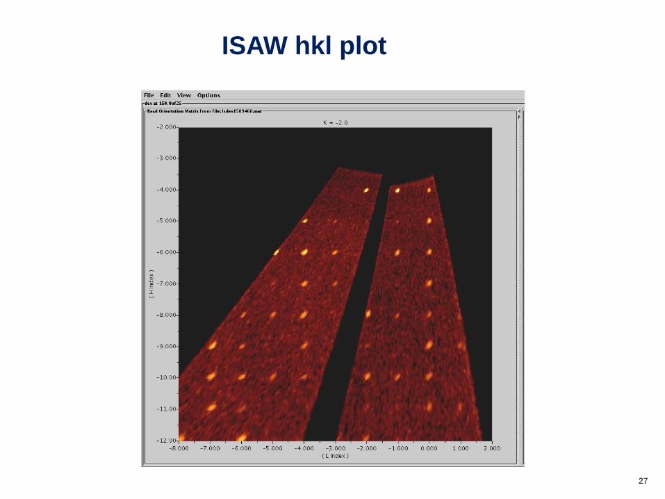
ISAW hkl plot
27

28
Analysis of ZnMn2O4 by William Ratcliff II (NIST).
ISAW 3D Reciprocal Space ViewerDiffuse Magnetic Scattering
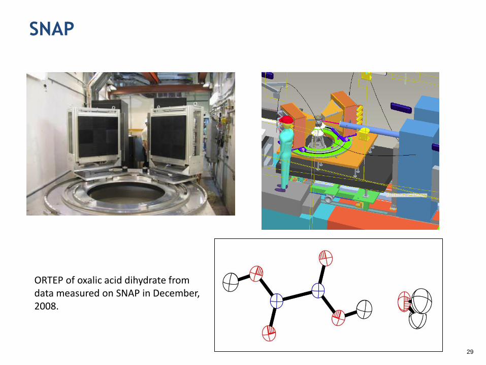
SNAP
29
ORTEP of oxalic acid dihydrate from data measured on SNAP in December, 2008.
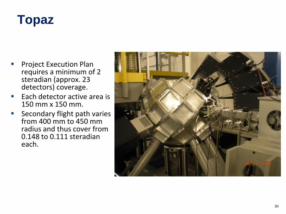
Topaz
Project Execution Plan requires a minimum of 2 steradian (approx. 23 detectors) coverage.
Each detector active area is 150 mm x 150 mm.
Secondary flight path varies from 400 mm to 450 mm radius and thus cover from 0.148 to 0.111 steradianeach.
30

Natrolite structure from TOPAZ data
31

Outline of single crystal structure analysis
Collect some initial data to determine the unit cell and
the space group.
– Auto-index peaks to determine unit cell and orientation
– Examine symmetry of intensities and systematic absences
Measure a full data set of observed intensities.
Reduce the raw integrated intensities, Ihkl, to structure
factor amplitudes, |Fobs|2.
Solve the structure.
Refine the structure.
32

Data reduction
33
k = scale factor
f = incident flux spectrum, obtained by measuring the
incoherent scattering from a vanadium sample
e = detector efficiency calculated as a function of
wavelength A() = sample absorption; includes the wavelength
dependence of the linear absorption coefficients
Vs = sample volume
Vc = unit cell volume
Nc = number of unit cells in the sample
Data reduction: convert raw integrated intensities, Ihkl,
into relative structure factor amplitudes, |Fhkl|2.
Ihkl = k t() f() e() A() (Vs/Vc) (|Fhkl|2/Vc)
4/sin2Q
Ihkl = k t() f() e() A() Nc (|Fhkl|2/Vc)
4/sin2Q
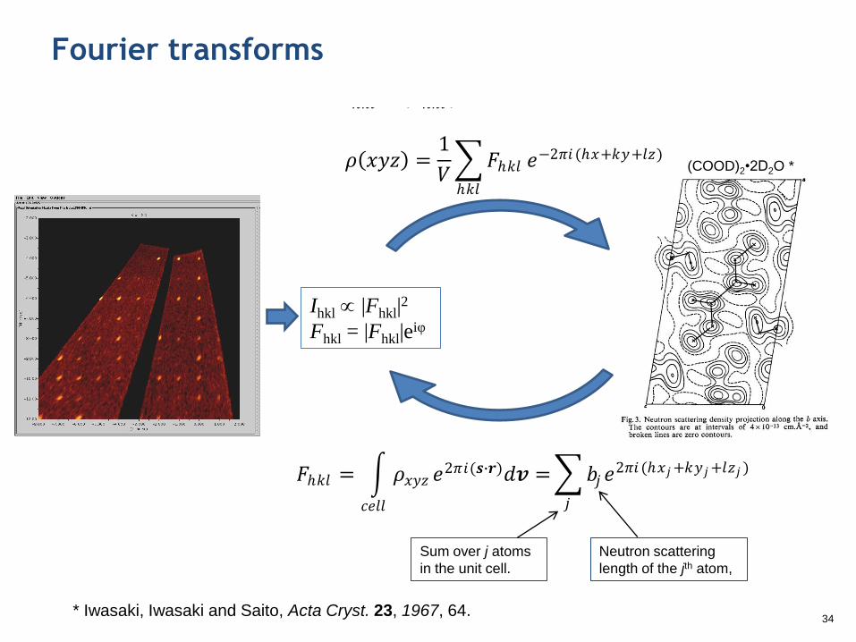
Fourier transforms
34
Ihkl |Fhkl|2
Fhkl = |Fhkl|eiφ
𝐼ℎ𝑘𝑙 ∝ 𝐹ℎ𝑘𝑙 2
𝜌 𝑥𝑦𝑧 =1
𝑉 𝐹ℎ𝑘𝑙
ℎ𝑘𝑙
𝑒−2𝜋𝑖 (ℎ𝑥+𝑘𝑦+𝑙𝑧)
𝐹ℎ𝑘𝑙 = 𝐹ℎ𝑘𝑙 𝑒−𝑖𝜙 = 𝐹ℎ𝑘𝑙 cos𝜙 + 𝑖 𝐹ℎ𝑘𝑙 sin𝜙 = 𝐴 + 𝑖𝐵
𝜙 = tan−1𝐵
𝐴
𝐹ℎ𝑘𝑙 = 𝜌𝑥𝑦𝑧 𝑒2𝜋𝑖(𝒔∙𝒓)𝑑𝒗 =
𝑐𝑒𝑙𝑙
𝑏𝑗𝑒2𝜋𝑖 (ℎ𝑥𝑗+𝑘𝑦𝑗+𝑙𝑧𝑗 )
𝑗
𝐼ℎ𝑘𝑙 ∝ 𝐹ℎ𝑘𝑙 2
𝜌 𝑥𝑦𝑧 =1
𝑉 𝐹ℎ𝑘𝑙
ℎ𝑘𝑙
𝑒−2𝜋𝑖 (ℎ𝑥+𝑘𝑦+𝑙𝑧)
𝐹ℎ𝑘𝑙 = 𝐹ℎ𝑘𝑙 𝑒−𝑖𝜙 = 𝐹ℎ𝑘𝑙 cos𝜙 + 𝑖 𝐹ℎ𝑘𝑙 sin𝜙 = 𝐴 + 𝑖𝐵
𝜙 = tan−1𝐵
𝐴
𝐹ℎ𝑘𝑙 = 𝜌𝑥𝑦𝑧 𝑒2𝜋𝑖(𝒔∙𝒓)𝑑𝒗 =
𝑐𝑒𝑙𝑙
𝑏𝑗𝑒2𝜋𝑖 (ℎ𝑥𝑗+𝑘𝑦𝑗+𝑙𝑧𝑗 )
𝑗
Sum over j atoms
in the unit cell.
Neutron scattering
length of the jth atom,
* Iwasaki, Iwasaki and Saito, Acta Cryst. 23, 1967, 64.
(COOD)2•2D2O *

The phase problem
35
Measured intensity
Electron (X-ray) or nuclear (neutron) density at point x,y,z in the unit cell
Phase angle
Neutron scattering length or X-ray form factor for jth atom
Sum over j atoms in the unit cell

Solutions to the phase problem
Patterson synthesis using the |Fobs|2 values as Fourier coefficients
– Map of inter-atom vectors
– Also called the heavy atom method
Direct methods
– Based on probability that the phase of a third peak is equal to the sum of the
phases of two other related peaks.
– J. Karle and H. Hauptman received the 1985 Nobel Prize in Chemistry
Shake-and-bake
– Alternate between modifying a starting model and phase refinement
Charge flipping
– Start out with random phases.
– Peaks below a threshold in a Fourier map are flipped up.
– Repeat until a solution is obtained
MAD
– Multiple-wavelength anomalous dispersion phasing
Molecular replacement
– Based on the existence of a previously solved structure with of a similar protein
– Rotate the molecular to fit the two Patterson maps
– Translate the molecule36

Structure Refinement
37
222
2
0
2
/sin8exp2exp
iiii
i
ihkl
hkl
c
UlzkyhxibF
FFw
GSAS, SHELX, CRYSTALS, OLEX2, WinGX…
Nonlinear least squares programs. Vary atomic
fractional coordinates x,y,z and temperature factors U
(isotropic) or uij (anisotropic) to obtain best fit between
observed and calculated structure factors.
Neutron single crystal instruments in the US
SNAP @ SNS: high pressure sample environment (http://neutrons.ornl.gov/instruments/SNS/SNAP/)
TOPAZ @ SNS: small molecule to small protein, magnetism, future polarized neutron capabilities (http://neutrons.ornl.gov/instruments/SNS/TOPAZ/)
Four-Circle Diffractometer (HB-3A) @ HFIR: small molecule, high precision, magnetism (http://neutrons.ornl.gov/instruments/HFIR/HB3A/)
MaNDi (Macromolecular Neutron Diffractometer) @ SNS: neutron protein crystallography, commissioning in 2012 (http://neutrons.ornl.gov/instruments/SNS/MaNDi/)
IMAGINE (Image-Plate Single-Crystal Diffractometer) @ HFIR: small molecule to macromolecule crystallography , commissioning in 2012 (http://neutrons.ornl.gov/instruments/HFIR/imagine/)
SCD @ Lujan Center, Los Alamos: general purpose instrument, currently not available due to budget constraints (http://lansce.lanl.gov/lujan/instruments/SCD/index.html)
PCS (Protein Crystallography Station) @ Lujan Center, Los Alamos: neutron protein crystallography (http://lansce.lanl.gov/lujan/instruments/PCS/index.html)
38

Books and on-line tutorials
George E. Bacon, Neutron Diffraction, 3rd ed., Clarendon Press, 1975.
Colin G. Windsor, Pulsed Neutron Scattering, Taylor & Francis, 1981.
Chick C. Wilson, Single Crystal Neutron Diffraction From Molecular Crystals, World
Scientific, 2000.
M. F. C. Ladd and R. A. Palmer, Structure Determination by X-ray Crystallography,
Third Edition, Plenum Press, 1994.
J. P. Glusker and K. N. Trueblood, Crystal Structure Analysis: A Primer, 2nd ed., Oxford
University Press, 1985.
Interactive Tutorial about Diffraction: www.totalscattering.org/teaching/
IPNS SCD tutorial by Paula Piccoli: www.pns.anl.gov/instruments/scd/subscd/scd.shtml
39


![ELECTRON BACKSCATTER DIFFRACTION CRYSTAL … · 2014. 8. 8. · Electron backscatter diffraction (EBSD) measurement [1,2] is generally very useful in analyzing crystal morphology](https://img.pdfslide.net/doc/110x75/6020ac1488b59757b674b100/electron-backscatter-diffraction-crystal-2014-8-8-electron-backscatter-diffraction.jpg)