Embed Size (px)
Citation preview
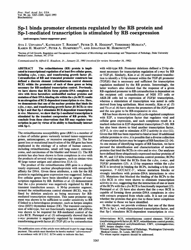
Proc. Natl. Acad. Sci. USAVol. 90, pp. 3265-3269, April 1993Biochemistry
Sp-1 binds promoter elements regulated by the RB protein andSp-1-mediated transcription is stimulated by RB coexpression
(anti-oncogene/tumor-suppressor gene)
AVA J. UDVADIA*, KATHLEEN T. ROGERS*, PETER D. R. HIGGINS*, YOSHIHIKO MURATA*,KAREN H. MARTIN*, PETER A. HUMPHREYtt, AND JONATHAN M. HoROWITZ*§*Section of Cell Growth, Regulation and Oncogenesis and Department of Microbiology and Immunology and tDepartment of Pathology, Duke UniversityMedical Center, Durham, NC 27710
Communicated by Alfred G. Knudson, Jr., January 22, 1993 (received for review November 16, 1992)
ABSTRACT The retinoblastoma (RB) protein is impli-cated in transcriptional regulation ofat least five cellular genes,including c-fos, c-myc, and transforming growth factor 31.Cotransfection of RB and truncated promoter constructs hasdefrmed a discrete element (retinoblastoma control element;RCE) within the promoters of each of these genes as beingnecessary for RB-mediated transcription control. Previously,we have shown that RCEs form protein-DNA complexes invitro with three heretofore unidentified nuclear proteins andmutation oftheir DNA-binding site within the c-fosRCE resultsin an abrogation ofRCE-dependent transcription in vivo. Here,we demonstrate that one of the nuclear proteins that binds thec-fos, c-myc, and transforming growth factor 13 RCEs in vitrois Sp-1 and that Sp-1 stimulates RCE-dependent transcriptionin vivo. Moreover, we show that Sp-l-mediated transcription isstimulated by the transient coexpression of RB protein. Weconclude from these observations that RB may regulate tran-scription in part by virtue of its ability to functionally interactwith Sp-l.
The retinoblastoma susceptibility gene (RBI) is a member ofa class of cellular genes variously termed tumor-suppressorgenes, antioncogenes, or recessive oncogenes (1). The fre-quent loss or mutational inactivation of the RB gene has beenimplicated in the etiology of a subset of human cancers,including retinoblastoma, small cell lung cancer, osteosar-coma, and carcinomas of the bladder and breast (1). The RBprotein has also been shown to form complexes in vivo withthe products of several viral oncogenes, such as simian virus40 large tumor antigen and adenovirus ElA (1).The product of the retinoblastoma (RB) gene is a ubiqui-
tously expressed, nuclear phosphoprotein with nonspecificaffinity for DNA. Given these attributes, a role for the RBprotein in regulating gene expression was suggested. Indeed,five cellular genes have been identified as targets of tran-scription regulation by RB. Robbins et al. (2) demonstratedthat the c-fos promoter was negatively regulated by RB intransient transfection assays. A 30-bp promoter segment,termed the retinoblastoma control element (RCE), was de-fined by deletion analysis as being necessary for RB-mediated transcription repression. This same promoter seg-ment was shown to be sufficient to confer sensitivity to RBif linked to a heterologous promoter, such as herpes simplexvirus (HSV) thymidine kinase (TK). The transient coexpres-sion of wild-type, but not functionally impaired, RB cDNAswas shown to be required for RB-mediated control of thec-fos RCE. Pietenpol et al. (3) subsequently showed that thec-myc promoter is negatively regulated by treatment withtransforming growth factor ,81 (TGF-,B1) or by cotransfection
with wild-type RB. Promoter deletions defined a 23-bp ele-ment necessary for transcription regulation of c-myc by RBor TGF-(31. Similarly, Kim et al. (4) used transient transfec-tion to identify a 32-bp element within the TGF-,B1 promoter(TGF,81) that is necessary and sufficient for transcriptionregulation mediated by the RB protein. Interestingly, thelatter workers also showed that the response of a givenRB-regulated promoter to RB cotransfection is dependent onthe recipient cell: cotransfections of NIH 3T3 cells orAKR-2B cells led to repression of TGF/31 transcription,whereas a stimulation of transcription was noted in cellsderived from lung epithelium. Most recently, Kim et al. (5)and Yu et al. (6) have shown the promoters of the IGF2 geneand the neu gene to be RB-responsive in vivo.RB has been shown to form cell cycle-regulated complexes
with E2F, a transcription factor that regulates viral andcellular gene expression, and such complexes result in amarked reduction in E2F-dependent transcription (7-10). RBhas also been shown to bind another transcription factor,ATF-2, in vitro and to stimulate ATF-2 activity in vivo (11).Given that RB has been reported to bind at least 10 additionalcellular proteins in vitro, it is possible that a diverse collectionoftranscription factors may be targets ofRB function (12-14).As one means of identifying targets of RB function, we havepursued the identification and characterization of nuclearproteins that bind the RCEs in vitro and in vivo. Our analyseshave defined three ubiquitously expressed nuclear proteins of80, 95, and 115 kDa (retinoblastoma control proteins; RCPs)that specifically bind the RCEs from the c-fos, c-myc, andTGF,81 promoters (15). A 13-bp sequence within the c-fosRCE is involved in binding the RCPs, including a 6-bp coresequence, 5'-GCCACC-3', whose mutation blocks orstrongly interferes with protein-DNA interactions in vitro(15). Mutations that blocked the binding of the RCPs to thec-fos RCE in vitro were shown to eliminate transcriptionactivity in vivo, suggesting that the interaction ofone or moreofthe RCPs with the c-fos RCE is functionally important (15).Pietenpol et al. (3) have also shown that the c-myc RCE iscapable of forming three protein-DNA complexes in vitrowith extracts prepared from whole cells. It is not yet clearwhether the proteins that give rise to these latter complexesare similar to those we have identified.
In this report, we provide evidence that one of the threeRCPs is Sp-1, a well-characterized transcription factor, andthat Sp-1 stimulates RCE-dependent transcription in vivo.
Abbreviations: RCE, retinoblastoma control element; TGF-/1,transforming growth factor ,81; RCP, retinoblastoma control protein;CAT, chloramphenicol acetyltransferase; CMV, cytomegalovirus;HSV, herpes simplex virus.tPresent address: Department of Pathology, Washington UniversityMedical Center, St. Louis, MO 63110.§To whom reprint requests should be addressed.
3265
The publication costs of this article were defrayed in part by page chargepayment. This article must therefore be hereby marked "advertisement"in accordance with 18 U.S.C. §1734 solely to indicate this fact.
3266 Biochemistry: Udvadia et al.
Furthermore, we show that the stimulation ofRCE transcrip-tion by Sp-l is enhanced by coexpression of RB protein.
MATERIALS AND METHODSCell Culture. NIH 3T3, EJ, C-33A, and A549 cells were
acquired from the American Type Culture Collection; PC12cells were a gift from Luis Parada (National Cancer Institute,Frederick, MD); Schneider SL2 cells were a gift of CheaptipBenyajati (University of Rochester); and COS cells were agift of Bryan Cullen (Duke University Medical Center). Cellswere grown in Dulbecco's modified Eagle's medium(DMEM) supplemented with l1o heat-inactivated fetal calfserum, except PC12 cells were grown in DMEM supple-mented with 10% horse serum/5% calf serum and SchneiderSL2 cells were grown in Schneider's medium supplementedwith 10% heat-inactivated fetal calf serum. Mammalian cellswere grown in humidified incubators under 5% C02/95% airand Schneider SL2 cells were grown at room temperature onlaboratory bench tops.
Oligonucleotides and Protein-DNA Binding Assays. Oligo-nucleotides were synthesized on an Applied Biosystemsautomated DNA synthesizer, deprotected, and then partiallypurified through Sephadex G-25. To ensure sequence fidelity,each oligonucleotide pair was cloned into an appropriatevector and sequenced by dideoxynucleotide chain termina-tion (16). The following oligonucleotide sequences were usedin these studies:
Fos,
5'Fos-4,5'Fos-5,RCP-,dbl RCP-,AP-1,Myc,TGF-81,Sp-1,
5'-CCCGCGCGCCACCCCTCTGGCGCCACCGTG-3'(15);5'-CCCTTGCGCCACCCCTCT-3'(15);5'-CCCGCGCGCCATTCCTCT-3'(15);5'-CCCGCGCGAAATTCCTCTGGCGCCACCGTG-3'(15);5'-CCCGCAAAAAACCCCTCTGAAAAAACCGTG-3';5'-TAAAATGAGTCAAGTGG-3'(17);5'-GCAGAGGGCGTGGGGGAAAAGAA-3'(3,15);5'-GGAGCCCGCCCACGCGAGATGAGGACGGTGGC-3'(4,15);5'-GATGGGCGGAGTTAGGGGCGGGACTATC-3'(18).
Oligonucleotides were labeled with [32P]dNTPs and purifiedfrom unincorporated radioactivity as described (15). Nuclearextracts were prepared and DNA-binding assays were per-formed as described (15). For DNA-binding assays in whichantibodies were included, antibodies were added to bindingassay mixtures before addition of radiolabeled oligonucleo-tides. Monoclonal antibodies 5M3 and M73 are an anti-synthetic peptide antibody prepared against human RB pro-tein and an anti-ElA antibody (19), respectively. Sp-1 proteinpurified from HeLa cells was obtained from Promega andused in DNA-binding assays as suggested by the supplier.
Anti-Sp-1 Antibody Preparation, Immunoprecipitations,and Western Blotting. To generate polyclonal antisera againstSp-1, a full-length human Sp-1 cDNA (kindly provided byRobert Tijan, University of California, Berkeley) supplied inplasmid pBSK+ was cleaved with BamHI and a 1.8-kbpfragment was inserted in-frame into pGEX-1, a bacterialfusion protein expression vector (20). This portion ofthe Sp-1cDNA encodes the N-terminal 603 aa of Sp-1 protein (21).After bacterial transformation, fusion proteins were inducedwith isopropyl ,B-D-thiogalactopyranoside and a 105-kDaGST-Sp-1 fusion protein was purified as described (20). Forimmunizations, a single New Zealand White rabbit was
sequentially immunized with 150 ,ug of affinity-purified fusionprotein in Freund's complete and incomplete adjuvants.Anti-Sp-1 immunoreactivity, as judged by immunoprecipita-tion and Western blotting, was detected in serum harvestedafter the first booster injection with immunogen.For anti-Sp-l immunoprecipitations, cells were metaboli-
cally labeled, and extracts were prepared and immunopre-cipitated as described (22). For Western blotting, denaturedprotein extracts were resolved on SDS/polyacrylamide gels,
transferred to nitrocellulose using a semidry transfer appa-ratus, and incubated with a 1:5000 dilution of rabbit anti-Sp-1antibody. After incubation with a 1:20,000 dilution of horse-radish peroxidase-conjugated goat anti-rabbit secondary an-tibody, Sp-1 was detected by using an enhanced chemilumi-nescent system (ECL; Amersham) and exposure at ambienttemperature to Hyperfilm.
Expression Constructs, Transfections, and ChloramphenicolAcetyltransferase (CAT) Assays. Plasmid pPaSp-1, a Sp-1expression construct, was obtained from Robert Tjian (23).PCR was used to generate an epitope-tagged Sp-1 cDNA byDNA amplification with primers immediately flanking theSp-l cDNA open reading frame. Each primer resulted inaddition of an EcoRI site to the Sp-1 cDNA and the 3' PCRprimer incorporated a 10-aa influenza hemagglutinin epitope(N-YPYDVPDYAS-C), recognized by monoclonal antibody12CA5 (gift of Rene Bernards, Massachusetts General Hos-pital Cancer Center, Charlestown), at the C terminus of Sp-1.After PCR and cloning of amplified DNA into pUC12,dideoxynucleotide sequencing was performed to ensure thatthe fusion of Sp-1 and hemagglutinin sequences had occurredin-frame (16). The epitope-tagged Sp-1 cDNA was thenrecloned into the HindIII site of pCMV4, a cytomegalovirus(CMV) immediate-early promoter expression vector (gift ofStefan Doerrer, Duke University Medical Center) after ad-dition of EcoRI/HindIII adapters. Reporter constructs wereprepared by cloning dimers of RCE-containing oligonucleo-tides upstream of the HSV TK promoter and the bacterialCAT gene as described (15). Wild-type and mutated DHFR-CAT constructs (24) were kindly provided by J. Azizkhan(University of North Carolina, Chapel Hill). A human RBexpression construct, pCMV-HRb, driven by the CMV im-mediate-early promoter was prepared by cloning a wild-typehuman RB cDNA into plasmid pIENH (gift of Jeffrey Marks,Duke University Medical Center). Transient transfections ofCOS cells were performed by a protocol incorporatingDEAE-dextran followed by treatment with chloroquine anda dimethyl sulfoxide shock as described (25). COS whole cellextracts were prepared 48 hr posttransfection for immuno-precipitation and DNA-binding assays following solubiliza-tion in ELB+ and removal of cell debris (22). Transfection ofSchneider SL2 cells was performed as described (26). CATassays were performed as described and results were nor-malized against the abundance of total cell protein in aportion of each extract.
RESULTSNuclear Factors That Bind RCEs Also Bind to Oligonude-
otides Containing Sp-1 Binding Sites. Given that the c-fos,c-myc, and TGF/31 RCEs are G+C-rich promoter elementsresembling Sp-1-binding sites and that two of three RCPs wehave identified (95 and 115 kDa) approximate Sp-1 in molec-ular mass, we performed a series of experiments to testwhether Sp-1 encodes one or more RCE-binding proteins. Inthe first experiment, an oligonucleotide containing two Sp-1-binding sites from the simian virus 40 early promoter wasused as an unlabeled competitor for the formation of RCP-RCE complexes in a DNA-binding assay. We have previ-ously shown that a full-length c-fos RCE probe forms threedistinct protein-DNA complexes in vitro (15). These threecomplexes are also formed by a radiolabeled probe encom-passing the 5' half of the c-fos RCE (labeled 1A, 1B, and 2 inFig. 1A; ref. 15). Also, as reported (15), these protein-DNAcomplexes are abolished by inclusion of a 200-fold molarexcess of unlabeled oligonucleotides containing RCEs fromthe c-fos, c-myc, and TGF/31 promoters but not with aheterologous, unlabeled oligonucleotide (AP-1 in Fig. 1A;ref. 15). As shown in Fig. 1A (Left), an oligonucleotidecontaining two Sp-l-binding sites also abolished the appear-
Proc. Natl. Acad. Sci. USA 90 (1993)
Proc. Natl. Acad. Sci. USA 90 (1993) 3267
5 Fos-4 Sp- 1O L o6- X o -X..0 L m x
0 C>- C* 0 x X 0 0 0 0L. IL o a OL L0 0 0 0 0 I.- x0 0 0 0
0IL < 2 Co a. - 00- a. o to
B "t e)cn u co co
Co) 0 0 Co) 0 0
0 Lo o o o o+ + CL + +
IA- _
aum
NuclearExtract
Pure Sp-1
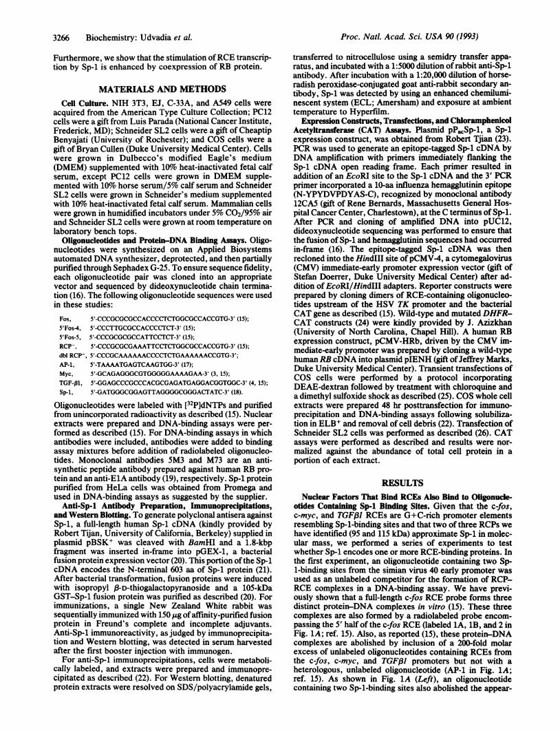
FIG. 1. c-fos RCE-RCP complexes are abolished by RCE and Sp-1 oligonucleotides and are formed by purified Sp-1. (A) (Left) DNA-bindingassays were performed with NIH 3T3 nuclear extracts and a 5'Fos-4 probe. A 200-fold molar excess of unlabeled RCE-containing (Myc, Fos,and TGF-f31), heterologous (AP-1), or Sp-1 oligonucleotides was included as competitor DNA. RCE-specific complexes 1A, 1B, and 2 areindicated by arrows. (Right) Increasing concentrations of unlabeled 5'Fos-4 oligonucleotides (10- to 200-fold molar excess) or Sp-1oligonucleotides (2- to 100-fold molar excess) were included in DNA-binding assays with a 5'Fos-4 probe (Probe). DNA-binding assays wereexposed to Hyperfilm for 18 hr at -80'C. (B) A 5'Fos-4 probe was included in DNA-binding assays with nuclear extracts prepared from C-33Acells (18 ,ug; Nuclear Extract) or purified Sp-1 protein (30 ng; Pure Sp-1). Unlabeled oligonucleotides that form complexes with the RCPs (5'Fos-4,Fos) or do not form complexes with the RCPs (5'Fos-5, RCP-, dbl RCP-) were included as competitor DNAs. DNA-binding assays were exposedto Hyperfilm for 18 hr at -80'C.
ance of complexes 1A, 1B, and 2. Not apparent from thisexperiment is the result, shown in Fig. 1A (Right), thatcomplete competition for RCE-RCP binding is achieved byincorporation of only a 10-fold molar excess of unlabeledSp-1 oligonucleotides, suggesting that the simian virus 40-derived oligonucleotide is a more efficient competitor DNAthan is the c-fos-derived RCE.We next investigated whether purified Sp-1 would bind to
the c-fos RCE and result in a protein-DNA complex thatcomigrates with one or more of the RCP-RCE complexesrecovered from nuclear extracts. As shown in Fig. 1B,protein-DNA complexes formed in DNA-binding assayswith purified Sp-1 resulted in the appearance of a singleRCE-Sp-1 complex that comigrated with RCE complex 1Afrom nuclear extracts. These results are consistent withprevious UV cross-linking experiments showing that com-plex 1A is composed of a single photoaffinity-labeled proteinof 95 kDa (15). As shown in Fig. 1B, the Sp-1-inducedprotein-DNA complex was sensitive to competition withwild-type but not with mutated partial RCE oligonucleotides(5'Fos-5) that do not form complexes with the RCPs (15).Surprisingly, the complex formed by Sp-1 was abolished byfull-length c-fos RCE oligonucleotides with a mutated RCP-binding site (RCP-). We have previously shown that thismutated oligonucleotide does not form RCE-RCP complexesin DNA-binding assays using nuclear extracts and is func-tionally inactive as measured in transient transfections (15).This unexpected result suggested that purified Sp-1 can bindto an alternative binding site within the c-fos RCE in vitro,perhaps at a closely related 3' site we have previouslyidentified (15). To test this proposition, Sp-1 DNA-bindingassays were performed with another RCE oligonucleotidethat is mutated within each of two directly repeated 5'-GCGCCACC-3' sequences (dbl RCP-). As predicted, dblRCP- is not able to compete for Sp-1 binding in vitro (Fig.1B). Taken together, we conclude that (i) the nuclear factorsthat interact with the c-fos RCE in vitro also bind to aheterologous oligonucleotide containing bona fide Sp-l-binding sites, (ii) purified Sp-1 forms a protein-DNA complexwith the c-fos RCE that comigrates with RCP-RCE complex1A, (iii) purified Sp-1 and the RCPs share a c-fos RCE-binding site, and (iv) purified Sp-l can bind to a specific sitewithin the c-fos RCE that is not bound in vitro by proteins innuclear extracts.One of Three RCPs Is Bound by Antibodies Reactive with
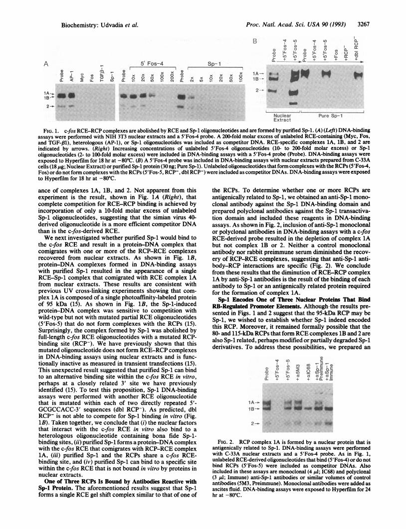
Sp-1 Protein. The aforementioned results suggest that Sp-1forms a single RCE gel shift complex similar to that of one of
the RCPs. To determine whether one or more RCPs areantigenically related to Sp-1, we obtained an anti-Sp-1 mono-clonal antibody against the Sp-1 DNA-binding domain andprepared polyclonal antibodies against the Sp-1 transactiva-tion domain and included these reagents in DNA-bindingassays. As shown in Fig. 2, inclusion of anti-Sp-1 monoclonalor polyclonal antibodies in DNA-binding assays with a c-fosRCE-derived probe resulted in the depletion of complex 1Abut not complex 1B or 2. Neither a control monoclonalantibody nor rabbit preimmune serum diminished the recov-ery of RCP-RCE complexes, suggesting that anti-Sp-1 anti-body-RCP interactions are specific (Fig. 2). We concludefrom these results that the diminution of RCE-RCP complex1A by anti-Sp-1 antibodies is the result of the binding of eachantibody to Sp-1 or an antigenically related protein requiredfor the formation of complex 1A.
Sp-1 Encodes One of Three Nuclear Proteins That BindRB-Regulated Promoter Elements. Although the results pre-sented in Figs. 1 and 2 suggest that the 95-kDa RCP may beSp-1, we wished to establish whether Sp-1 indeed encodedthis RCP. Moreover, it remained formally possible that the80- and 115-kDa RCPs that form RCE complexes 1B and 2 arealso Sp-1 related, perhaps modified or partially degraded Sp-1derivatives. To address these possibilities, we prepared an
a)1t LO c
CL+ + + + CL + E1B>.
2---
FIG. 2. RCP complex 1A is formed by a nuclear protein that isantigenically related to Sp-1. DNA-binding assays were performedwith C-33A nuclear extracts and a 5'Fos-4 probe. As in Fig. 1,unlabeled RCE-derived oligonucleotides that bind (5'Fos-4) or do notbind RCPs (5'Fos-5) were included as competitor DNAs. Alsoincluded in these assays are monoclonal (4 ,l; IC68) and polyclonal(3 IlI; Immune) anti-Sp-1 antibodies or similar volumes of controlantibodies (5M3, Preimmune). Monoclonal antibodies were added asascites fluid. DNA-binding assays were exposed to Hyperfilm for 24hr at -80'C.
A
2 -_ ..
0.
o O -LL Er
Biochemistry: Udvadia et al.

3268 Biochemistry: Udvadia et al.
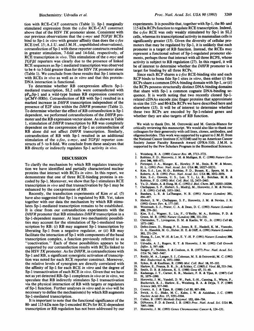
epitope-tagged Sp-1 cDNA and used a CMV promoter ex-pression vector to transiently express this recombinant pro-tein in COS cells. We chose as an epitope tag for theseexperiments a 10-aa influenza hemagglutinin peptide againstwhich a monoclonal antibody (12CA5) was available. Thishemagglutinin peptide was linked in-frame to the C terminusof Sp-1 as the result of DNA amplification and cloning byPCR. To ensure that this Sp-1/flu construct would lead to theexpression ofan epitope-tagged Sp-1 protein, COS cells weretransfected, incubated with [35S]methionine, and immuno-precipitated with anti-Sp-1 polyclonal or anti-hemagglutininmonoclonal antibody. As shown in Fig. 3A, transient trans-fection of COS cells with the epitope-tagged Sp-1 cDNAresulted in overexpression of 95- to 105-kDa proteins that areimmunoprecipitable by rabbit anti-Sp-1 antiserum as well asmonoclonal antibody 12CA5. Neither antibody detected asimilarly abundant protein in mock-transfected COS cells,suggesting that transient transfection of the Sp-1/flu con-struct results in overexpression of bona fide fusion protein(Fig. 3A).To determine whether Sp-1 encodes one or more RCPs,
DNA-binding assays were performed with a radiolabeledRCE probe. As shown in Fig. 3B, DNA-binding assays using
-AI
k0 0
_kD o 0
o- s U 2 0
B c
z z E na D -B E
n o o 'F1- L. ULL a CL = _
- co O - wUYc toe a+ C+
CL + + + + + 0CD .
200-
97.4- g;-105 v-95
68.0-^
- e
43.0-
FIG. 3. Immunoprecipitation and DNA-binding assays of COScells transfected with an epitope-tagged Sp-1 cDNA. (A) Immuno-precipitation of [35S]methionine-labeled COS cell extracts with rab-bit polyclonal anti-Sp-1 antibodies (10 ,ul; aSp-1) or a monoclonalanti-hemagglutinin antibody (150-1l hybridoma supematant; aflu).Extracts were prepared from untransfected (Mock) COS cells orCOS cells transfected with an epitope-tagged Sp-1 cbNA (Sp-1/flu).Immunoprecipitates (from 5 x 10⁶ cell equivalents per lane) wereresolved on a SDS/8%o/4% polyacrylamide gel; the gel was preparedfor fluorography, dried, and exposed to Hyperfilm for 20 hr at -80°C.Upon extended exposure times, endogenous Sp-1 was detected in theleftmost lane (data not shown). Molecular mass markers are shownon the left and arrows indicate unphosphorylated (95 kDa) andphosphorylated Sp-1 (105 kDa) species. (B) DNA-binding assaysperformed with extracts prepared from COS cells transfected with anepitope-tagged Sp-1 cDNA. Whole cell extracts (8 ,g) were preparedfrom mock-transfected COS cells (Mock), or after transfection witha Sp-1/flu recombinant cDNA, and incubated with a 5'Fos-4 probe(Sp-1/flu). A 200-fold molar excess of unlabeled wild-type (5'Fos-4)or mutated c-fos RCE oligonucleotides (5'Fos-5) was included ascompetitor DNA. Rabbit preimmune (1 ,ul) and immune (1 IlI)anti-Sp-1 antiserum or monoclonal antibodies (10-,l hybridomasupernatant) against influenza hemagglutinin (aflu) or adenovirusElA (aElA, M73; ref. 19) were included to identify transientlyexpressed Sp-1/flu fusion protein. Binding assays were exposed toHyperfilm for 15 hr at -80°C.
a c-fos RCE-derived oligonucleotide gave rise to one abun-dant RCE-protein complex whose detection was eliminatedby appropriate competitor oligonucleotides (5'Fos4 but not5'Fos-5 DNA) and whose gel mobility was coincident withRCP-RCE complex 1 (data not shown). This complex wasnot recovered in DNA-binding assays using similarly pre-pared extracts from mock-transfected COS cells (Fig. 3B).Moreover, the appearance of the Sp-l-RCE complex wasunquestionably the result of overexpression of the Sp-1/fluconstruct, since this complex was abolished both by anti-Sp-1and anti-hemagglutinin antibodies but not by control anti-bodies (Fig. 3B). We conclude from these results that Sp-1encodes only one of three nuclear factors that complex withRCEs in vitro. The identity of the 80- and 115-kDa RCPsremains to be established.
Sp-1 Stimulates Transcription of RCEs in Vivo and Sp-l-Mediated Transcription Is Stimulated by the RB Protein.Given that Sp-1 interacts with RCEs in vitro, we wished todetermine whether Sp-1 influences RCE transcription in vivo.Since the expression of Sp-1 and other RCE-binding proteinsin most cells would obscure transcription mediated by exog-enous Sp-1, we chose to transfect Drosophila Schneider SL2cells, a Sp-1-deficient cell type that does not express detect-able in vitro RCE-binding activity (ref. 23; A.J.U. andJ.M.H., unpublished observations). For Schneider SL2transfections, we used a Sp-1 expression construct driven bythe Drosophila actin promoter (pPcSp-1; ref. 23). To ensurethat exogenous expression ofSp-1 would transactivate a bonafide Sp-l-dependent promoter, we performed cotransfectionswith a wild-type reporter construct prepared from theDHFRpromoter and a mutated derivative lacking E2F-binding sites.Cotransfection with the Sp-1 expression vector significantlystimulated (30- to 40-fold) DHFR-CAT and DHFR-E2F--CAT expression in SL2 cells (Table 1). In contrast, differ-ential levels of transcription resulted from Sp-1 cotransfec-
Table 1. Drosophila Schneider SL2 transfection resultsReporterconstruct Addition
DHFR NoneRBSp-1RB + Sp-1
DHFR-E2F- NoneRBSp-1RB + Sp-1
TK NoneSp-1RB + Sp-1
FOS NoneSp-1RB + Sp-i
MYC NoneSp-1RB + Sp-1
TGF-,81 NoneSp-1RB + Sp-1
% acetylation,mean (SD)0.5 (0.4)0.7 (0.6)
18.1 (2.3)79.0 (12.6)0.40.613.260.05.8 (3.8)
15.5 (1.3)60.2 (25.0)5.2 (3.8)
23.5 (16.0)120.0 (63.0)
6.7 (1.0)44.7 (7.3)
247.9 (96.0)2.0 (0.42)
27.9 (4.6)218.1 (72.0)
Schneider SL2 cells were transfected with 5 jig of TK-CAT,FOS-CAT, MYC-CAT, TGFP-CAT, or 2 ,g of DHFR-CAT orDHFR-E2F--CAT reporter constructs alone or with 0.1 Lg ofpP.Sp-1, 20 pg ofpCMV-HRB, or both. ControlDNA (pUC12) wasincluded in each transfection to bring the final total DNA concen-tration to 30 pg. Expression values are expressed as mean percentageacetylation (SD) of [14C]chloramphenicol perA600 of 1 ,1 of total cellextract. Control DNAs consisting of equivalent amounts of Sp-1 andRB expression vectors lacking their respective cDNAs were includedin transfections to determine basal transcription activities.
Proc. Natl. Acad. Sci. USA 90 (1993)
:A--f:.i! Aiiii.- ANiiiiik -.16I.M ;.Iplpppw --- .:,Akdb
Proc. Natl. Acad. Sci. USA 90 (1993) 3269
tion with RCE-CAT constructs (Table 1). Sp-1 marginallystimulated expression from the c-fos RCE-CAT constructabove that of the HSV TK promoter alone. Consistent withour previous observations that the c-myc and TGF/31 RCEsbind to Sp-1 in vitro with greater affinity than does the c-fosRCE (ref. 15; A.J.U. and J.M.H., unpublished observations),cotransfection of Sp-1 with these reporter constructs resultedin greater stimulation, 7-fold and 14-fold, respectively, ofRCE transcription in vivo. This stimulation of the c-myc andTGF/I reporters was clearly due to the presence of linkedRCE sequences as Sp-1 mediated transcription was observedto be 4- to 5-fold greater than for the HSV TK promoter alone(Table 1). We conclude from these results that Sp-1 interactswith RCEs in vivo as well as in vitro and that this protein-DNA interaction is functional.To determine whether RB coexpression affects Sp-1-
mediated transcription, SL2 cells were cotransfected withPPacSp-1 and a wild-type human RB expression construct(pCMV-HRb). Cotransfection of RB with Sp-1 resulted in amarked increase in DHFR transcription independent of thepresence of E2F sites within the DHFR promoter (Table 1).To determine whether the effect of RB cotransfection is Sp-1dependent, we performed cotransfections of the DHFR pro-moter and the RB expression vector alone. As shown in Table1, stimulation ofDHFR transcription by RB was completelydependent on the coexpression of Sp-1 as cotransfection ofRB alone did not affect DHFR transcription. Similarly,cotransfection of RB with Sp-1 resulted in an additionalstimulation of the c-fos, c-myc, and TGF.31 reporter con-structs of 5- to 8-fold. We conclude from these analyses thatRB directly or indirectly regulates Sp-1 activity in vivo.
DISCUSSIONTo clarify the mechanism by which RB regulates transcrip-tion we have identified and partially characterized nuclearproteins that interact with RCEs in vitro. In this report, wedemonstrate that one of three RCE-binding proteins is en-coded by Sp-1. Moreover, we show that Sp-1 stimulates RCEtranscription in vivo and that transactivation by Sp-1 may beenhanced by the coexpression of RB.
Recently, the transfection experiments of Kim et al. (5)have also suggested that Sp-1 is regulated by RB. Yet, takentogether with our data the mechanism by which RB stimu-lates Sp-1-mediated transcription remains to be established.It is clear from our cotransfection experiments with theDHFR promoter that RB stimulates DHFR transcription in aSp-l-dependent manner. At least two mechanistic possibili-ties may account for the stimulation of Sp-1-mediated tran-scription by RB: (i) RB may augment Sp-1 transcription byliberating Sp-1 from a negative regulator, or (ii) RB mayfacilitate the interaction of Sp-l with components of the basaltranscription complex, a function previously referred to as"coactivation." Each of these possibilities appears to besupported by our cotransfection results with RCEs linked tothe HSV TK promoter. As for DHFR, in cotransfections withSp-1 and RB, a significant synergistic activation of transcrip-tion was noted for each RCE reporter construct. Moreover,the relative levels of synergism are directly correlated withthe affinity of Sp-1 for each RCE in vitro and the degree ofSp-1 transactivation of each RCE in vivo. Given that we havenot as yet detected RB-Sp-1 complexes in vivo or in vitro, wespeculate that RB indirectly stimulates Sp-1 transactivationvia the physical interaction of RB with targets or regulatorsof Sp-l function. Further analyses in vitro and in vivo will benecessary to define the mechanism(s) by which RB augmentsSp-l-mediated transcription.
It is important to note that the functional significance of the80- and 115-kDa non-Sp-1-encoded RCPs for RCE-dependenttranscription or RB regulation has not been addressed by our
experiments. It is possible that, together with Sp-1, the 80- and115-kDa RCPs function to regulate RCE transcription. Indeed,the c-fos RCE was only weakly stimulated by Sp-1 in SL2cells, whereas its transcriptional activity in mammalian cells issignificantly greater (15). Given the diversity of cellular pro-moters that may be regulated by Sp-1, it is unlikely that eachpromoter is a target of RB function. Instead, the RCEs mayrepresent a functional subset of Sp-1-regulated promoter ele-ments, perhaps those that interact with all three RCPS, whoseactivity is subject to RB regulation (27). In this regard, it willbe of interest to determine whether the DHFR promoter is atarget for binding by all three RCPs.
Since each RCP shares a c-fos RCE-binding site and eachRCP binds to bona fide Sp-1 sites in vitro, then either (i) theRCPs share a common DNA-binding domain with Sp-1, or (ii)the RCPs possess structurally distinct DNA-binding domainsthat share with Sp-1 a common cognate DNA-binding se-quence. It is worth noting that two recently cloned Sp-l-related cDNAs encode zinc finger proteins that approximatein size the 115- and 80-kDa RCPs we have described here andelsewhere (15). It will be of interest to determine whetherthese two RCPs are encoded by Sp-1-related genes andwhether they are also targets of RB function.
We wish to thank Drs. M. Ostrowski and M. Garcia-Blanco forcritically reviewing this manuscript. We would also like to thank ourcolleagues for their generosity with cell lines, clones, antibodies, andoligonucleotides. This work was supported by a grant to J.M.H. fromthe National Cancer Institute (CA53248) and by an American CancerSociety Junior Faculty Research Award (JFRA-310). J.M.H. issupported by the Pew Scholars Program in the Biomedical Sciences.
1. Weinberg, R. A. (1989) Cancer Res. 49, 3713-3721.2. Robbins, P. D., Horowitz, J. M. & Mulligan, R. C. (1990) Nature (Lon-
don) 346, 668-671.3. Pietenpol, J. A., Munger, K., Howley, P. M., Stein, R. W. & Moses,
H. L. (1991) Proc. Natl. Acad. Sci. USA 88, 10227-10231.4. Kim, S.-J., Lee, H.-D., Robbins, P. D., Busam, K., Sporn, M. B. &
Roberts, A. B. (1991) Proc. Natl. Acad. Sci. USA 88, 3052-3056.5. Kim, S.-J., Onwuta, U. S., Lee, Y. I., Li, R., Botchan, M. R. &
Robbins, P. D. (1992) Mol. Cell. Biol. 12, 2455-2463.6. Yu, D., Matin, A. & Hung, M.-C. (1992)J. Biol. Chem. 267,10203-10206.7. Chellappan, S. P., Hiebert, S., Mudryj, M., Horowitz, J. M. & Nevins,
J. R. (1991) Cell 65, 1053-1061.8. Bandara, L. R. & LaThangue, N. B. (1991) Nature (London) 351,
494-497.9. Hiebert, S. W., Chellappan, S. P., Horowitz, J. M. & Nevins, J. R.
(1992) Genes Dev. 6, 177-185.10. Weintraub, S. J., Prater, C. A. & Dean, D. C. (1992) Nature (London)
358, 259-261.11. Kim, S.-J., Wagner, S., Liu, F., O'Reilly, M. A., Robbins, P. D. &
Green, M. R. (1992) Nature (London) 358, 331-334.12. Chittenden, T., Livingston, D. M. & Kaelin, W. G., Jr. (1991) Cell 65,
1073-1082.13. Defeo-Jones, D., Huang, P. S., Jones, R. E., Haskell, K. M., Vuocolo,
G. A., Hanobik, M. G., Huber, H. E. & Oliff, A. (1991) Nature (London)352, 251-254.
14. Huang, S., Lee, W.-H. & Lee, E. Y.-H. P. (1991) Nature (London) 350,160-162.
15. Udvadia, A. J., Rogers, K. T. & Horowitz, J. M. (1992) Cell GrowthDiffer. 3, 597-608.
16. Sanger, F., Nicklen, S. & Coulson, A. R. (1977) Proc. Natl. Acad. Sci.USA 74, 5463-5467.
17. Reddy, M. A., Langer, S. J., Coleman, M. S. & Ostrowski, M. C. (1992)Mol. Endocrinol. 6, 1051-1060.
18. Sykes, K. & Kaufman, R. (1990) Mol. Cell. Biol. 10, 95-102.19. Harlow, E., Franza, B. R., Jr., & Schley, C. (1985) J. Virol. 55, 533-546.20. Smith, D. B. & Johnson, K. S. (1988) Gene 67, 31-40.21. Kadanoga, J. T., Carner, K. R., Masiarz, F. R. & Tijan, R. (1987) Cell
51, 1079-1090.22. Horowitz, J. M., Yandell, D. W., Park, S.-H., Canning, S., Whyte, P.,
Buchovich, K. J., Harlow, E., Weinberg, R. A. & Dryja, T. P. (1989)Science 243, 937-940.
23. Courey, A. J. & Tijan, R. (1988) Cell 55, 887-898.24. Swick, A. G., Blake, M. C., Kahn, J. W. & Azizkhan, J. C. (1989)
Nucleic Acids Res. 17, 9291-9304.25. Cullen, B. (1987) Methods Enzymol. 152, 684-704.26. DiNocera, P. D. & Dawid, I. B. (1983) Proc. Natl. Acad. Sci. USA 80,
7095-7098.27. Horowitz, J. M. (1993) Genes Chromosomes Cancer 6, 124-131.
Biochemistry: Udvadia et al.
![Chandler Unified School District / Home Page · Web viewTranscription begins when [RNA / RNA polymerase] binds to the gene’s promoter. The promoter region contains the sequence](https://img.pdfslide.net/doc/110x75/6006f3e28ecd7a613e1d7184/chandler-unified-school-district-home-page-web-view-transcription-begins-when.jpg)


![BMC Bioinformatics - Home | Computer Science...binds to the TF complex and to RNA Polymerase II, bringing the CRM into close proximity to the promoter to start transcription [1]. Experimental](https://img.pdfslide.net/doc/110x75/5f03df377e708231d40b2e6b/bmc-bioinformatics-home-computer-science-binds-to-the-tf-complex-and-to.jpg)