Embed Size (px)
Citation preview
22
Stereo
Microscopy
Stereo Microscope Part 2b: Greenough Microscopes 3rd Edition
R. Jordan Kreindler (USA) ___________________________________________________________________
Horatio S. Greenough signature, and last portion of one of his letters sent to Zeiss. c. 1890s Courtesy of Herr Berndt-Joachim Lau, Carl Zeiss Microscopy GmbH (Lau, 2012).
23
Stereo
Microscopy
Some Specialized Applications Greenough stereo microscopes in addition to general use have also been designed as instruments for use in some specialized applications.
Ophthalmology One type of stereo microscope used daily in clinical practice is found in slit lamp instruments, Fig. 33 and Fig. 34, seen in most ophthalmologist's and optometrist's offices. These instruments contain stereo Greenough microscopes, (e.g., Haag Streit, Topcon), or CMOs (to be discussed in Part 3), adjustable slit lamp illumination, usually a tonometer, a device for measuring intraocular pressure (IOP) in mm of mercury to test for glaucoma, a chin brace, and forehead rest on a single adjustable stand. As Manuel del Cerro explained to the author (del Cerro, 2012), the name slit lamp is perhaps inappropriate, as this assemblage is named for only one of its components, which it can be reasonably argued, is not as important as its microscope. This is likely the reason a slit lamp is sometimes referred to by a longer and more descriptive name, slit-lamp biomicroscope. Slit lamps are usually used in conjunction with a Hruby lens, typically -55/56 diopter (-55/56D), to allow examination of the retina. Slit lamps are used to examine the eye's interior, iris, cornea, vitreous humor, and retina to allow for anatomical diagnosis. As they are built using high quality optical and mechanical components designed for continuous clinical use, slit lamps are expensive, but appear virtually indestructible and long functioning.
Figure 33. Topcon SL-2E Slit Lamp (Bryant, 2012)

24
Stereo
Microscopy
A slit lamp changed from a purely observational device to a measuring instrument with the inclusion of a tonometer to evaluate intraocular pressure (IOP). It was further extended as a measuring tool when additional capabilities were added to measure the distance from the cornea to the lens, and the thickness of the cornea. Used functioning models, of relatively recent slit lamps, are usually on the market only a short time, as there is a constant demand from eye care specialists. Models from the major slit lamp manufacturers such as Zeiss (CMOs), the original manufacturer of the modern stereo microscope, and Haag Streit (Greenough), generally retain good resale values as used equipment. For example, a used Haag-Streit S350 slit lamp, depending upon condition and completeness, often sells for between USD $3,000 to $8,500. On trade-in for an improved Haag-Streit slit lamp, the BM 900 pictured here can reach $5,000. Fig. 34 shows a Haag-Streit Greenough microscope from various angles. Fig. 35 shows an eye as seen through this instrument, and the slit lamp's illumination can be seen reflected by the eye (Ozment, 2012).

25
Stereo
Microscopy
Figure 34. Haag-Streit slit lamp

26
Stereo
Microscopy
Figure 35. Human eye as seen through BM 900 Haag-Streit Greenough Microscope

27
Stereo
Microscopy
Photoreconnaissance Another specialized application was film photoreconnaissance analysis. One example of this is the Bausch & Lomb Greenough-style stereo zoom 240 photoreconnaissance microscope, c. 1970. This was used for photo interpretation of film from, often still classified, flights of Corona satellites, SR-71 Blackbirds, and other U.S. photoreconnaissance resources. With the arrival of high-resolution digital imaging, this microscope was removed from use. However, during the early digital imaging age film still had higher resolution, so film photoreconnaissance analysis persisted. Only after the development of higher resolution digital imaging was film finally replaced. In spite of the availability of later optical instruments, while film continued in use this B&L microscope was still used, and often preferred, for the analysis of film images.
Figure 36. Bausch and Lomb 240 aerial photo interpretation stereo zoom microscope, in its laboratory storage case, with some accessories

28
Stereo
Microscopy
The B&L 240 Aerial Photo Interpretation Stereo Zoom microscope, with some of its accessories, is shown in its storage case in Fig. 36, and assembled in Fig 37. The microscope has a maximum magnification of 120x and can resolve images up to 400 lines per mm. Although this microscope is c. 1970s, its resolution is greater than that of some modern high-quality camera lenses.
Figure 37. Assembled Bausch and Lomb 240 aerial photo interpretation stereo zoom microscope

29
Stereo
Microscopy
The Stereozoom 240, shown here, has two rhomboid arms and stereo objective lenses that at the ends of these arms. When used for photoreconnaissance analysis, the B&L 240 pod with attached rhomboid arms and objectives was installed over a light table, typically made by Richards or Bausch and Lomb. The light intensity provided by different table models varied significantly, from a luminance of approximately 2,200 to 90,000 foot-Lamberts (about 700 to 28,650 candles/square foot). The transmitted light, from most tables, followed the movement of the rhomboid arms either magnetically or mechanically, and so provided lighting where needed. Separate illumination for each stereo microscope objective had been introduced by Riddell over 100 years earlier.
30
Stereo
Microscopy
Quality Control - Solid State Devices
Most semiconductor devices are fabricated onto a wafer typically made from silicon, although
other compounds are also used. These semiconductor devices are made in relatively expensive
facilities called "fabs".
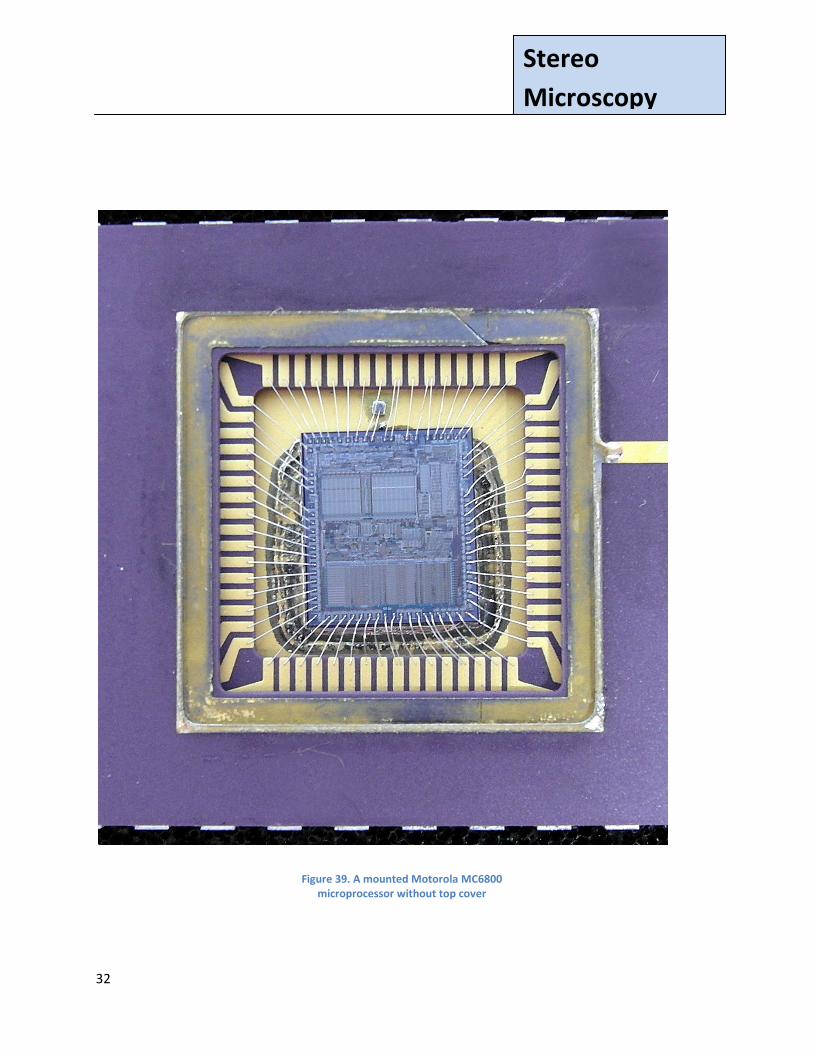
Before the 1970s about 3/4 of stereomicroscope applications were in the life sciences. The 1970s saw the rapid growth of the semiconductor industry. Coincident with the growth of fabs was the acquisition and use of Greenough zoom stereo microscopes for the examination of thin sheets of semiconductor material. These sheets are called wafers and contain fabricated integrated circuits (ICs). These ICs are removed from the wafers and installed in packages. The rapid growth of the semiconductor industry lead to a concurrent and rapid growth in the production of Greenough microscopes. The new semiconductor industry was probably the single greatest impetus to growth that Greenough microscopes had ever experienced. Fig. 38 shows a 3 inch wafer, typical of c. 1970s, containing many fabricated Motorola MC6800 chips. In general, the larger the chip the greater the chance for a flaw/damage and the lower the yield. Quality control using stereo microscopes was, and is, an important resource for identifying damaged chips Wafers diameters were initially measured in inches, up to about 5 inches. For larger wafers, dimensions are measured in millimeters (mms). Today 300mm is considered to be the standard for state-of-the-art wafers, with the next standard expected to be 450mm. Fig. 39 shows a Motorola MC 6900 microprocessor, an early 70,000 transistor microprocessor, shown mounted in its package, before the package is sealed. Fig. 40 shows a portion of a printed circuit board with some soldered components. Items such as those in Figs. 38, 39 and 40 were often viewed through a Greenough microscope for quality control.

31
Stereo
Microscopy
Figure 38. A wafer with Motorola 6800 ICs
32
Stereo
Microscopy
Figure 39. A mounted Motorola MC6800 microprocessor without top cover

33
Stereo
Microscopy
Figure 40. PC Board with soldered components as seen through a Greenough microscope
34
Stereo
Microscopy
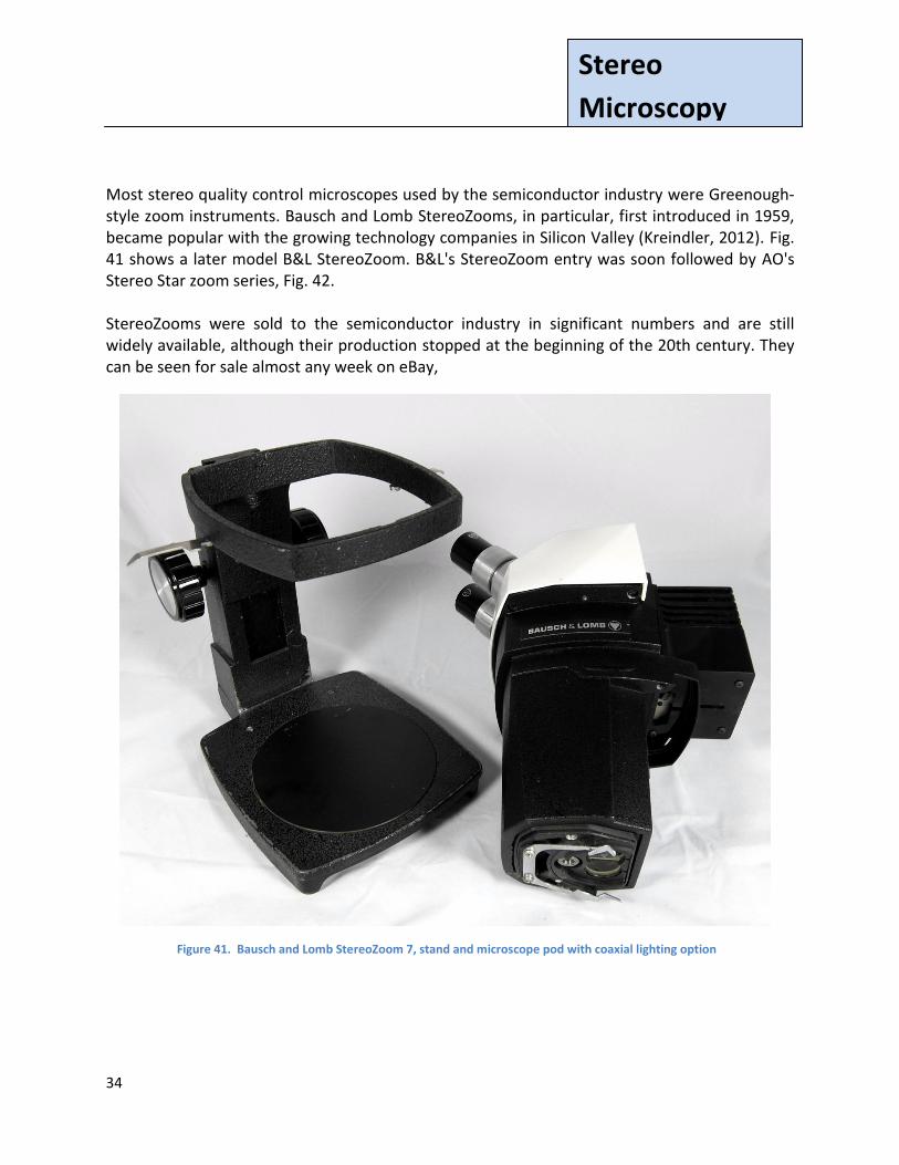
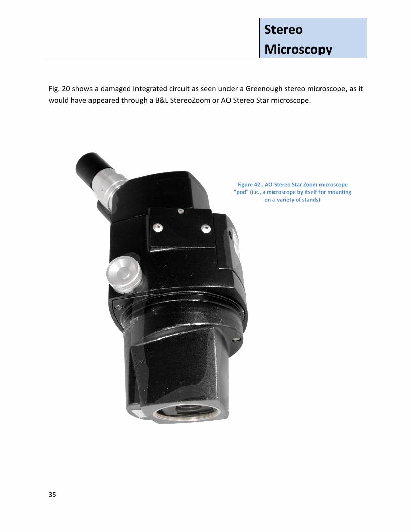
Most stereo quality control microscopes used by the semiconductor industry were Greenough-style zoom instruments. Bausch and Lomb StereoZooms, in particular, first introduced in 1959, became popular with the growing technology companies in Silicon Valley (Kreindler, 2012). Fig. 41 shows a later model B&L StereoZoom. B&L's StereoZoom entry was soon followed by AO's Stereo Star zoom series, Fig. 42. StereoZooms were sold to the semiconductor industry in significant numbers and are still widely available, although their production stopped at the beginning of the 20th century. They can be seen for sale almost any week on eBay,
Figure 41. Bausch and Lomb StereoZoom 7, stand and microscope pod with coaxial lighting option
35
Stereo
Microscopy
Fig. 20 shows a damaged integrated circuit as seen under a Greenough stereo microscope, as it
would have appeared through a B&L StereoZoom or AO Stereo Star microscope.
Figure 42.. AO Stereo Star Zoom microscope "pod" (i.e., a microscope by itself for mounting
on a variety of stands)

36
Stereo
Microscopy
Today, higher zoom ratios are common. The first, double digit, 10:1 zoom stereo microscope
was the Zeiss Citoval c. 1975 (Lau, 2012). Zoom ratios have continued to expand beyond this for
many top-of-the-line instruments, e.g., the Nikon SMZ1500 with a 15:1 (0.75 - 11.25x) zoom.
The Bausch and Lomb Optical Systems Division and the American Optical (AO) company after a
series of corporate acquisitions and mergers came together in one company. A company that
also owned Reichert and Leica. This led to the rebranding of many stereo instruments. See Part
4 for a further discussion of stereo microscope rebranding.
37
Stereo
Microscopy
Surgery
Operating room microscopes are usually stereoscopic and are often stable, floor standing instruments. Stereo microscopes are used for a variety of surgical procedures. The surgical applications are too numerous for a comprehensive list to be present here, but they include the medical specialties of cardiology, cardiac electrophysiology, dentistry, ENT (Ear, Nose and Throat), neurology, oncology, ophthalmology, orthopedics, plastic and reconstructive surgery, and urology. Many operating room microscopes have straight or only slightly tilted binocular tubes. However, microscopes used for ophthalmologic and other specialized surgeries are often inclined at 45 degrees (although occasionally at other angles). Most high-quality operating room microscopes have electronic controls for focusing and positioning, which are usually foot or head-mounted. These microscopes are frequently equipped with dual or triple heads, and/or with a video output channel for simultaneous viewing of the surgical procedure by operating room personnel. However, the video is only two dimensional, while the images through the microscope are three dimensional. As these microscope are usually equipped with their own independent light sources, they can provide the spot illumination needed to see inside small openings. Not all operating room microscopes are floor-standing. Zeiss makes surgical head-mounted loupes, in powers from about 4x to 8x. Leica now sells a head-mounted surgical microscope, model HM500. These head-mounted stereo microscopes allow surgeons greater mobility than possible with a floor standing unit. The HM500 comes with zoom and autofocus capabilities, similar in many ways to modern digital cameras, and with from 2 - 9x magnification. The HM500 uses rechargeable batteries for mobility. It provides foot pedal controls for zooming and manual focusing if needed. Most operating room microscopes are registered and/or certified. In the US registration is done by the Food and Drug Administration (FDA). In Europe Conformité Européenne (CE) certification, indicating compliance with EU regulations, is common. Unfortunately, some countries do not require registration or certification. In these countries surgical room microscopes are usually less expensive, but issues of optical and mechanical performance can arise. All surgical microscopes are relatively expensive, even head-mounted loupes.
38
Stereo
Microscopy
A Small Sampling of Zeiss Greenough Microscopes
Introduction
After Zeiss' introduction of the Greenough stereo microscope at the end of the 19th century,
other companies started manufacturing similar instruments. It would be very difficult, perhaps
impossible, to list all Greenough microscopes manufactured, even if restricted to just modern
times and "top" makers. With only modest descriptions, that list would likely exceed the length
of this paper. Many previous Greenough microscope makers are no longer in business. Thus,
confirming the accuracy of model designations and release dates would be difficult, and likely
impossible. Even for companies I've contacted still in business, some manufacturing records
are no longer available.
Therefore, rather than attempt to cover all the Greenoughs instruments manufactured, only a
small sampling of general purpose instruments from the original Greenough developer, Zeiss, is
presented here. This sampling serves to illustrate the evolution of Greenough microscopes. For
competitive reasons, microscope makers often copied each other's "newest" concepts. Thus,
the evolution presented here is somewhat synchronized with the evolution of Greenough
microscopes by other makers.
Zeiss continued to produced Greenough microscopes after their first in 1897, and the company
still manufactures and sells them today, e.g., the Stemi DV4, 2000/C/CS. Many of these
Greenoughs are general purpose instruments, used for a variety of applications. The discussion
that follows presents a few of these general purpose Zeiss models, spanning the interim from
Zeiss' first Greenough to the present.
Some Zeiss Greenough Models
Fig. 43 shows a Zeiss Stand X, similar to that of Fig. 24. However, the Stand here has a triple
turret, to allow easier magnification changes. This model had some of the earliest turrets made
for Greenough microscopes. The turret here is thin and requires care in changing
magnifications to avoid bending the assembly. This potential problem was eliminated by Zeiss
in later models.

39
Stereo
Microscopy
A contemporary Zeiss catalog notes,
If the observer can always make do with as few as three paired objectives, a still more
rapid exchange may be had by arming the double tube X with the triple revolving nose
piece ... In this case, all that is necessary is to turn the disk of the nosepiece in order to
swing any pair of objectives into line with the axes of the double tube.
If the revolving nosepiece is to be employed, room must be provided for it by a recess in
the prism body. The revolving nosepiece cannot be used with a double tube not having
the recess.
(Zeiss, 1937).
Figure 43. Zeiss 'Double tube X with revolving nosepiece', c. 1935.
40
Stereo
Microscopy
Although the turret design is somewhat delicate, this model is fully functional and the
arrangement provides exceptional images. The inserts in Fig. 43 show this turret from above
and below, illustrating both its flexibility and fragility. The turret, in addition to providing
magnification changes using the mounted lens sets, allowed the objective lens pairs on dovetail
sliders to be inserted and removed, offering magnification options beyond those available in
the installed sets. These objective pairs were similar to the objective sets in the single
magnification Stand X of Fig. 24, so many magnification choices were available.
Below the stage, of Fig. 43 is a large circular rotating disk providing three backgrounds: a black
background or white background for incident illumination, and a cylindrical opening for
transmitted illumination. To reflect transmitted light the microscope has both plane and convex
substage mirrors. The short, open cylinder for transmitted light allows for the insertion of a lens
or condenser in the light path. This arrangement again demonstrates the heritage Stand X, and
other relatively early Greenoughs, owe to the biological compound microscope.
This microscope could be used for dissecting with the attached hand rests, but with its multiple
magnifications it was commonly used as a general purpose instrument. As noted in Part 2a,
Stand X was manufactured from 1926 to 1942.
Fig. 44 shows a Zeiss Greenough Stereomicroscope III c. 1965 with magnifications of 1 - 4x and a
working distance of 74mm (about 3 inches), that can be used for a variety of applications. It has
the capability of seeing objects with either incident or transmitted light. A "stripped down"
version of this stand was available with only incident light capabilities. It replaced the Zeiss
Model II, and was itself replaced itself replaced by Zeiss' Model IVb. The model IVb, c 1976, had
over double the magnification range, 0.8 - 5x, of the Stereomicroscope III. [If you're using the
pictures in this article for model identification, please note that the Zeiss Greenough
Stereomicroscopes I, III, and IV look almost identical. However, the toroid (doughnut-shaped
ring) directly below the prisms on the Stereomicroscope III has a large black knob at its front
center. This knob is not present on Stereomicroscope Models I and IV.]

41
Stereo
Microscopy
Figure 44. Zeiss Greenough-style
Stereomicroscope III, c. 1965, used for a variety of applications. Two extra eyepieces are shown on the bottom
left
42
Stereo
Microscopy
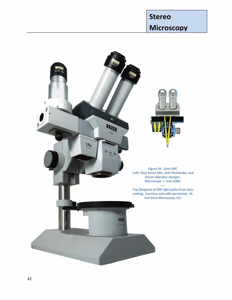
Figure 45. Zeiss DRC -Left. Zeiss Stemi DRC, with Phototube, and
stereo objective changer, Microscope c. mid-1980s
----- -Top [Diagram of DRC light paths from Zeiss catalog, Courtesy and with permission of,
Carl Zeiss Microscopy, LLC
(Zeiss, 1984)]
43
Stereo
Microscopy
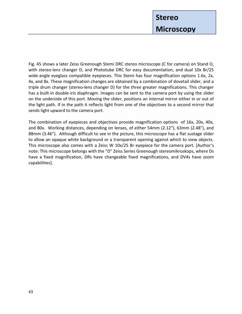
Fig. 45 shows a later Zeiss Greenough Stemi DRC stereo microscope (C for camera) on Stand O, with stereo-lens changer D, and Phototube DRC for easy documentation, and dual 10x Br/25 wide-angle eyeglass compatible eyepieces. This Stemi has four magnification options 1.6x, 2x, 4x, and 8x. These magnification changes are obtained by a combination of dovetail slider, and a triple drum changer (stereo-lens changer D) for the three greater magnifications. This changer has a built-in double-iris diaphragm. Images can be sent to the camera port by using the slider on the underside of this port. Moving the slider, positions an internal mirror either in or out of the light path. If in the path it reflects light from one of the objectives to a second mirror that sends light upward to the camera port. The combination of eyepieces and objectives provide magnification options of 16x, 20x, 40x, and 80x. Working distances, depending on lenses, of either 54mm (2.12"), 63mm (2.48"), and 88mm (3.46"). Although difficult to see in the picture, this microscope has a flat sustage slider to allow an opaque white background or a transparent opening against which to view objects. This microscope also comes with a Zeiss W 10x/25 Br eyepiece for the camera port. [Author's note: This microscope belongs with the "D" Zeiss Series Greenough stereomikroskops, where Ds have a fixed magnification, DRs have changeable fixed magnifications, and DV4s have zoom capabilities].
44
Stereo
Microscopy
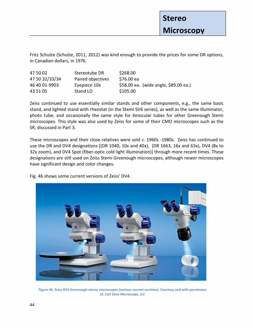
Fritz Schulze (Schulze, 2011, 2012) was kind enough to provide the prices for some DR options, in Canadian dollars, in 1976. 47 50 02 Stereotube DR $268.00 47 50 32/33/34 Paired objectives $76.00 ea 46 40 01-9903 Eyepiece 10x $58.00 ea. (wide angle, $89.00 ea.) 43 51 05 Stand LO $105.00 Zeiss continued to use essentially similar stands and other components, e.g., the same basic stand, and lighted stand with rheostat (in the Stemi SV6 series), as well as the same illuminator, photo tube, and occasionally the same style for binocular tubes for other Greenough Stemi microscopes. This style was also used by Zeiss for some of their CMO microscopes such as the SR, discussed in Part 3. These microscopes and their close relatives were sold c. 1960s -1980s. Zeiss has continued to use the DR and DV4 designations [(DR 1040, 10x and 40x), (DR 1663, 16x and 63x), DV4 (8x to 32x zoom), and DV4 Spot (fiber-optic cold light illumination)] through more recent times. These designations are still used on Zeiss Stemi Greenough microscopes, although newer microscopes have significant design and color changes. Fig. 46 shows some current versions of Zeiss' DV4.
Figure 46. Zeiss DV4 Greenough stereo microscopes (various current versions). Courtesy and with permission of, Carl Zeiss Microscopy, LLC
45
Stereo
Microscopy
Zeiss 'aus Jena'
In 1945 the US Army (USA) occupied Jena before it was to be turned over to the Russians [part
of what was to become the GDR (East Germany)] as reparations. So, in 1945 the US Army
requested that many of Zeiss' top scientists and senior management move to what was to
become West Germany. This relocated team built a new Zeiss company in West Germany using
designs from many existing Zeiss microscopes (and other items) built in Jena. It became
profitable sometime in the mid-1950s.
Some Zeiss personal not relocated to West Germany were sent to Russia to
help the Russians start new technology businesses. The Zeiss Jena business restarted quickly in
1946, although without those personal sent to assist the Russians. When these specialists
returned from Russia c. 1950/51 after being away about four years, the restarted Zeiss Jena
enterprise was already in full operation. On June 1, 1948 the Zeiss Jena company became a VEB
("peoples owned enterprise") with the Zeiss foundation no longer Zeiss' owner. Sometime later
it became the Kombinat VEB Carl Zeiss Jena, a conglomerate of companies.
This is from Zeiss on-line (it appears translated from original German text),
It was not until 1971 [that] an agreement [was reached] in London whereby ...
For example, VEB Carl Zeiss JENA was permitted to offer its products in the
Eastern Bloc countries, Syria, in the Lebanon and Kuwait using the agreed
trademarks. Carl Zeiss Oberkochen, on the other hand, had the right to distribute
the products bearing the name Carl Zeiss in West Germany, West Berlin, the
Benelux countries, Italy, Greece and the USA.
-- (Zeiss, undated History)

46
Stereo
Microscopy
As the Zeiss name in the West was to be used by Zeiss West Germany, microscopes sold in the
West and built by the competitive East German Zeiss company carried the name 'aus Jena'
(from Jena).
Berndt-Joachim Lau, Carl Zeiss Microscopy GmbH, a long time Zeiss employee who began work
for VEB Carl Zeiss Jena in East Germany in 1973 (Lau, 2012), explained to the author that there
are still strong and somewhat differing opinions about this time held by East and West German
"Zeissians", making the whole truth somewhat difficult to find. From after WW II to the
dissolution of the Soviet Union, the two Zeiss companies were competitors rather than
partners. Mr. Lau was also kind enough to send the author a short account of this period in
Zeiss history. That account is presented in Part 3.
Fig 47 presents an East German Zeiss (aus Jena) microscope from the time when there were
competing products from the two Zeiss companies, and before the two companies merged
after the Soviet Union ceased in 1989. In the late 1980s, VEB Carl Zeiss Jena had almost four
times as many employees as Carl Zeiss Oberkochen.
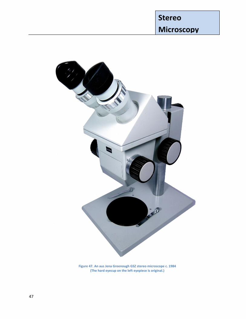
The microscope in Fig. 47 can be approximately dated from a Zeiss East German publication,
GSM Stereo Microscopes, # 30-735-1 (Zeiss 1984-GDR). GSM and GSZ microscopes were
contemporary instruments of similar design (see the quote below from a Zeiss GSM/GSZ
manual).
The GSM and GSZ are
largely standardized with regard to their mechanical and optical construction. They
are mainly differing through the type of the magnification changer. While the
magnification of the GSM is made by changeable objectives in fixed magnification
steps, the GSZ is equipped with a pancratic, i.e., step-less magnification changer. In
addition to this the GSZ had diopter adjusting rings for balancing accommodation
and defective vision. Tripod, eyepieces as well as the lighting and almost all other
supplementary equipment are interchangeable between the two microscopes.
(Zeiss, Undated GDR-2)
47
Stereo
Microscopy
Figure 47. An aus Jena Greenough GSZ stereo microscope c. 1984 (The hard eyecup on the left eyepiece is original.)
48
Stereo
Microscopy
Some Greenough Stereo Microscope Images
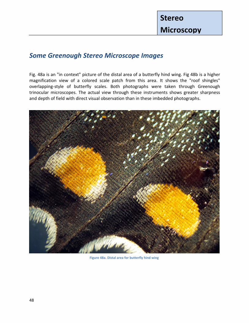
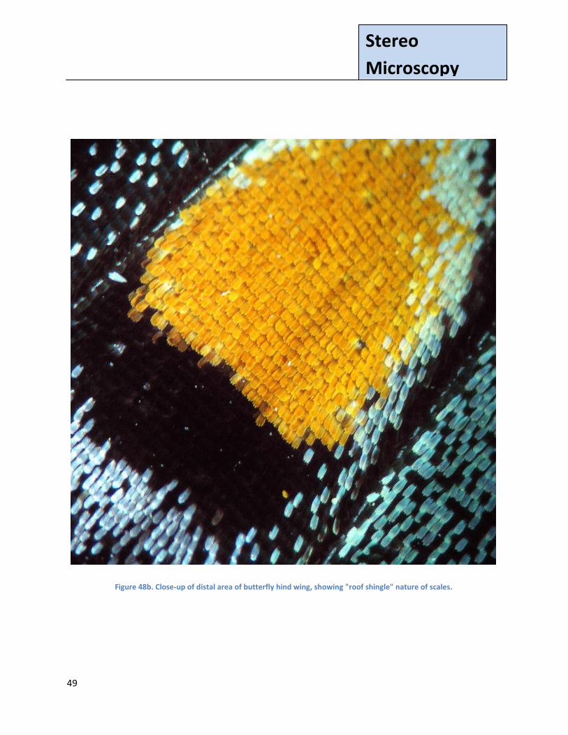
Fig. 48a is an "in context" picture of the distal area of a butterfly hind wing. Fig 48b is a higher magnification view of a colored scale patch from this area. It shows the "roof shingles" overlapping-style of butterfly scales. Both photographs were taken through Greenough trinocular microscopes. The actual view through these instruments shows greater sharpness and depth of field with direct visual observation than in these imbedded photographs.
Figure 48a. Distal area for butterfly hind wing
49
Stereo
Microscopy
Figure 48b. Close-up of distal area of butterfly hind wing, showing "roof shingle" nature of scales.
50
Stereo
Microscopy
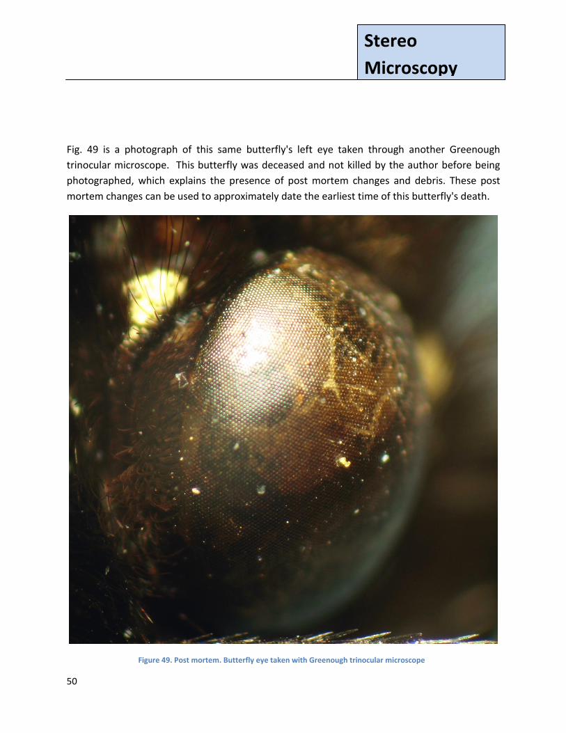
Fig. 49 is a photograph of this same butterfly's left eye taken through another Greenough
trinocular microscope. This butterfly was deceased and not killed by the author before being
photographed, which explains the presence of post mortem changes and debris. These post
mortem changes can be used to approximately date the earliest time of this butterfly's death.
Figure 49. Post mortem. Butterfly eye taken with Greenough trinocular microscope
51
Stereo
Microscopy
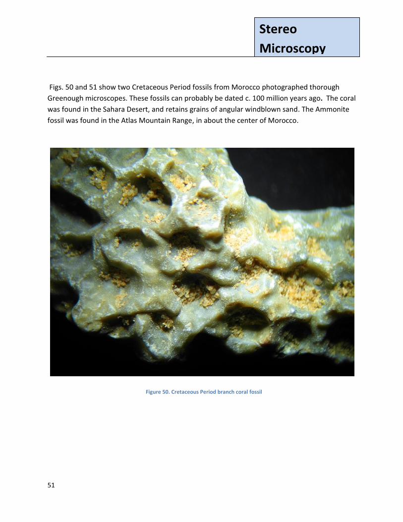
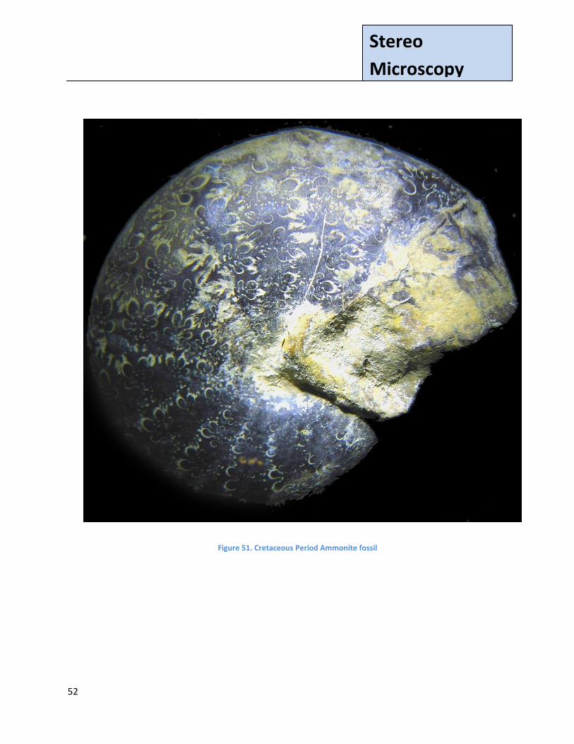
Figs. 50 and 51 show two Cretaceous Period fossils from Morocco photographed thorough
Greenough microscopes. These fossils can probably be dated c. 100 million years ago. The coral
was found in the Sahara Desert, and retains grains of angular windblown sand. The Ammonite
fossil was found in the Atlas Mountain Range, in about the center of Morocco.
Figure 50. Cretaceous Period branch coral fossil
52
Stereo
Microscopy
Figure 51. Cretaceous Period Ammonite fossil
53
Stereo
Microscopy
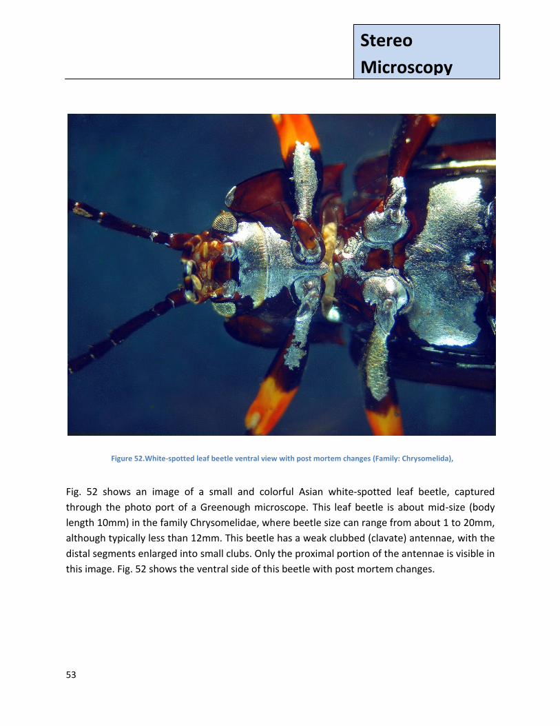
Fig. 52 shows an image of a small and colorful Asian white-spotted leaf beetle, captured
through the photo port of a Greenough microscope. This leaf beetle is about mid-size (body
length 10mm) in the family Chrysomelidae, where beetle size can range from about 1 to 20mm,
although typically less than 12mm. This beetle has a weak clubbed (clavate) antennae, with the
distal segments enlarged into small clubs. Only the proximal portion of the antennae is visible in
this image. Fig. 52 shows the ventral side of this beetle with post mortem changes.
Figure 52.White-spotted leaf beetle ventral view with post mortem changes (Family: Chrysomelida),
54
Stereo
Microscopy
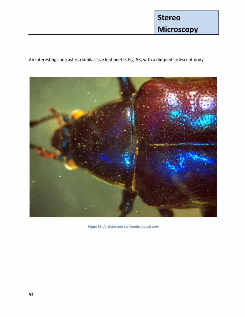
An interesting contrast is a similar-size leaf beetle, Fig. 53, with a dimpled iridescent body.
Figure 53. An iridescent leaf beetle, dorsal view
55
Stereo
Microscopy
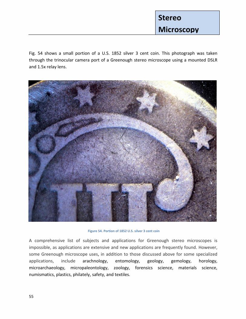
Fig. 54 shows a small portion of a U.S. 1852 silver 3 cent coin. This photograph was taken
through the trinocular camera port of a Greenough stereo microscope using a mounted DSLR
and 1.5x relay lens.
A comprehensive list of subjects and applications for Greenough stereo microscopes is
impossible, as applications are extensive and new applications are frequently found. However,
some Greenough microscope uses, in addition to those discussed above for some specialized
applications, include arachnology, entomology, geology, gemology, horology,
microarchaeology, micropaleontology, zoology, forensics science, materials science,
numismatics, plastics, philately, safety, and textiles.
Figure 54. Portion of 1852 U.S. silver 3 cent coin
56
Stereo
Microscopy
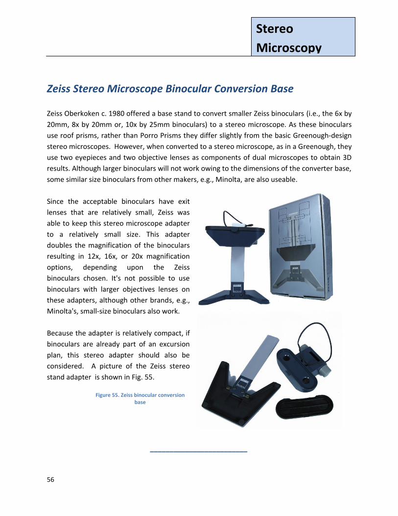
Zeiss Stereo Microscope Binocular Conversion Base
Zeiss Oberkoken c. 1980 offered a base stand to convert smaller Zeiss binoculars (i.e., the 6x by
20mm, 8x by 20mm or, 10x by 25mm binoculars) to a stereo microscope. As these binoculars
use roof prisms, rather than Porro Prisms they differ slightly from the basic Greenough-design
stereo microscopes. However, when converted to a stereo microscope, as in a Greenough, they
use two eyepieces and two objective lenses as components of dual microscopes to obtain 3D
results. Although larger binoculars will not work owing to the dimensions of the converter base,
some similar size binoculars from other makers, e.g., Minolta, are also useable.
Since the acceptable binoculars have exit
lenses that are relatively small, Zeiss was
able to keep this stereo microscope adapter
to a relatively small size. This adapter
doubles the magnification of the binoculars
resulting in 12x, 16x, or 20x magnification
options, depending upon the Zeiss
binoculars chosen. It's not possible to use
binoculars with larger objectives lenses on
these adapters, although other brands, e.g.,
Minolta's, small-size binoculars also work.
Because the adapter is relatively compact, if
binoculars are already part of an excursion
plan, this stereo adapter should also be
considered. A picture of the Zeiss stereo
stand adapter is shown in Fig. 55.
_________________________
Figure 55. Zeiss binocular conversion base

57
Stereo
Microscopy
©2011, 2012 Text and photographs (except as noted) by the author.
The author welcomes any suggestions for corrections or improvement. He has a continuing
interest in early and modern stereo microscopes from major manufacturers. He can be reached
at:
R. Jordan Kreindler: [email protected]
__________

58
Stereo
Microscopy
Combined References and End Notes (This list includes references/notes for the full paper. However, additional references may be added in later Parts)
Allen, R. M., (1940) The Microscope. Boston: D. Van Nostrand Company, Inc., p87. Bryant, Dr. Mark L., (2012) The author's thanks to Dr. Bryant and his staff for permission to photograph their Topcon slit lamp. Bausch & Lomb Optical Co, (1929) Microscopes & Accessories: Photomicrographic and Micro-Projection Apparatus Microtomes . Colorimeters Optical Measuring Instruments and Refractometers. Bausch & Lomb New York, p 81. Blocker (2012) Blocker History of Medicine, http://ar.utmb.edu/ar/Library/BlockerHistoryofMedicineCollection/BlockerHistoryofMedicineArtifacts/MicroscopeCollection/MicroscopesMakersandTheirInstruments/MicroscopeSwift/tabid/877/Default.aspx
Carpenter, William (with revisions by Rev. W. H. Dallinger) , (1901) The Microscope and Its Revelations.
Eighth Edition. Philadelphia: P. Blakiston's Son & Company, p 96.
Cherubin, d'Orleans. Père, (1677) La Dioptrique Oculaire ou La vision parfait ou le concours des deux axes de la vision en un seul point de l'objet , Paris: S. Mabre-Cramoisy del Cerro, Manual (2012) The author's thanks to Dr. del Cerro for his kindness in reviewing the
section on ophthalmology, and his helpful suggestions. However, all content is the sole
responsibility of the author.
Doherty, Glenn (2012) The author's thanks to Mr. Doherty, Support Representative, Carl Zeiss Microscopy, LLC for his help in identifying start and end manufacturing dates for some Zeiss stereomicroscopes.
Davis, George E., F.R.M. S. (1882) Practical Microscopy. London: David Bogue Encyclopaedia Britannica, (1910) A Dictionary of Arts, Sciences, Literature and General Information, 11th Edition, Volume 3, Binocular Instrument. New York, p 950.
59
Stereo
Microscopy
Ferraglio, Paul L., (2008) The Riddell-Stephenson Binocular Microscope. The Journal of the Microscope
Historical Society. Volume 16.
The author's thanks to Dr. Ferraglio, a leading authority on Prof. Riddell's microscope
and its successors. Dr. Ferraglio was kind enough to provide the author with reprints
of his papers, as well as helpful comments on an earlier version of this paper. However,
all content here is the sole responsibility of the author.
Ford, Brian, (1973) The Optical Microscope Manual. Past and Present Uses and Techniques. New York: Crane, Russet & Company, Inc. Goren, Yuval, The author's thanks to Dr. Goren for the many discussions we've had on historical
microscopes, and his emphasis on the importance of setting microscopes in their historical
context.
Gubas, Lawrence J., (2008) A Survey of Zeiss Microscopes 1846-1945. Las Vegas: Graphics 2000. This
book provides additional color photographs of a Model XV and its storage on page 253.
It can be highly recommended for its detailed and excpetional discussions of Zeiss microscopes.
Gubas, Lawrence J., (private correspondence, 2012) The author's thanks to Mr. Gubas for information on Zeiss instruments and employees, and pointers to Zeiss materials.
Hagan, Kevin (private correspondence, 2011) Thanks to Mr. Hagan of ALA industries Limited, Valparaiso, Indiana for providing a Contamikit brochure and PDF of the Instruction Manual. Hermann, Armin, Nur Der Name War Geb lieben: Die absenteuerliche Geschichte der Firma Carl Zeiss
Stuttgart: Deutsche Verlag-Anstalt, 1991, p. 37
Journal of the Society of Arts, Vol XXXIV, (November 1886). London: George Bell and Sons, for the Society of Arts, Fig. 16, p 1014.
Kreindler, R.J. and Yuval Goren, (March 2011),
Comparison of the Swift FM-31 Portable Field Microscope and an FM-31 Clone, Micscape, Figs.
11, 12, and 13.
Kreindler, R.J. and Yuval Goren, (May 2011), Baker's Traveller's Microscope, Micscape
Kreindler, R.J. and Yuval Goren, (November 2011), The TWX-1 Folded-Optics Microscope, Micscape

60
Stereo
Microscopy
Kreindler, R. J. (2012) The author worked in Silicon Valley for a number of years and saw the
extensive use, and occasional abuse, stereo microscopes in high-tech companies were
subjected to.
Lau, Berndt-Joachin (2012) The author 's thanks to Herr Lau of Carl Zeiss Microscopy GmbH.
His long experience at Zeiss combined with his personal knowledge of Zeiss
stereo microscopes and Zeiss history have truly been of immeasurable assistance to the author.
Maertin, Rainer (2012) www.photosrsenal.com. The author's thanks for his permission to use the
photo of the Brewster type stereo viewer.
Mappes, Timo (2005) The First Commercial Comparison Microscope, made after Wilhelm Thörner by
W. & H. Seibert, Wetzlar. The Journal of the Microscope Historical Society. Volume 13, No. 2.
Mappes, Timo (2005-2006) Museum optischer Instrumente, http://www.musoptin.com/seibert_15368.html
Moe, Harald, (2004) The Story of the Microscope. Denmark: Rhodes International Science and Art Publishers with the Collaboration of The Royal Microscopical Society, p. 176. Nikon Microscopy U (undated) Introduction to Stereomicroscopy states, "The first modern stereomicroscope was introduced in the United States by the American Optical Company in
1957. Named the Cycloptic®, this breakthrough design...". Although this was a landmark in American stereomicroscopes, the common objective concept was first used by Riddell in 1850s, and a common large objective was later implemented by Zeiss in their Citoplast, considerably before the Cycloptic® was introduced. NYMS (1957) The author's thanks to the NYMS for permission to reprint the advertisement from their 1957 Newsletter (See Pollinger, 1957) Orlowski, Kristen and Dr. Michael Zölffel (private correspondence, 2012)
- The author's thanks to both Kristen Orlowski, Product Marketing Manager, Light Microscopes, Carl Zeiss Microscopy, LLC and Dr. Michael Zölffel, Carl Zeiss MicroImaging Gmb, Jena, Germany for information and materials they provided regarding Zeiss history. Ozment, Randall R. (2012) The author's thanks to Dr. Ozment for permission to photograph his Haag- Streit slit lamp, and for his explanation of its use in clinical practice.
61
Stereo
Microscopy
Phillips, Jay. (private correspondence, 2011, 2012) Provided a copy of Zeiss' catalog Mikroskope für Wissenschaft und Technologie (Prob. 1951). Pollinger, Mel. (1957) The author's thanks to Mr. Pollinger, Editor NYMS Newsletter for permission to reprint the advertisement from The New York Microscopical Society (NYMS) Newsletter of 1957
(See NYMS, 1957) Phillips, Jay. (private correspondence, (2011, 2012) Provided a copy of Zeiss' catalog "Mikroskope für Wissenschaft und Technologie" (Prob. 1951). Purtle, Helen R. (Second Edition), (1987 reprint) The Billings Microscope Collection. Second Edition. Washington, D.C.: Armed Forces Institute of Pathology, p 228, Figure 458 (Catalog number: M- 030.00541, AFIP accession number: 518,969, MIS photograph: 73-3899) Riemer, Marvin F., (1962) Microscope and the World of Science. New York: SCOPE Instrument Corp. RMS (1898) Journal of the Royal Microscopical Society, Volume 18, pp 469-471 Sander, Klaus. (1994) An American in Paris and the origins of the stereomicroscope. Institut für Biologie I (Zoologie). Freiburg, Germany: Springer-Verlag Schulze, Fritz , (2011, 2012) The author's thanks to Mr. Schulze, former head of the Historical Microscopical Society of Canada for his extensive knowledge of Zeiss microscopes which he kindly shared, and our extended exchanges on stereo microscopes.
Schwabe, Ms. Marte (2012) The author's thanks to Ms. Schabe, Assistant to Dr. Wimmer, Carl . Zeiss Archiv for her assistance (see Wimmer below). Schwidefsky, Kurt,( 1950) Grundriss der Photogrammetrie, Verlag für Wissenschaft und Fachbuch: 1950 (Reference from Fritz Schulze). Stanley, Jay (2012) The author's thanks for permission to use photos from his web site Classic Optics. Wade Nicolas , (1998) A Natural History of Vision. Cambridge, Mass: MIT press,p 301. Waldsmith, John (1991) Stereo Views: An Illustrated History and Price Guide. Wallace-Homestead Book Company: Radnor, Pennsylvania.
62
Stereo
Microscopy
Walker, David (undated) This is a short no frills introduction to stereo microscopes. http://www.microscopy-uk.org.uk/dww/novice/choice3.htm
Wheatstone, Charles. (1838) Contributions to the Physiology of Vision.—Part the First. On some remarkable, and hitherto unobserved, Phenomena of Binocular Vision, June 21, 1838 Wise, F. C., Francis Edmund Jury Ockenden, P. K.Sartory, (1950) The binocular microscope: its development, illumination and manipulation. (Quekett Microscopical Club Monograph) London: Williams & Norgate Wimmer, Wolfgang. The author's thanks to Dr. Wimmer's office at the Carl Zeiss Archiv Jena, Germany for their help. Zeiss, (Microscopy, LLC, MicroImaging Gmb, Jena)
- Zeiss (1934) Zeiss 1934 catalog, English version - Zeiss (1937) Zeiss catalog - Zeiss (1951) Mikroskope für Wissenschaft und Technologie Catalog - Zeiss (1984) Catalog 41-603-e - Zeiss(1984-GDR) GSM Stereo Microscopes Publication # 30-735-1 - Zeiss (Undated) Citoplast brochure, East Germany - Zeiss (Undated GDR-2) GSM GSZ Stereomicroscopes - Zeiss (Undated History) - Two Zeiss Factories in Germany, http://corporate.zeiss.com/history/en_de/corporate-history /at-a-glance.html#inpagetabs-4 [The extended extract is available at the Zeiss site. It is reproduced with permission of Wolfgang Mühlfriedel and Edith Hellmuth (1996), from a publication of the Regional Center for Political Education, Thuringia] - Zeiss (Undated) Opton catalog,, West Germany - Zeiss (Undated) Stemi DR, Stemi DV4, Stemi Stereomicroscopes brochure Zölffel, Michael (2012) see Orlowski above
63
Stereo
Microscopy
_________________________
Published in the online magazine Micscape, August 2012,
www.microscopy-uk.org.uk/mag/artaug12/jk-stereo2b.pdf
www.microscopy-uk.org.uk.
Please report any Web problems or offer general comments to the Micscape Editor. Micscape is the on-line monthly magazine of the Microscopy UK web
site at Microscopy-UK