Embed Size (px)
Citation preview
LETTER
Structural basis of AimP signaling moleculerecognition by AimR in Spbeta groupof bacteriophages
Dear Editor,
Quorum sensing (QS) is a widespread phenomenon inbacteria which enables them to participate in cell-to-cellcommunication by producing and responding to small signalmolecules, thus synchronously altering their behaviordepending on population density (Singh et al., 2000; Millerand Bassler, 2001). Through QS, bacteria coordinate pro-cesses such as expression of virulence factors (Slamti andLereclus, 2002), biofilm formation (Parashar et al., 2011),sporulation (Perego et al., 1996), conjugation (Kozlowiczet al., 2006), antibiotic synthesis (Miller and Bassler, 2001;Whiteley et al., 2017) etc.
In Gram-positive bacteria, QS is mainly controlled by afamily of cytosolic peptide-sensing regulators known asRRNPP, which is named for its representative members, i.e.,Rap, Rgg, NprR, PlcR and PrgX. The reported structures ofRRNPP members (Parashar et al., 2011; Gallego del Soland Marina, 2013; Parashar et al., 2013) show a two-domainstructure with an N-terminal helix-turn-helix (HTH) motifDNA-binding domain (DBD), a C-terminal tetratricopeptiderepeat (TPR) domain containing the elements for signalpeptide binding and oligomerization, and a short linker helixconnecting the two domains. Despite the conserved tertiarystructure of these regulators, structural analyses revealunexpected diversity in the mechanism of activation andmolecular strategies that couple the peptide-induced allos-tery to gene expression (Do and Kumaraswami, 2016).
Remarkably, a recent report described the use of a QSsystem for regulation of entry into lytic or lysogenic cycle bythe Bacillus-infecting temperate phages phi3T and SPbeta(Erez et al., 2017). This system, termed arbitrium, is remi-niscent of RRNPP-mediated QS found in Gram-positivebacteria, and is the first known example of QS in bacterio-phages. The arbitrium system comprises three phage genes:(1) AimP, encoding the peptide that is processed into a 6 aasignaling peptide extracellularly, (2) AimR, encoding theintracellular receptor of AimP that is predicted to be struc-turally similar to RRNPP regulators (Fig. 1A), and (3) AimX,encoding a putative long non-coding DNA that functions asthe negative regulator of lysogeny (Erez et al., 2017). During
the initial stages of infection, the bacteriophages expressAimP and AimR. As a transcription factor, AimR dimerinduces expression of AimX, which promotes lytic cycle.Concurrently, the levels of mature AimP rise in extracellularmedium. The concentration of AimP will eventually reach thepoint that its uptake by newly infected bacteria will be highenough to bind AimR and abolish transcriptional activation ofAimX, thereby shifting the phage into lysogenic cycle. In thismanner, the bacteriophages of SPbeta group are able tocoordinate their reproduction, with preference for lytic cyclewhen there is abundance of host cells, and preference forlysogenic cycle in case of a dwindling bacterial population.However, the structures of AimR and AimR-AimP complexare still unknown, which impedes deeper understanding ofthe molecular mechanism underlying the switch betweenlytic and lysogenic cycles mediated by the arbitrium system.
Here, we used X-ray crystallography to determine thestructures of apo AimR (Fig. 1B) and AimR-AimP complex ofBacillus subtilis Spbeta group of bacteriophages (Fig. 1Cand 1D). The crystals of apo AimR were obtained followingextensive crystal screening and optimization assays. Due tothe high flexibility of N-terminus, the electron density of theapo AimR DBD was weak, which made it challenging to buildthe structure of residues 1–55. After a strenuous effort, thestructure of full-length apo AimR was solved at 2.63 Å res-olution using single-wavelength anomalous diffractionmethod. The electron density of the N-terminus of AimRwithin the AimR-AimP complex was of better quality; usingthe structure of apo AimR as a model, the binary complexAimR-AimP was solved by molecular replacement andrefined to 2.00 Å resolution. Detailed diffraction and refine-ment statistics are listed in Table S1.
The final structure of apo AimR contained two moleculesin the asymmetric unit spanning residues 1–386 and aC-terminal His-tag (Fig. 1B). The two protomers within theasymmetric unit are nearly identical, with an average rootmean square deviation (RMSD) of 0.159 Å (Fig. S1A). Aspredicted from its similarity with RRNPP proteins, thestructure of AimR is characterized by two domains, i.e., theN-terminal HTH DNA-binding domain (residues 1–73) andthe C-terminal TPR domain (residues 79–386), which are
© The Author(s) 2018
Protein Cell 2019, 10(2):131–136https://doi.org/10.1007/s13238-018-0588-6 Protein&Cell
Protein
&Cell
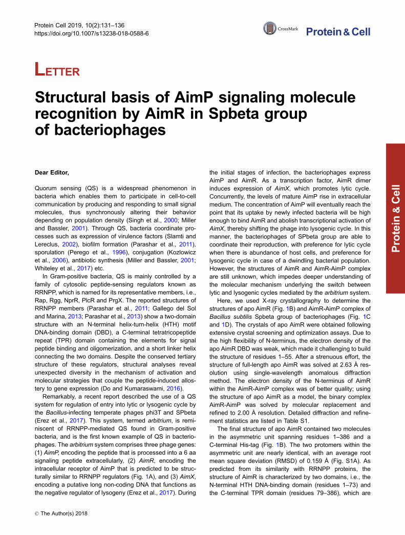
connected by a short linker (residues 74–78). Anotable feature of AimR structure is that it comprises a totalof 24 helical secondary structures, including 22 α-helicesand two 310-helices (Fig. S1C). While the first five helicesform the DNA binding domain, the remaining α-helices aregrouped into eight TPR motifs and a C-terminal cappinghelix. AimR and AimR-AimP are homodimers (Fig. 1E and
1F) and the dimerization is mediated by the C-terminalcapping helix, with an approximate solvent-accessible sur-face of 810 Å2. The residues L380, L383, L384 and L386contribute to dimerization by engaging in van der Waalsinteractions with matching residues from the other subunit(Fig. 1F). We tested an AimR deletion mutant lackingC-terminal capping helix (missing seven C-terminal amino
Figure 1. Overall structures of apo AimR and AimR-AimP complex. (A) Domain organization of AimR. (B) Structure of apo AimR.
Two AimR molecules are found in one asymmetric cell unit. The AimR molecules are shown in cartoon, with colors depicting DBD and
TPR domains as in Fig. 1A. (C) Structure of AimR-AimP complex in one symmetric cell unit. (D) The homodimer of AimR-AimP.
AimRs are colored as in Fig. 1B, whereas AimP molecules are presented as yellow sticks. (E) Surface features of AimR homodimer
by electrostatic potential (red, negative; white, neutral; blue, positive). (F) An overhead view of the leucine residues of C-terminal
capping helix that form AimR dimerization interface. (G) Analytical size-exclusion chromatography of AimRΔ379 and wild-type AimR-
AimP with Superdex 200 increase (GE Healthcare). Before the assay, AimP was incubated with AimR at 10:1 molar ratio for 30 min.
Wild-type AimR-AimP was eluted at 13.5 mL (blue), whereas AimRΔ379 was eluted at 14.9 mL (red).
LETTER Xiangkai Zhen et al.
132 © The Author(s) 2018
Protein
&Cell
acids, denoted AimRΔ379) by performing size-exclusionchromatography and found that dimerization was disrupted(Fig. 1G), thus confirming the importance of these residuesin dimerization of AimR. Analysis of the surface electrostaticpotential of AimR reveals a large positively charged areawith potential DNA-binding activity located on the surface ofDBD (Fig. S1A and S1B).
Although the overall structure of AimR shows high simi-larity to the classical RRNPP regulators, the sequencehomology between AimR and the members of RRNPP isvery low. However, a protein structure comparison on Daliserver with AimR as a query retrieved 11 structures, amongwhich RapJ, NprR, PlcR, RapF, RapI and RapH (all mem-bers of RRNPP family of proteins) had Z-scores higher than13.
In the structure of AimR-AimP complex, two molecules ofAimR are present in the asymmetric cell unit. One moleculeof the AimP hexapeptide (GMPRGA) binds to one moleculeAimR. The electron density of AimP was clearly visible withinthe complex (Fig. 2A). The TPR domain of AimR forms adeep concave pocket that accommodates a molecule ofAimP, with a buried interface area of 758.6 Å2. The C-ter-minus alanine of AimP is facing the interior of the pocket,whereas the N-terminal glycine is located at the entry point(Fig. 2B and 2C). In vitro ITC assay demonstrates that full-length AimR specifically binds AimP with a Kd of 40 nmol/L(Fig. 2D). The interaction between AimR and AimP includesan extensive network of hydrogen bonds and hydrophobiccontacts. The oxygen atoms from the peptide backbone ofAimR form hydrogen bonds with four AimP residues:Q299AimR and E300AimR interact with the nitrogen moiety ofG1AimP, N239AimR forms hydrogen bonds with P3AimP,N202AimR interacts with G5AimP, and R228AimR contacts theoxygen moieties of A6AimP, whereas five hydrophobic resi-dues from AimR (L205, L242, F276, F362 and L363) areinvolved in van der Waals interactions with the side chain ofM2AimP.
It is noteworthy to mention that AimR accommodates theGMRPGA peptide in a manner similar to the classicalRRNPP transcription factors in bacteria. However, the modeby which AimP interacts with AimR differs from the previ-ously studied interactions between RRNPP proteins and thecorresponding signaling peptides. As opposed to the clas-sical interaction between RRNPP and the signaling peptidein bacteria that does not include side chain-specific contacts,the guanidinium group of R4AimP forms hydrogen bonds withthree residues in AimR, namely N206, N329 and D360.
The importance of the AimR residues involved in AimPcoordination was further confirmed by carrying out ITC assaywith seven single point mutations (N202A, N206A, N239A,Q299A, E300A, N329A and D360A). While the value ofdissociation constant for the wild-type AimR-AimP complexwas 40 nmol/L, the abovementioned mutations reduced theAimP binding activity of AimR in the range between0.14 μmol/L and 6.7 μmol/L (Fig. S2A–G). A sequencealignment of three AimRs from Bacillus phages revealed that
all the residues except E300 are highly conserved(Fig. S1C). Thus, we conclude that the residues N202, N206,N239, N299, N329 and D360 of AimR play an essential rolein binding AimP. Since previous research demonstrated thatbinding of AimP to AimR leads to silencing of the AimR-mediated AimX transcription and subsequent entry intolysogenic pathway (Erez et al., 2017), we further speculatethat these residues form a switch that shifts phages betweenlytic and lysogenic cycle.
Active AimR dimer from the phi3T reportedly dissociatesinto inactive monomers upon AimP binding (Erez et al.,2017). However, our structure indicates that in the spBeta-derived AimR-AimP complex AimRs still exist as a dimer.This observation was corroborated by size-exclusion chro-matography (GE healthcare, Superdex 200 increase)(Fig. 1F). Further analysis with the PISA program revealedthat the AimR-AimP complex is most stable in heterote-tramer conformation (i.e., an AimR dimer with each subunitbinding one molecule of AimP), implying that in arbitriumsystem of spBETA phages AimP inactivates AimR through amechanism distinct from the corresponding mechanism inphi3T phages.
While we were preparing our manuscript for submission,Wang et al. reported the structures of apo AimR (aa 43–386)and AimR43-386-AimP complex (Wang et al., 2018). Theirstructures share high similarity with the structures frompresent study (average RMSD of 0.223 Å for aligned apoAimR structures) (Fig. S3A). Moreover, similarly to Wanget al., we also noticed subtle conformational changes inN-terminal domain of AimR caused by binding of AimP.Prompted by this observation, we analyzed the intramolec-ular B-factor of AimR. The results revealed that the B-factorvalue of the N-terminal DBD, especially the first three heli-ces, is notably higher than the rest of molecule, indicatingthat AimP binding may change the conformation of DBD(Fig. S3B–D), which in turn would reduce the DNA-bindingability of AimR. This is also in agreement with SAXS analysisindicating that AimP-bound AimR has an extended confor-mation when compared to AimP-free AimR (Wang et al.,2018).
Furthermore, our results are in line with another recentstudy, which found difference in infection dynamics betweenSPbeta phages and phi3T phages, and confirmed thatSPbeta-derived AimR does not dissociate upon bindingGMPRGA peptide (Dou et al., 2018). Altogether, thesefindings indicate that the molecular mechanism of AimRregulation does not only differ from bacterial RRNPP pro-teins but also from the more closely-related AimR in phi3Tphages. However, future studies should investigate thestructure of AimR bound to AimX gene in order to provide aclearer answer to how does AimP inhibit transcription ofAimX by binding to AimR.
In summary, we reported the crystal structures of apoAimR and AimR-AimP complex from arbitrium system ofSPbeta phage group. Our high resolution structures coupledwith biochemical analyses shed new light on the molecular
Structural basis of pheromone recognition by AimR LETTER
© The Author(s) 2018 133
Protein
&Cell

mechanisms underlying the switch between lysis and lyso-geny in arbitrium QS system of SPbeta phages, and are insupport of similar studies (Dou et al., 2018; Wang et al.,
2018). The main findings in our study are outlined as follows:(1) AimR consists of 22 α-helices and two 310-helices. Thefirst three α helices in N-terminal DNA-binding domain of apo
Figure 2. Recognition of AimP hexapeptide (GMPRGA) by AimR from SPbeta phage. (A) Electron density map of the AimP
bound to AimR. The 2Fo-Fc map, calculated by simulated annealing without peptide in the structure, is shown contoured at 1.5 σ as a
gray grid with the peptide in sticks colored by atom type. (B) The binding mode between AimR and AimP. AimR is shown as cartoon
colored in white, whereas the residues that interact with AimP (N202, N206, N239, Q299, E300, N329 and D360) are shown as sticks
colored by atom type. AimP is shown in stick representation with purple carbons. Hydrogen bonds are depicted as dotted yellow lines.
(C) Similar to (B), but showing only interacting residues from another angle of view. (D) Binding affinity of wild-type AimR for AimP
measured by ITC. The concentration of AimP used in assay was 200–500 μmol/L, and the concentration of AimR was 10–20 μmol/L.
LETTER Xiangkai Zhen et al.
134 © The Author(s) 2018
Protein
&Cell
AimR show a high degree of flexibility. (2) The TPR domainof AimR forms a concave pocket in which a number ofconserved residues bind the AimP hexapeptide GMPRGAwith high affinity through an extensive network of hydrogenbonds and hydrophobic interactions. Although AimRaccommodates the signaling peptide similarly to RRNPPproteins in bacteria, the nature of interactions between AimRand AimP is distinct. (3) The dimerization of AimR is medi-ated by the C-terminal capping helix. Unlike the AimR inphi3T phages, AimR dimer in SPbeta phages does not dis-sociate into monomers upon AimP binding, but insteadappears to be further stabilized. This was inferred fromobservations in crystallization assays, the crystal structuresof apo AimR and AimR-AimP complex, and computationalanalysis with PISA.
To conclude, we would like to propose an amendedmechanistic model of arbitrium system in SPbeta phages,which takes into consideration all three relevant studies (Douet al., 2018; Wang et al., 2018, and the present study): in theabsence of AimP hexapeptide GMPRGA, AimR possesses ahighly flexible N-terminal DNA-binding domain. When bind-ing the AimX gene locus, the DBD domain adopts a con-formation that allows specific interactions with DNA, whichare primarily mediated by the first three α helices in DBD.However, AimP binding induces a second conformationalchange in AimR that is characterized by an extended butmore rigid DBD, which distorts interactions between AimRand DNA, and results in decreased (rather than abolished)DNA binding. This, in turn, shifts the equilibrium betweenlysis and lysogeny in phage population towards lysogeny.Such model can more clearly explain not only the differencesin infection dynamics between SPbeta phages and phi3Tphages (Dou et al., 2018) but also the lack of obvious con-formational variations between apo AimR and AimR-AimPcomplex (Wang et al., 2018 and the present study).
FOOTNOTES
The atomic coordinates and structure factors have been deposited in
the Protein Data Bank (PDB) under the accession codes 6IPX for
apo AimR and 6IM4 for AimR-AimP complex.
We thank the staff at beamline BL-17U1 of Shanghai Syn-
chrotron Radiation Facility for their help with X-ray diffraction data
collection.
This work was supported by the Ministry of Science and Tech-
nology of China grants 2014CB910400 and the National Nature
Science Foundation of China grants 31770948, 31570875,
31800159 and 81590761. We thank Fujian Normal University for
financial support.
Xiangkai Zhen, Huan Zhou, Wei Ding, Biao Zhou, Xiaolong Xu,
Vanja Perčulija, Chun-Jung Chen, Ming-Xian Chang, Muhammad
Iqbal Choudhary and Songying Ouyang declare that they have no
conflict of interests.
This article does not contain any studies with human or animal
subjects performed by the authors.
Xiangkai Zhen1,2, Huan Zhou3, Wei Ding4, Biao Zhou1,2,Xiaolong Xu1,2, Vanja Perčulija1,2, Chun-Jung Chen6,Ming-Xian Chang7, Muhammad Iqbal Choudhary8,
Songying Ouyang1,2,5&
1 The Key Laboratory of Innate Immune Biology of Fujian Province,
Biomedical Research Center of South China, College of Life
Sciences, Fujian Normal University, Fuzhou 350117, China2 Provincial University Key Laboratory of Cellular Stress Response
and Metabolic Regulation, College of Life Sciences, Fujian Normal
University, Fuzhou 350117, China3 Shanghai Institute of Applied Physics, Chinese Academy of
Sciences, Shanghai 201204, China4 CAS Key Laboratory of Soft Matter Physics, Institute of Physics,
Chinese Academy of Sciences, Beijing 100190, China5 National Laboratory of Biomacromolecules, Institute of Biophysics,
Chinese Academy of Sciences, Beijing 100101, China6 Institute of Biotechnology, National Cheng Kung University, Tainan
701, China7 State Key Laboratory of Freshwater Ecology and Biotechnology,
Institute of Hydrobiology, Chinese Academy of Sciences, Wuhan
430072, China8 H.E.J. Research Institute of Chemistry, International Center for
Chemical and Biological Sciences, University of Karachi, Karachi
75270, Pakistan
& Correspondence: [email protected] (S. Ouyang)
OPEN ACCESS
This article is distributed under the terms of the Creative Commons
Attribution 4.0 International License (http://creativecommons.org/
licenses/by/4.0/), which permits unrestricted use, distribution, and
reproduction in any medium, provided you give appropriate credit to
the original author(s) and the source, provide a link to the Creative
Commons license, and indicate if changes were made.
REFERENCES
Do H, Kumaraswami M (2016) Structural mechanisms of peptide
recognition and allosteric modulation of gene regulation by the
RRNPP family of quorum-sensing regulators. J Mol Biol
428:2793–2804
Dou C, Xiong J, Gu Y, Yin K, Wang J, Hu Y, Zhou D, Fu X, Qi S, Zhu
X et al (2018) Structural and functional insights into the regulation
of the lysis–lysogeny decision in viral communities. Nat Microbiol
3:1285–1294
Erez Z, Steinberger-Levy I, Shamir M, Doron S, Stokar-Avihail A,
Peleg Y, Melamed S, Leavitt A, Savidor A, Albeck S et al (2017)
Communication between viruses guides lysis-lysogeny deci-
sions. Nature 541:488–493
Gallego del Sol F, Marina A (2013) Structural basis of Rap
phosphatase inhibition by Phr peptides. PLoS Biol 11:e1001511
Kozlowicz BK, Shi K, Gu ZY, Ohlendorf DH, Earhart CA, Dunny GM
(2006) Molecular basis for control of conjugation by bacterial
pheromone and inhibitor peptides. Mol Microbiol 62:958–969
Structural basis of pheromone recognition by AimR LETTER
© The Author(s) 2018 135
Protein
&Cell
Miller MB, Bassler BL (2001) Quorum sensing in bacteria. Annu Rev
Microbiol 55:165–199
Parashar V, Jeffrey PD, Neiditch MB (2013) Conformational change-
induced repeat domain expansion regulates Rap phosphatase
quorum-sensing signal receptors. PLoS Biol 11:e1001512
Parashar V, Mirouze N, Dubnau DA, Neiditch MB (2011) Structural
basis of response regulator dephosphorylation by Rap phos-
phatases. PLoS Biol 9:e1000589
Perego M, Glaser P, Hoch JA (1996) Aspartyl-phosphate phos-
phatases deactivate the response regulator components of the
sporulation signal transduction system in Bacillus subtilis. Mol
Microbiol 19:1151–1157
Singh PK, Schaefer AL, Parsek MR, Moninger TO, Welsh MJ,
Greenberg EP (2000) Quorum-sensing signals indicate that
cystic fibrosis lungs are infected with bacterial biofilms. Nature
407:762–764
Slamti L, Lereclus D (2002) A cell-cell signaling peptide activates the
PlcR virulence regulon in bacteria of the Bacillus cereus group.
EMBO J 21:4550–4559
Wang Q, Guan Z, Pei K, Wang J, Liu Z, Yin P, Peng D, Zou T (2018)
Structural basis of the arbitrium peptide–AimR communication
system in the phage lysis–lysogeny decision. Nat Microbiol
3:1266–1273
Whiteley M, Diggle SP, Greenberg EP (2017) Progress in and
promise of bacterial quorum sensing research. Nature 551:313–
320
Electronic supplementary material The online version of thisarticle (https://doi.org/10.1007/s13238-018-0588-6) contains sup-
plementary material, which is available to authorized users.
LETTER Xiangkai Zhen et al.
136 © The Author(s) 2018
Protein
&Cell


![A Passion for the Elements - AIMR, Tohoku Univ. · 019 ]AIMR Action Log[April 2015 - March 2016 Contents It is our great pleasure to publish Volume 8 of AIMR Magazine. Fiscal year](https://img.pdfslide.net/doc/110x75/5f32efe6caee3e246a54f6b7/a-passion-for-the-elements-aimr-tohoku-univ-019-aimr-action-logapril-2015.jpg)