Embed Size (px)
Citation preview

RESEARCH ARTICLE
Allopregnanolone Preclinical AcutePharmacokinetic and PharmacodynamicStudies to Predict Tolerability and Efficacy forAlzheimer’s DiseaseRonald W. Irwin1, Christine M. Solinsky2, Carlos M. Loya3, Francesco G. Salituro3,Kathleen E. Rodgers4, Gerhard Bauer5, Michael A. Rogawski6, Roberta Diaz Brinton1,7*
1 Department of Pharmacology and Pharmaceutical Science, School of Pharmacy, University of SouthernCalifornia, Los Angeles, California, United States of America, 2 Clinical and Experimental TherapeuticsProgram, University of Southern California, Los Angeles, California, United States of America, 3 SageTherapeutics, Cambridge, Massachusetts, United States of America, 4 Titus Family Department of ClinicalPharmacy and Pharmaceutical Economics & Policy, School of Pharmacy, University of Southern California,Los Angeles, California, United States of America, 5 Department of Internal Medicine, School of Medicine,University of California Davis, Sacramento, California, United States of America, 6 Department of Neurology,School of Medicine, University of California Davis, Sacramento, California, United States of America,7 Department of Neurology, Keck School of Medicine, University of Southern California Los Angeles, LosAngeles, California, United states of America
AbstractTo develop allopregnanolone as a therapeutic for Alzheimer’s disease, we investigated mul-
tiple formulations and routes of administration in translationally relevant animal models of
both sexes. Subcutaneous, topical (transdermal and intranasal), intramuscular, and intrave-
nous allopregnanolone were bolus-administered. Pharmacokinetic analyses of intravenous
allopregnanolone in rabbit and mouse indicated that peak plasma and brain levels (3-fold
brain/plasma ratios) at 5min were sufficient to activate neuroregenerative responses at sub-
sedative doses. Slow-release subcutaneous suspension of allopregnanolone displayed 5-
fold brain/plasma ratio at Cmax at 30min. At therapeutic doses by either subcutaneous or in-
travenous routes, allopregnanolone mouse plasma levels ranged between 34-51ng/ml by
30min, comparable to published endogenous human level in the third trimester of pregnan-
cy. Exposure to subcutaneous, topical, intramuscular, and intravenous allopregnanolone,
at safe and tolerable doses, increased hippocampal markers of neurogenesis including
BrdU and PCNA in young 3xTgAD and aged wildtype mice. Intravenous allopregnanolone
transiently and robustly phosphorylated CREB within 5min and increased levels of neuronal
differentiation transcription factor NeuroD within 4h. Neurogenic efficacy was achieved with
allopregnanolone brain exposure of 300-500hr*ng/g. Formulations were tested to deter-
mine the no observable adverse effect level (NOAEL) and maximally tolerated doses (MTD)
in male and female rats by sedation behavior time course. Sex differences were apparent,
males exhibited�40%more sedation time compared to females. Allopregnanolone formu-
lated in sulfobutyl-ether-beta-cyclodextrin at optimized complexation ratio maximized
PLOSONE | DOI:10.1371/journal.pone.0128313 June 3, 2015 1 / 31
a11111
OPEN ACCESS
Citation: Irwin RW, Solinsky CM, Loya CM, SalituroFG, Rodgers KE, Bauer G, et al. (2015)Allopregnanolone Preclinical Acute Pharmacokineticand Pharmacodynamic Studies to Predict Tolerabilityand Efficacy for Alzheimer’s Disease. PLoS ONE10(6): e0128313. doi:10.1371/journal.pone.0128313
Academic Editor: Grace E. Stutzmann, RosalindFranklin University, UNITED STATES
Received: September 8, 2014
Accepted: April 25, 2015
Published: June 3, 2015
Copyright: © 2015 Irwin et al. This is an openaccess article distributed under the terms of theCreative Commons Attribution License, which permitsunrestricted use, distribution, and reproduction in anymedium, provided the original author and source arecredited.
Data Availability Statement: All data are within thepaper.
Funding: This research was supported by theNational Institute on Aging (http://www.nia.nih.gov/)U01 AG031115 and U01 AG047222 to RDB.Allopregnanolone was supplied through support bythe Department of Defense under award numberW81XWH-09-1-0746 to MAR. Views and opinions of,and endorsements by the authors do not reflect thoseof the US Army or the Department of Defense. SageTherapeutics, Cambridge MA supported the mousepharmacokinetic LC-MS/MS data acquisition. The

allopregnanolone delivery and neurogenic efficacy. To establish the NOAEL and MTD for
Allo-induced sedation using a once-per-week intravenous regenerative treatment regimen:
In female rats the NOAEL was 0.5mg/kg and MTD 2mg/kg. The predicted MTD in human fe-
male is 0.37mg/kg. In male rats the NOAEL and MTD were less than those determined for
female. Outcomes of these PK/PD studies predict a safe and efficacious dose range for ini-
tial clinical trials of allopregnanolone for Alzheimer’s disease. These findings have transla-
tional relevance to multiple neurodegenerative conditions.
IntroductionTo date, no therapeutic intervention exists to prevent, delay, or treat Alzheimer's disease [1, 2].Recent failed Phase 3 trials targeting beta-amyloid plaques are indicative of the complexity ofthe multifactorial disease process and highlight the need for alternative innovative therapeutics[3, 4]. A novel therapeutic approach targets the regenerative neurogenic capacity of the brainto sustain neurological function and to prevent, delay or treat neurodegenerative diseases [5].In adults, the subgranular zone of the hippocampus dentate gyrus and the subventricular zoneof the lateral ventricle comprise the two most prolific neurogenic niches [6]. Multiple studiesindicate that adult human neurogenesis occurs and is sustained throughout the lifespan in thedisease-free brain [7–9]. Previously, we demonstrated that the neurosteroid allopregnanolone(Allo) promotes neurogenesis in vivo [10] and proliferation of rodent and human neural pro-genitor cells in vitro [11]. Allo increased neurogenesis within the hippocampus and restoredlearning and memory function to normal prior to and following the onset of Alzheimer's dis-ease pathology in the triple transgenic Alzheimer’s disease (3xTgAD) mouse [10, 12, 13]. Fur-ther, Allo was comparably efficacious in the aged wildtype mouse [13]. In 3xTgAD mice, Alloincreased markers of white matter regeneration and cholesterol homeostasis while simulta-neously reducing beta-amyloid burden and microglia inflammatory markers [12].
An optimal Allo dosing regimen of once per week significantly increased neurogenesiswhile simultaneously reducing Alzheimer’s related pathology [5, 12, 14]. Allo fulfills multiplerequirements for drugs targeting the brain including a small molecular weight (318.49 g/mol);low number of hydrogen bond donors (one) and acceptors (two). The logP 5.042 value forAllo, poses a solubility challenge for aqueous formulation and thus makes formulation for oraladministration difficult [15]. Parenteral (non-oral) routes of Allo administration are advanta-geous because they minimize first-pass metabolism through the liver. Allo is blood brain barri-er penetrant molecule with previous safety data in humans [16–21]. A regenerative therapeuticregimen of once per week Allo increases the margin of safety by allowing clearance and recov-ery of the neuro-regenerative system prior to the next dose.
The mechanism of action for Allo activates cell cycle gene expression in neural stem cellsvia GABAA receptor mediated chloride efflux. Allo potentiates the GABA-mediated chlorideion flux through GABAA receptors resulting in depolarization of the plasma membrane to acti-vate L-type voltage-dependent calcium channels followed by a rise in intracellular calcium andsubsequent activation of the cell cycle [5, 11, 22]. The rapid time scale of Allo-activated signal-ing cascades suggested that injectable administration, wherein maximal blood and brain con-centrations of Allo would be rapidly achieved and cleared, should be sufficient to activate theregenerative pathways required for Allo-induced neurogenesis [5].
Our preclinical data continue to indicate that the regenerative effect of Allo occurs optimallyat sub-sedative doses in a regimen consistent with the time course for regeneration, i.e. a once
Regenerative Therapeutic for Alzheimer's Disease
PLOS ONE | DOI:10.1371/journal.pone.0128313 June 3, 2015 2 / 31
funders had no role in study design, data collectionand analysis, decision to publish, or preparation ofthe manuscript.
Competing Interests: There are patents andproducts in development. Patents pending onallopregnanolone as a therapeutic for mild cognitiveimpairment, Alzheimer's disease, and other CNSdisorders. The authors report the filing of the followingpatents relevant to this research: 1) AGENTS,COMPOSITIONS AND METHODS FORENHANCING NEUROLOGICAL FUNCTION(Application number: 12/701,309, Publicationnumber: US 2010/0204192 A1, and Filing date: Feb5, 2010). 2) ALLOPREGNANOLONE IN A METHODFOR ENHANCING NEUROLOGICAL FUNCTION(Application number: 12/526,604, Publicationnumber: US 2010/0105646 A1, Filing date: Jun 11,2008, and patent #8,969,329 issued March 03, 2015).CML and FGS are employees of Sage Therapeutics.However, they had no role in the analyses orcollecting of data. This does not alter the authors'adherence to all the PLOS ONE policies on sharingdata and materials.

per week exposure and not daily delivery as is typical for most therapeutics. The aim of the cur-rent analyses was to bridge previous Allo subcutaneous suspension delivery studies [10, 13] tosoluble cyclodextrin-based formulations necessary for clinical translation. Formulations ofAllo were tested by multiple routes of administration through a series of pharmacokinetic (PK)and dose range-finding studies to evaluate efficacy and tolerability parameters. Results of thecurrent analyses indicated that by multiple routes, Allo rapidly promotes the regenerative ca-pacity of the brain. This translational research is important for Alzheimer’s disease and otherneurological disorders including Parkinson’s disease, multiple sclerosis, Niemann-Pick, Frag-ile-X syndrome, diabetic neuropathy, status epilepticus, and traumatic brain injury [2, 5].
Materials and Methods
ChemicalsAll chemicals were from Sigma (St. Louis, MO) unless otherwise noted. Allopregnanolone forthe rabbit pharmacokinetics study was purchased from Steraloids, Inc. (Newport, RI) and forall other studies was provided by Dr. M.A. Rogawski (University of California, Davis).(2-hydroxypropyl)-beta-cyclodextrin (HBCD) was obtained from Cyclodextrin TechnologiesDevelopment, Inc. (High Springs, FL). Sulfobutylether-beta-cyclodextrin (SBECD) obtainedfrom CycloLab Cyclodextrin Research and Development Laboratory, Ltd (Budapest, Hungary)was used for the intramuscular mouse efficacy study. SBECD obtained from CyDex, Pharma-ceuticals, Inc. (Lenexa, KS) was used for all other SBECD containing studies.
AnimalsEthical Treatment of Animals. All rodent experiments were performed following Nation-
al Institutes of Health guidelines on ethical care and use of laboratory animals and protocolsspecifically approved for this study by the Institutional Animal Care and Use Committee of theUniversity of Southern California (Los Angeles, CA) (Protocol numbers: 11156 and 20127). Allprocedures in rabbits were conducted at SRI, International (Menlo Park, CA) and specificallyapproved for this study by the SRI Institutional Animal Care and Use Committee in accor-dance with the National Research Council (NRC) Guide for the Care and Use of LaboratoryAnimals (1996), and the Animal Welfare Standards incorporated in 9 CFR Part 3, 1991. All an-imals were housed with food and water ad libitum under a 12-h light/dark cycle. All procedureswere performed with strict adherence to protocol and all efforts were made tominimize suffering.
Rabbits. Adult, female New Zealand White rabbits (5–14 months, 3.6–4.5 kg) were pur-chased from Harlan (Indianapolis, IN). Each rabbit was individually housed in the animal facil-ity at SRI, International (Menlo Park, CA) under standard conditions and cared for inaccordance with SRI Institutional Animal Care and Use Committee, the National ResearchCouncil (NRC) Guide for the Care and Use of Laboratory Animals (1996), and the Animal Wel-fare Standards incorporated in 9 CFR Part 3, 1991.
Transgenic mice. Colonies of triple-transgenic Alzheimer’s mouse model (3xTgAD) andtheir background strain nontransgenic wildtype mouse (C57BL/6/129S; a gift from Dr. F.M.LaFerla, University of California, Irvine) [23] were bred and maintained at the University ofSouthern California (Los Angeles, CA) following National Institutes of Health guidelines onuse of laboratory animals and an approved protocol for this study by the University of South-ern California Institutional Animal Care and Use Committee (Protocol Number: 11156). Micewere group-housed 2–5 per cage on 12-h light/dark cycles and provided ad libitum access tofood and water. The 3xTgADmouse model was created by a genetic knock-in of presenilin-1to single-cell mouse embryos with the PS1M146V mutation co-injected with two human
Regenerative Therapeutic for Alzheimer's Disease
PLOS ONE | DOI:10.1371/journal.pone.0128313 June 3, 2015 3 / 31

transgenes (APP with the Swedish mutation and Tau with the P301L mutation) [23]. The genemutations (human APPSWE, TauP301L, and PS1M146V) are linked to AD and fronto-temporaldementia and exhibits an age-related neuropathological phenotype including both beta-amy-loid deposition and tau hyperphosphorylation [23]. Translation of the overexpressed trans-genes is primarily restricted to the central nervous system, notably in the cerebral cortex,amygdala, and hippocampus. Transgenes integrated at a single locus under the control of themouse Thy1.2 promoter and these mice are homozygous, viable, fertile and display no initialgross physical or behavioral abnormalities. To ensure the stability of AD-like phenotype in the3xTgAD mouse colony, we performed routine immunohistochemical assays every 3 to 4 gener-ations. Only offspring from breeders that exhibited stable AD pathology were randomized intothe study. In parallel with the 3xTgAD mouse model, we tested the wildtype or nontransgenic(WT; nonTg) 129SvB6 background strain animals at 15 months of age as a model of aging. Wefound previously 3xTgAD mice younger than 12 months of age and nonTg mice at 15 monthsof age were responsive to Allo, determined by a hippocampal-dependent associative learningand memory task and survival of BrdU-labeled hippocampal cells [13].
Rats. Male and female Sprague-Dawley rats were purchased from Harlan (Indianapolis,IN) following National Institutes of Health guidelines on use of laboratory animals and an ap-proved protocol for this study by the University of Southern California Institutional AnimalCare and Use Committee (Protocol Number: 20127). Each rat was individually housed in theanimal facilities at the University of Southern California (Los Angeles, CA). Rats were housedon 12 h light/dark cycles and provided ad libitum access to food and water. All rats receivedfrom Harlan were proven breeders and females were ovariectomized prior to shipment at 6months of age. As a reference, endogenous Allo blood and brain levels in gonadally-intact fe-males have been reported 1.6 ng/g Allo [24]. Seven days after surgical removal of ovaries, thelevel of Allo in blood plasma has been reported to decrease to 0.2 ng/g, a level similar to malerats, and within 4 months of ovariectomy Allo blood levels only slightly increased to 0.33 ng/g[24]. Endogenous rat brain cortex Allo was previously reported at 4.19 ng/g prior to ovariecto-my and that level decreased to 2.46 ng/g, a level similar to males, within 4 months of ovariecto-my [24]. Upon arrival, all rats were acclimated for at least two weeks prior to behavioral test.At initiation of Allo dose administration, all rats were age-matched with mean age: 7.12 ± 0.15months old and body weight: male 483 ± 30 g and ovariectomized female 318 ± 19 g. Aftercompletion of all behavioral testing with intermittent dosing by multiple routes, rats were eu-thanized with 4% isoflurane anesthesia inhalant followed by dissection, with mean age11.12 ± 0.15 months old and body weight: male 533 ± 34 g and female 331 ± 17 g. Low uterineweight confirmed ovariectomy procedure: 0.158 ± 0.028 g. Brain weight was recorded: male1.88 ± 0.07 g and female 1.67 ± 0.07 g. No gross abnormalities in organs were observed and allrats survived to study completion with no clinical symptoms.
Drug preparationRabbit IV pharmacokinetics study. Allo was dissolved in 20%w/v HBCD solution at 1.5
mg/ml by brief sonication. The pH was recorded as 7.1. The formulation was filter sterilizedusing a 0.2 μm filter (Millipore, Billerica, MA, USA).
Rabbit TD pharmacokinetics study. Allo was added to dimethyl sulfoxide (DMSO; Mal-linckrodt, Phillipsburg, NJ), 20 mg/ml, and vigorously mixed on a vortex mixer and sonicateduntil Allo was visibly dissolved.
Mouse pharmacokinetics and efficacy studies. Allo was dissolved in 6%w/v HBCD solu-tion at 0.5 mg/ml by brief sonication and was administered intravenously (IV) to mice at 1.5mg/kg via lateral tail vein. Allo was dissolved in 20%w/v HBCD solution at 2.5 mg/ml by brief
Regenerative Therapeutic for Alzheimer's Disease
PLOS ONE | DOI:10.1371/journal.pone.0128313 June 3, 2015 4 / 31

sonication and was subcutaneously (SC) injected to mice at 0.5, 1, and 10 mg/kg. Additionally,Allo was dissolved in 6%w/v SBECD solution at 0.5 mg/ml and injected IV to mice at 0.1, 0.5,and 1 mg/kg. HBCD or SBECD alone were included as vehicle controls. Topical transdermal(TD) was applied on the shaved dorsal surface at 50mg/kg using a gel solution of 3.3% Allo (w/w), 45% DMSO, 30% EtOH, 2.5% Klucel MF, 19.2% PEG-300. Intranasal (IN) formulationswere prepared in both 100% castor oil and 20% HBCD. Intramuscular (IM) formulation was ad-ministered to mice as Allo 1.5 mg/ml in 6% SBECD. As a positive control to our previous stud-ies, SC 10 mg/kg 2.5 mg/ml; PBS/5%EtOH was administered as a suspension formulation. For24 h cell proliferation studies, the thymidine analogue, 5-Bromo-2’-deoxyuridine (BrdU), incor-porated into newly synthesized DNA of replicating cells during the S-Phase of the cell cycle, wasdissolved in PBS and intraperitoneally injected at 100 mg/kg 1 h following Allo treatment.
Rat sedation/formulations study. Allo was dissolved in 6%w/v SBECD at 1.5 mg/ml bybrief sonication and administered IV to rats at 0.5–2 mg/kg. Additionally, Allo was dissolved in24%w/v SBECD at 6 mg/ml, 1.5 mg/ml and 24 mg/ml by brief sonication and administered bySC and intramuscular (IM) routes to the rats in doses ranging from 2–8 mg/kg. As a controlcomparison to our previous Allo efficacy studies [10, 13], Allo 2.5 mg/ml dissolved in ethanol,was diluted to 5% solution 95% phosphate buffered saline, administered as a suspension SC torats at 8 mg/kg and a consistent sedation score of 4, no detectable sedation, was obtained(n = 4; data not shown). A serial dilution test in 24% SBECD, found that Allo reaches a satura-tion point between 8–10 mg/ml coinciding with a molar ratio of 4–5 SBECD molecules perAllo molecule in water at room temperature without pH adjustment [2].
Pharmacokinetic studiesRabbits. Rabbit studies were conducted and analyzed by SRI, International (Menlo Park,
CA), a contract research organization. Rabbits were randomly assigned to treatment groups bya computerized body weight stratification procedure (Labcat In-Life version 8.0 SP2). Bloodwas collected from ear vessel into tubes containing EDTA, processed to plasma, and thenstored frozen at�-60°C. Blood was not collected from the vein in which drug was adminis-tered. Rabbits were not fasted prior to blood collection. After administration of IV Allo 3 mg/kg and TD Allo ~5 mg/kg, blood samples were collected pre-dose, and at 5, 15, 30 minutes, and1, 2, 4, 8, 24 hours post-dose, or until sacrifice. For dermal topical administration, a ~10 cmx10 cm interscapular area on the back of each rabbit was clipped free of fur where the topicalformulation was to be applied. Animals were sacrificed through an overdose of sodium pento-barbital administered by IV injection. The brain was collected at necropsy (excluding olfactorybulbs), cut sagittally through the midline into two halves. The brain sections were immediatelysnap-frozen in liquid nitrogen. Rabbits were euthanized for brain collection at a variety of timepoints after Allo administration to facilitate brain sample collection (IV: 5 min, n = 1; 30 min,n = 3; 4 h, n = 3; 24 h, n = 2 and TD: 30 min, n = 3; 4 h, n = 3; 24 h, n = 3). IV-treated rabbitswere sedated and non-responsive for 20–30 min after dose administration. One rabbit treatedwith IV Allo 3 mg/kg bolus did not recover and died at 5 min after dose administration and itsbrain tissue was collected. The remaining rabbits survived and did not show clinical symptoms.
On Day 1, topically administered Allo formulated in 100% DMSO caused moderate to se-vere erythema/edema and moist weeping on the skin in 8 of 9 rabbits. On Day 2 (final studyday), the symptoms had not resolved in 6 rabbits. These observations are typical of topicallyapplied DMSO. No mortality occurred in this group with topical delivery. Individual animalplasma and brain sample analysis and quantification of Allo was done using LC-MS/MS. Phar-macokinetic (PK) parameters were determined using noncompartmental methods with thesparse sampling option, which provides a group analysis of limited plasma data from multiple
Regenerative Therapeutic for Alzheimer's Disease
PLOS ONE | DOI:10.1371/journal.pone.0128313 June 3, 2015 5 / 31

animals. Plasma and brain concentration were included if above the lower limit of quantitation(LLOQ; Allo 2 ng/ml (6.28 nanomolar) plasma; Allo 10 ng/g brain) of the bioanalytical assaybefore the first measurable concentration were assigned a value of 1 in the calculation of meanconcentrations and PK parameters. Allo brain levels were reported as ng per g of brain tissue(ng/g). LLOQ of 2 ng/ml in the assay of brain homogenates corresponds to an Allo concentra-tion of 10.01 ± 0.27 ng/g brain tissue. The Allo concentrations were obtained and data input toExcel 2010 (Microsoft, Redmond, WA) Data Analysis Toolpak, to calculate PK parametersusing a computer-assisted method (PK functions for Microsoft Excel; J. L. Usansky, A. Desai,and D. Tang-Liu, Department of Pharmacokinetics and Drug Metabolism, Allergan, Irvine,CA [http://www.boomer.org/pkin/soft.html]). PK data analysis was performed using a uniformweighting scheme, and AUC was calculated using the linear up/log down trapezoidal method.To calculate t1/2, time points from the terminal elimination phase were selected manually, witha minimum of three non-zero plasma concentration values after the Tmax required.
Bioanalytical Method. Rabbit plasma and brain samples were analyzed by LC-MS/MS(SRI, International Menlo Park, CA). Rabbit blood plasma (100 μl) and d4-Allo (500 ng/ml inwater; (100 μl)) were combined with 1000 μl tert-butyl methyl ether to precipitate proteins.Brain samples were prepared by addition of 4 volumes of phosphate-buffered saline to a pre-weighed brain in a 15 ml Falcon tube. The brain tissue was homogenized on ice using a Poly-tron tissue grinder. Duplicate 100 μl aliquots of the resulting homogenates were processed ex-actly as described for the preparation of plasma samples. Both plasma and brain samples werevortexed for 10 min on a multi-tube vortex mixer at maximal speed followed by centrifugation(18000 x g, 5 min). The supernatant (800 μl) were transferred to a 1.5 ml microfuge tube, anddried under vacuum centrifugation. The samples were reconstituted with 50 μl of methanoland briefly vortexed on a multi-tube vortex mixer at maximal speed. Phenyl hydrazine-HCl(10mM; 50 μl) was added to form the phenylhydrazone of Allo then briefly vortexed and incu-bated in the dark for 6 h. Samples were transferred to HPLC vials fitted with glass inserts forLC-MS/MS analysis. Study samples were quantitated using a set of calibration standards pre-pared in blank matrix that were processed in parallel. Samples were injected onto a WatersAquity UPLC System with Luna C8(2) column (50 × 2.00 mm; 5 μm; Phenomenex, Torrance,CA) with a mobile phase A (0.1% formic acid in water) and mobile phase B (0.1% formic acidin acetonitrile) and eluted at a flow rate of 0.35 ml/min with the following gradient conditions:0 min 20% A and 80% B; 6 min 50% A and 50% B. Quantification was performed using aMicromass Quattro Micro Mass Spectrometer (Waters, Milford, MA) operating in positive ionmode monitoring the multiple-reaction ion transition of m/z 409.2 to m/z 161.2 (plasma) orm/z 409.3 to m/z 160.9 (brain). The desolvation temperature was 375°C, the capillary voltagewas 1.25 kV, the cone voltage was 40 V, and the collision energy was -30 eV (plasma) or -25 eV(brain). Quantitation of Allo in plasma samples using calibration standards was as follows:Peak areas of the analyte (Allo) were divided by the peak area of d4-Allo (internal standard; C/D/N/ Isotopes, Inc., Point Claire, Quebec, Canada) to yield peak area ratios. The calibrationstandard curve for Allo was prepared by performing weighted linear regression (1/y) of thepeak area ratio of Allo as the dependent variable (y-axis) and concentration as the independentvariable (x-axis). Integration and quantitation was performed by the Quanlynx portion of Mas-slynx Software ver. 4.1 for plasma and 4.0 for brain.
Mice. Male mice were randomly assigned to treatment groups by an age and body weightstratification procedure. After administration of IV Allo 1.5 mg/kg, blood samples were collect-ed until sacrifice at pre-dose, 5, 15, 30 min, 4 h and 24 h post-dose. Blood samples were collect-ed pre-dose and after administration of SC Allo 10mg/kg, at 30 min, 4 h, and 24 h post-dose oruntil sacrifice. Mice were not fasted prior to blood collection. Mice were anesthetized with 100mg/kg ketamine and 10 mg/kg xylazine and euthanized by cervical dislocation. Trunk blood
Regenerative Therapeutic for Alzheimer's Disease
PLOS ONE | DOI:10.1371/journal.pone.0128313 June 3, 2015 6 / 31

was collected into tubes containing EDTA. Brains were immediately collected and dissectedalong the sagittal line into two hemispheres; the left hemisphere was snap frozen on dry ice andthen stored at -80°C for biochemical analysis. Mice were euthanized for brain collection at mul-tiple timepoints after Allo administration (IV: 5 min, 15min, 30 min, 4 h, 24 h, all n = 5–6)(SC: 30 min, 4 h, 24 h, all n = 3). Studies were conducted at the University of Southern Califor-nia and animal plasma and brain sample analysis and quantification of Allo was contracted toAgilux Laboratories (Worcester, MA) and conducted by LC-MS/MS. Brain samples were ho-mogenized in 80:20 water:acetonitrile (4:1 w:v) and analyzed against a brain tissue standardcurve. The Allo concentrations were obtained and data input to Excel 2010 (Microsoft, Red-mond, WA) Data Analysis Toolpak, to calculate PK parameters using a computer-assistedmethod (PK functions for Microsoft Excel; J. L. Usansky, A. Desai, and D. Tang-Liu, Depart-ment of Pharmacokinetics and Drug Metabolism, Allergan, Irvine, CA [http://www.boomer.org/pkin/soft.html]). Allo brain levels were reported as ng per g of brain tissue (ng/g). As a ref-erence point, endogenous Allo levels have been previously measured in brain to be ~2.25 ng/gin young wildtype mice and this level decreases to ~1.25 ng/g in wildtype 24-month male mice.In 3xTgAD male mice the brain levels range from 1.8–1.9 ng/g [25].
Bioanalytical Method. Mouse plasma and brain samples were processed by liquid-liquidextraction and the supernatants were analyzed by LC-MS/MS (Agilux Laboratories, Worcester,MA). Samples were injected onto a Waters Aquity UPLC System with BEH C18 column (2.1 x50mm; 1.7 μm) with a mobile phase A (95:5:0.1 H2O:ACN:FA) and mobile phase B (50:50:0.1MeOH:ACN:FA) and eluted at a flow rate of 0.5 ml/min with the following gradient condi-tions: 0 min 70% A and 30% B; 0.2 min 70% A and 30% B; 1.6 min 35% A and 65% B; 3.2 min15% A and 85% B; 3.5 min 15% A and 85% B; 3.6 min 70% A and 30% B; 4 min 70% A and30% B. Quantification was performed using a API 5500 Mass Spectrometer (AB SCIEX, Fra-mingham, MA) operating in positive ion mode monitoring the multiple-reaction ion transitionof m/z 319.1 to m/z 283.3. The desolvation temperature was 550°C. Dose forms were diluted1000X (10X in DMSO followed by another 100X dilution into mouse plasma) and analyzedagainst a matrix-matched calibration curve. Calculated concentrations for dose formulationswere within +15% of nominal in all cases with %CV for the three replicates<10%. Quantita-tion of Allo in plasma samples using calibration standards was as follows: Peak areas of the an-alyte (Allo) were divided by the peak area of testosterone (internal standard) to yield peak arearatios. The calibration standard curve for Allo was prepared by performing weighted linear re-gression (1/x2) of the peak area ratio of Allo as the dependent variable (y-axis) and concentra-tion as the independent variable (x-axis).
Pharmacokinetic parameters calculatedRabbits. The following plasma PK parameters were reported for Allo: maximal plasma
concentration after intravenous administration (Cmax), plasma concentration at the first sam-pling time point after IV administration (Cp), time to reach Cmax or Cp (Tmax), area under theplasma concentration time curve (AUC) up to the last sampling time point (AUClast), AUC ex-trapolated to infinity (AUCinf), mean residence time up to the last sampling time point(MRTlast), terminal elimination half-life (t1/2), apparent volume of distribution (V), and totalclearance (Cl). Allo exposure in brain was estimated by determining Cmax, Tmax, and AUClast
from Allo brain levels. Comparisons of brain-to-plasma exposure for each dose route were cal-culated as ratios of Cmax and AUClast.
Mice. PK parameters reported for Allo: maximal plasma concentration after intravenous ad-ministration (Cmax), time to reach Cmax or Cp (Tmax), area under the plasma concentrationtime curve (AUC) up to the last sampling time point (AUClast), AUC extrapolated to infinity
Regenerative Therapeutic for Alzheimer's Disease
PLOS ONE | DOI:10.1371/journal.pone.0128313 June 3, 2015 7 / 31

(AUCinf), terminal elimination half-life (t1/2z), and elimination constant (k). Allo exposure inbrain was estimated by determining Cmax, Tmax, and AUClast from Allo brain levels. Comparisonsof brain-to-plasma exposure for each dose route were calculated as ratios of Cmax and AUClast.
Western blot analysisAll comparisons were made within blots. Equal amounts of protein (20–40 μg depending onthe protein of interest) were separated by SDS-PAGE on a 12% or 10–20% Bis-Tris gel (Bio-Rad), transferred to 0.45 μm PVDF membrane (Millipore Corp., Bedford, MA). Nonspecificbinding sites were blocked with blocking buffer (5% nonfat milk in Tris-buffered saline, TBS,containing 0.1% Tween-20, TBST). After blocking, primary antibodies were incubated withmembrane in blocking buffer overnight at 4°C. The following antibodies were used in thisstudy: NeuroD (1:1000, Cell Signaling Technology), Doublecortin (1:300, AbCam), proliferat-ing cell nuclear antigen (PCNA; 1:1000, Millipore), CyclinD2 (1:1000, Cell Signaling Technolo-gy), pCREB (1:1000, Cell Signaling Technology), CREB (1:1000, Cell Signaling Technology).The membranes were then incubated with a horseradish peroxidase-conjugated anti-rabbit an-tibody or anti-mouse secondary antibody (1:5000–1:10,000) complementary to the primary an-tibody. Immunoreactive bands were visualized by Pierce SuperSignal ChemiluminescentSubstrates (Thermo Scientific) and captured by Molecular Imager ChemiDoc XRS System(Bio-Rad Laboratories). All band intensities were quantified using Un-Scan-it software. Proteinexpression was normalized by loading house keeping protein β-actin (1:6000, Millipore). It isnot possible to determine from whole hippocampus protein lysates, whether or not the proteinsassociated with cell cycle, proliferation, or differentiation in vivo are co-expressed or differen-tially expressed within each cell, cell type, or sub-hippocampal region.
Flow-cytometryIn order to reduce the labor-intensive and time-consuming demands of using immunohis-tochemistry combined with unbiased stereology to analyze the Allo-induced formation of newcells in hippocampus of 6-, 9-, 12-, and 15-month-old mice, we chose to assess the BrdUimmunopositive nuclei using flow cytometry assay, which we have demonstrated to more ob-jectively screen a large amount of samples in a high-throughput manner to evaluate the efficacyof a neurogenic agent versus immunohistochemistry combined with unbiased stereology [10,13, 26]. Hippocampi were dissected from the fixed hemispheres from the cell survival assess-ment experiment using consistent anatomical landmarks as criteria for dissection as describedbefore [27]. The fornix adjacent to the hippocampal lobe was removed to avoid the subventri-cular zone and rostral migratory stream proliferative pools. The extracted hippocampi werehomogenized using Next Advance 24-sample homogenizer (Next Advance, Inc., Averill Park,NY, USA) for 3 minutes on speed 7. This procedure lyses the plasma lemma while preservingthe integrity of the nuclear envelope. The nuclei were collected into a 1.5 mL microcentrifugetube by washing the beads and tube 4 times using 200 μL of PBS, and then centrifuged for 10minutes at 10,000 rpm. When all of the nuclei were collected in a pellet, the supernatant wasdiscarded. The pellet was then resuspended in 600 μL of PBS plus 0.5% Triton X-100. Thenumber of nuclei was estimated by staining with propidium iodide (PI), a fluorescent moleculethat stoichiometrically binds to DNA by intercalating between the bases with no sequence pref-erence. Aliquots of 25 μL were resuspended in 200 μL of a 0.2 M solution of boric acid, pH 9.0,and heated for 1 hour at 75°C for epitope retrieval. After washing in PBS, the nuclei were incu-bated for 24 hours at 4°C with primary mouse monoclonal anti-BrdU antibody (1:100, Abcam,Ab12219, Cambridge, UK) and subsequently incubated with FITC-conjugated goat anti-mouse IgG secondary antibody (1:100 in PBS; Vector Labs, FI-2001). The remainder of the cell
Regenerative Therapeutic for Alzheimer's Disease
PLOS ONE | DOI:10.1371/journal.pone.0128313 June 3, 2015 8 / 31

suspension was diluted to 500 uL and sent for flow cytometry assay using Beckman FC 500 Sys-tem with CXP Instrument Manager v.8.05 (Brea, CA, USA). PI cells were first gated on a histo-gram; the expressing cells were visualized on a forward/side scatter plot. PI cells were “back-gated” on the forward/side scatter plot to eliminate debris prior to analysis; this also eliminatedauto fluorescence of the sample. Gates were always set using dissociates with cell aliquotswhich lack of the first antibody, but which were incubated with secondary antibody and pro-cessed alongside the experimental procedure. PI-labeled cells in a fixed volume were gated, andthe number of cells showing BrdU+ signal was analyzed.
Balance beamThe effect of Allo on motor coordination and balance in rats was measured by the use of a bal-ance beam paradigm [28, 29]. The apparatus is a 60 cm long wooden dowel rod (diameter = 5cm) attached to a 25 cm x 35 cm smooth-surfaced escape platform, leveled and suspended by astable support column 60 cm above the floor, protected by a thick soft foam-padded surface toensure safety. The beam height was sufficient to deter the rat from jumping to the floor. Oneach experimental day, the rats were weighed and habituated to the balance beam apparatusprior to treatment injection as a baseline control test. At baseline, all rats were able to walkalong the rod to reach the platform within the time limit of 120 sec. After treatment, rats werethen re-tested for their ability to walk across the rod at 5 min intervals until recovery. For eachtrial, a rat was positioned with their head near the center point of the rod facing the platformand was scored for their ability to walk across the remaining 30 cm on top of the rod, to reachthe escape platform within a time limit of 120 sec. Measured from the hindlegs or base of theirtail, the test required the rat to walk approximately 60 cm to fully escape from the rod onto theplatform. Each rat was compared to its own baseline to determine its sedation level over time.The task was designed to be sufficiently simple enough that at baseline each rat could reach theplatform every time and that recovery from acute sedation could be accurately assessed attimed intervals. The balance beam was thoroughly cleaned in between trials. Doses were basedon individual body weight. A washout period of 3 days up to 1 week between experiments wasallowed to avoid acute tolerance. Rats were individually tested in a room with dimmed lighting,white noise background, and ranked by two observers. A rank score was determined using thefollowing 5-point scale: 0-fall, 1-clasp, 2-all paws on top, 3-takes steps, 4-reach the platform[28]. Area bound by the sedation curve (AUCsed) was calculated using the trapezoidal methodand expressed as behavioral score (a rank score between 4 and 0 described above) multipliedby minutes post-treatment.
Statistical analysisStatistical significance for group comparison was performed by a planned one-way ANOVA orStudent’s t-test followed by a Newman-Keuls post-hoc analysis. The non-parametric Wilcoxonsigned-rank test or Mann-Whitney U-test analyzed ranked behavioral scores. The differencebetween groups was considered significant when the P value was< 0.05.
Results
Pharmacokinetics of allopregnanolone in two speciesPK parameters of Allo were determined in plasma and brain of female rabbits after intravenous(IV) administration and male mice after IV and subcutaneous (SC) administration. Across twospecies and routes of administration, the PK profiles were consistent (Figs 1 and 2; Table 1).Plasma and brain samples of both species tested showed equal Tmax values (0.08 h). Brain Allo
Regenerative Therapeutic for Alzheimer's Disease
PLOS ONE | DOI:10.1371/journal.pone.0128313 June 3, 2015 9 / 31

Fig 1. Rabbit plasma and brain concentration-time profiles of allopregnanolone following intravenous(IV) bolus or transdermal (TD) administration. A. Dosing at 3 mg/kg IV bolus using 1.5 mg/ml Allo solutionin 20% HBCD in nine female New Zealand rabbits (n = 1–9 per time point) and B. Dosing 5 mg/kg TD using20 mg/ml Allo solution in DMSO administered to nine adult female New Zealand rabbits (n = 1–9 per timepoint). Data points represented as mean ± SD. LLOQ: Lower Limit of Quantification.
doi:10.1371/journal.pone.0128313.g001
Regenerative Therapeutic for Alzheimer's Disease
PLOS ONE | DOI:10.1371/journal.pone.0128313 June 3, 2015 10 / 31

Fig 2. Mouse plasma and brain concentration-time profiles of allopregnanolone following intravenous(IV), subcutaneous (SC) and transdermal (TD) administration. A. Dosing at 1.5 mg/kg IV using Allosolution in 6% HBCD vehicle in male non-transgenic mice (15-months-old, n = 5–6). B. Dosing at 10 mg/kgSC using Allo suspension in 5% EtOH/PBS vehicle in male non-transgenic mice (15-months-old, n = 1–3), at10 mg/kg SC using Allo suspension in 5% EtOH/PBS vehicle in male 3xTgADmice (5-months-old, n = 6) and50 mg/kg TD using Allo solution 45% DMSO, 30% EtOH, 2.5% Klucel MF, 19.2% PEG-300 (5-months-old,n = 8). Data points represented as mean ± SD. LLOQ: Lower Limit of Quantification.
doi:10.1371/journal.pone.0128313.g002
Regenerative Therapeutic for Alzheimer's Disease
PLOS ONE | DOI:10.1371/journal.pone.0128313 June 3, 2015 11 / 31

levels reached as high as Cmax 3550 ng/ml at 5 min post-IV bolus measured post-mortem inone of the 9 rabbits that received IV Allo 3 mg/kg. At 30 min post-dose, whole brain Allo in 3rabbits was 1182±241 ng/ml. The IV Allo elimination half-life (T1/2) in plasma was 3.3 h and4.0 h and similar in brain, 3.7 h and 4.0 h, in rabbits and mice respectively. The eliminationrate constants were also similar between species and tissues, ranging from 0.209–0.189 hr-1 forrabbits and from 0.174–0.224 hr-1 for mice.
Bridging subcutaneous to intravenous route of administrationPK analysis was conducted in 15-month-old wildtype male mice, administered Allo 10 mg/kgSC or 1.5 mg/kg IV in a 24 h time course (Fig 1B; Table 1). At 30 min following SC administra-tion, the Allo plasma concentration was 34±20.4 ng/ml (107 nmol/L), which is comparable tothe plasma concentration 51 ng/ml (160 nmol/L) 30min after IV administration. Brain cortexconcentrations were also comparable at 159 ng/g, 30min after SC administration and 192 ng/g,30min after IV administration. The mean absolute bioavailability of SC administered Allo was44% in plasma with a T1/2 of 9.6 h. For the SC time course conducted in aged wildtype mice,plasma Allo concentrations were 27.6±8.7 ng/ml 4 h after treatment, which is comparable to5-month-old 3xTgAD male mice Allo 36.2±18.2 ng/ml treated with the same SC formulation(10 mg/kg; 5%EtOH/PBS) (Fig 2B). By comparison, at 4 h the IV dosing group plasma andbrain levels were below the quantifiable limits (Fig 2A).
Transdermal deliveryIn rabbits, transdermal (TD; topical) Allo 5 mg/kg dose reached peak plasma Cmax of 9.6 ng/ml(30 nmol/L) at 15 min, a much lower peak concentration compared to IV Allo 3 mg/kg dose(Fig 1A and 1B; Table 1). Systemic exposure to Allo was 6.8% following TD administration(Fig 1B; Table 1), compared to the 100% for the IV dose route (Fig 1A). The concentration ofTD administered Allo in the brain was higher than IV administered plasma Allo level after
Table 1. Pharmacokinetic parameters for allopregnanolone in both rabbit andmouse.
PLASMA
Species DoseRoute
Dose Level(mg/kg)
Cmax (ng/ml)
Tmax
(hr)AUClast
(hr*ng/ml)AUCinf
(hr*ng/ml)t1/2, 1(hr)
t1/2, z(hr)
kelimin(hr-1)
V Cl F
L/kg ml/hr/kg
%
Rabbit IV 3 1176 0.08 611.9 625.1 0.26 3.3 0.209 23.0 4799.2 N/A
Mouse IV 1.5 214.6 0.08 149.9 155.6 0.21 4 0.174 55.4 9640.1 N/A
Mouse SC 10 33.5 0.5 445.9 533.1 12.46 9.6 0.072 132.9 9566.7 51
Rabbit TD 5 9.6 0.25 58.9 71.2 2.14 8.5 0.082 58.2 4775.3 6.8
BRAIN
Species DoseRoute
Dose Level(mg/kg)
Cmax (ng/g)
Tmax
(hr)AUClast
(hr*ng/g)AUCinf
(hr*ng/g)t1/2, 1(hr)
t1/2, z(hr)
kelimin(hr-1)
V Cl F
L/kg ml/hr/kg
%
Rabbit IV 3 3550 0.08 5464.7 5554.5 0.27 3.7 0.189 25.4 4799.2 N/A
Mouse IV 1.5 639.2 0.08 496.5 501 0.25 4 0.224 44.7 10006.7 N/A
Mouse SC 10 159 0.5 300 307 0.48 4.9 0.142 80.5 11437.5 9
Rabbit TD 5 151.0 4.0 3261.5 Insufficient data to calculate 35.8
LC-MS/MS measurements of allopregnanolone in plasma (top panel) or brain (bottom panel) after a single bolus dose (mg/kg body weight) by intravenous
(IV), subcutaneous (SC), or transdermal (TD) administration.
doi:10.1371/journal.pone.0128313.t001
Regenerative Therapeutic for Alzheimer's Disease
PLOS ONE | DOI:10.1371/journal.pone.0128313 June 3, 2015 12 / 31

Fig 3. Subcutaneous Allo increased BrdU incorporation and PCNA protein expression in male mouseADmodel. A. In 5-month-old 3xTgADmouse hippocampus, BrdU+ nuclei increased significantly in 24 h aftera single subcutaneous (SC) dose of Allo 0.5, 1, or 10 mg/kg. B. In 5-month-old 3xTgADmouse hippocampus,protein expression of ~29 kDa PCNA increased significantly at 24 h after SC Allo 0.5 and 1 mg/kg doseswhereas 10 mg/kg dose trended towards increase but did not reach significance. Transdermal and
Regenerative Therapeutic for Alzheimer's Disease
PLOS ONE | DOI:10.1371/journal.pone.0128313 June 3, 2015 13 / 31

approximately 8 h (Fig 1A and 1B). Interestingly, Allo levels above 100 ng/g were maintainedin the brain for the full 24 h study time course, resulting in brain exposure (AUC) in the TDgroup that was 36% of the IV group. In 5-month-old 3xTgADmale mice, plasma levels were35.5±19.8 ng/ml 4 h after TD dose of 50 mg/kg using a topical gel formulation (Fig 2B).
Effect of subcutaneous Allo on markers of neurogenesis in 3xTgADmiceTo develop the optimal therapeutic dose of Allo for AD by the SC route of administration, weinvestigated dose-ranges with cyclodextrin-solubilized formulations (Fig 3; Table 2). While asuspension formulation would be acceptable by SC route, only soluble particle-free solutionsare clinically acceptable by IV route [30]. To bridge our previous studies with SC soluble 20%2-hydroxyl-beta-cyclodextrin (HBCD) [12] to IV route solubilized with cyclodextrins (HBCDor sulfobutyl-ether-beta-cyclodextrin (SBECD)) in a model relevant to human disease, we con-ducted analyses in both aged wildtype and young 3xTgAD mouse models that were previouslyconfirmed to respond to SC Allo administration (Wang et al 2010; Chen et al. 2011, Singh et al.2011). To further evaluate the neurogenic efficacy of Allo in mice, a single SC bolus treatmentparadigm with thymidine analog BrdU was injected 1h following Allo administration. Resultsfrom flow cytometry analysis indicated that SC Allo treatment in 5-month-old 3xTgAD mousehippocampus significantly increased S-phase DNA synthesis labeled by BrdU+ nuclei (Fig 3A;Table 2). Relative to vehicle control, SC Allo increased proliferation in a dose dependent man-ner with 0.5 mg/kg inducing a 95 ± 35% increase (p<0.05), 1 mg/kg inducing a 215 ± 83% in-crease (p<0.01), and 10 mg/kg increasing 360 ± 68% (p<0.001). At the same 24 h time point,Allo SC 0.5 mg/kg increased proliferating cell nuclear antigen (PCNA) expression relative tovehicle by 122 ± 24% (p<0.001) and 1 mg/kg increased expression by 160 ± 25% (p<0.001) inthe 3xTgAD mouse hippocampus (Fig 3B). At 24 h, PCNA expression did not reach statisticalsignificance with SC Allo 10 mg/kg, although there was a trend towards increased expression.PCNA is a cofactor of DNA polymerases and is an S-phase cell cycle marker that we have pre-viously shown to be a surrogate indicator of Allo-induced neurogenesis [10].
subcutaneous Allo increased PCNA protein expression in male mouse ADmodel. C. In 5-month-old 3xTgADmouse hippocampus, protein expression of PCNA increased significantly at 4 h after transdermal (TD) Allo50 mg/kg and SC Allo 10 mg/kg doses. D. In 15-month-old nonTg mouse hippocampus, protein expression ofPCNA increased significantly at 4 h after TD Allo 50 mg/kg dose. E. In 5-month-old 3xTAD and 15-month-oldnonTgmouse hippocampus, BrdU+ nuclei increased significantly in 24 h after intranasal (IN) dose of Allo 3mg/kg 100% Castor Oil and Allo 10 mg/kg 20% HBCD suspension doses. F. In 5-month-old 3xTgAD and15-month-old nonTg mouse hippocampus, protein expression of PCNA increased significantly at 24 h afterIN Allo 3 mg/kg 100% Castor Oil and Allo 10 mg/kg 20% HBCD suspension doses. Intramuscular Allo-induced increase in cell cycle marker in male mouse ADmodel. G. In 5-month-old 3xTgADmousehippocampus, BrdU+ nuclei increased significantly in 24 h after a single intramuscular (IM) dose of Allo 2 mg/kg and SC Allo 10 mg/kg dose. H. In 5-month-old 3xTgADmouse hippocampus, protein expression of PCNAincreased significantly at 24 h post-IM Allo 2 mg/kg dose and SC Allo 10 mg/kg dose. Intravenous Allo-induced increase in cell cycle and neurodifferentiation markers in male mouse ADmodel. I. In 5-month-old3xTgADmouse hippocampus, protein expression of 30 kDa cyclinD2 increased significantly at 4 h post-intravenous (IV) Allo 0.1 and 0.5 mg/kg dose whereas 1 mg/kg dose trended towards increase but did notreach significance. J. In 5-month-old 3xTgADmouse hippocampus, protein expression of PCNA increasedsignificantly at 4 h after IV Allo 0.5 mg/kg dose whereas 0.1 and 1 mg/kg dose did not reach significance. K. In5-month-old 3xTgADmouse hippocampus, protein expression of ~40 kDa doublecortin (DCX) increasedsignificantly at 4 h after IV Allo 0.5 and 1 mg/kg doses. L. In 5-month-old 3xTgADmouse hippocampus,protein expression of 49 kDa NeuroD increased significantly at 4 h after IV Allo 0.5 mg/kg, whereas 0.1 and 1mg doses did not reach significance. Intravenous Allo-induced rapid transient increase in CREBphosphorylation in male mouse aging model. M. In 15-month-old nonTg mouse hippocampus, proteinexpression of 43 kDa serine 133 phosphorylated CREB (pCREB) increased significantly 5 min after IV Allo1.5 mg/kg dose. N. In 15-month-old nonTg mouse hippocampus, protein expression of 49 kDa NeuroD1(NeuroD) increased significantly at 4 h then decreased at 24 h after intravenous Allo 1.5 mg/kg dose. *p<0.05, ** p<0.01, *** p<0.001, **** p<0.0001, n = 4–6, bars represent mean value ± SEM.
doi:10.1371/journal.pone.0128313.g003
Regenerative Therapeutic for Alzheimer's Disease
PLOS ONE | DOI:10.1371/journal.pone.0128313 June 3, 2015 14 / 31

Table 2. Summary of mouse efficacy data and statistical comparisons.
Route of Administration Mouse Model Allo Dose Timepoint Biomarker Allo-induced % Change Relative to Vehicle P-value
Study 1: Soluble SC dose response, 24h (Fig 3A and 3B)
SC 3xTgAD, 5mo 0.5mg/kg 24h BrdU 95 ± 35% incr. <0.05
3xTgAD, 5mo 1mg/kg 24h BrdU 215 ± 83% incr. <0.01
3xTgAD, 5mo 10mg/kg 24h BrdU 360 ± 68% incr. <0.001
3xTgAD, 5mo 0.5mg/kg 24h PCNA 122 ± 24% incr. <0.001
3xTgAD, 5mo 1mg/kg 24h PCNA 160 ± 25% incr. <0.001
Study 2A: TD vs. susp. SC single dose, 4h (Fig 3C)
TD 3xTgAD, 5mo 50mg/kg 4h PCNA 55 ± 6% incr. <0.001
SC 3xTgAD, 5mo 10mg/kg 4h PCNA 60 ± 10% incr. <0.01
Study 2B: TD single dose, 4h (Fig 3D)
TD WT, 15mo 50mg/kg 4h PCNA 24 ± 6% incr. <0.01
Study 3: Soluble IN vs. susp. IN single dose, 24h (Fig 3E and 3F)
IN 3xTgAD, 5mo 3mg/kg 24h BrdU 42 ± 12% incr. <0.05
WT, 15mo 10mg/kg 24h BrdU 160 ± 51% incr. <0.05
3xTgAD, 5mo 3mg/kg 24h PCNA 22 ± 3% incr. <0.001
WT, 15mo 10mg/kg 24h PCNA 15 ± 5% incr. <0.05
Study 4: IM vs. susp. SC single dose, 24h (Fig 3G and 3H)
IM 3xTgAD, 5mo 2mg/kg 24h BrdU 86 ± 22% incr. <0.05
SC 3xTgAD, 5mo 10mg/kg 24h BrdU 95 ± 23% incr. <0.05
IM 3xTgAD, 5mo 2mg/kg 24h PCNA 130 ± 10% incr. <0.001
SC 3xTgAD, 5mo 10mg/kg 24h PCNA 123 ± 14% incr. <0.001
Study 5: IV dose response, 4h (Fig 3I, 3J, 3K and 3L)
IV 3xTgAD, 5mo 0.1mg/kg 4h CyclinD2 32 ± 11% incr. <0.05
3xTgAD, 5mo 0.5mg/kg 4h CyclinD2 40 ± 8% incr. <0.01
3xTgAD, 5mo 0.5mg/kg 4h PCNA 25 ± 3% incr. <0.05
3xTgAD, 5mo 0.5mg/kg 4h DCX 60 ± 17% incr. <0.05
3xTgAD, 5mo 1mg/kg 4h DCX 50 ± 23% incr. <0.05
3xTgAD, 5mo 0.5mg/kg 4h NeuroD 18 ± 4% incr. <0.05
Study 6: IV time course (Fig 3M and 3N)
IV WT, 15mo 1.5mg/kg 5min pCREB 90 ± 9% incr. <0.001
WT, 15mo 1.5mg/kg 15min pCREB 130 ± 11% (decr. relative to peak pCREB) <0.0001
WT, 15mo 1.5mg/kg 15min pCREB 40 ± 11% (decr. relative to basal pCREB) <0.05
WT, 15mo 1.5mg/kg 4h NeuroD 43 ± 10% incr. <0.01
WT, 15mo 1.5mg/kg 24h NeuroD 34 ± 7% incr. <0.01
Summary of data presented in Fig 3. Study 1 Allo subcutaneous (SC) dose response demonstrated increased hippocampal BrdU-labeled nuclei and
PCNA protein levels in early pathology-stage 3xTgAD mice. Study 2A demonstrated comparable hippocampal PCNA increases 4 h after transdermal (TD)
and our previously reported (Wang et al. 2010) suspension SC Allo. Study 2B extended TD Allo-induced hippocampal PCNA increase at 4 h to 15-month-
old wildtype (nontransgenic background strain) mice. Study 3 demonstrated that both 3 mg/kg and 10 mg/kg intranasal (IN) Allo increased hippocampal
BrdU-labeled nuclei and PCNA protein levels. Study 4 demonstrated that intravenous (IV) sub-sedative doses ranging from 0.1 mg/kg to 1 mg/kg
increased cell proliferation and differentiation markers cyclinD2, PCNA, doublecortin, and NeuroD with 0.5 mg/kg dose efficacious across all markers in
young adult 3xTgAD mice. Study 5 was conducted with the same wildtype 15-month-old mice that were used to conduct the PK study (see Fig 2B;
Table 1). Allo 1.5 mg/kg IV induced a rapid rise in CREB phosphorylation that followed the PK Allo peak and rapid clearance. NeuroD, an important
transcription factor in cell maturation to neurons was expressed within 4 h after a single bolus dose of Allo.
doi:10.1371/journal.pone.0128313.t002
Regenerative Therapeutic for Alzheimer's Disease
PLOS ONE | DOI:10.1371/journal.pone.0128313 June 3, 2015 15 / 31

Effect of transdermal and intranasal Allo on neurogenesis markers inboth mouse Alzheimer’s disease and aging modelsIn 5-month-old 3xTgADmouse hippocampus, Allo TD 50 mg/kg increased PCNA expressionrelative to vehicle by 55 ± 6% (p<0.001) at 4 h after topical gel application, had a comparablePCNA increase of 60 ± 10% (p<0.01) relative to vehicle compared to suspension SC Allo 10mg/kg dose (Fig 3C). Basal or untreated mice had the same expression of PCNA as the TD ve-hicle group. In 15-month-old nonTg mouse hippocampus, protein expression of PCNA in-creased 24 ± 6% (p<0.01) at 4 h (Fig 3D) after TD Allo 50 mg/kg dose, comparable to theexpression response observed in young 3xTgADmice (Fig 3C). In addition to TD as a topicalroute, intranasal (IN) was tested in both 5-month-old 3xTgAD and 15-month-old nonTgmouse hippocampus. BrdU+ nuclei increased significantly 42 ± 12% (p<0.05) after IN dose ofAllo 3 mg/kg 100% castor oil and by 160 ± 51% (p<0.05) after IN Allo 10 mg/kg 20% HBCDsuspension, measured 24 h after dosing (Fig 3E). Hippocampal protein expression of PCNA in-creased 22 ± 3% (p<0.001) at 24 h after IN Allo 3 mg/kg 100% castor oil and 15 ± 5% (p<0.05)after Allo 10 mg/kg 20% HBCD suspension doses (Fig 3F).
Effect of intramuscular Allo on markers of neurogenesis in 3xTgADmiceWe investigated neurogenic efficacy of Allo administered by IM route in 6% SBECD (Cyclolab,Ltd) molar ratio 5.89. Results from flow cytometry analysis indicated that IM Allo treatment in5-month-old 3xTgAD mouse hippocampus significantly increased BrdU+ labeled nuclei (Fig3G; Table 2). Relative to vehicle control, IM Allo increased proliferation 2 mg/kg inducing an86 ± 22% increase (p<0.05) and comparable to SC suspension 10 mg/kg 95 ± 23% increase(p<0.05) within 24 h. At the same time point, Allo IM 2 mg/kg increased PCNA expressionrelative to vehicle by 130 ± 10% (p<0.001) and comparable to SC suspension 10 mg/kg by123 ± 14% (p<0.001) in the 3xTgAD mouse hippocampus (Fig 3H). Neither IM 2 mg/kg norSC suspension 10 mg/kg were sedative.
Effect of intravenous Allo on markers of neurogenesis and neuronaldifferentiation in 3xTgADmiceTo bridge from SC to IV route of administration, Allo dose-response studies in 5-month-old3xTgAD mice after SC and IV administration, and in 15-month old wildtype mice after IV ad-ministration were conducted. Neurogenic efficacy was measured by hippocampus protein ex-pression of cell cycle proliferation and differentiation markers. Our previous studies indicateddose-dependent efficacy of Allo on neurogenesis [10, 11].
Neurogenic efficacy of Allo was assessed in the hippocampus of 5-month-old 3xTgADmiceby Western blot analyses of several rapidly induced cell cycle proteins. Expression of 30 kDacyclinD2 increased 32 ± 11% (p<0.05) and 40 ± 8% (p<0.01) at 4 h with IV Allo 0.1 and 0.5mg/kg doses respectively (Fig 3I). CyclinD2 is the only D-type cyclin expressed in dividingcells derived from neuronal precursors present in the adult hippocampus [31]. Protein expres-sion of PCNA increased 25 ± 3% (p<0.05) at 4 h after an IV Allo 0.5 mg/kg dose whereas 0.1and 1mg/kg doses did not reach significance (Fig 3J).
DCX is a microtubule-associated protein that is expressed specifically in migrating neuronalprecursors of the developing CNS [32] and correlates with the level of cellular proliferation inthe dentate gyrus [33] [34]. Protein expression of DCX increased at 4 h after IV Allo 0.5 and1mg/kg doses by 60 ± 17% and 50 ± 23% respectively (Fig 3K).
To assess impact of Allo on phenotypic differentiation to neurons, protein expression ofNeuroD, a transcription factor required for differentiation of immature hippocampal granule
Regenerative Therapeutic for Alzheimer's Disease
PLOS ONE | DOI:10.1371/journal.pone.0128313 June 3, 2015 16 / 31
cells to neurons was determined [35]. NeuroD increased by 18 ± 4% after IV Allo 0.5 mg/kg,whereas 0.1 and 1mg doses did not reach statistical significance (Fig 3L).
Collectively, these data indicate that 0.5 mg/kg IV Allo in a cyclodextrin-based formulationwas the optimal sub-sedative dose for inducing protein expression of multiple neuroregenera-tive efficacy indicators in the early-Alzheimer’s disease pathology stage of 3xTgAD mice.
Rapid effects of intravenous Allo on phospho-CREB and NeuroDexpression in aged wildtype micePreviously we established that 15-month-old wildtype mice had a significantly reduced basallevel of BrdU+ cells and learning performance comparable to that of 6-month-old 3xTgADmice [13]. Allo restored regenerative capacity of 15-month wildtype mice to neurogenic levelsof 6-month wildtype levels [13]. To determine Allo neurogenic efficacy, we performedWesternblot analyses and confirmed that hippocampal CREB activation was consistent with the AlloPK profile (Fig 2A) in the same 15-month-old wildtype male mice. IV Allo 1.5 mg/kg induceda rapid transient increase in phosphorylated CREB at 5 min (Cmax brain), 90 ± 9% (p<0.001)relative to basal (Fig 3M). Phospho-CREB expression followed the same rapid peak and rapidelimination of Allo from brain and blood (Fig 2A). By 15 min, phospho-CREB was significantlydecreased relative to peak (p<0.0001) and decreased 40 ± 11% (p<0.05) below basal. Collec-tively, these data indicate that Allo induced a transient activation of phospho-CREB followedby deactivation.
To determine whether induction of phospho-CREB resulted in downstream indicators ofneurogenesis we investigated expression of a marker of neuronal differentiation, NeuroD. At 4h post-IV infusion, Allo induced a significant increase (43 ± 10%; p<0.01) in NeuroD expres-sion. As with induction of phospho-CREB, expression of NeuroD was transient with a signifi-cant decline (34 ± 7%; p<0.01) at 24 h relative to vehicle (Fig 3N).
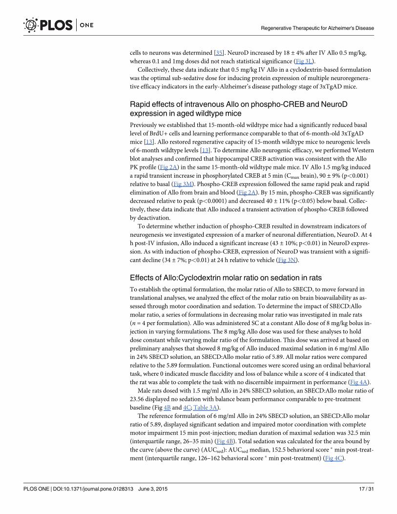
Effects of Allo:Cyclodextrin molar ratio on sedation in ratsTo establish the optimal formulation, the molar ratio of Allo to SBECD, to move forward intranslational analyses, we analyzed the effect of the molar ratio on brain bioavailability as as-sessed through motor coordination and sedation. To determine the impact of SBECD:Allomolar ratio, a series of formulations in decreasing molar ratio was investigated in male rats(n = 4 per formulation). Allo was administered SC at a constant Allo dose of 8 mg/kg bolus in-jection in varying formulations. The 8 mg/kg Allo dose was used for these analyses to holddose constant while varying molar ratio of the formulation. This dose was arrived at based onpreliminary analyses that showed 8 mg/kg of Allo induced maximal sedation in 6 mg/ml Alloin 24% SBECD solution, an SBECD:Allo molar ratio of 5.89. All molar ratios were comparedrelative to the 5.89 formulation. Functional outcomes were scored using an ordinal behavioraltask, where 0 indicated muscle flaccidity and loss of balance while a score of 4 indicated thatthe rat was able to complete the task with no discernible impairment in performance (Fig 4A).
Male rats dosed with 1.5 mg/ml Allo in 24% SBECD solution, an SBECD:Allo molar ratio of23.56 displayed no sedation with balance beam performance comparable to pre-treatmentbaseline (Fig 4B and 4C; Table 3A).
The reference formulation of 6 mg/ml Allo in 24% SBECD solution, an SBECD:Allo molarratio of 5.89, displayed significant sedation and impaired motor coordination with completemotor impairment 15 min post-injection; median duration of maximal sedation was 32.5 min(interquartile range, 26–35 min) (Fig 4B). Total sedation was calculated for the area bound bythe curve (above the curve) (AUCsed): AUCsed median, 152.5 behavioral score � min post-treat-ment (interquartile range, 126–162 behavioral score � min post-treatment) (Fig 4C).
Regenerative Therapeutic for Alzheimer's Disease
PLOS ONE | DOI:10.1371/journal.pone.0128313 June 3, 2015 17 / 31

Fig 4. Formulation development, route of administration, and rat behavioral task study components. Study design. A. Four different formulations wereadministered by subcutaneous (SC) route to male rats at the maximally tolerated dose (MTD) for sedation, determined to be 8 mg/kg for the leadingformulation Allo 6 mg/ml, 24% sulfobutyl-ether-β-cyclodextrin sodium (SBECD). Optimal release rate formulations for SC were then tested via dose-responsefor SC, intramuscular (IM), and intravenous (IV) routes using the rat balance beam behavioral task to score motor incoordination due to Allo-inducedsedation, a biomarker of Allo target engagement and tolerability. Sedation score as a biomarker of Allo target engagement. B. The sedative component ofGABAA receptor activation in the brain was used as a biomarker outcome of Allo delivery and tolerability. Male rats (Age 7 months at study initiation) weredosed by SC route of administration with multiple formulations, to determine effect on sedation. Doses were administered at Allo 8 mg/kg SC to compare
Regenerative Therapeutic for Alzheimer's Disease
PLOS ONE | DOI:10.1371/journal.pone.0128313 June 3, 2015 18 / 31

At 1.5 mg/ml Allo in 6% SBECD solution, an SBECD:Allo molar ratio of 5.89, displayed sig-nificant sedation and impaired motor coordination with complete motor impairment 20 minpost-injection; median duration of maximal sedation was 20 min (interquartile range, 12.5–27.5 min) (Fig 4B). Total sedation was calculated: AUCsed median, 120 behavioral score � minpost-treatment (interquartile range, 70–142 behavioral score � min post-treatment) (Fig 4C).
At 24 mg/ml Allo in 24% SBECD suspension, an SBECD:Allo molar ratio of 1.47 displayedminor impairment (median, 2.5 min; interquartile range, 0–8.75 min), peaking at 15 min post-injection, with complete recovery by 30 min (Fig 4B). Total sedation was calculated: AUCsed
median, 20 behavioral score � min post-treatment (interquartile range, 0–62.5 behavioral score� min post-treatment) (Fig 4C).
Collectively, results of these analyses indicate that the SBECD:Allo formulation at a 5.89molar ratio was optimal. This conclusion is based on the rapid induction of maximal sedationwith greatest duration indicating optimal brain delivery relative to the other molar ratios.
Effects of route of administration on sedation in ratsTo investigate the impact of route of administration on sedation, we utilized the optimalSBECD:Allo 5.89 molar ratio formulation. Investigated routes of administration include SC,IM, and IV. Functional outcomes were scored using the same balance beam behavioral taskused to determine the optimal formulation.
SC administration of 2 mg/kg or 4 mg/kg Allo to female (n = 4; Fig 5A and 5G) and male(n = 4; Fig 5B and 5G) rats did not induce sedation (behavioral score 4) whereas 8 mg/kg Alloinduced significant sedation with median maximal behavioral score for female 0 (interquartilerange, 0–3); duration of maximal sedation 12.5 min (interquartile range, 1.25–23.75 min);maximal behavioral score for male 0 (interquartile range, 0); duration 32.5 min (interquartilerange, 26.25–35 min). Total sedation was calculated: females AUCsed median, 97.5 behavioralscore � min post-treatment (interquartile range, 23–115.5 behavioral score � min post-treat-ment) and males AUCsed, 152.5 behavioral score � min post-treatment (interquartile range,126.5–162 behavioral score � min post-treatment) (Fig 5G).
IM administration of 2 mg/kg Allo to female (n = 4; Fig 5C and 5G) and male (n = 4; Fig 5Dand 5G) rats did not induce appreciable sedation (median maximal behavioral score for female4 (interquartile range, 3.25–4); maximal behavioral score for male 4 (interquartile range, 2.5–4)). IM 4 mg/kg Allo had sedative impact (maximal behavioral score for female 4 (interquartilerange, 1–4); duration of maximal sedation 0 min (interquartile range, 0–7.5 min); maximal be-havioral score for male 0 (interquartile range, 0); duration of maximal sedation 25 min (inter-quartile range, 17.5–25 min)). Total sedation was calculated: females AUCsed median, 0behavioral score � min post-treatment (interquartile range, 0–58 behavioral score � min post-treatment) and males AUCsed, 118.8 behavioral score � min post-treatment (interquartilerange, 110.5–132.5 behavioral score � min post-treatment). Maximal sedative impact occurredwith 8 mg/kg Allo IM administration (maximal behavioral score for female 0 (interquartilerange, 0); duration of maximal sedation 32.5 min (interquartile range, 21.25–40 min); maximal
sedation responses at 5 min intervals. The formulations were of either Allo solutions or suspensions (in 0.9% sodium chloride with SBECD) designed todetermine the Allo delivery rate as it relates to SBECD:Allo molar ratio. Balance Beam Behavioral Scoring System: 4 = reach platform; 3 = takes steps; 2 = allpaws on top; 1 = clasp; 0 = fall. n = 4, interval points represent mean value ± SEM. Area bound by sedation curve as an indicator of rapid Allo targetengagement. C. The area bound by the sedation curve (AUCsed; area above the curve as graphically represented) was calculated to determine the sedativecomponent of GABAA receptor activation in the brain as an indicator of Allo delivery. Male rats (Age 7 months at study initiation) were administered Allo 8 mg/kg SC bolus to compare delivery of multiple formulations of either Allo solutions or suspensions (in 0.9% sodium chloride with SBECD) designed to determinethe Allo delivery rate as it relates to SBECD:Allo molar ratio. Area bound by sedation curve was calculated and expressed as behavioral score (a scorebetween 4 and 0) multiplied by minutes post-treatment. n = 4, bars represent mean value ± SEM.
doi:10.1371/journal.pone.0128313.g004
Regenerative Therapeutic for Alzheimer's Disease
PLOS ONE | DOI:10.1371/journal.pone.0128313 June 3, 2015 19 / 31

Table 3. Summary of rat sedation data and statistical comparisons.
Study 1: Formulation
Route ofAdministration
Sex Dose Endpoint Comparison Mean ± SEM Mean ± SEM Median(InterquartileRange)
Median(InterquartileRange)
p-value
SC M 8mg/kg Deepestsedationscore
1.5mg/ml 6% (Molar Ratio6) vs 1.5mg/ml 24%(Molar Ratio 24)
4 ± 0 0 ± 0 4(0) 0(0) <0.0001
M 8mg/kg Deepestsedationscore
6mg/ml 24% (Molar Ratio6) vs 1.5mg/ml 24%(Molar Ratio 24)
4 ± 0 0 ± 0 4(0) 0(0) <0.0001
Study 2: Route of Administration
Route ofAdministration
Sex Dose Endpoint Mean ± SEM Median(InterquartileRange)
Comparison Mean ± SEM Median(InterquartileRange)
p-value
SC M&F 4mg/kg Deepestsedationscore
2.75 ± 0.9 4(0.25) 8mg/kg(M&F)
0 ± 0 0(0) 0.0313
SC M&F 4mg/kg Duration ofdeepestsedation
13.75 ± 4.8 0(1.25) 8mg/kg(M&F)
31.25 ± 2.4 25(15) 0.0156
SC F 8mg/kg AUCsed 78.75 ± 26.9 97.5(37.5) M 146.88 ± 9.8 132.5(46.1) 0.0571
SC F 8mg/kg Duration ofdeepestsedation
12.5 ± 6.0 12.5(17.5) M 31.25 ± 2.4 32.5(6.25) 0.0571
IM M&F 2mg/kg Deepestsedationscore
3.625 ± 0.4 4(0.25) 4mg/kg(M&F)
1.5 ± 1.1 0(4) 0.0625
IM M&F 2mg/kg Duration ofdeepestsedation
1.25 ± 1.2 0(1.25) 4mg/kg(M&F)
12.5 ± 5.9 12.5(25) 0.0625
IM F 4mg/kg AUCsed 19.38 ± 19.38 0(19.4) M 120.63 ± 5.8 118.75(15.6) 0.0286
IM M 4mg/kg Duration ofdeepestsedation
22.5 ± 2.5 25(2.5) F 2.5 ± 2.5 0(2.5) 0.0286
IM M&F 4mg/kg Duration ofdeepestsedation
12.5 ± 5.8 12.5(25) 8mg/kg(M&F)
44.38 ± 8.1 45(20) 0.0078
IM F 8mg/kg AUCsed 153.75 ± 24.8 150(71.25) M 245 ± 17.1 240(35) 0.0571
IM F 8mg/kg Duration ofdeepestsedation
31.25 ± 5.2 32.5(16.25) M 57.5 ± 3.2 57.5(7.5) 0.0286
IV M&F 0.5mg/kg
Deepestsedationscore
3.75 ± 0.3 4(0.25) 2mg/kg(M&F)
0 ± 0 0(0) 0.0078
IV M&F 0.5mg/kg
Duration ofdeepestsedation
12.5 ± 3.2 0(0.625) 2mg/kg(M&F)
10 ± 1.8 10(3.75) 0.0078
IV M&F 0.5mg/kg
AUCsed 0.78 ± 0.52 0(0.625) 2mg/kg(M&F)
58.75 ± 4.2 55(15) 0.0078
A. Four different formulations were administered by subcutaneous (SC) route to male rats at the maximally tolerated dose (MTD) for sedation, determined
to be 8 mg/kg for the leading formulation Allo 6 mg/ml, 24% sulfobutyl-ether-β-cyclodextrin sodium (SBECD) (See Fig 4). B. Optimal release rate
formulations determined via SC were then tested via dose-response for SC, intramuscular (IM), and intravenous (IV) routes using the rat balance beam
behavioral task to score motor incoordination due to Allo-induced sedation, a biomarker of Allo target engagement and tolerability (See Fig 5). Units for
comparison of mean ± SEM: Deepest sedation score (Balance Beam Behavioral Scoring System: 4 = reach platform; 3 = takes steps; 2 = all paws on top;
1 = clasp; 0 = fall); duration of deepest sedation (minutes post-treatment); Area bound by sedation curve (AUCsed; behavioral score * min post-treatment).
doi:10.1371/journal.pone.0128313.t003
Regenerative Therapeutic for Alzheimer's Disease
PLOS ONE | DOI:10.1371/journal.pone.0128313 June 3, 2015 20 / 31

Fig 5. Allopregnanolone (Allo) dose-response comparison by subcutaneous (SC), intramuscular (IM), and intravenous (IV) routes ofadministration.Ovariectomized female rats (left panel) were compared to age-matched male rats (right panel) following SC (A. and B.), IM (C. and D.), or IV(E. and F.) bolus injection and challenged with a motor coordination behavioral task to assess level of sedation at timed intervals until recovery. SC/IMformulation: 6 mg/ml Allo solution in 0.9% sodium chloride with 24% sulfobutyl ether β-cyclodextrin sodium (SBECD). IV formulation: 1.5 mg/ml Allo solutionin 0.9% sodium chloride with 6% SBECD. The volume of vehicle administered was equal to the volume administered with the highest dose, i.e. 8 mg/kg (SC/IM) or 2 mg/kg (IV). Balance Beam Behavioral Scoring System: 4 = reach platform; 3 = takes steps; 2 = all paws on top; 1 = clasp; 0 = fall. n = 4, interval pointsrepresent mean value ± SEM. Area bound by sedation curve as an indicator of rapid Allo target engagement. G. The area bound by the sedation curve(above the curve; Fig 5A–5F) (AUCsed) was calculated to determine the sedative component of GABAA receptor activation in the brain as an indicator of Allodelivery. AUCsed was calculated and expressed as behavioral score (a score between 4 and 0) multiplied by minutes post-treatment. Sex differences in theAllo-induced sedation response were determined. * p<0.05, ** p<0.01, n = 4, bars represent mean value ± SEM.
doi:10.1371/journal.pone.0128313.g005
Regenerative Therapeutic for Alzheimer's Disease
PLOS ONE | DOI:10.1371/journal.pone.0128313 June 3, 2015 21 / 31

behavioral score for male 0 (interquartile range, 0); duration of maximal sedation 57.5 min(interquartile range, 51.25–63.75 min)). Total sedation was calculated: females AUCsed median,150 behavioral score � min post-treatment (interquartile range, 109–202.5 behavioral score �
min post-treatment) and males AUCsed, 240 behavioral score � min post-treatment (interquar-tile range, 215–280 behavioral score � min post-treatment) (Fig 5G).
IV administration of 0.5 mg/kg Allo to female (n = 4; Fig 5E and 5G) and male (n = 4; Fig5F and 5G) rats did not induce sedation (median maximal behavioral score for female 4 (inter-quartile range, 4); maximal behavioral score for male 3.5 (interquartile range, 3–4)). At thehighest dose tested, 2 mg/kg IV Allo, maximal sedative impact occurred (maximal behavioralscore for female 0 (interquartile range, 0); duration of maximal sedation 7.5 min (interquartilerange, 5.5–9.5 min); maximal behavioral score for male 0 (interquartile range, 0); duration ofmaximal sedation 12.5 min (interquartile range, 10–15 min)). Total sedation was calculated: fe-males AUCsed median, 50 behavioral score � min post-treatment (interquartile range, 45–55behavioral score � min post-treatment) and males AUCsed, 70 behavioral score � min post-treatment (interquartile range, 57.5–75 behavioral score � min post-treatment) (Fig 5G).
Significant sex differences were apparent in duration of maximal sedation at IM 8 mg/kgAllo (female 32.5 min (range 21.25–40) vs males 57.5 min (range 51.25–63.75); p< 0.05) (Fig5A–5F). There were also significant sex differences in total sedation area (AUCsed) at IM 4 mg/kg Allo (female 0 (range 0–58) vs males 118.8 (range 110.5–132.5) behavioral score � min post-treatment; p< 0.05). Although the following differences did not reach significance, the datasuggests that with more animals a stronger difference may be detectable: duration of maximalsedation at SC 8 mg/kg Allo (female 12.5 min (range 1.25–23.75) vs males 32.5 min (range26.25–35); p = 0.0571), and IV 2 mg/kg Allo (female 7.5 min (range 5.5–9.5) vs males 12.5 min(range 10–15); p = 0.0857); in AUCsed SC 8 mg/kg Allo (female 97.5 (range 23–115.5) vs males152.5 (range 126.5–162) behavioral score � min post-treatment; p = 0.0571; Fig 5A and 5B), IM8 mg/kg Allo (female 150 (range 109–202.5) vs males 240 (range 215–280) behavioral score �
min post-treatment; p = 0.0571), and IV 2 mg/kg Allo (female 50 (range 45–55) vs males 70(range 57.5–75) behavioral score � min post-treatment; p = 0.0857) (Fig 5G).
Route of administration impacted the duration of sedation with 8 mg/kg SC resulting inshorter duration of maximal sedation relative to IM administration (p = 0.016, n = 8). Compar-ing IM to SC at equivalent doses, the level and extent of sedation was increased via the IMroute, likely due to the high vascularization of quadricep muscle tissue allowing for more rapidand extensive systemic absorption. Within 30 sec of bolus administration, IV 2 mg/kg Allo in-duced maximal sedation. Results of route of administration analyses indicate that SBECD:Alloformulation at a 5.89 molar ratio administered via IM bolus was optimal. This conclusion isbased on the rapid induction of maximal sedation with greatest duration indicating optimalbrain delivery relative to the other routes.
Allo via IV and IM routes of administration displayed rapid onset of sedation with IV in-ducing the earliest onset IM inducing the greatest duration of sedation (Fig 5; Table 3B). In thisregard, IV route of administration provides a method of determining the absolute bioavailabledose response. An IM bolus or IV slow infusion to mimic the PK profile of IM would provideclinical translatability with a longer time course for absorption and safe sedation profile.
Estimation of the dose range from no observable adverse effect level(NOAEL) to maximally tolerated dose (MTD) for each route ofadministration in ratsAllo dose range finding via the sedation balance beam behavioral task was used to estimate theno observable adverse effect level (NOAEL) and maximally tolerated dose to sedation (MTD).
Regenerative Therapeutic for Alzheimer's Disease
PLOS ONE | DOI:10.1371/journal.pone.0128313 June 3, 2015 22 / 31

The NOAEL was estimated to be Allo SC 4 mg/kg for female and SC 2 mg/kg for male rats (Fig5A, 5B and 5G). Also from the behavioral data, we estimated an Allo NOAEL of IM 2 mg/kgfor female and IM less than 2 mg/kg for male rats by bolus injection (Fig 5C, 5D and 5G).
For female rats treated with SC Allo, the MTD was estimated to be greater than 8 mg/kg. Fe-male rats treated with Allo SC 8 mg/kg had a maximal median score of 0 (range 0–3) on the be-havioral sedation scale indicating that full sedation was not reached in all animals and aslightly higher dose would be tolerated. For male rats treated with SC Allo, the MTD was esti-mated to be 8 mg/kg with a maximal behavioral score of 0 in all animals indicating loss ofmotor control. Similarly, the Allo IMMTD was estimated to be 8 mg/kg for female and lessthan 8 mg/kg for male rats with a maximal behavioral score of 0 in all animals.
To assess sedation following IV administration, the dose was adjusted to account for 100%bioavailability. Because Allo IV 1 mg/kg was found to be efficacious in mice and is equivalentto a 0.5 mg/kg dose in a rat (Table 4), 0.5 mg/kg was selected as the IV starting dose in rats.After administration of Allo IV 0.5 mg/kg, female rats showed no discernible signs of sedationand males showed very minimal sedation. From this we estimate the NOAEL to be Allo 0.5mg/kg IV for female and slightly less than 0.5 mg/kg IV for male rats (Fig 5E–5G; Table 5). TheMTD was estimated to be Allo IV 2 mg/kg for both female and male rats with a maximal be-havioral score of 0 in all animals. The MTD of 2 mg/kg IV Allo reached maximal sedation
Table 4. Allopregnanolone equivalent doses in mouse, rat, rabbit, and human.
Allopregnanolone equivalent dose estimates
Adult Species: Mouse Rat Rabbit Human Total Human Dose (mg/70kg)
NOAEL and MTD Based on Rat Sedation (Balance BeamTask)
Allo dose (mg/kg)
0.09 0.04 0.02 0.01 0.5
0.37 0.15 0.07 0.03 2.0
0.50 0.21 0.10 0.04 2.7
0.60 0.25 0.12 0.05 3.3 NOAEL Male IV
0.74 0.31 0.15 0.06 4.0
1.00 0.41 0.20 0.08 5.4
1.11 0.46 0.22 0.09 6.0
1.21 0.50 0.24 0.09 6.5 NOAEL Female IV
1.50 0.62 0.30 0.12 8
2.00 0.83 0.40 0.15 11
2.41 1.00 0.48 0.19 13 NOAEL Male IM
3.00 1.24 0.60 0.23 16
4.82 2.00 0.96 0.37 26 NOAEL Male SC, Female IM; MTD Male and Female IV
9.65 4.00 1.92 0.75 52 NOAEL Female SC
10.00 4.15 1.99 0.77 54
15.08 6.25 3.00 1.17 82
19.29 8.00 3.84 1.49 104 MTD Male and Female SC and IM
25.13 10.42 5.00 1.94 136
50.00 20.74 9.95 3.87 271
Equivalent dose estimates of allopregnanolone (Allo) for each species assume a comparison of identical formulation and route of administration.
Conversion is based on body surface area and computed with the following equation: Human equivalent dose = animal dose (mg/kg) x [animal weight (kg)
/ human weight (kg)]0.33. The exponent 0.33 is an allometric conversion constant [36]. Allo doses indicated in bold were administered during the course of
the preclinical studies (see Methods section for route of administration and formulation). Total human doses of Allo were calculated in milligrams per 70 kg
average body weight. Human doses indicated in bold are predicted to be within tolerable range for IV infusion to be tested in a multiple ascending dose
trial (Phase 1b clinical trial of Allo for early AD; ClinicalTrials.gov Identifier: NCT02221622 [37]). Note: IV doses should not approach or exceed 3 mg/kg
rabbit or the equivalent in other species.
doi:10.1371/journal.pone.0128313.t004
Regenerative Therapeutic for Alzheimer's Disease
PLOS ONE | DOI:10.1371/journal.pone.0128313 June 3, 2015 23 / 31

levels in both male and female within 30 sec (Fig 5), briefly matching depth of sedation com-pared to SC or IMMTD with loss of righting reflex observed. After IV bolus injection, a rapidrecovery from sedation was observed in rats which correlated with the Allo IV PK profile thatwas determined in rabbit and mouse (Fig 1; Table 1).
Collectively, these findings provide translational evidence for a dose range to establish inhumans a subsedative dose which would predict neuroregeneration in the targeted populationswith mild cognitive impairment or early AD.
Discussion
Preclinical pharmacokineticsIn this study, we conducted translational analyses to enable AD clinical trial development. PKanalyses, preclinical efficacy, clinical formulation development, and determination of MTDbased on sedation as the endpoint were compiled to evaluate Allo. To inform future toxicologystudies in non-rodent and rodent species that meet FDA toxicology requirements for therapeu-tic development, acute bolus dose PK analysis was conducted in female rabbits and male mice.Rabbit and mouse Allo PK profiles after IV administration were similar. Additionally, IV PKparameters from both species (Allo T1/2 rabbit 3.3 h; mouse 4 h) were comparable to reportedIV human exposure, where the Allo T1/2 has been reported to be 4.4 ± 1.7 h [20, 21]. In addi-tion to IV route, the TD route was tested in rabbits. TD Allo was modestly skin-penetrablewith 6.8% bioavailability to the plasma and 36% of the plasma-available Allo entered the brain.Although the rabbit IV dose tested was approximately 10-fold higher than the mouse IV dose,when considering differences in species [36], similar results from multiple PK parameters wereobtained. After allometric species conversion [36], the IV mouse dose of 1.5 mg/kg was compa-rable to the Allo dose used in a previous clinical trial, 0.09 mg/kg (see Table 4) [20, 21]. Thedosing studies conducted herein combined with the available scientific literature were used toestimate the maximum safe starting dose range [36] for a Phase 1 clinical trial for AD [37].
Preclinical efficacyTo determine the optimal regimen for therapeutic efficacy of Allo treatment in AD, we investi-gated the efficacy of acute treatment protocols designed to determine the optimal dose,
Table 5. Estimation of allopregnanolone no observable adverse effect level (NOAEL) andmaximally tolerated dose (MTD) in male and female ratsand predicted human NOAEL and MTD bymultiple routes of administration.
NOAEL (mg/kg) MTD (mg/kg)
Species Route of Administration Female Male Female Male
Rat Subcutaneous 4 2 > 8 8
Intramuscular 2 < 2 8 < 8
Intravenous 0.5 < 0.5 2 2
Human (predicted) Subcutaneous 0.75 0.37 > 1.49 1.49
Intramuscular 0.37 < 0.37 1.49 < 1.49
Intravenous 0.09 < 0.09 0.37 0.37
Data from rat sedation studies was used to determine NOAEL and MTD by subcutaneous, intramuscular, and intravenous routes of administration. Allo
was administered by bolus push injections within 30 sec via subcutaneous or intramuscular: 6 mg/ml Allo solution in 0.9% sodium chloride with 24%
sulfobutyl ether β-cyclodextrin sodium (SBECD); or intravenous: 1.5 mg/ml Allo solution in 0.9% sodium chloride with 6% SBECD. All formulations
contained SBECD:Allo molar ratio of 5.89. Sex differences found in rat sedation studies (see Fig 5; Table 3) suggest that males are more sensitive to Allo
on the basis of mg/kg body weight.
doi:10.1371/journal.pone.0128313.t005
Regenerative Therapeutic for Alzheimer's Disease
PLOS ONE | DOI:10.1371/journal.pone.0128313 June 3, 2015 24 / 31

formulation, and route for Allo treatment to promote the regenerative capacity of the brain.Previously published studies by our group have demonstrated Allo efficacy via the SC route inyoung 3xTgAD and aged nonTg mice [10, 12, 13]. We established that 3xTgAD mice at3-months of age and wildtype mice at 15-months have significantly reduced basal levels of hip-pocampal BrdU+ cells with 15-month wildtype mice exhibiting learning performance deficitscomparable to 6-month-old 3xTgADmice [13]. The age-associated deficits in neurogenesiswere rescued by SC Allo treatment [13]. Further, we demonstrated neurogenic efficacy in3xTgAD mice treated once per week SC Allo 20% HBCD solution for 6 months duration [12].SC Allo injections have also been effective in inherited developmental neurodegenerative disor-der rodent models [38] and the effect of Allo was partly associated with a reduction in oxidativestress [39]. Here, with 5-month-old 3xTgAD mice, prior to formation of extraneuronal plaque,results indicated 24 h SC Allo treatment dose-dependently increased BrdU labeled hippocam-pal nuclei and PCNA protein expression.
Bridging SC Allo results to IV Allo cyclodextrin-based formulations, we demonstrateddose-dependent neurogenic efficacy of IV Allo for the first time. The activation profile of hip-pocampal phosphorylated CREB was consistent with the Allo IV pharmacokinetic profile,peaking at 5 min followed by rapid clearance. Interestingly, in a mouse seizure model with theidentical formulation and dose used here to demonstrate CREB activation, within 1 min ofAllo dose, total protection was found. This response was no longer evident by 15 and 30 minafter the IV bolus whereas an IM dose prolonged efficacy for up to 1 h [40, 41]. In vitro experi-ments have demonstrated in cultured Schwann cells treated with 10 nM Allo that by 15 min,cAMP levels more than doubled followed by rapid CREB phosphorylation [42]. The mecha-nism of Allo-induced neurogenesis takes advantage of the developmentally regulated reversedchloride gradient to activate a calcium-to-CREB signaling cascade to induce mitosis in neuralstem cells [5]. In neural stem cells, Allo promotes a rapid, dose-dependent, stereo-specific, anddevelopmentally regulated increase of intracellular calcium concentration via a mechanismthat requires both the GABAAR and L-type calcium channel [22]. Calcium influx via the volt-age-gated L-type calcium channels activates CaMKIV that phosphorylates CREB [43, 44]. Neu-ral progenitor-derived neurons express L-type calcium ion channels within days after celldivision and show spontaneous calcium oscillations that support neuronal differentiation [22,45]. It has been shown that GABA-induced phosphorylation of CREB overlaps with NeuroDexpression within one hour [46]. NeuroD increased relative to vehicle at 4 h then decreased by24 h after IV Allo 1.5 mg/kg. The transient expression of NeuroD is a critical neurodifferentia-tion element and IV administration of Allo 0.5 mg/kg was efficacious (upregulation of cellcycle proliferation and differentiation markers pCREB, cyclin D2, PCNA, DCX, and NeuroD),well tolerated, and translates to an approximate human dose of Allo 0.04 mg/kg. CharacterizingAllo exposure and efficacy after IV administration was used to validate the planned Allo Phase1b trial design [37], which will utilize the IV route of administration to evaluate safety in hu-mans and will set the tolerable dose level and PK for subsequent efficacy studies.
Allo administered by IM route in 6% SBECD molar ratio 5.89, demonstrated neurogenic ef-ficacy in 5-month-old 3xTgAD mouse hippocampus. IM Allo increased proliferation at 2 mg/kg inducing an increase comparable to SC suspension at 10 mg/kg within 24 h. Both IM 2 mg/kg and SC suspension 10 mg/kg were efficacious, subsedative, and well tolerated.
The formulations tested were intended to evaluate the potential of Allo to be deliveredthrough multiple routes with tailored solvents for each route. Regardless of the route of admin-istration, Allo crosses the blood-brain barrier as evidenced by multiple indicators of target en-gagement. These include markers of neurogenesis at low doses and profiles of sedation athigher doses. Collectively the data show that route of administration determines dosing withIV requiring the least dose of Allo and TD/IN/SC requiring the highest doses for target
Regenerative Therapeutic for Alzheimer's Disease
PLOS ONE | DOI:10.1371/journal.pone.0128313 June 3, 2015 25 / 31

engagement. Thus the safety and efficacy profile of Allo can be modified by both dose androute of administration. Clinically, establishing the MTD in a Phase 1 by IV administration isfeasible within a restricted dose range. Based on IV data, MTD can be established for alterna-tive routes of administration that may have greater translational feasibility for clinical chronictreatment regimens.
Formulation developmentThe complexation ratio of Allo with cyclodextrins such as SBECD is a major determinant of re-lease of Allo into the blood and brain. The cyclodextrin vehicle SBECD does not cross theblood brain barrier [47] but instead facilitates Allo release to steroid carrier proteins in blood.Cyclodextrins represent an optimal vehicle for solubility of Allo in aqueous formulations andSBECD has been tested extensively for safety in animals and humans and is an excipient in sev-eral approved drugs [47]. To develop formulations with clinical utility, we tested multipleSBECD:Allo complexation ratios on behavior in adult rats. Rats display more human-like seda-tion responses compared to mice and thus were used for these analyses. In rats, Allo inducesdose-related sleep changes, including a reduced sleep onset latency and an increase in pre-rapid-eye-movement sleep [48]. Allo has a greater tolerance profile than most agonistic modu-lators of GABAARs, including benzodiazepine hypnotics [49]. To functionally detect and quan-tify sedation, a sensitive balance beam motor coordination task was used to determinedifferences in the delivery and release rate of multiple Allo formulations. Acute motor im-pairment was used as a surrogate marker, translatable to clinical studies, to assess that Alloreached its target in the CNS/brain.
Soluble SBECD:Allo formulations estimated between 7:1 and 3:1, maximally deliver Allo tosystemic circulation resulting in rapid brain uptake observed by altered motor control. In theoptimal SBECD:Allo formulation (molar ratio of 5.89), Allo is fully soluble and rapidly bio-available, indicated by rapid prolonged sedation at the MTD of 8 mg/kg. When the ratio is in-creased (molar ratio of 23.56), the rate of Allo release into the brain is reduced, indicated by alack of sedation. Likewise, when the ratio is decreased (molar ratio of 1.47) relative to optimal,the rate of Allo release into the brain was also reduced. The volume of soluble Allo adminis-tered in the saturated suspension formulation was limited when dosing 8 mg/kg to rats and re-sults in mild sedative effects likely due to the relatively small fully soluble fraction of Allo,giving the suspension formulation dual properties. Based on these data, it was postulated thatthe small but fully soluble fraction of Allo was rapidly delivered to the brain in a subfractiondose followed by a slowly absorbed suspension fraction that does not possess the release rate re-quired for sedation at the 8 mg/kg dose level. These pharmacodynamic studies served to identi-fy three distinct formulations that provide rationale for multiple potential release rate options:1) Slow release 2) Rapid release 3) Rapid release followed by slow release.
Route of administration and dose-dependent behaviorThe sub-sedative (NOAEL) and MTD Allo doses were determined in male and female rats forSC, IM, and IV routes of administration. For clinical translatability, the NOAEL we targetedwas chosen to minimize sedation and to ensure an exposure to Allo that was below 50 ng/ml or157 nmol/L, i.e. equivalent to the blood level measured in normal pregnant women with milddrowsiness during the third trimester [19]. We observed gender differences in the onset andduration of sedation in Allo dose-response studies. Male rats administered 2 mg/kg IV or 8mg/kg SC/IM displayed the deepest level of sedation. Through SC, IM, and IV routes of admin-istration females administered the same dose as males were less sedated and recovered morerapidly. This sex difference could be explained by the higher number of GABAA receptor
Regenerative Therapeutic for Alzheimer's Disease
PLOS ONE | DOI:10.1371/journal.pone.0128313 June 3, 2015 26 / 31

binding sites in male brains relative to female brains [50]. Alternatively, a sex difference couldbe attributed to the observed peak blood levels observed in previous human studies of IV dos-ing of Allo 0.09 mg/kg with maximum plasma levels of 150 nmol/L (48 ng/ml) in men and 100nmol/L (32 ng/ml) in women [21].
For therapeutic use of Allo, it was imperative to determine the optimal dose, formulation,and dosing regimen [5]. Weekly exposure to Allo was optimally efficacious in 3xTgAD mice[12] and this regimen will move forward to definitive FDA-compliant chronic toxicology stud-ies in rodent and non-rodent species to meet or exceed the duration of early phase clinical trials[51]. In human phase 1 studies, mild sedation as an indicator of tolerability will establish theMTD. Endogenous Allo levels are increased during stress [52], the menstrual cycle [53], andpregnancy [19]. Use of anti-depressants [54] can also elevate Allo levels whereas depression isassociated with lower circulating Allo [55, 56]. Allo levels are reduced in Alzheimer’s diseasebrains compared to age-matched controls [54, 57]. Currently, the upper physiological Alloblood concentration of 50 ng/ml reached during the third trimester of pregnancy sets the safeboundary for human dosing until chronic toxicology studies conducted under Good Laborato-ry Practice standards in rodent and non-rodent species are completed and evaluated byregulatory agencies.
Our preclinical results provide useful estimates for the safest starting dose in humans withconsideration of the formulation, route of administration, neurogenic efficacy, and tolerability.Keeping the formulation constant, an SC Allo NOAEL dose of 2 mg/kg to rats is equivalent toa human dose of approximately 0.37 mg/kg or ~26 mg for a 70 kg adult (Table 4). Of the for-mulations tested, Allo 6 mg/ml would require 4.3 ml, which poses a challenge for human SCadministration. However, the 24 mg/ml suspension formulation would be a viable option todeliver Allo 26 mg/1.1 ml. For human translation, SC is advantageous due to ease of adminis-tration, patient compliance, and tolerability. However, the volume of formulation required toreach the appropriate dose is a limiting factor. Assessed by rat motor coordination test, SC Alloresponse was more variable than IM Allo response. The data indicated that at high doses withidentical formulation, IM route is less variable than SC and approximately twice as potent at in-duction of sedation, a surrogate indicator of brain uptake. An IM Allo NOAEL dose of 2 mg/kgto mice is equivalent to a human IM dose of approximately 0.15 mg/kg or ~11 mg Allo for a 70kg adult (Table 4). Translatable to humans, Allo 6 mg/ml in 24% SBECD would require an esti-mated 2 ml volume IM. PK studies are often conducted via the IV route of exposure to providedata on maximal bioavailability and inform further studies by alternative routes of administra-tion. An IV Allo NOAEL dose of 0.5 mg/kg in mice or 0.21 mg/kg to rats is equivalent to ahuman IV dose of approximately 0.042 mg/kg or ~3 mg for a 70 kg adult. The vehicle androute of administration were demonstrated to influence PK and pharmacodynamic outcomesand thereby the therapeutic range of Allo.
ConclusionsPK, pharmacological efficacy, and behavioral studies were conducted to inform Allo formula-tion development, safety monitoring, and pharmacokinetic interpretation in clinical trials(Phase 1b clinical trial of Allo for early AD; ClinicalTrials.gov Identifier: NCT02221622 [37]).Results of the rabbit and mouse PK studies, demonstrated that soluble cyclodextrin-basedbolus IV injections rapidly delivered Allo to blood and brain and reached peak levels in brainwithin 5min which was then rapidly cleared. While the rabbit PK dose was at MTD level, themouse PK dose was near NOAEL and was accompanied by indicators of efficacy in hippocam-pal neurogenesis indicating that efficacy can be achieved within safe dosing limits. In mouse,the suspension SC Allo peak levels were much lower than IV with slower uptake to the brain.
Regenerative Therapeutic for Alzheimer's Disease
PLOS ONE | DOI:10.1371/journal.pone.0128313 June 3, 2015 27 / 31

Data from our studies demonstrate that Allo SC, administered in cyclodextrin-solublized form,and IV Allo were efficacious at NOAEL doses in both aged wildtype mice and young 3xTgADmice. The dose-responses in these studies indicated that lower sub-sedative doses were optimalfor both efficacy and safety. The dose response curve for Allo exhibits an inverted U-shapedfunction for neuroregeneration where lower doses are on the ascending arm of the dose re-sponse curve whereas higher doses are on the descending arm [5, 10–12]. The rat behaviorstudies allowed for formulation development and were used to compare sedation responses asa surrogate detection method to brain Allo PK across different routes of administration. TheIV route is standard for early phase safety and PK clinical trials and control of IV infusion rateto mimic the IM or SC delivery rate of Allo can be achieved. Other options explored here, theTD and IN routes are less-invasive alternatives that may prove to be most clinically translat-able. In cyclodextrin-based formulations, oral Allo bioavailability is generally not sufficient al-though food oils may enhance solubility for the treatment of medical conditions [58]. Previoushuman trials with IV Allo were formulated in albumin and tested in young adults [21]. Clinicaldose-escalating studies with a cyclodextrin-based Allo formulation in elderly people are war-ranted to determine safety and tolerability for use in early AD and other unmet neurologicaldisorders [2, 5, 59]. Collectively, the translational research described herein build upon our pre-vious preclinical results in relevant animal models [10, 12, 13] to provide key insights that pre-dict the maximally safe starting dose for humans, efficacy by several routes of administration,and the optimal Allo formulation complex necessary to advance to proof of concept clinicalstudies of Allo as a regenerative and disease modifying therapeutic for Alzheimer’s disease.
AcknowledgmentsWe are grateful for the contributions of C. Green, K. Steinmetz, A. Miklos, R. Swezey and J.Gow of SRI, International (Menlo Park, CA) in conduct of the rabbit PK study; T. Zmetra ofAgilux Laboratories (Worcester, MA) in conduct of LC-MS/MS for the mouse PK study; andK. Johnson of Adroit Pharmaceuticals (Durham, NC) for topical Allo formulation develop-ment. We gratefully acknowledge our colleagues at the University of Southern California: S.Chen and H.Y. She for assistance with flow-cytometry.
Author ContributionsConceived and designed the experiments: RWI RDB. Performed the experiments: RWI CMS.Analyzed the data: RWI CMS KER RDB. Contributed reagents/materials/analysis tools: CMLFGS GBMAR RDB. Wrote the paper: RWI CMS KER RDB.
References1. Alzheimer's_Association. Alzheimer's Disease Facts and Figures. 2013.
2. Irwin RW, Solinsky CM, Brinton RD. Frontiers in therapeutic development of allopregnanolone for Alz-heimer's disease and other neurological disorders. Frontiers in cellular neuroscience. 2014; 8:203.Epub 2014/08/16. doi: 10.3389/fncel.2014.00203 PMID: 25126056; PubMed Central PMCID:PMC4115668.
3. Doody RS, Thomas RG, Farlow M, Iwatsubo T, Vellas B, Joffe S, et al. Phase 3 trials of solanezumabfor mild-to-moderate Alzheimer's disease. N Engl J Med. 2014; 370(4):311–21. Epub 2014/01/24. doi:10.1056/NEJMoa1312889 PMID: 24450890.
4. Salloway S, Sperling R, Fox NC, Blennow K, KlunkW, Raskind M, et al. Two phase 3 trials of bapineu-zumab in mild-to-moderate Alzheimer's disease. N Engl J Med. 2014; 370(4):322–33. Epub 2014/01/24. doi: 10.1056/NEJMoa1304839 PMID: 24450891.
5. Brinton RD. Neurosteroids as regenerative agents in the brain: therapeutic implications. Nat Rev Endo-crinol. 2013; 9(4):241–50. Epub 2013/02/27. doi: 10.1038/nrendo.2013.31 PMID: 23438839.
Regenerative Therapeutic for Alzheimer's Disease
PLOS ONE | DOI:10.1371/journal.pone.0128313 June 3, 2015 28 / 31

6. Lazarov O, Marr RA. Of mice and men: neurogenesis, cognition and Alzheimer's disease. Frontiers inaging neuroscience. 2013; 5:43. Epub 2013/08/30. doi: 10.3389/fnagi.2013.00043 PMID: 23986699;PubMed Central PMCID: PMC3753540.
7. Knoth R, Singec I, Ditter M, Pantazis G, Capetian P, Meyer RP, et al. Murine features of neurogenesisin the human hippocampus across the lifespan from 0 to 100 years. PLoS One. 2010; 5(1):e8809. Epub2010/02/04. doi: 10.1371/journal.pone.0008809 PMID: 20126454; PubMed Central PMCID:PMC2813284.
8. Spalding KL, Bergmann O, Alkass K, Bernard S, Salehpour M, Huttner HB, et al. Dynamics of hippo-campal neurogenesis in adult humans. Cell. 2013; 153(6):1219–27. Epub 2013/06/12. doi: 10.1016/j.cell.2013.05.002 PMID: 23746839.
9. Ernst A, Alkass K, Bernard S, Salehpour M, Perl S, Tisdale J, et al. Neurogenesis in the striatum of theadult human brain. Cell. 2014; 156(5):1072–83. Epub 2014/02/25. doi: 10.1016/j.cell.2014.01.044PMID: 24561062.
10. Wang JM, Singh C, Liu L, Irwin RW, Chen S, Chung EJ, et al. Allopregnanolone reverses neurogenicand cognitive deficits in mouse model of Alzheimer's disease. Proc Natl Acad Sci U S A. 2010; 107(14):6498–503. Epub 2010/03/17. Doi: 1001422107 [pii] doi: 10.1073/pnas.1001422107 PMID:20231471; PubMed Central PMCID: PMC2851948.
11. Wang JM, Johnston PB, Ball BG, Brinton RD. The neurosteroid allopregnanolone promotes prolifera-tion of rodent and human neural progenitor cells and regulates cell-cycle gene and protein expression.J Neurosci. 2005; 25(19):4706–18. PMID: 15888646.
12. Chen S, Wang JM, Irwin RW, Yao J, Liu L, Brinton RD. Allopregnanolone Promotes Regeneration andReduces beta-Amyloid Burden in a Preclinical Model of Alzheimer's Disease. PLoS One. 2011; 6(8):e24293. Epub 2011/09/16. doi: 10.1371/journal.pone.0024293 PMID: 21918687; PubMed CentralPMCID: PMC3168882.
13. Singh C, Liu L, Wang JM, Irwin RW, Yao J, Chen S, et al. Allopregnanolone restores hippocampal-de-pendent learning and memory and neural progenitor survival in aging 3xTgAD and nonTgmice. Neuro-biol Aging. 2011. Epub 2011/08/02. doi: 10.1016/j.neurobiolaging.2011.06.008 PMID: 21803451.
14. Irwin RW,Wang JM, Chen S, Brinton RD. Neuroregenerative mechanisms of allopregnanolone in Alz-heimer's disease. Front Endocrinol (Lausanne). 2011; 2:117. Epub 2011/01/01. doi: 10.3389/fendo.2011.00117 PMID: 22654847; PubMed Central PMCID: PMC3356095.
15. Lipinski CA. Drug-like properties and the causes of poor solubility and poor permeability. Journal ofpharmacological and toxicological methods. 2000; 44(1):235–49. Epub 2001/03/29. PMID: 11274893.
16. Grant KA, Helms CM, Rogers LS, Purdy RH. Neuroactive steroid stereospecificity of ethanol-like dis-criminative stimulus effects in monkeys. The Journal of pharmacology and experimental therapeutics.2008; 326(1):354–61. PMID: 18436788. doi: 10.1124/jpet.108.137315
17. Kask K, Backstrom T, Lundgren P, Sundstrom Poromaa I. Allopregnanolone has no effect on startle re-sponse and prepulse inhibition of startle response in patients with premenstrual dysphoric disorder orhealthy controls. Pharmacol Biochem Behav. 2009; 92(4):608–13. Epub 2009/03/10. doi: S0091-3057(09)00079-3 [pii] doi: 10.1016/j.pbb.2009.02.014 PMID: 19268499.
18. Kask K, Backstrom T, Nilsson LG, Sundstrom-Poromaa I. Allopregnanolone impairs episodic memoryin healthy women. Psychopharmacology (Berl). 2008; 199(2):161–8. Epub 2008/06/14. doi: 10.1007/s00213-008-1150-7 PMID: 18551282.
19. Luisi S, Petraglia F, Benedetto C, Nappi RE, Bernardi F, Fadalti M, et al. Serum allopregnanolone levelsin pregnant women: changes during pregnancy, at delivery, and in hypertensive patients. J Clin Endo-crinol Metab. 2000; 85(7):2429–33. Epub 2000/07/21. PMID: 10902789.
20. Timby E, Balgard M, Nyberg S, Spigset O, Andersson A, Porankiewicz-Asplund J, et al. Pharmacoki-netic and behavioral effects of allopregnanolone in healthy women. Psychopharmacology (Berl). 2006;186(3):414–24. Epub 2005/09/24. doi: 10.1007/s00213-005-0148-7 PMID: 16177884.
21. van Broekhoven F, Backstrom T, van Luijtelaar G, Buitelaar JK, Smits P, Verkes RJ. Effects of allopreg-nanolone on sedation in men, and in women on oral contraceptives. Psychoneuroendocrinology. 2007;32(5):555–64. Epub 2007/05/02. doi: 10.1016/j.psyneuen.2007.03.009 PMID: 17470385.
22. Wang JM, Brinton RD. Allopregnanolone-induced rise in intracellular calcium in embryonic hippocam-pal neurons parallels their proliferative potential. BMC Neurosci. 2008; 9 Suppl 2:S11. Epub 2009/01/06. doi: 1471-2202-9-S2-S11 [pii] doi: 10.1186/1471-2202-9-S2-S11 PMID: 19090984; PubMed Cen-tral PMCID: PMC2604895.
23. Oddo S, Caccamo A, Shepherd JD, Murphy MP, Golde TE, Kayed R, et al. Triple-transgenic model ofAlzheimer's disease with plaques and tangles: intracellular Abeta and synaptic dysfunction. Neuron.2003; 39(3):409–21. Epub 2003/08/05. PMID: 12895417.
24. Caruso D, Pesaresi M, Maschi O, Giatti S, Garcia-Segura LM, Melcangi RC. Effect of short-and long-term gonadectomy on neuroactive steroid levels in the central and peripheral nervous system of male
Regenerative Therapeutic for Alzheimer's Disease
PLOS ONE | DOI:10.1371/journal.pone.0128313 June 3, 2015 29 / 31

and female rats. J Neuroendocrinol. 2010; 22(11):1137–47. Epub 2010/09/08. doi: 10.1111/j.1365-2826.2010.02064.x PMID: 20819120.
25. Caruso D, Barron AM, BrownMA, Abbiati F, Carrero P, Pike CJ, et al. Age-related changes in neuroac-tive steroid levels in 3xTg-ADmice. Neurobiol Aging. 2013; 34(4):1080–9. Epub 2012/11/06. doi: 10.1016/j.neurobiolaging.2012.10.007 PMID: 23122920; PubMed Central PMCID: PMC3545103.
26. Henry S, Bigler S, Wang J. High throughput analysis of neural progenitor cell proliferation in adult ro-dent hippocampus. Bioscience trends. 2009; 3(6):233–8. Epub 2010/01/28. PMID: 20103852; PubMedCentral PMCID: PMC2830061.
27. Bilsland JG, Haldon C, Goddard J, Oliver K, Murray F, Wheeldon A, et al. A rapid method for the quanti-fication of mouse hippocampal neurogenesis in vivo by flow cytometry. Validation with conventionaland enhanced immunohistochemical methods. Journal of neuroscience methods. 2006; 157(1):54–63.Epub 2006/08/01. doi: 10.1016/j.jneumeth.2006.03.026 PMID: 16876875.
28. Friedemann MN, Gerhardt GA. Regional effects of aging on dopaminergic function in the Fischer-344rat. Neurobiol Aging. 1992; 13(2):325–32. Epub 1992/03/01. PMID: 1522947.
29. Wallace JE, Krauter EE, Campbell BA. Motor and reflexive behavior in the aging rat. Journal of geron-tology. 1980; 35(3):364–70. Epub 1980/05/01. PMID: 7410787.
30. Stella VJ, He Q. Cyclodextrins. Toxicol Pathol. 2008; 36(1):30–42. Epub 2008/03/14. doi: 10.1177/0192623307310945 PMID: 18337219.
31. Ben-Ari Y, Gaiarsa JL, Tyzio R, Khazipov R. GABA: a pioneer transmitter that excites immature neu-rons and generates primitive oscillations. Physiological reviews. 2007; 87(4):1215–84. Epub 2007/10/12. doi: 10.1152/physrev.00017.2006 PMID: 17928584.
32. Gleeson JG, Lin PT, Flanagan LA, Walsh CA. Doublecortin is a microtubule-associated protein and isexpressed widely by migrating neurons. Neuron. 1999; 23(2):257–71. Epub 1999/07/10. PMID:10399933.
33. Rao MS, Hattiangady B, Shetty AK. The window and mechanisms of major age-related decline in theproduction of new neurons within the dentate gyrus of the hippocampus. Aging Cell. 2006; 5(6):545–58. Epub 2006/11/30. doi: ACE243 [pii] doi: 10.1111/j.1474-9726.2006.00243.x PMID: 17129216.
34. Couillard-Despres S, Winner B, Schaubeck S, Aigner R, VroemenM, Weidner N, et al. Doublecortin ex-pression levels in adult brain reflect neurogenesis. Eur J Neurosci. 2005; 21(1):1–14. Epub 2005/01/19.doi: 10.1111/j.1460-9568.2004.03813.x PMID: 15654838.
35. Miyata T, Maeda T, Lee JE. NeuroD is required for differentiation of the granule cells in the cerebellumand hippocampus. Genes Dev. 1999; 13(13):1647–52. Epub 1999/07/10. PMID: 10398678; PubMedCentral PMCID: PMC316850.
36. FDA-CDER. Guidance for industry: Estimating the maximum safe starting dose in initial clinical trials fortherapeutics in adult healthy volunteers. 2005.
37. ClinicalTrials.gov. Allopregnanolone for Mild Cognitive Impairment Due to Alzheimer's Disease or MildAD [online] 2014. Available from: http://clinicaltrials.gov/show/NCT02221622.
38. Griffin LD, GongW, Verot L, Mellon SH. Niemann-Pick type C disease involves disrupted neurosteroi-dogenesis and responds to allopregnanolone. Nature medicine. 2004; 10(7):704–11. Epub 2004/06/23. doi: 10.1038/nm1073 PMID: 15208706.
39. Zampieri S, Mellon SH, Butters TD, Nevyjel M, Covey DF, Bembi B, et al. Oxidative stress in NPC1 defi-cient cells: protective effect of allopregnanolone. J Cell Mol Med. 2009; 13(9B):3786–96. Epub 2008/09/09. doi: JCMM493 [pii] doi: 10.1111/j.1582-4934.2008.00493.x PMID: 18774957; PubMed CentralPMCID: PMC2832077.
40. Zolkowska D, Dhir A, Cooke GR,Wu C, Zhu L, Wulff H, et al. Anticonvulsant activity of intravenous andintramuscular allopregnenalone. Epilepsy Curr. 2013; 13(Suppl.1):11 (Abst 1.023).
41. Rogawski MA, Loya CM, Reddy K, Zolkowska D, Lossin C. Neuroactive steroids for the treatment ofstatus epilepticus. Epilepsia. 2013; 54 Suppl 6:93–8. Epub 2013/09/06. doi: 10.1111/epi.12289 PMID:24001085; PubMed Central PMCID: PMC3772544.
42. Magnaghi V, Parducz A, Frasca A, Ballabio M, Procacci P, Racagni G, et al. GABA synthesis inSchwann cells is induced by the neuroactive steroid allopregnanolone. J Neurochem. 2010; 112(4):980–90. Epub 2009/12/01. doi: 10.1111/j.1471-4159.2009.06512.x PMID: 19943853.
43. Lepski G, Jannes CE, Nikkhah G, Bischofberger J. cAMP promotes the differentiation of neural progen-itor cells in vitro via modulation of voltage-gated calcium channels. Frontiers in cellular neuroscience.2013; 7:155. Epub 2013/09/26. doi: 10.3389/fncel.2013.00155 PMID: 24065885; PubMed CentralPMCID: PMC3777016.
44. Redmond L, Kashani AH, Ghosh A. Calcium regulation of dendritic growth via CaM kinase IV andCREB-mediated transcription. Neuron. 2002; 34(6):999–1010. Epub 2002/06/28. PMID: 12086646.
Regenerative Therapeutic for Alzheimer's Disease
PLOS ONE | DOI:10.1371/journal.pone.0128313 June 3, 2015 30 / 31

45. D'Ascenzo M, Piacentini R, Casalbore P, Budoni M, Pallini R, Azzena GB, et al. Role of L-type Ca2+channels in neural stem/progenitor cell differentiation. Eur J Neurosci. 2006; 23(4):935–44. Epub 2006/03/08. doi: 10.1111/j.1460-9568.2006.04628.x PMID: 16519658.
46. Ramirez M, Hernandez-Montoya J, Sanchez-Serrano SL, Ordaz B, Ferraro S, Quintero H, et al. GABA-mediated induction of early neuronal markers expression in postnatal rat progenitor cells in culture.Neuroscience. 2012; 224:210–22. Epub 2012/09/04. doi: 10.1016/j.neuroscience.2012.08.044 PMID:22940108.
47. Luke DR, Tomaszewski K, Damle B, SchlammHT. Review of the basic and clinical pharmacology ofsulfobutylether-beta-cyclodextrin (SBECD). Journal of pharmaceutical sciences. 2010; 99(8):3291–301. Epub 2010/03/10. doi: 10.1002/jps.22109 PMID: 20213839.
48. Lancel M, Faulhaber J, Schiffelholz T, Romeo E, Di Michele F, Holsboer F, et al. Allopregnanolone af-fects sleep in a benzodiazepine-like fashion. The Journal of pharmacology and experimental therapeu-tics. 1997; 282(3):1213–8. Epub 1997/10/08. PMID: 9316828.
49. Damianisch K, Rupprecht R, Lancel M. The influence of subchronic administration of the neurosteroidallopregnanolone on sleep in the rat. Neuropsychopharmacology. 2001; 25(4):576–84. Epub 2001/09/15. doi: 10.1016/S0893-133X(01)00242-1 PMID: 11557171.
50. Juptner M, Hiemke C. Sex differences in GABAA receptor binding in rat brain measured by an improvedin vitro binding assay. Experimental brain research. 1990; 81(2):297–302. Epub 1990/01/01. PMID:2168842.
51. Steinmetz KL, Spack EG. The basics of preclinical drug development for neurodegenerative disease in-dications. BMC Neurol. 2009; 9 Suppl 1:S2. Epub 2009/06/26. doi: 10.1186/1471-2377-9-S1-S2 PMID:19534731; PubMed Central PMCID: PMC2697630.
52. Purdy RH, Morrow AL, Moore PH Jr., Paul SM. Stress-induced elevations of gamma-aminobutyric acidtype A receptor-active steroids in the rat brain. Proc Natl Acad Sci U S A. 1991; 88(10):4553–7. Epub1991/05/25. PMID: 1852011; PubMed Central PMCID: PMC51699.
53. Girdler SS, Straneva PA, Light KC, Pedersen CA, Morrow AL. Allopregnanolone levels and reactivity tomental stress in premenstrual dysphoric disorder. Biol Psychiatry. 2001; 49(9):788–97. Epub 2001/05/02. PMID: 11331087.
54. Marx CE, VanDoren MJ, Duncan GE, Lieberman JA, Morrow AL. Olanzapine and clozapine increasethe GABAergic neuroactive steroid allopregnanolone in rodents. Neuropsychopharmacology. 2003; 28(1):1–13. Epub 2002/12/24. doi: 10.1038/sj.npp.1300015 PMID: 12496935.
55. Girdler SS, Lindgren M, Porcu P, Rubinow DR, Johnson JL, Morrow AL. A history of depression inwomen is associated with an altered GABAergic neuroactive steroid profile. Psychoneuroendocrinol-ogy. 2012; 37(4):543–53. Epub 2011/09/06. doi: 10.1016/j.psyneuen.2011.08.004 PMID: 21890277;PubMed Central PMCID: PMC3233657.
56. Zorumski CF, Paul SM, Izumi Y, Covey DF, Mennerick S. Neurosteroids, stress and depression: poten-tial therapeutic opportunities. Neuroscience and biobehavioral reviews. 2013; 37(1):109–22. Epub2012/10/23. doi: 10.1016/j.neubiorev.2012.10.005 PMID: 23085210; PubMed Central PMCID:PMC3591791.
57. Weill-Engerer S, David JP, Sazdovitch V, Liere P, Eychenne B, Pianos A, et al. Neurosteroid quantifica-tion in human brain regions: comparison between Alzheimer's and nondemented patients. J Clin Endo-crinol Metab. 2002; 87(11):5138–43. Epub 2002/11/05. PMID: 12414884.
58. Rogawski MA, inventor; Google Patents, assignee. Edible oils to enhance delivery of orally adminis-tered steroids patent WO2013043985 A1. 2013.
59. Irwin RW, Brinton RD. Allopregnanolone as regenerative therapeutic for Alzheimer's disease: Transla-tional development and clinical promise. Prog Neurobiol. 2014; 113:40–55. Epub 2013/09/21. doi: 10.1016/j.pneurobio.2013.08.004 PMID: 24044981.
Regenerative Therapeutic for Alzheimer's Disease
PLOS ONE | DOI:10.1371/journal.pone.0128313 June 3, 2015 31 / 31

![GÖQ - tip.kocaeli.edu.trtip.kocaeli.edu.tr/.../NIEMANN-PICKTIPC.pdf · x Genetik inceleme Niemann Pick d] Z ofRfvf }R µo v fX. Niemann Pick , ofRf (NPH) Niemann Pick , ofRfV ol](https://img.pdfslide.net/doc/110x75/5c6c994f09d3f2fe088b4cea/goeq-tip-x-genetik-inceleme-niemann-pick-d-z-ofrfvf-r-o-v-fx-niemann.jpg)