Embed Size (px)
Citation preview
Volume 13 Number 20 1985 Nucleic Acids Research
Study of the expression of myelin proteolipid protein (lipophilin) using a cloned complementaryDNA
A.L.Naismith1, E.Hoffman-Chudzik', L.-C.Tsui2 and J.R.Riordan1
'Departments of Biochemistry and Clinical Biochemistry, 'Department of Medical Genetics andMedical Biophysics, University of Toronto, Toronto M5S 1A8, and '•'The Research Institute, Hospitalfor Sick Children, Toronto M5G 1X8, Canada
Received 20 May 1985; Revised 6 September 1985; Accepted 9 September 1985
ABSTRACTWe have prepared a AgtlO cDNA library with the mRNA isolated fromfetal calf brains which were actively myelinatlng. Using twooligonucleotides made according to the known amino acid sequence ofmyelin proteolipid protein (PLP or lipophilin), we have isolatedseveral cDNA clones for this major intrinsic membrane protein ofmyelin. One of these clones, designated as pLPl, is found to contain444 bp of coding sequence for the C-terminal half of PLP and 486 bp of3' untranslated sequence. Using pLPl as a hybridization probe, we havestudied the regulation of PLP at the mRNA level during rat braindevelopment. Our results show that the relative amounts of mRNA forPLP and that for the major extrinsic protein of the myelin membrane,myelin basic protein, increase coordinately during the course ofmyelination in the brain.
INTRODUCTION
Myelin of the central nervous system contains two major and
several minor membrane proteins (1). A considerable amount of
information has been accumulated in understanding the importance of
these proteins in nervous function. The two abundant ones, the
extrinsic myelin basic protein (MBP) and the intrinsic proteolipid
(PLP) or lipophilin (2), have been most extensively investigated.
Studies have provided the complete primary structures of these two
proteins (3-5,26), considerable insight into the arrangement of these
proteins in association with the myelin membrane (1,6) and interesting
suggestions as to their possible functions (7,8). However, little is
known about the regulation of MBP and PLP synthesis and control of
gene expression during myelination. In addition, several genetically
determined disorders affecting myelin are known in both animals and
man (9). To understand the basic defect in these diseases and the
regulation of MBP and PLP expression, studies have been initiated to
examine these proteins at the gene level. Recently the isolation of
©IRLPnw Limited, Oxford, England. 7 4 1 3
Downloaded from https://academic.oup.com/nar/article-abstract/13/20/7413/2377658by gueston 08 February 2018
Nucleic Acids Research
cDNA clones for MBP have been reported (10-12). We here describe the
molecular cloning of a complementary DNA (cDNA) for PLP and provide
preliminary evidence that the two proteins are coordinately regulated
at the mRNA level during development.
MATERIALS AND METHODS
RNA Isolation
Total RNA was isolated from third trimester fetal calf or
postnatal rat brains using the procedure described by Chirgwin et al
(13). Poly A+ RNA was selected from total RNA preparations by
chromatography on oligo dT-cellulose (Type 3, Collaborative Research)
according to Aviv and Leder (14).
cDNA Library Construction
cDNA was synthesized and cloned in the bacteriophage vector AgtlO
following the procedures of Huynh et al (15). Briefly, single stranded
cDNA was synthesized from 1 ug of poly A RNA using oligo-dT as primer
and AMV reverse transciptase (Life Science). The second DNA strand was
synthesized using DNA polymerase I (prepared from E. coli 594
according to reference 16) by formation of a hairpin which was
subsequently cleaved and removed by using nuclease SI (Boehringer
Mannheim). Following methylation with EcoRI methylase (prepared from
E. coli RY13 as in reference 17) and filling of staggered ends with
DNA polymerase I, the DNA was ligated to synthetic EcoRI linkers
(Collaborative Research). After cleavage with EcoRI the cDNA was size
fractionated on Sephacryl S-1000 (Pharmacia). Species constituting the
leading edge of the cDNA peak were ligated into the EcoRI site of
XgtlO. The DNA was packaged in vitro and the resulting phages plated
with Escherichia coli Y1O73 cells. (Phage lacking inserts do not form
plaques on Y1073). A cDNA library of approximately 1.5 x 10
independent XgtlO recombinant phages were obtained.
Oligonucleotide screening of recombinant phage
Two mixtures of oligonucleotide probes were used for the screening
of PLP-specific cDNA (see below). They were synthesized by the solid
phase phosphoramidite method (18) and purified by gel filtration and
polyacrylamide gel electrophoresis. For hybridization screening of the
cDNA library, the oligonucleotides were end-labelled using [Y P]-ATP
(New England Nuclear) and T4 polynucleotide kinase (Pharmacia P-L
Biochemicals).
7414
Downloaded from https://academic.oup.com/nar/article-abstract/13/20/7413/2377658by gueston 08 February 2018
Nucleic Acids Research
DNA from individual bacteriophage plaques was transferred to
nitrocellulose filters according to the method of Benton and Davis
(18). After prehybridization (4 x 90rain) in 5 x SSC (1 x SSC is 0.15M
sodium chloride and 0.015M sodium citrate), 5 x Denhardt's solution
(1 x Denhardt's is 0.022 Ficoll 400,000, 0.02% polyvinyl pyrrolidone
360,000 and 0.02% bovine serum albumin) and 0.05 % sodium
pyrophosphate at 32°, the filters were hybridized to each of the 3 2P-
labelled mixed oligonucleotide probes (2 x 10 cpm/ml) for 60 h at 32°
in the same solution. The filters were then washed 3 times, 15 min
each, at 32° in 5 x SSC and 0.05 Z sodium pyrophosphate.
The recombinant phages which hybridized with both mixed
oligonucleotide probes were purified and the inserts in these phages
were subcloned in the plasmid pUC9 (20) for restriction enzyme mapping
and DNA sequencing analyses.
DNA Sequencing
The cDNA inserts were excised from the pUC vectors using
appropiate restriction enzymes, purified by polyacrylamide gel
electrophoresis, end-labelled and subjected to DNA sequencing analysis
using the chemical cleavage method of Maxam and Gilbert (21).
Blot Hybridization of DNA and RNA
Bovine, human and rat DNA was isolated from cultured cells or
indicated tissues using standard procedures (22). DNA samples were
digested with restriction endonucleases under conditions recommended
by the suppliers and subjected to agarose gel electrophoresis (23).
Blot-hybridization analyses of DNA and RNA were performed as described
by Southern (24) and Thomas (25), respectively.
RESULTS
Isolation and Characterization of PLP cDNA Clones
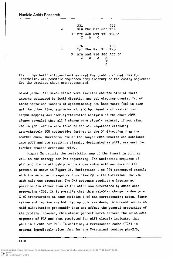
To facilitate isolation of PLP-specific cDNA clones, synthetic
oligonucleotides corresponding to two selected pentapeptides were
prepared (Figure 1). These two regions were chosen on the basis of
minimal codon redundancy upon reverse translation of the known amino
acid sequence of bovine PLP (1). Accordingly, mixtures of 8 different
14-mers corresponding to residues 176-180 and 32 different 15-mers
corresponding to residues 231-235 were end-labelled and used
sequentially to screen the fetal bovine brain cDNA library. Twenty-
four "positive" clones were obtained using the "231-235" mixed probe
but only 7 of them were also found to hydridize with the "176-180"
7415
Downloaded from https://academic.oup.com/nar/article-abstract/13/20/7413/2377658by gueston 08 February 2018
Nucleic Acids Research
231 235Glu Phe Gin Met Thr
31 CTC AAG GTT TAC TG-5'T A C
176 180Tyr Phe Asn Thr Trp
31 ATA AAG TTG TGG ACC 5'G A A A
TC
Fig 1. Synthetic oligonucleotides used for probing cloned cDNA forlipophilin. All possible sequences complimentary to the coding sequencesfor the peptides shown are represented.
mixed probe. A H seven clones were isolated and the size of their
inserts estimated by EcoRI digestion and gel electrophoresis. Two of
these contained inserts of approximately 850 base pairs (bp) in size
and the other five, approximately 950 bp. Results of restriction
enzyme mapping and blot-hjrbridization analysis of the above cDNA
clones revealed that all 7 clones were closely related, if not sibs.
The longer inserts were found to contain sequences extending
approximately 100 nucleotides further in the 51 direction than the
shorter ones. Therefore, one of the longer cDNA inserts was subcloned
into pUC9 and the resulting plasmid, designated as pLPl, was used for
further studies described below.
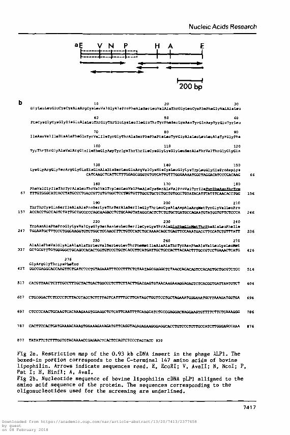
Figure 2a depicts the restriction map of the insert in pLPl as
well as the stategy for DNA sequencing. The nucleotide sequence of
pLPl and its relationship to the known amino acid sequence of the
protein is shown in Figure 2b. Nucleotides 1 to 444 correspond exactly
with the amino acid sequence from his-129 to the C-termlnal phe-276
with only one exception: The DNA sequence predicts a leucine at
position 254 rather than valine which was determined by amino acid
sequencing (26). It is possible that this val->leu change is due to a
G->C transversion at base position 1 of the corresponding codon. Since
valine and leucine are both hydrophobic residues, this conserved amino
acid substitution presumably does not affect the general properties of
the protein. However, this almost perfect match between the amino acid
sequence of PLP and that predicted for pLPl clearly indicates that
pLPl is a cDNA for PLP. In addition, a termination codon (TGA) is
present immediately after that for the C-terminal residue phe-276,
7416
Downloaded from https://academic.oup.com/nar/article-abstract/13/20/7413/2377658by gueston 08 February 2018
Nucleic Acids Research
V N P HAt
200 bp
b 10 20 30GlyL«uL«uGluCyaCyaAlaArgCyaIj«'iiValGlyAlaProPhaAlaS«rL«uValAlAThrt;iyL«uCyaPh«PhaGlyValAlaL«-u
40 SO 60PhaCyaClyCyaClyHiaGluAlaL«uThrGlyThrGluLyaL«uIl«GluThrryxPh«S«rLyBABnTyrGlnAapTyrGluTyrLBU
70 80 90I l«AanValI l«HiaAlaPh«GlnTyrVal I l«Tyr<: iyThrAlaS«rPh«Ph^h«L«uTyrGlyAiaL«oL«uL»i iAlaTyrGlyPh«
100 110 120TyrThiThrGlyAlaValAr9G]jiIl«Pha<:lyAjpTyTLyaThrThrIl»CyaGlyLyaGlyL«uS«rAlaIhrValThrGlyGlyCln
130 140 150LyaGlyAT9<:iyS«rArgGlyGljiHiaGlnAlaHia3*rL«uGluAxqValCyaBiaCyaL«uGlyLyaTlpL«uSlyEiaProAapLya
CATCAAGCTCArrCTTrG<aGCCGGTCTGTCArrCTIT«BaUUkATCGCTAaaCATCCCCACAAG 66
ISO 170 180Ph«ValClvIlaThrTvr*IaL«uThrVa.lValTrpL«nL«uValPh«*laCv«««rAlaValProVa)TvrT1«TvT-ph»».nTlM-Tni
67 TTTGTGG<XATCACCTATGCCCTGACCGTrGTCTGGCTCCTKrarTTCCCTCCTCTGCTCT^ 156
190 200 210TfaxThrCyaGln£«rIlaAlaAlaProS«rLyaThr5«rAlaS«rIl*GlyThrL«aCyaAlaAapAlAAr9ttatTyrGlyValL«aPro
157 ACCAXXTGCCAGTCTATTGCTWXCCCACCAACACCTCTayU^GTATAGCCACTCTCTGTlK^̂ 246
220 230 240TrpAanAlaPh*ProClvI.vValryClyS«- rA«nI^Ql^pB»T-Tl»Cy T.yThrAl •GlnPfrCl n f̂ai-T>irPhaFH «T^uPh>Tl«
247 TGGAATGCTTICCCTGGCAAGGI<̂ CTGGCTCCAACCTTCTGTCCATCTCCAAAACACCTa«̂ TCCAAATiaCCTT^ 336
250 260 270AlaAIaPh*VaiGlyAIaAlaAIaThrL«uValS«rL«ijLaiiThrPhaMatIl«AlaAI<T)irTyTAanflMAlaValL«uLyaL*u)tat
337 GCTGCCTTTGTCGO2XT<XAGCCACACTCGTt^CCCTGCTCACCTTCATGATTGCTGCCACn^ 426
276GlyAnjGlyThrLyaPh»End
421 aaXCAGGCACCAAGTTCTtaTCTCCTCTAGAAATTTCCCTTTCTCTAATKCGAGO^ 516
517 CATCrrAACTCTTTGCCTTTCCIACTCACrCGCCCTCTTCTTACTT<aC<»CTGTAACAACAAAOCJ^^ 606
607 CTGCGGACTCTCCCCTCTTACGTACCTCTTTTAGTCATTTrGCTTCATAGCTW^^ 696
697 CTCCCCAACr<XAA<^CACAAAauSGT<aaG<B^CTCATTCAATITTau^CCATCTCCCCAGGAC>«CCAAC^ 186
t76
8 7 7 TATATTCTCTTTGGTGTACAAAACCGASAACTCACTCCAGTCTCCCTACTACC 9 3 0
Fig 2a. Restriction map of the 0.93 kb cDNA insert in the phage ALP1. Theboxed-in portion corresponds to the C-tenninal 147 amino acids of bovinelipophllin. Arrows indicate sequences read. E, EcoRI; V, Avail; N, Ncol; P,Pst I; H, Hinfl; A, Aval.Fig 2b. Nucleotide sequence of bovine lipophilin cDNA pLPl alligned to theamino acid sequence of the protein. The sequences corresponding to theoligonucleotides used for the screening are underlined.
7417
Downloaded from https://academic.oup.com/nar/article-abstract/13/20/7413/2377658by gueston 08 February 2018
Nucleic Acids Research
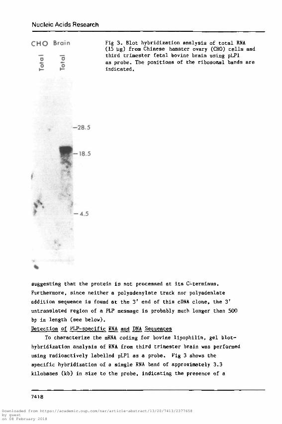
C H O Brain Fig 3. Blot hybridization analysis of total RNA(15 ug) from Chinese hamster ovary (CHO) cells and
— — third trimester fetal bovine brain using pLPl*• •— as probe. The positions of the ribosomal bands areH- t- indicated.
-28.5
t -4.5
suggesting that the protein is not processed at its C-terminus.
Furthermore, since neither a polyadenylate track nor polyadenlate
addition sequence is found at the 3' end of this cDNA clone, the 31
untranslated region of a PLP message is probably much longer than 500
bp in length (see below).
Detection of_ PLP-specific RNA and DNA Sequences
To characterize the mRNA coding for bovine llpophilin, gel blot-
hybridization analysis of RNA from third trimester brain was performed
using radioactively labelled pLPl as a probe. Fig 3 shows the
specific hybridization of a single RNA band of approximately 3.3
kilobases (kb) in size to the probe, indicating the presence of a
7418
Downloaded from https://academic.oup.com/nar/article-abstract/13/20/7413/2377658by gueston 08 February 2018
Nucleic Acids Research
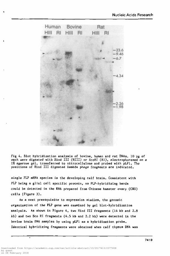
Human Bovine RatHIM Rl Hill Rl Hill Rl
m -23.6-9.46-6.7
* -4.34
-2.26-1.98
Fig 4. Blot hybridization analysis of bovine, human and rat DNAs. 10 ug ofeach were digested with Hind III (HIII) or EcoRI (Rl), electrophoresed on a1% agarose gel, transferred to nitrocellulose and probed with pLPl. Thepositions of Hind III digested lambda phage fragments are indicated.
single PLP mRNA species in the developing calf brain. Consistent with
PLP being a glial cell specific protein, no PLP-hybridizing bands
could be detected in the RNA prepared from Chinese hamster ovary (CHO)
cells (Figure 3).
As a next prerequisite to expression studies, the genomic
organization of the PLP gene was examined by gel blot-hybridization
analysis. As shown in Figure 4, two Hind III fragments (14 kb and 2.8
kb) and two Eco Rl fragments (4.5 kb and 2.2 kb) were detected in the
bovine brain DNA samples by using pLPl as a hybridization probe.
Identical hybridizing fragments were obtained when calf thymus DNA was
7419
Downloaded from https://academic.oup.com/nar/article-abstract/13/20/7413/2377658by gueston 08 February 2018
Nucleic Acids Research
-28
-18
- b p b
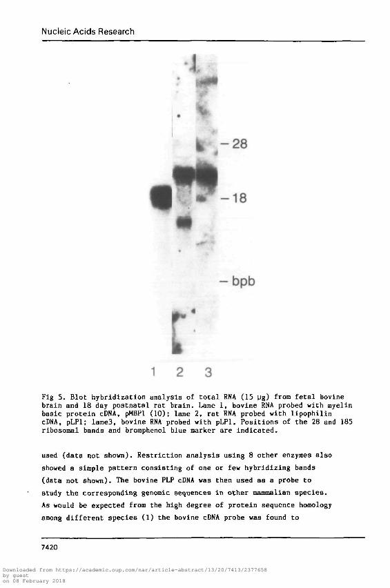
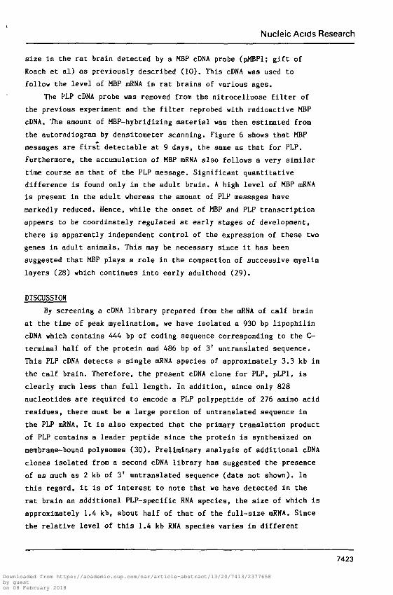
Fig 5. Blot hybridization analysis of total RNA (15 ug) from fetal bovinebrain and 18 day postnatal rat brain. Lane 1, bovine RNA probed with myelinbasic protein cDNA, pMBPl (10); lane 2, rat RNA probed with lipophilincDNA, pLPl; Iane3, bovine RNA probed with pLPl. Positions of the 28 and 185ribosomal bands and bromphenol blue marker are indicated.
used (data not shown). Restriction analysis using 8 other enzymes also
showed a simple pattern consisting of one or few hybridizing bands
(data not shown). The bovine PLP cDNA was then used as a probe to
study the corresponding genomic sequences in other mammalian species.
As would be expected from the high degree of protein sequence homology
among different species (1) the bovine cDNA probe was found to
7420
Downloaded from https://academic.oup.com/nar/article-abstract/13/20/7413/2377658by gueston 08 February 2018
Nucleic Acids Research
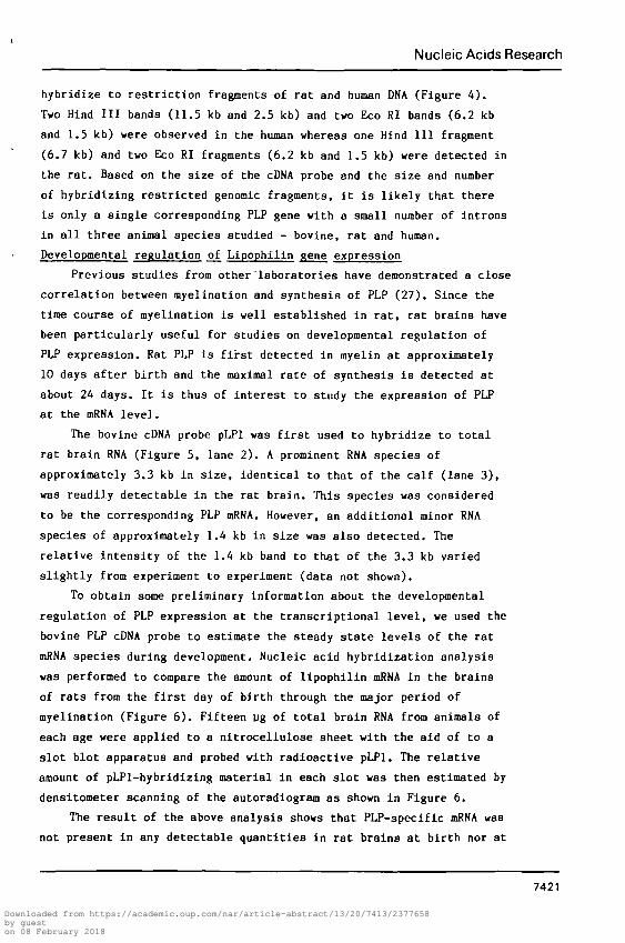
hybridize to restriction fragments of rat and human DNA (Figure 4).
Two Hind III bands (11.5 kb and 2.5 kb) and two Eco RI bands (6.2 kb
and 1.5 kb) were observed in the human whereas one Hind III fragment
(6.7 kb) and two Eco RI fragments (6.2 kb and 1.5 kb) were detected in
the rat. Based on the size of the cDNA probe and the size and number
of hybridizing restricted genomic fragments, it is likely that there
is only a single corresponding PLP gene with a small number of introns
in all three animal species studied - bovine, rat and human.
Developmental regulation of Lipophilin Rene expression
Previous studies from other laboratories have demonstrated a close
correlation between myelination and synthesis of PLP (27). Since the
time course of myelination is well established in rat, rat brains have
been particularly useful for studies on developmental regulation of
PLP expression. Rat PLP is first detected in myelin at approximately
10 days after birth and the maximal rate of synthesis is detected at
about 24 days. It is thus of interest to study the expression of PLP
at the mRNA level.
The bovine cDNA probe pLPl was first used to hybridize to total
rat brain RNA (Figure 5, lane 2). A prominent RNA species of
approximately 3.3 kb in size, identical to that of the calf (lane 3),
was readily detectable in the rat brain. This species was considered
to be the corresponding PLP mRNA. However, an additional minor RNA
species of approximately 1.4 kb in size was also detected. The
relative intensity of the 1.4 kb band to that of the 3.3 kb varied
slightly from experiment to experiment (data not shown).
To obtain some preliminary information about the developmental
regulation of PLP expression at the transcriptional level, we used the
bovine PLP cDNA probe to estimate the steady state levels of the rat
mRNA species during development. Nucleic acid hybridization analysis
was performed to compare the amount of lipophilin mRNA in the brains
of rats from the first day of birth through the major period of
myelination (Figure 6). Fifteen ug of total brain RNA from animals of
each age were applied to a nitrocellulose sheet with the aid of to a
slot blot apparatus and probed with radioactive pLPl. The relative
amount of pLPl-hybridizing material in each slot was then estimated by
densitometer scanning of the autoradiogram as shown in Figure 6.
The result of the above analysis shows that PLP-specific mRNA was
not present in any detectable quantities in rat brains at birth nor at
7421
Downloaded from https://academic.oup.com/nar/article-abstract/13/20/7413/2377658by gueston 08 February 2018
Nucleic Acids Research
0 5 9 11 1315 171920 21 23 28 30At i i i i i i i i i i i i i
I I I I I I MM II I I M I M M
PLP
a.
a.CD
oQ._l0.
28 ADULT
DAYS AFTER BIRTH
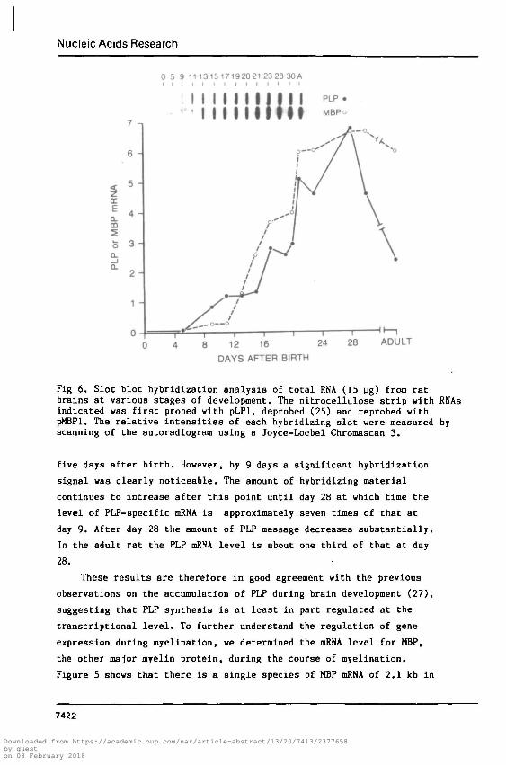
Fig 6. Slot blot hybridization analysis of total RNA (15 ug) from ratbrains at various stages of development. The nitrocellulose strip with RNAsindicated was first probed with pLPl, deprobed (25) and reprobed withpMBPl. The relative intensities of each hybridizing slot were measured byscanning of the autoradiogram using a Joyce-Loebel Chromascan 3.
five days after birth. However, by 9 days a significant hybridization
signal was clearly noticeable. The amount of hybridizing material
continues to increase after this point until day 28 at which time the
level of PLP-specific mRNA is approximately seven times of that at
day 9. After day 28 the amount of PLP message decreases substantially.
In the adult rat the PLP mRNA level is about one third of that at day
28.
These results are therefore in good agreement with the previous
observations on the accumulation of PLP during brain development (27),
suggesting that PLP synthesis is at least in part regulated at the
transcriptional level. To further understand the regulation of gene
expression during myelination, we determined the mRNA level for MBP,
the other major myelin protein, during the course of myelination.
Figure 5 shows that there is a single species of MBP mRNA of 2.1 kb in
7422
Downloaded from https://academic.oup.com/nar/article-abstract/13/20/7413/2377658by gueston 08 February 2018
Nucleic Acids Research
size in the rat brain detected by a MBP cDNA probe (pMBPl; gift of
Roach et al) as previously described (10). This cDNA was used to
follow the level of MBP mRNA in rat brains of various ages.
The PLP cDNA probe was removed from the nitrocelluose filter of
the previous experiment and the filter reprobed with radioactive MBP
cDNA. The amount of MBP-hybridizing material was then estimated from
the autoradiogram by densitometer scanning. Figure 6 shows that MBP
messages are first detectable at 9 days, the same as that for PLP.
Furthermore, the accumulation of MBP mRNA also follows a very similar
time course as that of the PLP message. Significant quantitative
difference is found only in the adult brain. A high level of MBP mRNA
is present in the adult whereas the amount of PLP messages have
markedly reduced. Hence, while the onset of MBP and PLP transcription
appears to be coordinately regulated at early stages of development,
there is apparently independent control of the expression of these two
genes in adult animals. This may be necessary since it has been
suggested that MBP plays a role in the compaction of successive myelin
layers (28) which continues into early adulthood (29).
DISCUSSION
By screening a cDNA library prepared from the mRNA of calf brain
at the time of peak myelination, we have isolated a 930 bp lipophilin
cDNA which contains 444 bp of coding sequence corresponding to the C—
terminal half of the protein and 486 bp of 3' untranslated sequence.
This PLP cDNA detects a single mRNA species of approximately 3.3 kb in
the calf brain. Therefore, the present cDNA clone for PLP, pLPl, is
clearly much less than full length. In addition, since only 828
nucleotides are required to encode a PLP polypeptide of 276 amino acid
residues, there must be a large portion of untranslated sequence in
the PLP mRNA. It is also expected that the primary translation product
of PLP contains a leader peptide since the protein is synthesized on
membrane-bound polysomes (30). Preliminary analysis of additional cDNA
clones isolated from a second cDNA library has suggested the presence
of as much as 2 kb of 3' untranslated sequence (data not shown). In
this regard, it is of interest to note that we have detected in the
rat brain an additional PLP-specific RNA species, the size of which is
approximately 1.4 kb, about half of that of the full-size mRNA. Since
the relative level of this 1.4 kb RNA species varies in different
7423
Downloaded from https://academic.oup.com/nar/article-abstract/13/20/7413/2377658by gueston 08 February 2018
Nucleic Acids Research
samples derived from animals of the same age (data not shown), it is
possible that it represents a partial degradation product of the full-
size transcript. However, it is not known at this moment whether this
snaller RNA species is still capable of directing PLP synthesis.
The availability of cDNA clones for both PLP (this study) and MBP
(10-12) has provided an opportunity for studying the developmental
regulation of the genes that encode the two major membrane proteins
in myelin. Our results show that the onset of transcription ifor both
genes appears to be coordinately regulated during development. Both
messages are first detectible at 9 days after birth and then continue
to accumulate until approximately 30 days; after this time, however,
the amount of PLP message declines more rapidly than that of MBP
mRNA. This may reflect either a differential reduction in the
transcription rate or in the stability of the messages for the two
proteins. Interestingly, this difference in mRNA levels correlates
well with reports that MBP turns over more rapidly than PLP (31,32).
REFERENCES1. Lees, M.B. and Brostoff, S.W. (1984) in Myelin, 2nd Edition, ed.
P. Morell (Plenum Press, New York), Chapter 6, pp 197-221.2. Boggs.J.M., Moscarello, M.A. and Papahadjopoulos, D. (1982) in
Lipid-Protein Interactions, eds. P. Jost and O.H. Griffith,(Academic Press, New York).
3. Eylar, E.H. (1972) in Functional and Structural Proteins of theNervous System, eds. A. Davison, P. Mandel and I. Morgan(Plenum Press, New York) pp 215-240.
4. Lees, M.B., Chao, B., Lin, L.H., Samiullah, M. and Laursen, R.(1983) Arch. Biochem. Biophys. 226:643-656.
5. Jolles,J.,Nussbaum, M.-L. and Jolles, P. (1983) Biochim. Biophys.Acta 742:33-38.
6. Laursen, R.L., Samiullah, M. and Lees.M. (1983) FEBS Lett 161:71-74.
7. Lin, L-F.H. and Lees, M.B. (1982) Proc. Natl. Acad. Sci. U.S.A.79:941-945.
8. Boggs, J.M. and Moacarello, M.A. (1978) Biochim. Biophys. Acta515:1-21.
9. Raine, C.E. (1984) in: Myelin, 2nd Edition, ed. P. Morell (PlenumPress, N.Y.), Chapter 8, pp 259-310.
10. Roach, A., Boylan, K., Horvath, S., Prusiner, S.B. and Hood, L.E.(1983) Cell 34:799-806.
11. Zeller, N.K., Sprague, J.A., Lazzarini, R.A., Yu, Y-T. andCampagnoni, A.T. (1983) Trans. Am. Soc. Neurochem. 14:255.
12. Amorese, P. A., Ellis, S. B., Harphold, M. M. and Linthlcum, D. S.(1983) Trans. Am. Soc. Neurochem. 14:165.
13. Chirgwin, J.M., Przybyla, A.E., MacDonald, R.J. and Rutter, W.J.(1979) Biochemistry 18:5294-5299.
14. Aviv, H. and Leder, P. (1972) Proc. Natl. Acad. Sci. U.S.A.69:1408-1412.
7424
Downloaded from https://academic.oup.com/nar/article-abstract/13/20/7413/2377658by gueston 08 February 2018
Nucleic Acids Research
15. Huynh, T.V., Young, R.A. and Davis, R.W. (1984) in DNA Cloning:A Practical Approach, ed. D. Glover (IRL Press, Oxford).
16. Davis, R. W., Botstein, D. and Roth, J. R. (1980)Advanced Bacterial Genetics. Cold Spring Harbor Laboratory,Cold Spring Harbor, New York
17. Modrich, P. and Zabel, D. (1976) J. Biol. Chem. 251:5866-5874.18. Beaucage, S.L. and Carruthers, M.H. (1981) Tet. Lett. 22:1859-1862.19. Benton, W.D. and Davis, R.W. (1977) Science 196:180-182.20. Vieira, J. and Messing, J. (1982) Gene 19:259-268.21. Haxam, A.M. and Gilbert, W. (1980) Meth. Enzymol. 65:499-560.22. Kaplan, B.B., Schacter, B., Osterburg, H.H., de Vellis, J.C. and
Finch, C.E. (1978) Biochemistry 17:5516-5524.23. Maniatis, T., Fritsch, E. F. and Saabrook, J. (1982) Molecular
Cloning. Cold Spring Harbor Laboratory, Cold Spring Harbor,New York
24. Southern, E.M. (1975) J. Mol. Biol. 98:503-517.25. Thomas, P.S. (1983) in Meth. Enzymol. eds. R. Wu, L. Grossman
and I. Moldave (Academic Press, New York) pp 255-266.26. Stoffel, W., Hillen, H., Schroder, W. and Deutzmann, R. (1982)
Hoppe-Seyler's Z. Physiol. Chem. 363:1297-1407.27. Benjamins, J. A. and Smith, M. E. (1984) in Myelin. 2nd Edition,
ed. P. Morell (Plenum Press, New York), Chapter 7, pp 225-258.28. Brady, G. W., Murthy, N. S., Fein, D. B., Wood, D. D. and
Moscarello, M. A. (1981) Biophys. J. 34: 345-350.29. Chia, L. S., Thompson, J. E. and Moscarello, M A.(1983) FEBS
Letters 157: 155-158.30. Colman, D.R., Kreibich, G., Frey, A.B. and Sabatini, D.D. (1982)
J. Cell Biol. 95:598-608.31. Sabri, M.I., Bone, A. H. and Davison, A.N. (1974) Biochem J.
142:499-507.32. Lajtha, A., Toth, J., Fujimoto, K. and Agrawal, H. C. (1977)
Biochem. J.164:323-329.
7425
Downloaded from https://academic.oup.com/nar/article-abstract/13/20/7413/2377658by gueston 08 February 2018
Nucleic Acids Research
Downloaded from https://academic.oup.com/nar/article-abstract/13/20/7413/2377658by gueston 08 February 2018