Embed Size (px)
Citation preview

119I. Shergill et al. (eds.), Surgical Emergencies in Clinical Practice,DOI 10.1007/978-1-4471-2876-2_7, © Springer-Verlag London 2013
Introduction
It is only recently that maxillofacial surgery ceased to be just a dental specialty and now requires formal surgical/medical training for specialist registration. It remains, however, founded
Chapter 7 Maxillofacial Emergencies Tahwinder Upile , Waseem Jerjes , Colin Hopper , N. Patel , and Janavikulam Thiruchelvam
T. Upile , B.Sc. (hons), M.Sc., M.S., M.D., FRCS (Gen Surg), FRCS (OTO), FRCS (ORL-HNS), DFFP, FHEA, MRCGP (�) Department of ENT , Barnet and Chase Farm Hospital , En fi eld, Greater London , UK
Head and Neck Unit , University College London Hospitals , London , UK e-mail: [email protected]
W. Jerjes , B.Sc. (hons), Ph.D., M.Sc., BDS, MBBS Department of ENT , Barnet and Chase Farm Hospital , En fi eld, Greater London , UK
C. Hopper , FRCS, M.D. Head and Neck Unit , University College London Hospitals , London , UK
N. Patel , FRCS Head and Neck Unit, Department of ENT , Southampton University Hospitals , Southampton , UK
J. Thiruchelvam , FRCS, M.D. Oral Maxillofacial Unit, Head and Neck Department , Barnet and Chase Farm Hospitals , Greater London , UK
120 T. Upile et al.
in dentistry requiring many of the skills and attributes learned in general dental or oral surgical practice. Medical specialists as a part of emergency treatment and patient management may be called upon to treat dental injuries and maxillofacial trauma. Once again, ATLS management protocols must be adhered to with early airways management (bearing in mind a potential cervical spine injury) from any signi fi cant injury. The multidisciplinary role of emergency surgical airways man-agement may include emergency, general, ENT, and maxillo-facial surgeons. The place of several transtracheal catheters (grey/brown ven fl on) connected to oxygen to buy time until a de fi nitive airway must not be forgotten.
Many undergraduate curricula do not include maxillofa-cial surgery teaching and now reserve OMF for postgraduate students; this leaves the junior surgeon poorly prepared for initial clinical duties. It is essential that multidisciplinary care is entered into early, and that when required, appropriate referral is made at the opportune time to more senior and/or specialist teams. We have selected the cases based upon their frequency of presentation, need for correct initial manage-ment, and due to the medicolegal consequences of poor man-agement. Other more complex surgical emergencies may be found in specialty-speci fi c texts.
Clinical Case Scenario 1: Dentoalveolar Injuries
Case Presentation
A 16-year-old male comes to A&E with history of injury in school with avulsion of upper anterior tooth. He was bullying one of the junior pupils, who punched him in the face. There was no loss of consciousness, and the teacher brought him to hospital immediately. He has no past history of medical problems and is not on any medication.

121Chapter 7. Maxillofacial Emergencies
Key Features in History and Examination
History
General questions pertaining to any other trauma victims (loss of consciousness, other injuries, immediate treatment, etc.). Speci fi c questions pertaining to avulsed teeth – time since avulsion, where was the tooth found, how was it brought to A&E (dry, milk, water, saline).
Examination
Make sure the tooth is intact. Exclude other head and neck injuries and fractures. Check cheek sensation.
Principles of Acute Management
Initially ATLS protocol, then concentrate on management speci fi c to maxillofacial surgery. Clean the tooth with saline. Hold the tooth with the crown. Do not scrub the root surface, just clean it with saline. If indicated, X-rays may be taken. Earliest implantation and stabilization of the tooth into its socket is essential.
Discussion
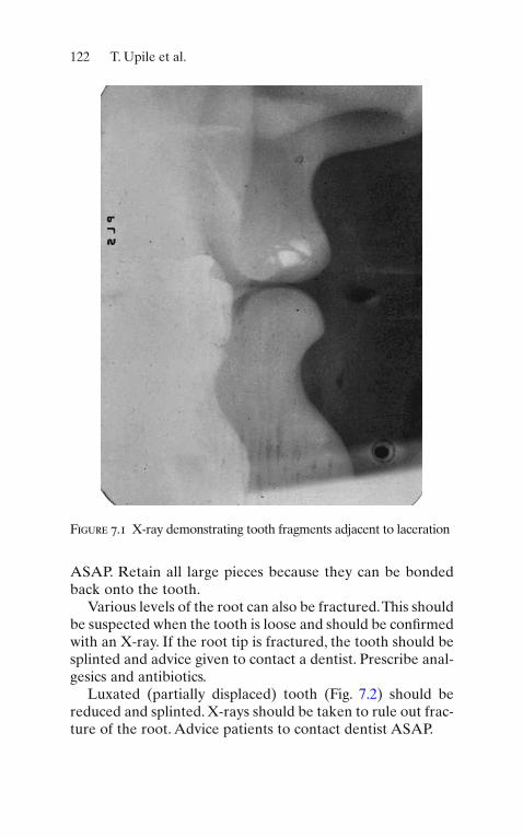
Dental injuries are common and are frequently associated with other facial trauma. The spectrum of dental injury is varied (including Crown fracture, crown root fracture, root fracture, periodontal ligament concussion, subluxation, luxation). Injury to the crown can range from chipping of enamel to exposure of pulp. This can be very painful and is best treated by a dentist. Remember to look for tooth frag-ments in the adjacent lacerations, con fi rmed by X-rays (Fig. 7.1 ). Prescribe analgesic and advice to see a dentist
122 T. Upile et al.
ASAP. Retain all large pieces because they can be bonded back onto the tooth.
Various levels of the root can also be fractured. This should be suspected when the tooth is loose and should be con fi rmed with an X-ray. If the root tip is fractured, the tooth should be splinted and advice given to contact a dentist. Prescribe anal-gesics and antibiotics.
Luxated (partially displaced) tooth (Fig. 7.2 ) should be reduced and splinted. X-rays should be taken to rule out frac-ture of the root. Advice patients to contact dentist ASAP.
Figure 7.1 X-ray demonstrating tooth fragments adjacent to laceration

123Chapter 7. Maxillofacial Emergencies
Figure 7.2 Luxated tooth and X-ray excluded fracture of the root
Avulsed teeth (out of the socket) needs rapid treatment. Tooth is best stored in cold milk or patients own mouth until it is reimplanted. Wash with cold running saline and hold it with the crown.
If a whole dentoalveolar segment is fractured, the whole segment has to be reduced and splinted. Any associated gin-gival tear should be sutured. Impressions can be taken to guide the reduction of teeth and also useful to make a cus-tom-made splint.
Clinical Case Scenario 2: Dental Infection
Case Presentation
A 14-year-old patient presents with a 6-day history of swell-ing in his left cheek. It was preceded by toothache and is now progressively getting worse. On arrival, he is apyrexial and otherwise well, but looks fl ushed. He was unable to attend for

124 T. Upile et al.
PE lesson today and his mother took him to the dentist, who immediately referred him to the on call ENT team.
Key Features in History and Examination
History
Ask for duration, source of infection, tooth pain, dif fi culty in breathing, or swallowing. Ask about diabetes and immunode-pression. Previous dental history and imaging taken.
Examination
Assess the extent of the swelling (determine which dental space is involved). Look out for the source of infection (skin, teeth, etc.). Grave signs include trismus, raised fl oor of mouth, unable to swallow, and drooling of saliva.
Principles of Acute Management
If airway is potentially compromised, one needs urgent airway management. Inform anesthetist for assessment. Try to avoid a tracheostomy. Steroids can be given to reduce edema.
Appropriate antibiotics should be given immediately (to cover cocci and anaerobes). Necessary radiographs (Fig. 7.3 ) should be taken to identify the source. Drainage is indicated if there is collection of pus. At the same time, the source of infection should be dealt with (e.g. extraction).
Blood tests should be done to rule out undiagnosed diabe-tes or other hematological conditions (leukemia).
Discussion
Acute facial swelling due to dental infection can be life threatening. It can progress rapidly in some cases. Therefore, urgent management is critical. In early stages, oral antibiotics are enough with urgent attention to the source of infection.

125Chapter 7. Maxillofacial Emergencies
In severe cases, urgent admission for IV antibiotics is important. Continuous monitoring of progress or airway compromise is necessary till the time of de fi nitive management. If there is pus collection, drainage at the earliest is necessary with removal of the source of infection. Depending on the teeth, various spaces can be involved. These include:
1. Canine space 2. Buccal space 3. Submandibular space 4. Submental space 5. Sublingual space 6. Submasseteric space 7. Pterygomandibular space 8. Parapharyngeal space 9. Temporal space
Ludwigs Angina
Ludwig’s angina is an acute and potentially life-threatening cellulitis involving, sublingual, submental, and submandibular spaces often due to dental infection. The tongue is swollen, fl oor of mouth is raised, and the patient will have dif fi culty in swallowing (Fig. 7.4 ). This will lead to airway obstruction if not intervened early. This condition therefore needs immediate management of airway and drainage of the above spaces.
Figure 7.3 Orthopantomogram ( OPG ) showing source of infection

126 T. Upile et al.
Figure 7.4 Image of patient with Ludwig’s angina
Admission to ITU may be necessary. General principles of dental infection management as above apply, but urgent attention to airway and drainage is critical.
Clinical Case Scenario 3: Mandibular Fractures
Case Presentation
A 25-year-old male comes to accident and emergency department with pain and dif fi culty in closing the mouth after a fi ght in a nightclub. He was punched once after he insulted his friend’s

127Chapter 7. Maxillofacial Emergencies
partner. There was no loss of consciousness and he remembers the incident vividly.
Key Features of History and Examination
Acute examination according to ATLS principles. A head injury of some form is to be expected and should be managed appropriately.
History
After initial assessment, essential history to be obtained are nature of injury, altered occlusion, numbness of lip and tongue, loss of teeth, and location of pain. Also, presence of previous skeletal deformity and temporomandibular joint problems should be determined.
Examination
Once other injuries are ruled out, look out for deformity, site of swelling, and tenderness; document the teeth present or missing, presence of loose teeth, step deformity (Fig. 7.5 ),
Figure 7.5 Step deformity in a patient with mandibular fracture

128 T. Upile et al.
altered occlusion, sublingual hematoma, numbness of lower lip and tongue, reduced mouth opening, and mobility of frac-ture site if other fi ndings are not convincing.
Principles of Acute Management
After initial management of ATLS is through and once a clinical diagnosis of a fractured mandible is made, con fi rmation is required with radiographs. Two X-rays are required orthopantomogram (Fig. 7.6 ) and PA mandible (Fig. 7.7 ).
In some cases, CT may be required. Start the patient on antibiotics and give adequate pain killers. If the fracture site is grossly displaced, reduction and temporary splinting with wires around the teeth will reduce the pain and bleeding.
Discussion
Mandibular fracture is one of the common bony maxillofacial injuries. The common cause is interpersonal violence. The other causes include road traf fi c accidents, sports injuries, fall, and industrial accidents. All these patients should be examined
Figure 7.6 Orthopantomogram showing mandibular fractures

129Chapter 7. Maxillofacial Emergencies
Figure 7.7 PA mandible X-ray
for other bony and dental injuries. The common sites of man-dibular fracture are shown in Fig. 7.8 .
This includes condyle, coronoid, ramus, angle, body, para-symphysis, and symphysis. Commonly, the mandible will frac-ture in two places. Therefore, if one fracture is identi fi ed, always look carefully at other sites. Due to the presence of teeth, most mandibular fractures are compound fractures and have a potential to get infected. Therefore, antibiotics should be prescribed.
There are several methods of fi xation of the fracture. This includes closed reduction and fi xation (intermaxillary fi xation) and open reduction and internal fi xation (ORIF) with plates. With the advent of mini-plates and screws, ORIF has been widely accepted as the treatment of choice. In general con-dyle, ramus and coronoid are treated with closed reduction and fi xation. Angle, body, symphysis and parasymphysis are treated with ORIF with mini-plates. Two plates are required

130 T. Upile et al.
in front of the mental foramen and one plate behind it (AO principle). Usually the fracture is approached intraorally. In selected cases, extraoral approach is indicated.
Clinical Case Scenario 4: Soft Tissue Injuries
Case Presentation
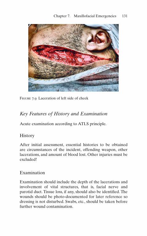
A 30-year-old male comes with a laceration to his left cheek (Fig. 7.9 ). He denies any knowledge of how he received the injury but is accompanied by a group of friends, with various injuries to their face and limbs.
14%
3%
21%20%
3%
2% 36%
Figure 7.8 Common sites of mandibular fracture
131Chapter 7. Maxillofacial Emergencies
Figure 7.9 Laceration of left side of cheek
Key Features of History and Examination
Acute examination according to ATLS principle.
History
After initial assessment, essential histories to be obtained are circumstances of the incident, offending weapon, other lacerations, and amount of blood lost. Other injuries must be excluded!
Examination
Examination should include the depth of the lacerations and involvement of vital structures, that is, facial nerve and parotid duct. Tissue loss, if any, should also be identi fi ed. The wounds should be photo-documented for later reference so dressing is not disturbed. Swabs, etc., should be taken before further wound contamination.

132 T. Upile et al.
Principles of Acute Management
Initially, according to ATLS principles, and then speci fi c to laceration, control of bleeding is essential prior to thorough examination to identify the structures involved.
Antibiotic and tetanus cover should be given. Thorough debridement of the wound is essential. The vital structures such as nerves, parotid duct should be repaired prior to sutur-ing the tissue in layers.
Discussion
Soft tissue injuries can be classi fi ed into contusion, abrasion, laceration, avulsion, burns (chemical, thermal). Circumstances of the injury are essential as they will give insight into the associated injuries and contamination.
All particulate matters and foreign bodies in the wound should be removed or they will result in traumatic tattooing. Petroleum-based liquids such as grease and oil should be removed by solvents like acetone or ether. Type of laceration will give an idea of the nature of injury. Sharp objects pro-duce a clean straight cut. Stellate laceration is usually due to blunt injuries.
Most lacerations to the head and neck are simple one, caused by a fall. These wounds are cleaned and closed primarily. Some lacerations are large and can be associated with tissue loss or damage to vital structures. In the neck, any laceration deep to the platysma should be evaluated with care to identify damage to other vital structures. Stab wounds should be examined with care as the depth is unpredictable with a small entry wound.
Bite Wounds
Bite wounds (Fig. 7.10 ) are associated with high infective risk. This is not only due to the presence of highly infective bacte-rial fl ora in the mouth (animals and human beings) but also due to the multiple types of injury. The animal bites usually
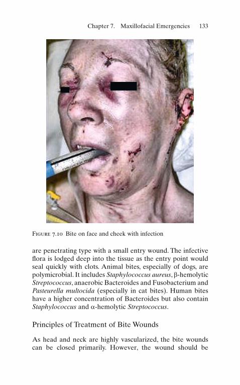
133Chapter 7. Maxillofacial Emergencies
are penetrating type with a small entry wound. The infective fl ora is lodged deep into the tissue as the entry point would seal quickly with clots. Animal bites, especially of dogs, are polymicrobial. It includes Staphylococcus aureus , b -hemolytic Streptococcus , anaerobic Bacteroides and Fusobacterium and Pasteurella multocida (especially in cat bites). Human bites have a higher concentration of Bacteroides but also contain Staphylococcus and a -hemolytic Streptococcus .
Principles of Treatment of Bite Wounds
As head and neck are highly vascularized, the bite wounds can be closed primarily. However, the wound should be
Figure 7.10 Bite on face and cheek with infection

134 T. Upile et al.
irrigated copiously and debrided appropriately. Especially the small punctured wound should be opened and irrigated well to its depth. Broad-spectrum antibiotics should be pre-scribed in addition to tetanus cover.
Clinical Case Scenario 5: Post – Dental Extraction Bleeding
Case Presentation
A 55-year-old patient comes to accident and emergency depart-ment with H/O bleeding from the mouth. He had a tooth extracted by his dentist in the morning. It has not stopped since, and he gives a history, only after prompting, of shaving injuries taking an extremely long time to stop bleeding.
Key Features in History and Examination
History
Has it been continuous/profuse? History of bleeding disor-ders, hypertension. Medications the patient is on (anticoagu-lants, aspirin, clopidogrel, etc.).
Examination
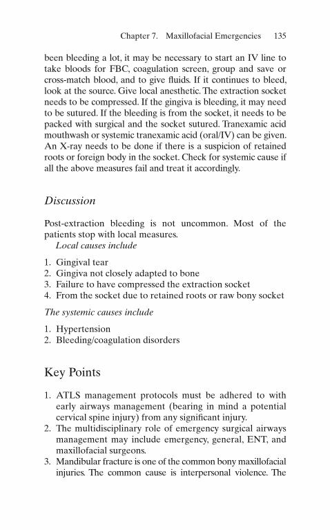
Examine to make sure the patient is hemodynamically stable. Measure blood pressure/pulse. Under a good light, examine the socket after suctioning. Look at the status of the extraction socket and look out for the source of bleeding (from gingival/extraction socket). Is there an obvious bleeding vessel?
Principles of Acute Management
Consider emergency circulatory management scenario. Measure pulse, BP, and control the local hemorrhage with local pressure (biting on a moist/adrenaline-soaked gauze). If the patient has
135Chapter 7. Maxillofacial Emergencies
been bleeding a lot, it may be necessary to start an IV line to take bloods for FBC, coagulation screen, group and save or cross-match blood, and to give fl uids. If it continues to bleed, look at the source. Give local anesthetic. The extraction socket needs to be compressed. If the gingiva is bleeding, it may need to be sutured. If the bleeding is from the socket, it needs to be packed with surgical and the socket sutured. Tranexamic acid mouthwash or systemic tranexamic acid (oral/IV) can be given. An X-ray needs to be done if there is a suspicion of retained roots or foreign body in the socket. Check for systemic cause if all the above measures fail and treat it accordingly.
Discussion
Post-extraction bleeding is not uncommon. Most of the patients stop with local measures.
Local causes include
1. Gingival tear 2. Gingiva not closely adapted to bone 3. Failure to have compressed the extraction socket 4. From the socket due to retained roots or raw bony socket
The systemic causes include
1. Hypertension 2. Bleeding/coagulation disorders
Key Points
1. ATLS management protocols must be adhered to with early airways management (bearing in mind a potential cervical spine injury) from any signi fi cant injury.
2. The multidisciplinary role of emergency surgical airways management may include emergency, general, ENT, and maxillofacial surgeons.
3. Mandibular fracture is one of the common bony maxillofacial injuries. The common cause is interpersonal violence. The

136 T. Upile et al.
other causes include road traf fi c accidents, sports injuries, fall, and industrial accidents. All these patients should be exam-ined for other bony and dental injuries. The common sites of mandibular fracture are shown in Fig. 7.8 .
Acute facial swelling due to dental infection can be life threatening.
Bite wounds (Fig. 7.10 ) are associated with high infective risk. This is not only due to the presence of highly infective bacterial fl ora in the mouth (animals and human beings) but also due to multiple types of injury.