Embed Size (px)
Citation preview
U.S. ARMY MEDICAL DEPARTMENT CENTER AND SCHOOL FORT SAM HOUSTON, TEXAS 78234-6100
SURGICAL METHODS
SUBCOURSE MD0574 EDITION 200
DEVELOPMENT
This subcourse is approved for resident and correspondence course instruction. It reflects the current thought of the Academy of Health Sciences and conforms to printed Department of the Army doctrine as closely as currently possible. Development and progress render such doctrine continuously subject to change.
ADMINISTRATION
For comments or questions regarding enrollment, student records, or shipments, contact the Nonresident Instruction Section at DSN 471-5877, commercial (210) 221-5877, toll-free 1-800-344-2380; fax: 210-221-4012 or DSN 471-4012, e-mail [email protected], or write to: COMMANDER AMEDDC&S ATTN MCCS HSN 2105 11TH STREET SUITE 4192 FORT SAM HOUSTON TX 78234-5064 Approved students whose enrollments remain in good standing may apply to the Nonresident Instruction Section for subsequent courses by telephone, letter, or e-mail. Be sure your social security number is on all correspondence sent to the Academy of Health Sciences.
CLARIFICATION OF TRAINING LITERATURE TERMINOLOGY When used in this publication, words such as "he," "him," "his," and "men" are intended to include both the masculine and feminine genders, unless specifically stated otherwise or when obvious in context. .
USE OF PROPRIETARY NAMES
The initial letters of the names of some products are capitalized in this subcourse. Such names are proprietary names, that is, brand names or trademarks. Proprietary names have been used in this subcourse only to make it a more effective learning aid. The use of any name, proprietary or otherwise, should not be interpreted as an endorsement, deprecation, or criticism of a product; nor should such use be considered to interpret the validity of proprietary rights in a name, whether it is registered or not. .
MD0574 i
TABLE OF CONTENTS Lesson Paragraphs INTRODUCTION 1 MINOR SURGICAL PROCEDURES............................................ 1-1--1-10 Exercises 2 WOUND CLOSURE AND SUTURES .......................................... 2-1--2-9 Exercises 3 EMERGENCY SURGICAL PROCEDURES................................. 3-1--3-6 Exercises
MD0574 ii
CORRESPONDENCE COURSE OF THE ACADEMY OF HEALTH SCIENCES, UNITED STATES ARMY
SUBCOURSE MD0574
SURGICAL METHODS
INTRODUCTION
As a Medical NCO, you need to be familiar with some surgical procedures. You may perform minor surgical procedures, wound closure and suturing, and you may assist in emergency surgical procedures. This subcourse will give you basic knowledge in each of these areas. Subcourse Components: The subcourse instructional material consists of the following:
Lesson 1, Minor Surgical Procedures. Lesson 2, Wound Closure and Sutures. Lesson 3, Emergency Surgical Procedures.
Here are some suggestions that may be helpful to you in completing this subcourse: --Read and study each lesson carefully. --Complete the subcourse lesson by lesson. After completing each lesson, work the exercises at the end of the lesson. --After completing each set of lesson exercises, compare your answers with those on the solution sheet that follows the exercises. If you have answered an exercise incorrectly, check the reference cited after the answer on the solution sheet to determine why your response was not the correct one. Credit Awarded: Upon successful completion of the examination for this subcourse, you will be awarded 6 credit hours. To receive credit hours, you must be officially enrolled and complete an examination furnished by the Nonresident Instruction Section at Fort Sam Houston, Texas. You can enroll by going to the web site http://atrrs.army.mil and enrolling under "Self Development" (School Code 555).
MD0574 1-1
LESSON ASSIGNMENT LESSON 1 Minor Surgical Procedures. LESSON ASSIGNMENT Paragraphs 1-1 through 1-10. LESSON OBJECTIVES After completing this lesson, you should be able to: 1-1. Identify the correct basic procedural steps for preparing the skin for a minor surgical procedure. 1-2. Identify the correct steps for preparing a traumatic wound for treatment. 1-3. Identify the appropriate fluid, the efficiency of the irrigation, and the method of irrigating a wound. 1-4. Identify the general considerations, preparation of the skin, and procedures of follow-up care for an abscessed wound requiring incision and drainage. 1-5. Describe the general characteristics, contraindications, treatment procedures indicated, and follow-up treatment for the following: a. Paronychia. b. Toenail removal. c. Subungual hematoma. d. Wart removal. e. Ring removal. 1-6. Describe the general guidelines for removal of a foreign body from the soft tissue and the treatment which follows the removal. SUGGESTION After completing the assignment, complete the exercises at the end of this lesson. These exercises will help you to achieve the lesson objectives.
MD0574 1-2
LESSON 1
MINOR SURGICAL PROCEDURES 1-1. INTRODUCTION One of the functions of the Medical NCO is to assist the physician assistant or the physician in performing minor surgical procedures. Eventually, you may be required to perform these procedures yourself. The procedures will be performed primarily in the emergency room, the troop medical clinic, and the battalion aid station so the patient will be able to return to duty. Basic knowledge of the procedures may be obtained from this lesson. 1-2. PREPARATION OF THE OPERATIVE SITE a. The "Skin-Prep." Preparation of the operative site is more commonly known as "skin-prep." The purpose of preparing the site is to render that area as free as possible from transient and resident microorganisms, dirt, and skin oil. All or any of these could infect an open wound. The goal of this preparation is to allow the surgical procedure to be performed with a minimal danger of infection. b. Basic Prep: Initial Procedures. The basic preparatory procedures at the site are as follow. (1) Expose the skin to be prepared. (2) Don sterile gloves. (3) Place sterile towels above and below the area to be cleaned. (4) Place sterile, absorbent towels along each side of the area. These towels act as an absorber for any solution that has run off. Remove these towels after the site preparation is completed. c. Basic Prep: Skin Scrub Procedures. Scrub the skin in this manner: (1) Wet a sponge with antiseptic solution (or use a prepackaged scrub brush). Squeeze out the excess solution to prevent run off of fluid. (2) Starting at the intended site of incision, scrub the skin using circular motions in ever-widening circles. Scrub for at least ten minutes. Use enough pressure and friction to remove dirt and microorganisms. Remember it takes both chemical (contact time) and mechanical (scrubbing) action to cleanse the area. (3) Discard the sponge after you reach the outside of the area.
MD0574 1-3
(4) Repeat this scrubbing procedure with a clean sponge. (5) Scrub the incision site for a minimum of ten minutes. CAUTION: Never bring a soiled sponge back toward the center of an area. d. Preparation of Traumatic Wounds: Procedures. A traumatic wound is any wound that occurs as a result of injury or other damage. The wound is considered contaminated. (1) Procedures. A variety of procedures may be needed in preparing a traumatic wound for incision. The wound may need to be irrigated or the wound may require packing or covering with sterile gauze. (2) Type of procedure. The wound can be cleansed and irrigated after you change to sterile gloves. The extent and type of injury will determine what preparatory procedure you choose. e. Preparation of Traumatic Wounds: General Guidelines. Note these guidelines: (1) Do not use solutions such as detergents and alcohols that can irritate an area in which tissue has been lost. (2) You may irrigate small areas with a warm sterile solution, usually normal saline, in a bulb syringe. (3) The purpose of irrigating a wound is to flush out debris gently. (4) When flushing out a large wound, you may need to use copious amounts of a warm saline solution. (5) A bottle of warm saline or Ringer's solution attached to IV tubing may be used to irrigate a wound. (6) Following irrigation, a wound is usually debrided. f. Hair Removal. Remove hair carefully to avoid injuring the skin. A break in the skin, even though caused by only hair removal, can provide an opportunity for entry and colonization of microorganisms with the potential for infection. Shaving an area should be done as close to the time of the incision as possible. The longer the time between the shaving and the incision, the greater is the chance of infection. CAUTION: NEVER shave or clip eyebrows.
MD0574 1-4
g. Irrigation. The irrigation fluid of choice is normal saline since this fluid is nonirritating to body tissue. (1) Efficiency of irrigation. Efficient irrigation is achieved at 7 to 9 pounds per square inch (psi). This is not easy to gauge when conducting irrigation in the field. It is commonly agreed that to achieve optimal irrigation pressure without excessive pressure you should use an 18-gauge (ga) needle on the end of a syringe or intravenous (IV) tubing. (2) Methods of irrigation. There are three commonly used methods of irrigating wounds: by bulb or asepto syringe, by 35cc syringe and a 18-gauge needle, and by a mechanical jet device. Irrigation using a 35cc syringe and an 18-gauge needle is the preferred method for irrigating contaminated wounds and uses intermediate pressure. Normal saline is the solution of choice, but any potable water can be used. Mechanical jet and pulse irrigation should not be used because they tend to push debris deeper in to the wound rather than out of the wound, thus causing more damage and increasing the risk of infection. 1-3. ABSCESS INCISION AND DRAINAGE An abscess is an infection that results in a collection of purulent material in a circumscribed and closed cavity. When an abscess is in the early stages of development, it may be treated with warm compresses. If this treatment is unsuccessful, incision and drainage (I & D) may need to be performed on a wound that has abscessed. Incision and drainage is the release of the collection of pus by making an incision in the skin and draining the pus. An I and D is commonly performed in a clinic setting. Indication of the need for an I and D is an abscess that is localized, erythematous, tender, and fluctuant. a. General Considerations. There are few, if any, contraindications to the procedure of abscess incision and drainage. Recurrent episodes of abscess may indicate an underlying problem, such as malnutrition, poor hygiene, diabetes, or immune deficiencies. These are not considered contraindications. Additionally, follow the considerations listed below. (1) Be sure sterile procedure is used to avoid secondary contamination. (2) Obtain informed consent before performing an incision and drainage procedure. b. Equipment Needed. Gather the following equipment. (1) Sterile gloves, drapes, and surgical gowns. (2) Antiseptic cleansing solutions such as povidone-iodine (Betadine®), 3% hydrogen peroxide, or isopropyl alcohol.
MD0574 1-5
(3) Syringe containing 0.5 percent or 1 percent lidocaine (Xylocaine®). (4) Disposable 3 ml or 10 ml syringe (depending on the size of the abscess). (5) Disposable 25 gauge needle. (6) One-fourth or one-half inch iodoform or plain sterile gauze packing or silverlon packing material. (7) 4-inch by 4-inch (4x4) gauze pads for dressing. (8) Nonallergenic adhesive tape for dressing. (9) Hydrogen peroxide. (10) Safety razor. (11) Scalpel with a #11 pointed I & D blade. (12) Hemostat (curved or straight). (13) Plain forceps. (14) Surgical scissors. (15) Cotton-tipped sterile applicators. (16) Culture swabs. c. Preparation of Area of Abscess and Surrounding Skin. Proceed in this manner. (1) Shave the area, if needed. Only shave the area if absolutely necessary to observe the wound. (2) Wash the area. (3) Prepare the area with povidone-iodine or isopropyl alcohol. (4) Drape the area.
MD0574 1-6
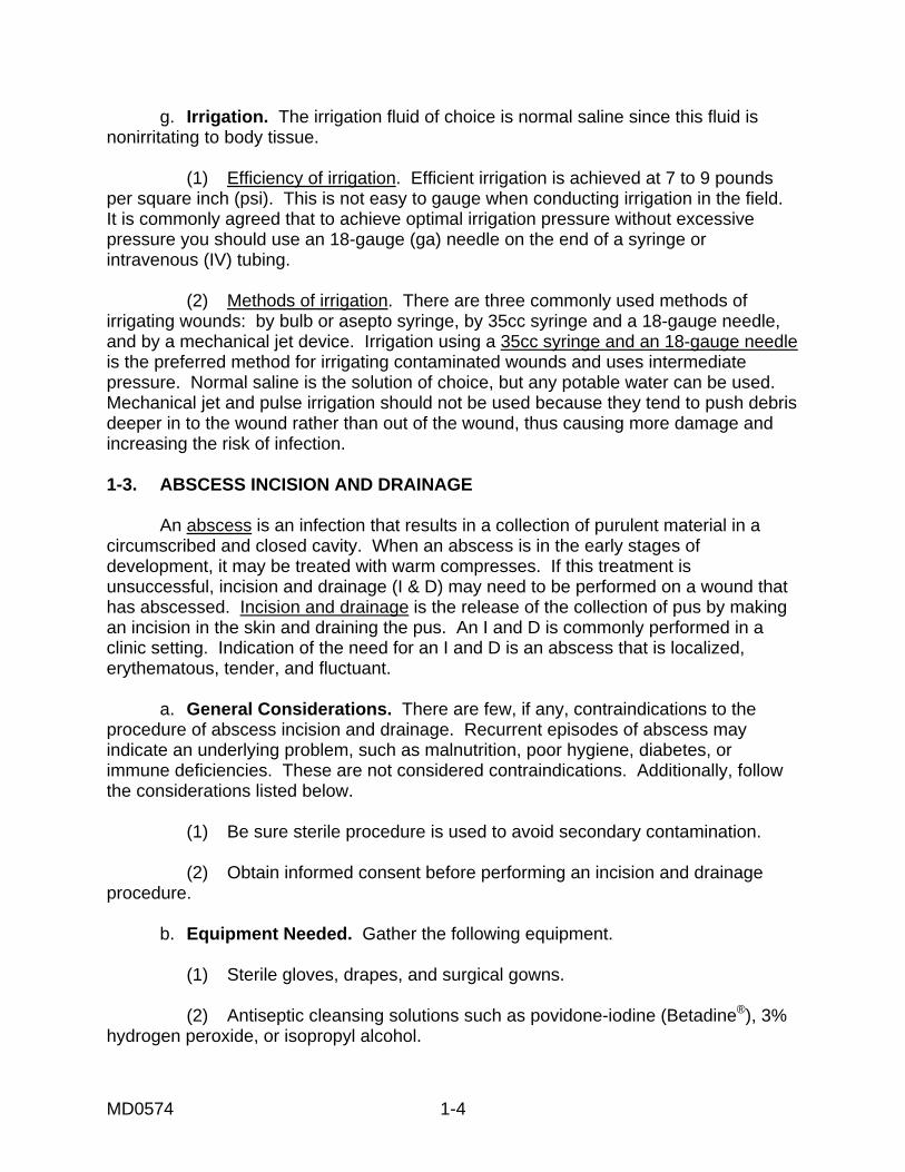
d. Anesthesia. Follow the procedure given below. (1) Infiltrate 0.5 percent or 1 percent lidocaine into the incision site over the abscess. (2) Anesthetize the area well beyond the incisional area so that drainage can occur without the hindrance of pain. (3) Delay the incision for several minutes after the injection to be sure there is a complete anesthetic block. e. Procedure. Use the following procedure for abscess incision and drainage. (1) Using the #11 blade, incise the abscess deeply from one side of the fluctuant area, to the opposite side of the area of fluctuance (figure 1-1). This is necessary to ensure complete evacuation of the purulent drainage
Figure 1-1. Making the incision.
(2) Express purulent material from wound. (3) Obtain cultures from the drainage. (4) Perform intra-cavity exploration to break up any adhesions. For smaller abscesses, soak a cotton-tipped applicator with hydrogen peroxide. Then, explore the cavity with the applicator to remove all pus, debris, and sebaceous materials. (5) Following exploration, clean the cavity with four to six hydrogen peroxide soaked, cotton-tipped applicators. You may also irrigate the cavity with a sterile saline solution. (6) Observe the incision for hemostasis. Hemostasis should occur spontaneously, but may be aided by subsequent packing.
MD0574 1-7
(7) The abscess may be loosely packed using one-fourth or one-half inch iodoform or plain gauze packing. This helps in keeping the cavity open and permits adequate drainage. (8) Apply a sterile gauze dressing and secure the dressing with nonallergenic adhesive tape. f. Follow up Care. (1) Initial patient education. Care the day of surgery entails advising the patient that the initial dressing should be left in place until the next day. Also tell him to elevate the affected extremity and that analgesics are seldom necessary. The day after surgery, the patient should remove the external dressing but leave the packing in place. He should soak the site in warm water compresses or take a tub bath for 20 to 30 minutes. The site should be submerged during soaks. A sterile dressing should be reapplied after each soak. CAUTION: If the packing falls out, DO NOT reinsert it! (2) Follow up: health care provider. Reevaluate the patient 36 to 48 hours after the incision and drainage procedure has been done. Wound care at this point includes the following steps. (a) Remove the external dressing. (b) Gently remove the packing from the I & D cavity. (c) Cleanse the abscess cavity with a cotton-tipped applicator soaked in hydrogen peroxide. Anesthesia is not necessary, but analgesics should be provided to the patient. (d) DO NOT repack the cavity, especially if it is clean and if pain and tenderness have significantly diminished. (e) Reapply a sterile gauze dressing to the open wound site. (3) Follow up: patient education. (a) Instruct the patient to continue soaks three to four times daily. (b) Continue these soaks for five to seven days or until the incision has healed. (c) If the abscess was adequately drained, the I & D will close spontaneously by secondary intention within five to seven days post-procedure.
MD0574 1-8
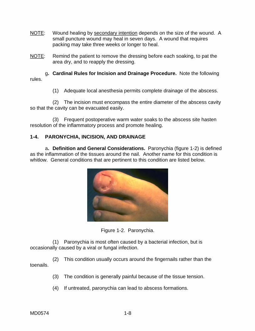
NOTE: Wound healing by secondary intention depends on the size of the wound. A small puncture wound may heal in seven days. A wound that requires packing may take three weeks or longer to heal. NOTE: Remind the patient to remove the dressing before each soaking, to pat the area dry, and to reapply the dressing. g. Cardinal Rules for Incision and Drainage Procedure. Note the following rules. (1) Adequate local anesthesia permits complete drainage of the abscess. (2) The incision must encompass the entire diameter of the abscess cavity so that the cavity can be evacuated easily. (3) Frequent postoperative warm water soaks to the abscess site hasten resolution of the inflammatory process and promote healing. 1-4. PARONYCHIA, INCISION, AND DRAINAGE a. Definition and General Considerations. Paronychia (figure 1-2) is defined as the inflammation of the tissues around the nail. Another name for this condition is whitlow. General conditions that are pertinent to this condition are listed below.
Figure 1-2. Paronychia. (1) Paronychia is most often caused by a bacterial infection, but is occasionally caused by a viral or fungal infection. (2) This condition usually occurs around the fingernails rather than the toenails. (3) The condition is generally painful because of the tissue tension. (4) If untreated, paronychia can lead to abscess formations.
MD0574 1-9
b. Conservative Treatment. If the condition is treated early, conservative treatment may be all that is necessary. Such treatment includes: (1) Soaks. (2) Zinc oxide dressing. (3) Elevation of the hand (if a fingernail is affected). (4) Antibiotics. c. Indications for Incision and Drainage. Incision and drainage procedure is indicated to: (1) Control pain. (2) Speed healing. (3) Prevent the spread of infection. NOTE: The patient usually feels immediate relief as the pressure of pus is relieved. d. Preparation for Incision and Drainage of Active Paronychia. A minimum of preparation and supplies is required. The I & D procedure can be performed painlessly through the necrotic tissue at the cuticle with a needle point scalpel or an 18 gauge needle. Gather this equipment: (1) Syringe with 25 and 21 gauge needle. (2) One percent lidocaine (Xylocaine®) without epinephrine for digital block. (3) Scalpel with a #11 blade. (4) Small scissors. (5) Mosquito forceps. (6) Gauze packing. e. Procedure. Use the following procedure for the incision and drainage. (1) Anesthesia. Use the cutaneous nerve block rather than local infiltration. The digital cutaneous nerves run along the medial and lateral aspects of each finger. These nerves can be blocked at any level above the distal phalanx. See figure 1-3.
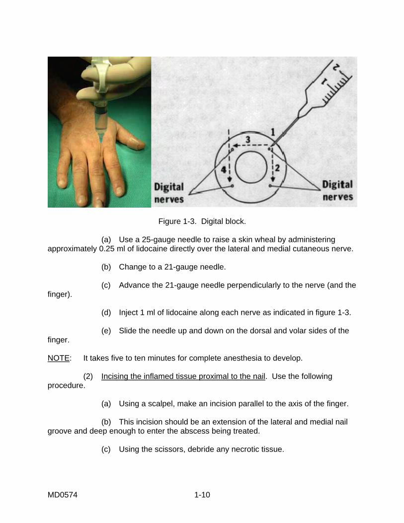
MD0574 1-10
Figure 1-3. Digital block.
(a) Use a 25-gauge needle to raise a skin wheal by administering approximately 0.25 ml of lidocaine directly over the lateral and medial cutaneous nerve. (b) Change to a 21-gauge needle. (c) Advance the 21-gauge needle perpendicularly to the nerve (and the finger). (d) Inject 1 ml of lidocaine along each nerve as indicated in figure 1-3. (e) Slide the needle up and down on the dorsal and volar sides of the finger. NOTE: It takes five to ten minutes for complete anesthesia to develop. (2) Incising the inflamed tissue proximal to the nail. Use the following procedure. (a) Using a scalpel, make an incision parallel to the axis of the finger. (b) This incision should be an extension of the lateral and medial nail groove and deep enough to enter the abscess being treated. (c) Using the scissors, debride any necrotic tissue.
MD0574 1-11
(3) Infection under the nail. For an infection that has spread under the nail, you must remove the proximal nail in the following manner. (a) Use mosquito forceps to lever up and hold the nail. (b) Cut the nail off in a straight line using the scissors. (c) Place gauze packing under the flap of the overhanging tissue and the cuticle. (4) Culturing the infected material. To determine what caused the infection, culture the infected material you have removed from under the nail. (5) Antibiotics. Usually drainage is sufficient to clear up the infection. Antibiotics may be considered, however. f. Follow-up Care. (1) Short term care. Tell the patient to elevate his hand for one to two days to prevent throbbing from the dependent position. The patient should return in two to three days for the packing to be removed. After the packing is removed, he should soak the affected finger in warm water for 15 minutes, three or four times a day. After each soaking, a dry, nonstick dressing should be applied. (2) Long term care. The nail must be protected from being torn away from the nail bed until it regrows from its base. This regrowth process may take several months. After the healing process is complete, the nail and cuticle may be deformed. 1-5. TOENAIL REMOVAL a. General Considerations. The removal of a toenail is a simple and safe procedure. This procedure requires a minimum of skill. b. Indications for Toenail Removal. A toenail may need to be removed in any of the circumstances given below. See figure 1-4. (1) Ingrown nail (ohychoptosis). (2) Ringworm or fungus infection of the nail (onychomycosis). (3) Inflammation of the nail fold (chronic or recurrent paronychia). (4) Deformed, enlarged, curved nail (onychogryposis).
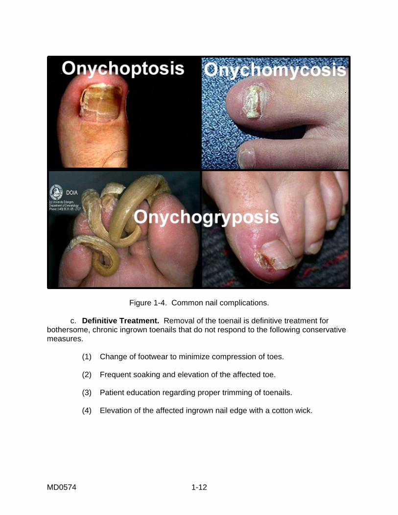
MD0574 1-12
Figure 1-4. Common nail complications.
c. Definitive Treatment. Removal of the toenail is definitive treatment for bothersome, chronic ingrown toenails that do not respond to the following conservative measures. (1) Change of footwear to minimize compression of toes. (2) Frequent soaking and elevation of the affected toe. (3) Patient education regarding proper trimming of toenails. (4) Elevation of the affected ingrown nail edge with a cotton wick.
MD0574 1-13
d. Contraindications for Removal of the Toenail. (1) The toenail should not be unnecessarily removed if the patient has: (a) Diabetes mellitus. (b) Peripheral vascular disease. (c) Bleeding disorders. (d) Allergy to local anesthetics (relative contraindication). (2) Presence of soft-tissue infection or paronychia may be a relative contraindication. It is recommended that the infection be treated prior to removing the toenail. e. Equipment. Gather the following equipment. (1) A 3 or 5 ml syringe. (2) 2 percent lidocaine without epinephrine. (3) Sterile scissors with straight blades (or narrow periosteal elevator). (4) A sterile rubber band. (5) Two sterile straight hemostats. (6) Phenol solution (88 percent) for permanent removal of the nail. (7) Isopropyl alcohol swabs. (8) Sterile cotton swabs. (9) Antibacterial or antibiotic ointment (for example, Betadine®, Bacitracin®). (10) Sterile gauze pads (4 x 4). f. Procedure. Use the following procedure for removal of the toenail. (1) With the patient supine, scrub and drape the toe in a sterile fashion.
MD0574 1-14
(2) Administer local anesthetic in ring-block fashion as described below. (a) The total solution should be 5 ml. (b) Raise a wheal at the base of the toe on the extensor surface on the affected side. (c) Direct the injection toward the plantar surface to envelop both the extension and plantar branches of true digital nerve on that side. (d) Deposit 1 ml at each site. (e) Retract the needle slightly. (f) Redirect the needle horizontally across the dorsal surface of the toe. (g) Inject 0.5 ml under the skin at the base of the toe on the opposite side. (h) Perform a second puncture at that site. (i) Advance the needle in the plantar direction. (j) Deliver 1 ml of anesthetic to each branch of the digital nerve. (3) When the anesthesia is achieved, secure a sterile rubber band with a straight hemostat to serve as a tourniquet. (4) Remove the nail from the nail bed using the following procedure (see figure 1-5). (a) Using a flat pointed blade of scissor, straight hemostat, or narrow periosteal elevator, introduce and advance the instrument upward and against the nail and away from the nail bed. This minimizes injury and bleeding. (b) Completely free the nail at its base under the edge of the cuticle. This allows the nail to be completely removed and provides exposure to the germinal tissue of the nail bed. (c) Using scissors, completely split the nail in a longitudinal direction. The split should include the base of the nail that rests against the cuticle. (d) Using a straight hemostat, grasp the portion of the nail to be removed lengthwise.
MD0574 1-15
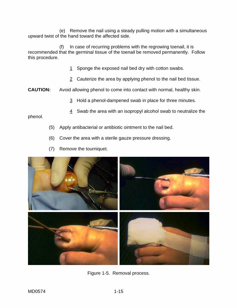
(e) Remove the nail using a steady pulling motion with a simultaneous upward twist of the hand toward the affected side. (f) In case of recurring problems with the regrowing toenail, it is recommended that the germinal tissue of the toenail be removed permanently. Follow this procedure. 1 Sponge the exposed nail bed dry with cotton swabs. 2 Cauterize the area by applying phenol to the nail bed tissue. CAUTION: Avoid allowing phenol to come into contact with normal, healthy skin. 3 Hold a phenol-dampened swab in place for three minutes. 4 Swab the area with an isopropyl alcohol swab to neutralize the phenol. (5) Apply antibacterial or antibiotic ointment to the nail bed. (6) Cover the area with a sterile gauze pressure dressing. (7) Remove the tourniquet.
Figure 1-5. Removal process.
MD0574 1-16
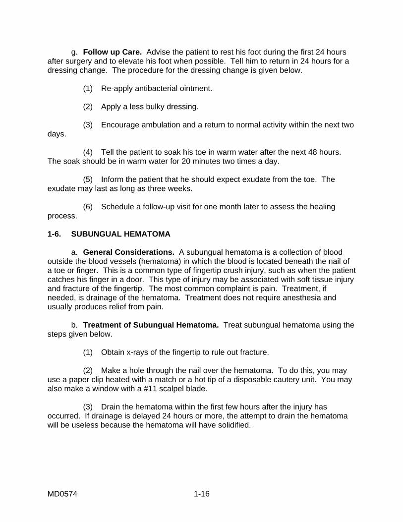
g. Follow up Care. Advise the patient to rest his foot during the first 24 hours after surgery and to elevate his foot when possible. Tell him to return in 24 hours for a dressing change. The procedure for the dressing change is given below. (1) Re-apply antibacterial ointment. (2) Apply a less bulky dressing. (3) Encourage ambulation and a return to normal activity within the next two days. (4) Tell the patient to soak his toe in warm water after the next 48 hours. The soak should be in warm water for 20 minutes two times a day. (5) Inform the patient that he should expect exudate from the toe. The exudate may last as long as three weeks. (6) Schedule a follow-up visit for one month later to assess the healing process. 1-6. SUBUNGUAL HEMATOMA a. General Considerations. A subungual hematoma is a collection of blood outside the blood vessels (hematoma) in which the blood is located beneath the nail of a toe or finger. This is a common type of fingertip crush injury, such as when the patient catches his finger in a door. This type of injury may be associated with soft tissue injury and fracture of the fingertip. The most common complaint is pain. Treatment, if needed, is drainage of the hematoma. Treatment does not require anesthesia and usually produces relief from pain. b. Treatment of Subungual Hematoma. Treat subungual hematoma using the steps given below. (1) Obtain x-rays of the fingertip to rule out fracture. (2) Make a hole through the nail over the hematoma. To do this, you may use a paper clip heated with a match or a hot tip of a disposable cautery unit. You may also make a window with a #11 scalpel blade. (3) Drain the hematoma within the first few hours after the injury has occurred. If drainage is delayed 24 hours or more, the attempt to drain the hematoma will be useless because the hematoma will have solidified.
MD0574 1-17
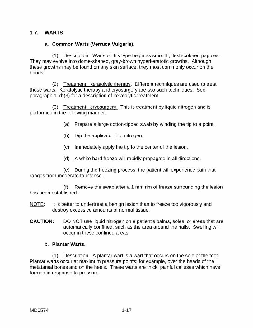
1-7. WARTS a. Common Warts (Verruca Vulgaris). (1) Description. Warts of this type begin as smooth, flesh-colored papules. They may evolve into dome-shaped, gray-brown hyperkeratotic growths. Although these growths may be found on any skin surface, they most commonly occur on the hands. (2) Treatment: keratolytic therapy. Different techniques are used to treat those warts. Keratolytic therapy and cryosurgery are two such techniques. See paragraph 1-7b(3) for a description of keratolytic treatment. (3) Treatment: cryosurgery. This is treatment by liquid nitrogen and is performed in the following manner. (a) Prepare a large cotton-tipped swab by winding the tip to a point. (b) Dip the applicator into nitrogen. (c) Immediately apply the tip to the center of the lesion. (d) A white hard freeze will rapidly propagate in all directions. (e) During the freezing process, the patient will experience pain that ranges from moderate to intense. (f) Remove the swab after a 1 mm rim of freeze surrounding the lesion has been established. NOTE: It is better to undertreat a benign lesion than to freeze too vigorously and destroy excessive amounts of normal tissue. CAUTION: DO NOT use liquid nitrogen on a patient's palms, soles, or areas that are automatically confined, such as the area around the nails. Swelling will occur in these confined areas. b. Plantar Warts. (1) Description. A plantar wart is a wart that occurs on the sole of the foot. Plantar warts occur at maximum pressure points; for example, over the heads of the metatarsal bones and on the heels. These warts are thick, painful calluses which have formed in response to pressure.
MD0574 1-18
(2) Treatment: general. Treatment is not required as long as the warts are painless. It may be better not to subject the patient to a course of treatment but to let the wart go through the normal evolution. Severely painful plantar warts may be treated by keratolytic therapy (duofilm) or blunt dissection. (3) Keratolytic therapy (duofilm). This type of treatment is conservative initial therapy. The treatment is nonscarring and relatively effective. It does require persistent application of medication once each day for several weeks. The procedure is given below. (a) Pare down the wart with pumice stone or sandpaper. (b) Soak the area in warm water to aid in the absorption of the medicine. (c) Apply medicine with the glass rod and allow the medicine to dry. (d) Cover the entire surface of the wart. NOTE: Penetration of the medication is increased if the treated wart is covered with a piece of adhesive tape. (e) After a few days, white, pliable keratin forms. Pare down this substance with sandpaper or a pumice stone. (f) Eventually, you will expose pink skin. (4) Blunt dissection. Perform the following steps. (a) Inject 2% lidocaine with epinephrine directly into the substance of the wart. (b) Insert the tip of a blunt-tipped scissors between the wart and normal skin. (c) Cut the skin circumferentially. (d) Insert a blunt dissector into the plane of cleavage. (e) Separate the lesion with short firm strokes. (f) Draw the blunt dissector firmly back and forth over the exposed surface of the bed to assure that no tissue fragments remain.
MD0574 1-19
(g) Apply a small sterile dressing over the wound. (h) Advise the patient to change the dressing daily for three to four days. 1-8. REMOVAL OF RINGS If the finger swells, it may be necessary to remove a ring. Three types of procedures for ring removal are given below. a. Lubricate the Finger. You can lubricate the finger with soap or K-Y jelly. Then, slip the ring off the finger. b. "Milk" the Finger. Wrap the finger snugly with string from the distal tip to just below the ring. This "milks" the edema out of the finger. You can then slide the ring off the finger. c. Cut the Ring. Cut the ring with a commercial ring cutter. Spread the ring with two pliers and remove the ring. 1-9. SOFT TISSUE FOREIGN BODY REMOVAL a. General Guidelines. (1) Take a history of the patient, including information about any unusual medical problems. (2) Determine the specific characteristics of the foreign body. (3) Devise the best plan for removing the foreign body. An object such as wood needs to be removed immediately since it can cause inflammation and infection. Objects such as glass or plastic may be removed on an elective basis. Metallic foreign bodies which are causing no additional damage need never be removed. CAUTION: DO NOT attempt a hasty exploration for the item. Consider other possibilities of injury rather than the patient's explanation. (4) Equipment to gather includes a standard suture tray, tissue retractors, and special pick-ups. Remember to have good direct light. b. Operative Technique. The operative technique to use is tailored to each clinical situation. CAUTION: DO NOT grab blindly with a hemostat in an effort to remove a foreign object.
MD0574 1-20
(1) Ground-in foreign material or tattooing removal. Use a local anesthetic and meticulous debridement with a sponge, scrub brush, or a tooth brush. Removing of such material or a tattoo may cause permanent disfigurement. It may be impossible to remove all pieces of ground-in foreign matter. (2) Removal of foreign bodies in fatty tissue. Follow these steps. (a) Make an elliptical incision surrounding the entrance of the wound. (b) Grasp the skin of the ellipse loosely with an Allis forceps. (c) Undercut the incision until the foreign body is contacted. (d) Remove the foreign body, skin, and entrance tract in one block. (3) Removal of foreign bodies in the sole of the feet. Assume that foreign matter has been introduced into the wound along with the foreign body. An example of such an occurrence would be a nail going into the foot through a rubber sole of a shoe. You may want to use a magnifying glass to see the foreign body. An ischemic tourniquet is mandatory when you are exploring the foot for a foreign body. Proceed in the following manner. (a) Enlarge the entrance wound, if necessary, with an adequate incision. (b) Explore the wound carefully by spreading the soft tissue with a hemostat. (c) Grasp the foreign body and remove it through the original wound tract. (d) Irrigate the wound. (4) Removal of subungual foreign bodies. Removing foreign bodies that are under a nail may require partial or complete removal of the nail. If the nail or the nail bed is to be manipulated, you will need a digital block. Techniques for removing a foreign object from under a nail are given below. (a) Use a hypodermic needle, bent at the tip. Slide the needle under the nail, hook the object, and withdraw the object. (b) Use a 19-gauge hypodermic needle to slide under the nail and surround the splinter. Bring the needle tip against the underside of the nail to secure the splinter. Withdraw the needle and splinter as a unit.
MD0574 1-21
(5) Removal of fishhooks. The condition of the fishhook in the body determines the manner used to withdraw the fishhook. Removal techniques are given below. (a) Infiltrate the area with 1% lidocaine. Force the barb of the fishhook through the anesthetized skin. Clip off the barb and remove the rest of the hook along the direction of entry. (b) Loop a piece of string or fishing line around the balley of the hook at which the hook enters the skin. Allow about one foot of string for traction. Hold the shank of the fishhook parallel to and in approximation with the skin by the index finger of the left hand. When you have disengaged the barb of the fishhook, pull sharply on the string to remove the hook through the wound entry. (c) After adequate anesthetic, use an 18-gauge needle to cover the barb. Pass the needle through the wound entrance parallel to the shank of the fishhook. Sheath the barb and allow the fishhook to be backed out. 1-10. CLOSING The injuries and problems addressed in this lesson can be quickly resolved by relatively minor surgical procedures. The important role you play is to ensure that the patient does not sustain additional injury or infection from the procedure.
Continue with Exercises
MD0574 1-22
EXERCISES, LESSON 1 INSTRUCTIONS. The following exercises are to be answered by writing the answer in the space provided or by marking the correct response. After you have completed all the exercises, turn to "Solutions to Exercises, Lesson 1" at the end of the exercises and check your answers. 1. What is the purpose of preparing the operative site? ______________________ ________________________________________________________________ 2. List five major steps in the basic preparation procedures for minor surgery. a. ____________________________________________. b. ____________________________________________. c. ____________________________________________. d. ____________________________________________. e. ____________________________________________. 3. To scrub the patient's skin effectively, you should scrub in a ____________ motion for a minimum of ________ minutes. 4. Complete the following statements (statements refer to preparation of traumatic wounds). a. It may be necessary to _____________ or cover the wound while you scrub and shave the area around the wound. b. Do not clean a traumatic wound with substances which might irritate the wound; substances such as ____________ or _______________. c. A common, nonirritating substance which can be used to irrigate a traumatic wound is _______________. d. The next step after irrigation of a traumatic wound is usually _____________.
MD0574 1-23
5. List three methods of irrigating a wound. a. ____________________________________________. b. ____________________________________________. c. ____________________________________________. 6. Never shave or clip ______________________. 7. Is it possible to irrigate contaminated wounds successfully by attaching IV tubing to a bag of normal saline and irrigating under the force of gravity? a. Yes. b. No. 8. Abscess is _______________________________________________________ ________________________________________________________________ 9. Incision and drainage (I & D) refers to __________________________________ ________________________________________________________________ ________________________________________________________________. 10. When an abscess is _______________________________________, treatment can be warm compresses. 11. A patient who has recurrent episodes of abscesses may have an underlying health problem. List three possible such problems. a. ____________________________________________. b. ____________________________________________. c. ____________________________________________.
MD0574 1-24
12. List eight major steps in the procedure of draining an abscess. a. ____________________________________________. b. ____________________________________________. c. ____________________________________________. d. ____________________________________________. e. ____________________________________________. f. ____________________________________________. g. ____________________________________________. h. ____________________________________________. 13. List three cardinal rules for irrigation and drainage (I & D) that must be remembered. a. ____________________________________________. b. ____________________________________________. c. ____________________________________________. 14. Paronychia is ____________________________________________________. 15. List four possible methods of treating paronychia, if the condition is treated early in its development. a. ____________________________________________. b. ____________________________________________. c. ____________________________________________. d. ____________________________________________.
MD0574 1-25
16. List the five major steps in the procedure of incising and draining paronychia. a. ____________________________________________ b. ____________________________________________ c. ____________________________________________ d. ____________________________________________ e. ____________________________________________ 17. Among the conditions which might require that a toenail be removed are these. Define these conditions. a. Onychoptosis--_____________________________________________. b. Onychomycosis--___________________________________________. c. Chronic/recurrent paronychia--_________________________________. d. Onychogryposis--___________________________________________. 18. List four contraindications for toenail removal. a. ____________________________________________. b. ____________________________________________. c. ____________________________________________. d. ____________________________________________.
MD0574 1-26
19. List the seven major steps in the procedure of toenail removal. a. ____________________________________________. b. ____________________________________________. c. ____________________________________________. d. ____________________________________________. e. ____________________________________________. f. ____________________________________________. g. ____________________________________________. 20. What is a subungual hematoma? _____________________________________ ________________________________________________________________ ________________________________________________________________ 21. Verruca vulgaris are commonly known as _____________________. 22. Cryosurgery is a method of treating verruca vulgaris using liquid ____________. 23. Where on the human body do plantar warts usually occur? ________________________________________________________________. 24. List three ways to remove a ring. a. ____________________________________________. b. ____________________________________________. c. ____________________________________________.
Check Your Answers on Next Page
MD0574 1-27
SOLUTIONS TO EXERCISES, LESSON 1 1. The purpose of preparing the operative site is to make that site as free as possible from microorganisms, dirt, and skin oil. (para 1-2a) 2. Expose the skin area to be prepared. Don sterile gloves. Place sterile towels above and below the area to be cleaned. Place sterile absorbent towels along each side of the area. Scrub the patient's skin. (para 1-2b) 3. Circular 10 (para 1-2c(2)) 4. a. Pack. b. Alcohol or detergents. c. Normal saline. d. Debridement. (para 1-2d, e) 5. Use a bulb or asepto syringe. Use a 35cc syringe and a #19 gauge needle. Use a mechanical jet device. (para 1-2g(2)) 6. Eyebrows. (para 1-2f, CAUTION) 7. a (para 1-2e (5)) 8. Abscess is an infection that results in a collection of purulent material in a circumscribed and closed cavity. (para 1-3) 9. Incision and drainage (I & D) refers to the release of the collection of pus by making an incision in the skin and draining the pus. (para 1-3) 10. In the early stage of development. (para 1-3) 11. You are correct if you listed any three of the following: Malnutrition. Poor hygiene. Diabetes. Immune deficiencies. (para 1-3a) 12. Make a straight incision. Express purulent material from wound. Obtain cultures from the drainage. Perform intracavity exploration. Clean the cavity. Observe the incision for hemostasis. Pack the abscess loosely. Apply a sterile gauze dressing. (para 1-3e(1) through (8))
MD0574 1-28
13. Local anesthesia permits complete drainage. Incision must go across the entire abscess. Postoperative warm water soaks. (para 1-3g) 14. Paronychia is an inflammation of the folds around the nail. (para 1-4a) 15. Soaks. Zinc oxide dressing. Elevation of the affected area (hand or foot). Antibiotics. (para 1-4b(1) through (4)) 16. Anesthetize the area. Incise the inflamed tissue next to the nail. If the infection has spread under the nail, remove the proximal nail. Culture the infected material to determine the causative organism. If there is not sufficient drainage, consider antibiotics. (para 1-4e(1) through (5)) 17. a. Ingrown nail. b. Ringworm or fungus infection. c. Inflammation of the nail fold. d. Deformed, enlarged, curved nail. (para 1-5b) 18. Patient has a history of: Diabetes mellitus. Peripheral vascular disease. Bleeding disorder. Allergy to local anesthetics. (para 1-5d(1)(a) through (d)) 19. Scrub and drape the patient's toe in a sterile fashion. Administer local anesthetic in a ring-block fashion. Secure a sterile rubber band with a straight hemostat as a tourniquet. Remove the nail from the nail bed. Apply antibacterial or antibiotic ointment to the nail bed. Cover the area with a sterile gauze pressure dressing. Remove the tourniquet. (para 1-5f) 20. A subungual hematoma is blood outside the blood vessels such as blood located beneath the nail of a toe or finger. (para 1-6a) 21. Warts. (para 1-7a) 22. Nitrogen. (para 1-7a(3)) 23. Plantar warts usually occur at the maximum pressure points. (para 1-7b(1)) 24. Lubricate the finger. "Milk" the ring. Cut the ring off. (para 1-8a through c)
End of Lesson 1
MD0574 2-1
LESSON ASSIGNMENT LESSON 2 Wound Closure and Suturing. TEXT ASSIGNMENT Paragraphs 2-1 through 2-9. LESSON OBJECTIVES After completing this lesson, you should be able to: 2-1. Identify specific types of lacerations. 2-2. Identify the phases of normal wound healing. 2-3. Identify the advantages and disadvantages of
the following suture materials: Nonabsorbable sutures. Metal sutures. Absorbable sutures. Skin tapes. 2-4. Identify the characteristics of the three types of
wound closure: Closure by primary intent. Closure by secondary intent. Closure by tertiary intent. 2-5. Identify the steps in the procedure of wound
debridement. 2-6. Identify the steps in the procedure of simple skin
suturing. 2-7. Identify the following advanced suturing
techniques with the advantages and disadvantages of each:
Running suture. Vertical mattress suture. Horizontal mattress suture. Subcuticular suture. 2-8. Identify the general considerations and steps in
the procedure of suture removal. SUGGESTION After completing the assignment, complete the
exercises of this lesson. These exercises will help you to achieve the lesson objectives.
MD0574 2-2
LESSON 2
WOUND CLOSURE AND SUTURING 2-1. INTRODUCTION One of the body's defenses is the integumentary system, the skin. A wound is a break in the continuity of the tissues of the skin. A small, surface wound may heal by itself. A larger, deeper wound may require closure and suturing. Information about wound closure and suturing will help you to deal with these more serious wounds. 2-2. REVIEW OF THE ANATOMY AND PHYSIOLOGY OF THE INTEGUMENTARY SYSTEM a. The integumentary system consists of the skin and its derivatives. This is the largest and one of the most complex systems of the body. The surface area of the skin covers about 1.8 square meters (16.2 square feet) of the body of the average male adult. The skin weighs about six pounds and receives roughly one-third of all blood circulating through the body. It is difficult to think of the skin as a system, but it is a complex of organs (sweat glands, oil glands, and so forth). The skin is elastic, regenerates, and functions in protection, thermoregulation, and sensation. b. The protection, sensations, secretions, and the other functions which the integument gives to the rest of the body are essential for life. Changes in the normal appearance of the skin often indicate abnormalities or disease of body function. c. Skin consists of three distinct layers: the epidermis, the dermis, and the subcutaneous layers (see figure 2-1). The top layer, the epidermis, is attached to the second layer, the dermis. The dermis is thick, connective tissue. The subcutaneous layer, the third layer of skin, is located beneath the dermis and consists of areolar (minute spaces in tissue) and adipose (fat) tissues. The first skin layer is fixed to the second skin layer as though the two were glued together. The second and third skin layers are attached in a different way. Fibers from the second layer (the dermis) extend down into the third layer (subcutaneous), anchoring the two layers together. The third layer is firmly attached to underlying deep fasciae. (1) Epidermis. The epidermis is composed of stratified, squamous (scale-like), epithelial cells which are organized in four or five layers. The number of cell layers depends on the location of the skin on the body. The epidermis has five layers on the palms of the hands and the soles of the feet because these areas have more wear and tear. Skin on other parts of the body has four layers of epidermis because there is less exposure to frictions. The layers of the epidermis from the deepest to the most superficial are described below.
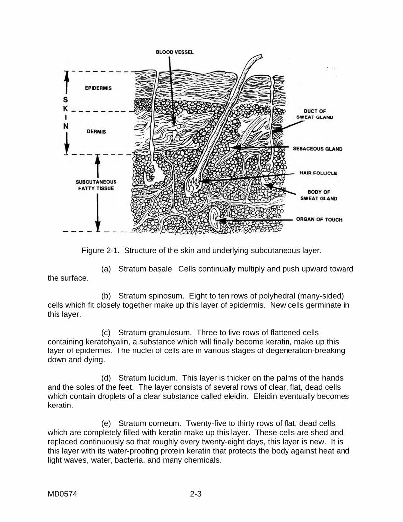
MD0574 2-3
Figure 2-1. Structure of the skin and underlying subcutaneous layer. (a) Stratum basale. Cells continually multiply and push upward toward the surface. (b) Stratum spinosum. Eight to ten rows of polyhedral (many-sided) cells which fit closely together make up this layer of epidermis. New cells germinate in this layer. (c) Stratum granulosum. Three to five rows of flattened cells containing keratohyalin, a substance which will finally become keratin, make up this layer of epidermis. The nuclei of cells are in various stages of degeneration-breaking down and dying. (d) Stratum lucidum. This layer is thicker on the palms of the hands and the soles of the feet. The layer consists of several rows of clear, flat, dead cells which contain droplets of a clear substance called eleidin. Eleidin eventually becomes keratin. (e) Stratum corneum. Twenty-five to thirty rows of flat, dead cells which are completely filled with keratin make up this layer. These cells are shed and replaced continuously so that roughly every twenty-eight days, this layer is new. It is this layer with its water-proofing protein keratin that protects the body against heat and light waves, water, bacteria, and many chemicals.
MD0574 2-4
(2) Dermis. (a) Characteristics. The second layer of skin, the dermis or corium, is sometimes called the true skin. This layer holds the epidermis in place by connective tissue and elastic fiber. The dermis is very thick on the palms of the hands and the soles of the feet but very thin on the eyelids, penis, and scrotum. The dermis contains the following: numerous blood vessels, nerves, lymph vessels, hair follicles, sweat glands, and sensory receptors. (b) Papillary layer. This upper one-fifth of the dermis has small, finger-like projections called dermal papillae. These projections reach into the concavities between ridges in the deep surface of the epidermis. This region or layer consists of loose connective tissue containing fine, elastic fibers. (c) Reticular layer. This layer makes up the rest of the dermis. The reticular layer consists of dense, irregularly arranged connective tissue that has interlacing bundles of collagenous and coarse fibers. Between the fibers are adipose (fat) tissue, hair follicles, nerves, oil glands, and the ducts of sweat glands. The collagenous and elastic fibers together give the skin strength, extensibility, and elasticity. The skin stretches during pregnancy, obesity, or edema. Elasticity allows the skin to contract after such stretching. If the skin has been stretched severely, small tears may occur. Initially, the tears are red; they lose the redness but remain visible as silvery white streaks called striae. NOTE: Extensibility is the ability to stretch. Elasticity is the ability to return to original shape after extension or contraction. (3) Subcutaneous-adipose. This layer is composed of loose connective tissue combined with adipose (fatty) tissue. The subcutaneous layer of skin has several important functions. The primary functions are listed below. (a) Storehouse for water and particularly for fat. Much of the fat in an overweight person is in this layer. (b) Layer of insulation protecting the body from heat loss. (c) Pads the body giving the body form and shape and cushioning and protecting the body from blows. (d) Provides a pathway for nerves and blood vessels.
MD0574 2-5
2-3. LACERATIONS A wound is a break in the continuity of the skin, the break caused by violence or trauma to the tissue. Types of wounds include abrasions, punctures, perforations, and lacerations. A laceration, which is our concern here, is a torn, jagged cut that has gone through the skin tissues and the blood vessels. Such a wound may have been made by a blunt instrument such as the fragments of a shell. A laceration may be very dirty and require cleaning. If only the epidermis layer of skin is involved, there will be no bleeding. If the dermis layer of skin is involved, there will be bleeding. A laceration may require wound closure and suturing. Look at the four major types of lacerations. a. Sheer Laceration. This type of laceration is caused by a sharp object such as a knife blade or the edge of glass. b. Tension Laceration. In a tension laceration, the skin strikes a flat surface, thus ripping because of the tissue stress caused by the impact. There is no bone directly below the region of the skin that is struck. Instead, there is contusion (bruising) of neighboring soft tissues. A tension laceration heals with more scarring than a sheered laceration. c. Compression Laceration. A compression laceration occurs when the tissue is caught between a bone and an external hard surface. The skin bursts, often causing a stellate (star-shaped) patterned wound to occur. There is a marked degree of injury adjacent to the laceration itself. This type of laceration heals the most poorly and with the greatest degree of scarring. d. Combined Laceration. Combined lacerations have the characteristics of both sheer and compression lacerations. An example of such an injury is the resultant injury when you walk into the corner of a desk and your hip bone hits the desk corner. If a laceration occurs, it will probably be a linear wound with wound edges that are crushed; in other words, a combined laceration. 2-4. WOUND HEALING Wound healing is a continuous process which begins at the time of injury. The process of normal healing can be divided into three phases: inflammation, repair, and maturation. a. Inflammation. The process of inflammation begins within minutes following a laceration. An increased blood supply with edema and engorgement of surrounding vessels accounts for the inflammatory appearance. b. Repair. A healthy patient with optimal wound care can expect a semblance of order in the wound to appear on the third day. The cellular and chemical activity during this phase results in "granulation tissue." Although signs of inflammation subside successively during this phase, the wound remains red, raised, and often itchy.
MD0574 2-6
c. Maturation. During this phase of wound healing, there is a progressive decrease in the vascularity of the scar and an increase in the strength of the scar. Maturation of a scar can occur up to two years after the injury took place. Ideal scarring occurs in three stages. (1) Stage I--0 to 4 weeks; the scar is soft, fine, and weak. (2) Stage II--4 to 12 weeks; the scar is red, hard, thick, and strong. (3) Stage III--12 to 40 weeks; the scar is soft, supple, white, and loose. d. Complications. Wound complication refers to anything abnormal in the healing process. The term also refers to the loss of function of a body organ, the function loss caused by the initial wound. Infection is the single most common wound complication. Other complications of wound healing include bleeding, dying tissue, and improper healing. (1) Continued bleeding. Bleeding must be stopped to allow the healing process to proceed. (2) Dying tissue. Tissues at the site of severe injuries may have been severely damaged by being deprived of their blood supply with its oxygen and nutrients. These tissues will die and must be removed or carried away in the capillaries for healing to take place properly. (3) Results of improper healing. Here are a number of possible results of wounds that have not healed properly. (a) Keloid. A keloid is excessive scar tissue growth. Keloids occur primarily in dark-skinned people. Given the proper conditions, anyone can develop a keloid, however. It can be removed surgically for cosmetic reasons. A keloid is the result of improper wound healing. (b) Abscess. An abscess is a localized infection in which there is an accumulation of pus. Pus is a liquid accumulation of phagocytes (also called leukocytes). An infecting microorganism causes the abscess. The particular microorganism involved determines whether the pus is white, yellow, pink, or green. (c) Cellulitis. Cellulitis is an inflammation of the deep, subcutaneous tissues and sometimes muscles, usually caused by infection of a wound or burn. Cellulitis sometimes occurs when an abscess is forming. This condition is serious because the infection can spread rapidly and extensively in the tissue spaces.
MD0574 2-7
(d) Empyema. Empyema is the collection of pus in an already existing cavity, such as in the gallbladder or the lung. (e) Fistula. A fistula is an abnormal passage between two internal organs. A wound that heals improperly could cause such a passage. (4) Blood supply. Since blood supplies the products used in healing, any factor that restricts blood circulation to a wound area interferes with healing. Dead or edematous tissue, restrictive bandages, and damaged arteries can all slow the healing process. e. Physiological Responses to Wounds. Once the skin and tissue have been injured, the process of healing begins. Many factors influence the body's ability to grow new tissue. (1) Age. Very young and very old people heal more slowly than those in other age groups. People in these age groups have less ability to fight infection, and fighting infection is a major part of the healing process. (2) Malnutrition. Malnourishment and obesity are both forms of malnutrition that affect wound healing. (a) A person who is undernourished has less fat and carbohydrate reserve; therefore, body protein (necessary for wound healing) must be used to provide energy needed for basic metabolic functions. This results in an imbalance of nitrogen which in turn depresses fibroblastic synthesis of collagen, the connective tissue for scar formation. A person suffering from Vitamin C deficiency may not be able to produce fibroblast, causing a delay in wound healing. (b) In obese individuals, fatty tissue may keep foreign matter from being seen. Fatty tissue has relatively few blood vessels, causing such tissue to separate easily. Tissue that separates easily heals slowly. (3) Abnormalities in endocrine function. Healing is slow if there are such abnormalities. In a person suffering from chronic vascular changes, the injured tissues of the wound may not get enough blood to heal at a normal rate. Persons having corticosteroid therapy will find that wounds heal more slowly. (4) Hormone production and carbohydrate metabolism. The combined effect of the increased hormone production is to increase the metabolism of carbohydrates. The metabolism of carbohydrates is important in the body's response to trauma. If the body's store of carbohydrates is depleted (severe crush injuries, starvation), the body will begin to use fats and proteins in place of carbohydrates. Eventually, there will not be enough carbohydrates to aid in the healing process.
MD0574 2-8
2-5. MATERIALS NEEDED FOR WOUND CLOSURE a. Instruments. Only a few basic instruments are required for the repair of most wounds. Gather the following equipment: (1) Needle holder. (2) Forceps. (3) Number 15 scalpel. (4) Scissors. b. Needles. (1) Straight needle/curved needle. There are two types of needles: the straight needle and the curved needle. The straight needle is used with hands, and the curved needle is used with needle holders. (2) Tapered needle/cutting needle. A tapered needle has a circular cross-sectional configuration and leaves a small hole. A cutting needle has a triangular cross-sectional configuration and is better able to pass through tough skin. (3) Grades of needles. Two grades of needles are the cuticular needle and the plastic needle. The cuticular needle is designated by the letter C and FS for skin. The plastic needle is designated by the letter P for plastic and PS for plastic surgery. The plastic needle is honed more sharply than the cuticular needle. Also, the plastic needle is more expensive than a cuticular needle. (4) Size of needles. The needle size is indicated by the number that follows the needle letter. Usually, the larger the number, the smaller the needle. Small needles are used for fine repair such as treating facial lacerations. Larger needles are used for taking bigger bites of tissue such as scalp lacerations. c. Suture Materials. One of the bases upon which surgery is founded is the suture of wounds. Many kinds of present day sutures have been known for thousands of years, but only since Lister's discoveries have the use of sutures been safe. Suture is a medical term for a thread-like material that is used to stitch or approximate (bring together) tissue edges until healing takes place. Other terms to know are gauge and tensile strength. Gauge refers to the diameter of the suture or the distance around the suture. Tensile strength refers to the amount of weight or pull that may be exerted on a suture before the suture will break.
MD0574 2-9
(1) Suture sizes. Suture sizes range from a fine number 9-0 to heavy number 5. Suture sizing is controlled by USP standards. Small sutures (number 0 through number 9-0) are in greater demand because the small diameter provides better handling qualities and smaller knots. Larger sutures (number 1 through number 5) are used as a retention stitch, that being a stitch used to reinforce a primary suture line. The kind and size of suture used depends on the patient, the type of tissue, the surgeon's preference, and the available suture material. (2) Nonabsorbable sutures. (a) Silk. Silk has a number of advantages as a suture material. Silk lies flat when it is tied. It is easy to handle and has the added advantage of forming a secure knot when tied. But there are also disadvantages. Silk is not the ideal suture material for routine emergency department use. Silk causes a host reaction since silk is a foreign protein. This means that there is a high risk of infection if silk is used as suture material. Therefore, use silk in uncontaminated wounds that are in well-perfused areas of the body; for example, wounds on the face. (b) Cotton. Briefly, the advantages of using cotton are the same as the advantages for using silk as a suture material. Similarly, the disadvantages of using cotton are the same as the disadvantages of using silk. (c) Nylon and polypropylene (synthetic materials). Among the advantages of using these synthetic materials as sutures are that these synthetic materials pose a lower risk of infection than silk or cotton. Also, these materials are the suture of choice for skin closure of most lacerations in the emergency room. Disadvantages include the following: 1 Synthetic materials do not lie flat during the suturing process. 2 Synthetic materials are more difficult to use. 3 There is less security of knots. (d) Dacron. The infection potential of Dacron is greater than that of nylon or polypropylene, but less than that of silk or cotton. Dacron is easier to work with and holds knots better than nylon or polypropylene. (3) Metal sutures. Staples are metal sutures. For many years, staples have been commonly used for surgical wound closure. Staples are used in emergency rooms for some types of lacerations. The advantages of metal sutures are that they are easier and quicker than other types of suture repair. The cost is lower, and the wound healing results are the same as for other types of suturing. The disadvantages are that an inexperienced person has a difficult time using these sutures. Additionally, metal sutures can be highly irritating to the patient.
MD0574 2-10
(4) Absorbable sutures. Absorbable suture material is digested and absorbed by body cells and fluids during and after healing of tissue. There are two types of regular absorbable suture--plain cat gut and chromic cat gut. Both of these indicate a surgical gut material that has not been treated to lengthen its absorption time in the tissue. (a) Plain catgut. Plain catgut holds tensile strength for about seven days. Sheep's intestine is the source of plain suture. This suture is used in tissue where rapid healing is expected to occur such as subcutaneous tissue and for tying superficial blood vessels. Plain catgut suture is pale yellow in color. A disadvantage of this type of suture is that it increases the formation of pus and has high tissue reactivity. (b) Chromic catgut. Chromic suture has been treated with chromic oxide so that it will resist digestion or absorption for longer periods of time. Chromic suture has the same source as plain suture. Chromic suture is used in tissue where rapid healing is not expected to take place, such as muscle fascia, peritoneum, and body organs. The advantage of this type of suture is that it retains its tensile strength for about two to three weeks. As with plain catgut, chromic suture increases the formation of pus and has high tissue reactivity. (c) Synthetic absorbable suture. Dexon® and Vyeril® are examples of synthetic absorbable suture. Advantages include that it retains tensile strength for sixty days or more. Also, there is low tissue reactivity and lower pus formation than with the use of plain or chromic cat gut. A disadvantage is that this type of suture material does not glide through tissue easily. Snags tend to occur, making knot tying more tedious. (5) Skin tapes. Steri-Strips, clearon, and skin-strips are examples of skin tapes. Skin tapes are often used in place of sutures to repair surface lacerations. Advantages of skin tapes are the low incidence of infection and no suture marks. Also, the patient need not return to have sutures removed. Disadvantages are that skin tapes are not practical in body areas that may become wet or that have motion. Young children have a tendency to pull off skin tapes. Also, at times wound edges invert after a skin tape has been applied. 2-6. BASIC LACERATION REPAIR a. Categories of Wound Closure. Closure of wounds is divided into three major categories: closure by primary intent, closure by secondary intent, and closure by tertiary intent.
MD0574 2-11
(1) Closure by primary intent. A wound which is repaired without delay after the injury is the definition of wound closure by primary intent. Such closure prevents the formation of granulation tissue and yields the fastest healing with the best cosmetic result. Closure by primary intent is the treatment of choice for a wound that is not infected or grossly contaminated. Closing the wound soon after the injury is important. The longer the time between injury and wound closure, the more bacteria can multiply. Most lacerations should be closed within eight hours from the time of injury. If the patient is debilitated, has poor circulation, has laceration due to crushing injuries, or is grossly contaminated, wound closure should be performed within four hours. (2) Closure by secondary intent. Here the wound is allowed to granulate on its own without surgical closure. The tissue is cleaned and dressed as usual, and the wound is covered with a sterile dressing. This is the procedure of choice for closing certain defects such as finger amputation and partial-thickness tissue loss. In the case of finger amputations, this type of closure usually gives better cosmetic and functional results. (3) Closure by tertiary intent. This is delayed primary closure. The wound is initially cleaned and dressed as in secondary intent. The patient returns in three to four days for definitive closure. This is the procedure of choice for contaminated lacerations that would leave unacceptable scars if not closed. Examples of lacerations are mammalian bites, contaminated crush-lacerations, and cases when the patient delayed too long for treatment to close primarily. b. Wound Debridement and Excision. (1) Debridement of adherent foreign material. Follow this procedure. (a) Irrigate the wound. Carefully explore the wound for any foreign material. NOTE: Foreign material serves as a source of infection and may "tattoo" the skin if the material is near the skin surface. (b) Remove the foreign material. The simplest method is to abrade the soiled region repeated with a 4 x 4 inch piece of gauze moistened with saline. An alternate method is to excise the soiled tissue using forceps and an iris scissors or scalpel. (2) Trimming the wound edge. Trim minute irregularities from the wound edges. This takes only a little time and often greatly improves the final appearance after the wound has healed. Often, only one millimeter of tissue needs to be trimmed off. Using sharp iris scissors, carefully trim off minor irregularities from the edge (figure 2-2A). A scalpel can also be used (figure 2-2B and C).
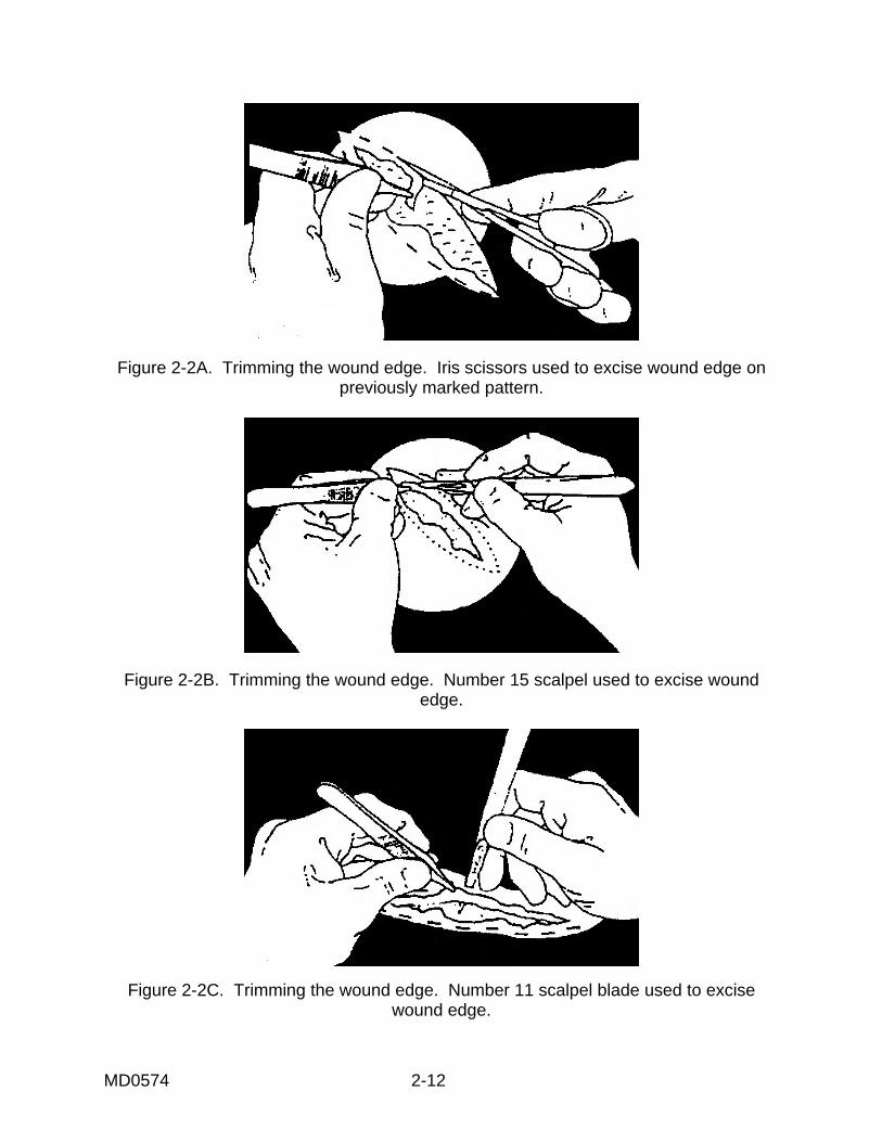
MD0574 2-12
Figure 2-2A. Trimming the wound edge. Iris scissors used to excise wound edge on previously marked pattern.
Figure 2-2B. Trimming the wound edge. Number 15 scalpel used to excise wound edge.
Figure 2-2C. Trimming the wound edge. Number 11 scalpel blade used to excise wound edge.
MD0574 2-13
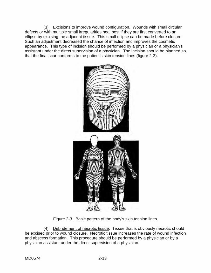
(3) Excisions to improve wound configuration. Wounds with small circular defects or with multiple small irregularities heal best if they are first converted to an ellipse by excising the adjacent tissue. This small ellipse can be made before closure. Such an adjustment decreased the chance of infection and improves the cosmetic appearance. This type of incision should be performed by a physician or a physician's assistant under the direct supervision of a physician. The incision should be planned so that the final scar conforms to the patient's skin tension lines (figure 2-3).
Figure 2-3. Basic pattern of the body's skin tension lines. (4) Debridement of necrotic tissue. Tissue that is obviously necrotic should be excised prior to wound closure. Necrotic tissue increases the rate of wound infection and abscess formation. This procedure should be performed by a physician or by a physician assistant under the direct supervision of a physician.
MD0574 2-14
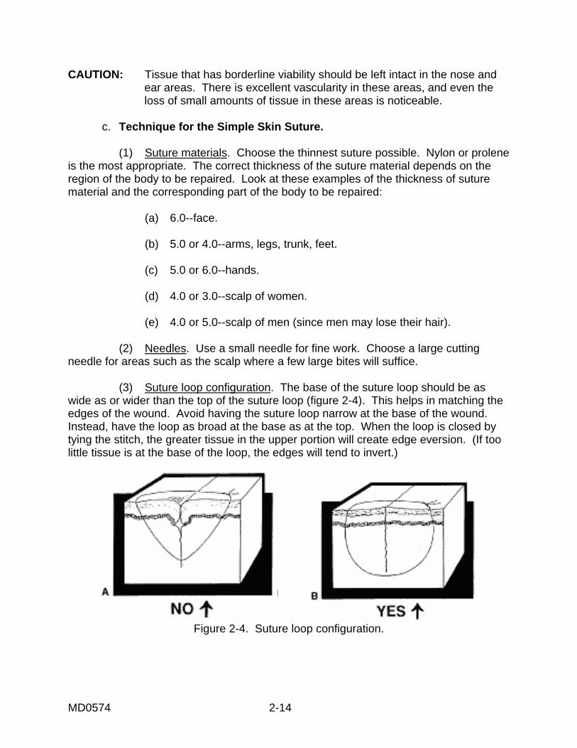
CAUTION: Tissue that has borderline viability should be left intact in the nose and ear areas. There is excellent vascularity in these areas, and even the loss of small amounts of tissue in these areas is noticeable. c. Technique for the Simple Skin Suture. (1) Suture materials. Choose the thinnest suture possible. Nylon or prolene is the most appropriate. The correct thickness of the suture material depends on the region of the body to be repaired. Look at these examples of the thickness of suture material and the corresponding part of the body to be repaired: (a) 6.0--face. (b) 5.0 or 4.0--arms, legs, trunk, feet. (c) 5.0 or 6.0--hands. (d) 4.0 or 3.0--scalp of women. (e) 4.0 or 5.0--scalp of men (since men may lose their hair). (2) Needles. Use a small needle for fine work. Choose a large cutting needle for areas such as the scalp where a few large bites will suffice. (3) Suture loop configuration. The base of the suture loop should be as wide as or wider than the top of the suture loop (figure 2-4). This helps in matching the edges of the wound. Avoid having the suture loop narrow at the base of the wound. Instead, have the loop as broad at the base as at the top. When the loop is closed by tying the stitch, the greater tissue in the upper portion will create edge eversion. (If too little tissue is at the base of the loop, the edges will tend to invert.)
Figure 2-4. Suture loop configuration.
MD0574 2-15
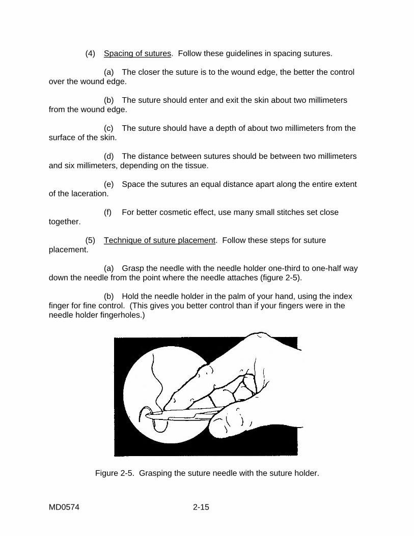
(4) Spacing of sutures. Follow these guidelines in spacing sutures. (a) The closer the suture is to the wound edge, the better the control over the wound edge. (b) The suture should enter and exit the skin about two millimeters from the wound edge. (c) The suture should have a depth of about two millimeters from the surface of the skin. (d) The distance between sutures should be between two millimeters and six millimeters, depending on the tissue. (e) Space the sutures an equal distance apart along the entire extent of the laceration. (f) For better cosmetic effect, use many small stitches set close together. (5) Technique of suture placement. Follow these steps for suture placement. (a) Grasp the needle with the needle holder one-third to one-half way down the needle from the point where the needle attaches (figure 2-5). (b) Hold the needle holder in the palm of your hand, using the index finger for fine control. (This gives you better control than if your fingers were in the needle holder fingerholes.)
Figure 2-5. Grasping the suture needle with the suture holder.
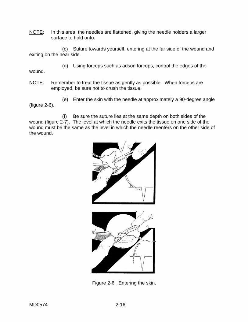
MD0574 2-16
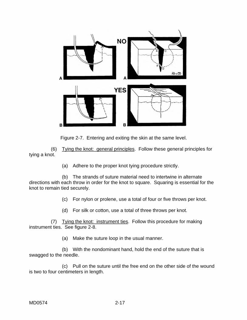
NOTE: In this area, the needles are flattened, giving the needle holders a larger surface to hold onto. (c) Suture towards yourself, entering at the far side of the wound and exiting on the near side. (d) Using forceps such as adson forceps, control the edges of the wound. NOTE: Remember to treat the tissue as gently as possible. When forceps are employed, be sure not to crush the tissue. (e) Enter the skin with the needle at approximately a 90-degree angle (figure 2-6). (f) Be sure the suture lies at the same depth on both sides of the wound (figure 2-7). The level at which the needle exits the tissue on one side of the wound must be the same as the level in which the needle reenters on the other side of the wound.
Figure 2-6. Entering the skin.
MD0574 2-17
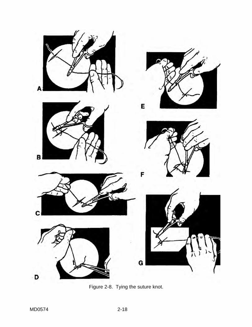
Figure 2-7. Entering and exiting the skin at the same level. (6) Tying the knot: general principles. Follow these general principles for tying a knot. (a) Adhere to the proper knot tying procedure strictly. (b) The strands of suture material need to intertwine in alternate directions with each throw in order for the knot to square. Squaring is essential for the knot to remain tied securely. (c) For nylon or prolene, use a total of four or five throws per knot. (d) For silk or cotton, use a total of three throws per knot. (7) Tying the knot: instrument ties. Follow this procedure for making instrument ties. See figure 2-8. (a) Make the suture loop in the usual manner. (b) With the nondominant hand, hold the end of the suture that is swagged to the needle. (c) Pull on the suture until the free end on the other side of the wound is two to four centimeters in length.
MD0574 2-18
Figure 2-8. Tying the suture knot.
MD0574 2-19
(d) With the dominant hand, hold the needle holder. (e) Loop the swaged end of the suture twice around the needle holder for nonfilament suture material. A single loop suffices for most braided sutures such as silk or Dexon®. (f) Grab the free end of the suture with the blades of the needle holder. (g) Cross the hands so that the hand holding the swaged end is on the far side, and the hand holding the needle holder and the free end are on the near side of the wound. (h) As you clinch the first throw of the knot, pull upward on the suture ends. (i) Adjust the tension of the first throw, so that the wound edges come together snugly, but not tightly. (j) For the second throw of the knot, the needle end is on the far side of the wound, and the free end is on the near side of the wound. (k) Hold the needle end of the suture in the non-dominant hand and lay the needle holder on top. (l) Loop the suture only once around the needle holder. (m) Grasp the free ends with the blades of the holder. (n) Cross the hands so that the sutures intertwine smoothly. (o) Cinch down the throw. CAUTION: Take care not to cinch down too tightly on the second throw, because the tightness will be transmitted to the wound. (p) Pull the knot to the side so that the knot will not lie directly over the wound. (q) Repeat the pattern of looping the suture around the holder on alternate sides of the wound. Do this until the desired number of throws have been completed. (r) Cut the ends of the suture material approximately three to five centimeters long.
MD0574 2-20
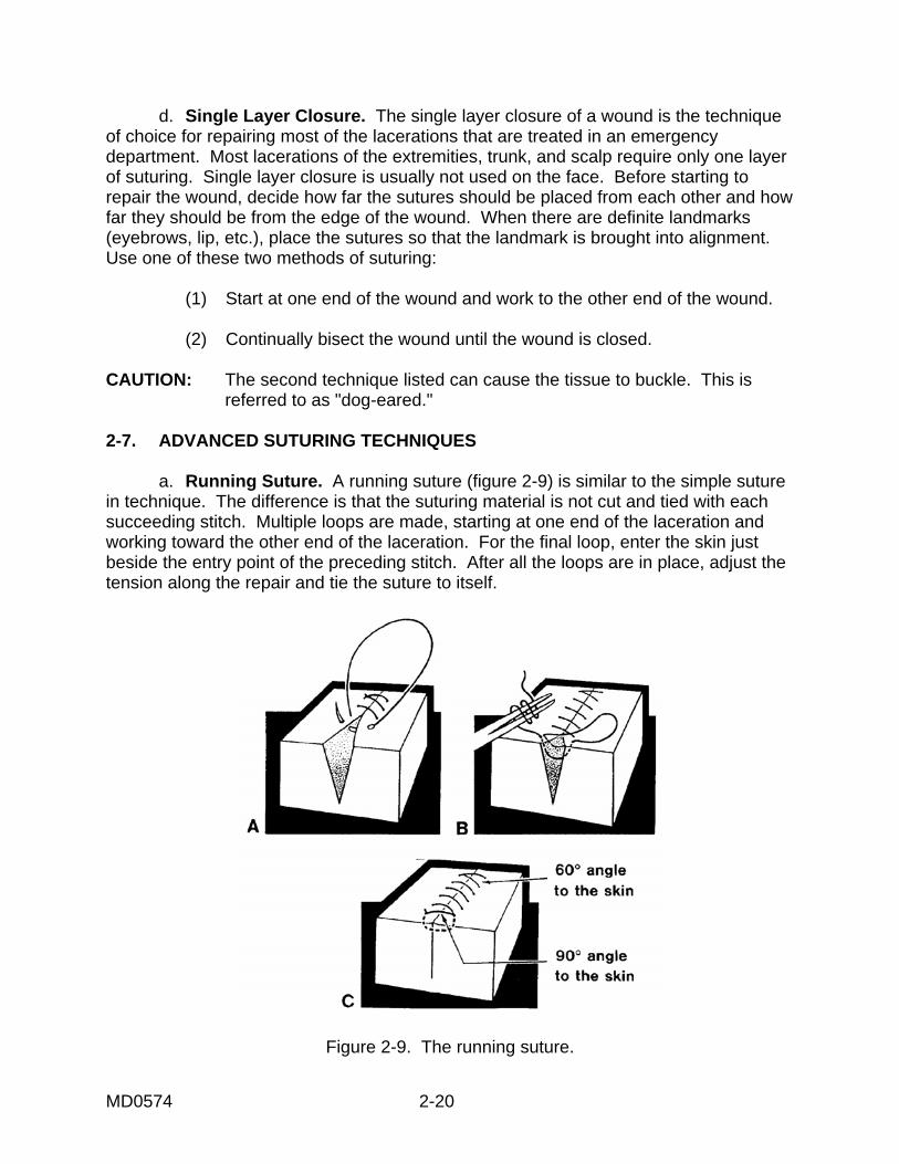
d. Single Layer Closure. The single layer closure of a wound is the technique of choice for repairing most of the lacerations that are treated in an emergency department. Most lacerations of the extremities, trunk, and scalp require only one layer of suturing. Single layer closure is usually not used on the face. Before starting to repair the wound, decide how far the sutures should be placed from each other and how far they should be from the edge of the wound. When there are definite landmarks (eyebrows, lip, etc.), place the sutures so that the landmark is brought into alignment. Use one of these two methods of suturing: (1) Start at one end of the wound and work to the other end of the wound. (2) Continually bisect the wound until the wound is closed. CAUTION: The second technique listed can cause the tissue to buckle. This is referred to as "dog-eared." 2-7. ADVANCED SUTURING TECHNIQUES a. Running Suture. A running suture (figure 2-9) is similar to the simple suture in technique. The difference is that the suturing material is not cut and tied with each succeeding stitch. Multiple loops are made, starting at one end of the laceration and working toward the other end of the laceration. For the final loop, enter the skin just beside the entry point of the preceding stitch. After all the loops are in place, adjust the tension along the repair and tie the suture to itself.
Figure 2-9. The running suture.
MD0574 2-21
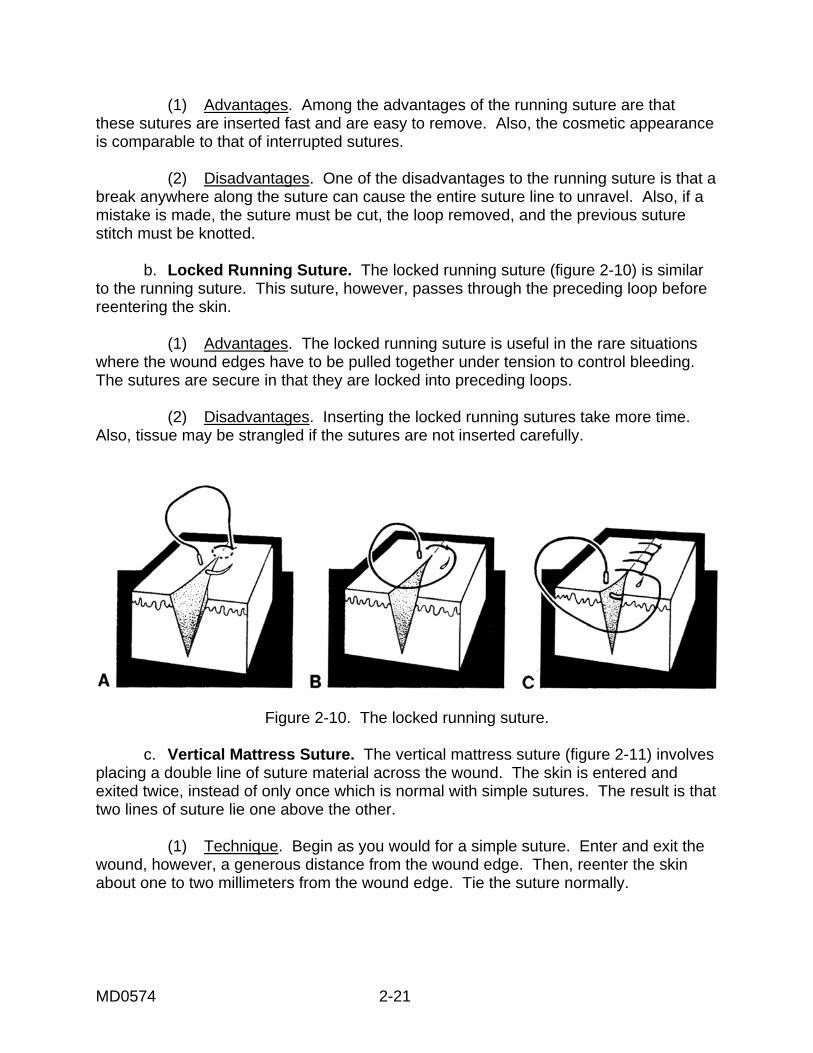
(1) Advantages. Among the advantages of the running suture are that these sutures are inserted fast and are easy to remove. Also, the cosmetic appearance is comparable to that of interrupted sutures. (2) Disadvantages. One of the disadvantages to the running suture is that a break anywhere along the suture can cause the entire suture line to unravel. Also, if a mistake is made, the suture must be cut, the loop removed, and the previous suture stitch must be knotted. b. Locked Running Suture. The locked running suture (figure 2-10) is similar to the running suture. This suture, however, passes through the preceding loop before reentering the skin. (1) Advantages. The locked running suture is useful in the rare situations where the wound edges have to be pulled together under tension to control bleeding. The sutures are secure in that they are locked into preceding loops. (2) Disadvantages. Inserting the locked running sutures take more time. Also, tissue may be strangled if the sutures are not inserted carefully.
Figure 2-10. The locked running suture. c. Vertical Mattress Suture. The vertical mattress suture (figure 2-11) involves placing a double line of suture material across the wound. The skin is entered and exited twice, instead of only once which is normal with simple sutures. The result is that two lines of suture lie one above the other. (1) Technique. Begin as you would for a simple suture. Enter and exit the wound, however, a generous distance from the wound edge. Then, reenter the skin about one to two millimeters from the wound edge. Tie the suture normally.
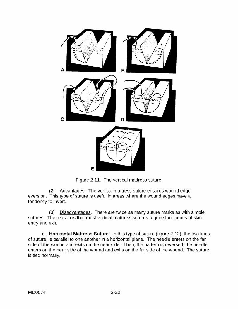
MD0574 2-22
Figure 2-11. The vertical mattress suture. (2) Advantages. The vertical mattress suture ensures wound edge eversion. This type of suture is useful in areas where the wound edges have a tendency to invert. (3) Disadvantages. There are twice as many suture marks as with simple sutures. The reason is that most vertical mattress sutures require four points of skin entry and exit. d. Horizontal Mattress Suture. In this type of suture (figure 2-12), the two lines of suture lie parallel to one another in a horizontal plane. The needle enters on the far side of the wound and exits on the near side. Then, the pattern is reversed; the needle enters on the near side of the wound and exits on the far side of the wound. The suture is tied normally.
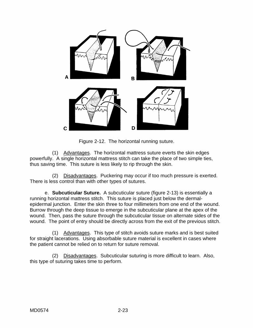
MD0574 2-23
Figure 2-12. The horizontal running suture. (1) Advantages. The horizontal mattress suture everts the skin edges powerfully. A single horizontal mattress stitch can take the place of two simple ties, thus saving time. This suture is less likely to rip through the skin. (2) Disadvantages. Puckering may occur if too much pressure is exerted. There is less control than with other types of sutures. e. Subcuticular Suture. A subcuticular suture (figure 2-13) is essentially a running horizontal mattress stitch. This suture is placed just below the dermal-epidermal junction. Enter the skin three to four millimeters from one end of the wound. Burrow through the deep tissue to emerge in the subcuticular plane at the apex of the wound. Then, pass the suture through the subcuticular tissue on alternate sides of the wound. The point of entry should be directly across from the exit of the previous stitch. (1) Advantages. This type of stitch avoids suture marks and is best suited for straight lacerations. Using absorbable suture material is excellent in cases where the patient cannot be relied on to return for suture removal. (2) Disadvantages. Subcuticular suturing is more difficult to learn. Also, this type of suturing takes time to perform.
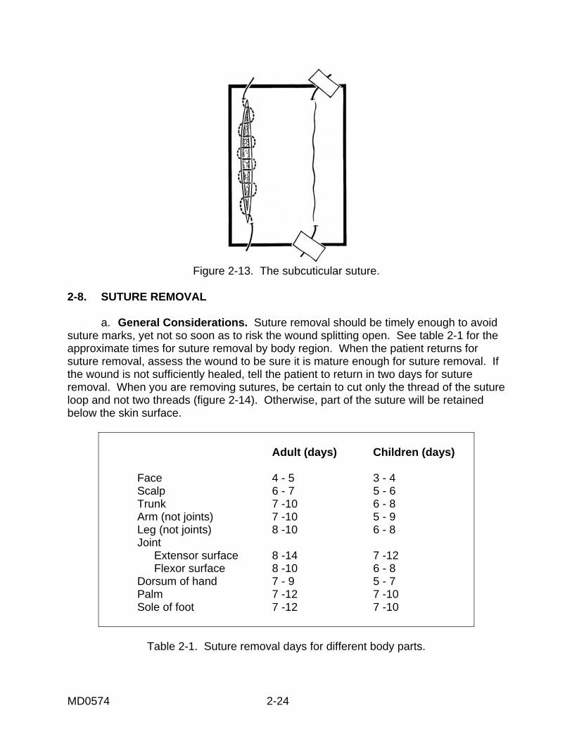
MD0574 2-24
Figure 2-13. The subcuticular suture.
2-8. SUTURE REMOVAL a. General Considerations. Suture removal should be timely enough to avoid suture marks, yet not so soon as to risk the wound splitting open. See table 2-1 for the approximate times for suture removal by body region. When the patient returns for suture removal, assess the wound to be sure it is mature enough for suture removal. If the wound is not sufficiently healed, tell the patient to return in two days for suture removal. When you are removing sutures, be certain to cut only the thread of the suture loop and not two threads (figure 2-14). Otherwise, part of the suture will be retained below the skin surface.
Adult (days) Children (days) Face 4 - 5 3 - 4 Scalp 6 - 7 5 - 6 Trunk 7 -10 6 - 8 Arm (not joints) 7 -10 5 - 9 Leg (not joints) 8 -10 6 - 8 Joint Extensor surface 8 -14 7 -12 Flexor surface 8 -10 6 - 8 Dorsum of hand 7 - 9 5 - 7 Palm 7 -12 7 -10 Sole of foot 7 -12 7 -10
Table 2-1. Suture removal days for different body parts.
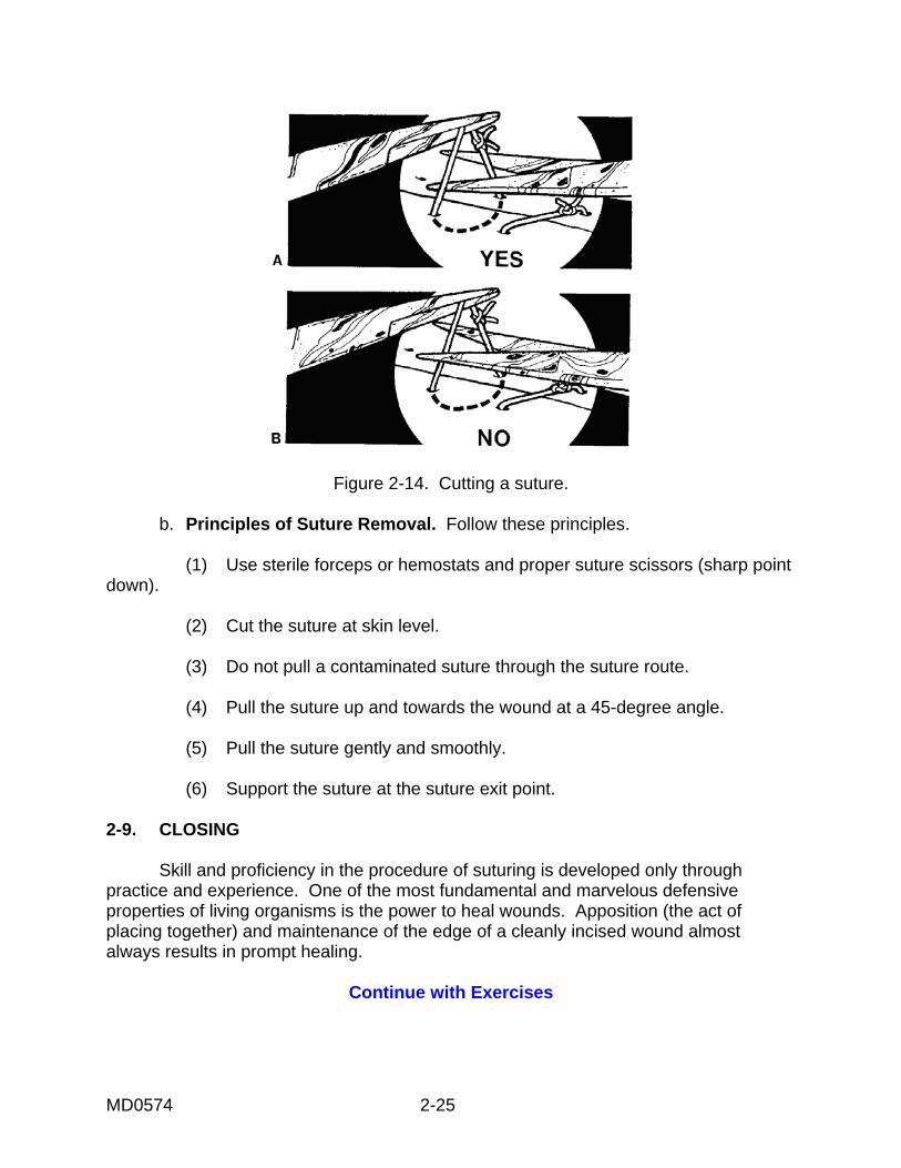
MD0574 2-25
Figure 2-14. Cutting a suture. b. Principles of Suture Removal. Follow these principles. (1) Use sterile forceps or hemostats and proper suture scissors (sharp point down). (2) Cut the suture at skin level. (3) Do not pull a contaminated suture through the suture route. (4) Pull the suture up and towards the wound at a 45-degree angle. (5) Pull the suture gently and smoothly. (6) Support the suture at the suture exit point. 2-9. CLOSING Skill and proficiency in the procedure of suturing is developed only through practice and experience. One of the most fundamental and marvelous defensive properties of living organisms is the power to heal wounds. Apposition (the act of placing together) and maintenance of the edge of a cleanly incised wound almost always results in prompt healing.
Continue with Exercises
MD0574 2-26
EXERCISES, LESSON 2 INSTRUCTIONS. The following exercises are to be answered by writing the answer in the space provided or by matching terms as directed. After you have completed all the exercises, turn to "Solutions to Exercises, Lesson 2" at the end of the exercises and check your answers. 1. How many square feet of skin cover the body of the average male? __________ 2. About _________ of all blood that circulates through the body goes to the skin. 3. List the three layers of skin. a. ___________________________________________. b. ___________________________________________. c. ___________________________________________. 4. A change in the normal appearance of an individual's skin could indicate an abnormality or _________________________________________________. 5. How many layers of epidermis are on the palms of the hands and the soles of the feet? _________________ 6. List four body parts contained in the second layer of skin. a. ___________________________________________. b. ___________________________________________. c. ___________________________________________. d. ___________________________________________. 7. Which layer of skin gives the skin its elasticity? _____________________.

MD0574 2-27
8. List three functions of the subcutaneous layer of skin. a. ___________________________________________ b. ___________________________________________ c. ___________________________________________ 9. A wound is _______________________________________________________ ________________________________________________________________ 10. List four types of wounds. a. ___________________________________________. b. ___________________________________________. c. ___________________________________________. d. ___________________________________________. 11. A ________________________ is a torn, jagged cut which has gone through the skin tissue and the blood vessels. 12. What is the body's first response to cell damage (in other words, a wound)? ________________________________________________________________ ________________________________________________________________ 13. What causes a wound abscess? ______________________________________ ________________________________________________________________
MD0574 2-28
14. List two possible complications of wound healing. a. ___________________________________________. b. ___________________________________________. 15. What is a keloid? __________________________________________________ 16. Why does dead tissue around a wound slow the healing process? ________________________________________________________________ ________________________________________________________________ 17. List four factors which influence the body's ability to grow new tissue; in other words, to heal a wound. a. ___________________________________________. b. ___________________________________________. c. ___________________________________________. d. ___________________________________________. 18. List the three stages of wound healing. a. ___________________________________________. b. ___________________________________________. c. ___________________________________________.
MD0574 2-29
19. A variety of needles is used in wound closure. Answer the following questions about such needles. a. What are two types of needles? (1) ___________________________________________. (2) ___________________________________________. b. List the two grades of needles and their use. (1) ___________________________________________. (2) ___________________________________________. c. Uses of different sized needles. (1) What are smaller needles used for? ________________________________________________________ . (2) What are larger needles used for? ________________________________________________________ 20. List the three major types of suture materials. a. ___________________________________________. b. ___________________________________________. c. ___________________________________________.
MD0574 2-30
21. Answer the following questions. a. On what part of the body should silk sutures be used? __________________ ______________________________________________________________ b. List one advantage and one disadvantage of using metal sutures. (1) Advantage = ______________________________________________ (2) Disadvantage = ___________________________________________. c. Synthetic absorbable sutures have many advantages; list one. List one disadvantage of these sutures. (1) Advantage = ______________________________________________ (2) Disadvantage = ____________________________________________ d. List one advantage and one disadvantage of using skin tapes. (1) Advantage = _______________________________________________ (2) Disadvantage = ____________________________________________ 22. Complete the statements about categories of wound closure. a. Wound closure by primary intent is _________________________________ _____________________________________________________________ b. Allowing the wound to granulate on its own without surgical repair is the definition of ___________________________________________________. c. Cleaning and dressing the wound with the patient returning for wound closure in three or four days is the definition of _______________________________ 23. List the two steps in debridement and excision of a wound. a. ___________________________________________. b. ___________________________________________. 24. Why is it necessary to trim the irregularities from the edge of a wound ________________________________________________________________ ________________________________________________________________
MD0574 2-31
25. List two reasons why necrotic tissue (dead tissue) around a wound should be excised. a. ___________________________________________. b. ___________________________________________. 26. A general principle of suturing is that the base of the suture loop configuration should be _________________________ the top of the suture loop.(how wide) 27. List four general principles of suture removal. a. ___________________________________________. b. ___________________________________________. c. ___________________________________________. d. ___________________________________________. 28. Match the definitions with the correct term by writing the letter of the definition next to the correct term. TERM DEFINITION ____ a. Sheer laceration W. Laceration caused by skin striking a flat surface. ____ b. Combined laceration. X. Laceration caused by skin being caught between the bone and an external surface. ____ c. Tension laceration. Y. Characteristics of sheer and compression lacerations. ____ d. Compression laceration. Z. A laceration made by a sharp object such as a knife.
MD0574 2-32
29. Match the definitions with the correct term by writing the letter of the definition next to the correct term. TERM DEFINITION ____ a. Cellulitis. W. Pus collecting in an already existing body cavity. ____ b. Empyema. X. Localized infection in which there is an accumulation of pus. ____ c. Abscess. Y. Abnormal passage between two internal organs. ____ d. Fistula. Z. Inflammation of the deep subcutaneous tissue. 30. In the process of maturation of wound healing, there are three stages. Write the letter of the appropriate description next to the appropriate stage. STAGE CHARACTERISTICS ____ a. Stage I. X. Scar is red, hard, thick, and strong. ____ b. Stage II. Y. Scar is soft, supple, white, and loose. ____ c. Stage III. Z. Scar is soft, fine, and weak. 31. Match the suturing technique in the first column with the advantage of the technique in the second column. Write the appropriate letter from the second column in the space provided next to the letter in the first column. COLUMN I COLUMN II SUTURING TECHNIQUE ADVANTAGE OF TECHNIQUE ____ a. Vertical mattress suture V. Fast to insert; easy to remove. ____ b. Subcuticular suture W. Useful in situations where wound edges have to be pulled together under tension to control bleeding. ____ c. Horizontal mattress suture X. Useful in areas where the wound edges tend to invert. ____ d. Running suture Y. One suture can take the place of two ties. ____ e. Locked running suture Z. Avoids suture marks; best suited for straight lacerations.
Check Your Answers on Next Page
MD0574 2-33
SOLUTIONS TO EXERCISES, LESSON 2 1. 16.2 square feet. (para 2-2a) 2. One-third. (para 2-2a) 3. Epidermis layer. Dermis layer. Subcutaneous layer. (para 2-2c) 4. A disease of body function. (para 2-2b) 5. 5 layers of epidermis. (para 2-2c(1)) 6. You are correct if you listed any four of the following: Blood vessels. Nerves. Lymph vessels. Hair follicles. Sweat glands. Sensory receptors. (para 2-2c(2)) 7. The dermis layer (the second layer). (para 2-2c(2)) 8. You are correct if you listed any three of the following: Storehouse for water. Storehouse for fat. Insulation function, protecting the body from heat loss. Pads the body from blows. Pathway for nerves. Pathway for blood vessels. (para 2-2c(3)) 9. A wound is a break in the continuity of the skin. (para 2-3) 10. Abrasions. Punctures. Perforations. Lacerations. (para 2-3) 11. Laceration. (para 2-3) 12. The body's first response is inflammation. Blood rushes to the site and the area feels warm to the touch. (para 2-4a) 13. An abscess is caused by an infecting organism. (para 2-4d(3)) 14. You are correct if you listed any two of the following: Infection. Continued bleeding. Dying tissue. Improper healing. para 2-4d(1) through (3))
MD0574 2-34
15. A keloid is excessive scar tissue. (para 2-4d(3)) 16. Dead tissue restricts blood circulation to a wound area, thus interfering with healing. (para 2-4d(4)) 17. You are correct if you listed any four of the following: Age. Malnutrition. Obesity. Abnormalities in endocrine function. Hormone production. Carbohydrate metabolism. (para 2-4e(1) through (4)) 18. Inflammation. Repair. Maturation. (para 2-4) 19. a. (1) Straight needles. (2) Curved needles. (para 2-5b(1)) b. (1) Cuticular needle - for use on the skin. (2) Plastic needle--a sharper needle, used for plastic surgery. (para 2-5b(3)) c. (1) Fine repair such as treating facial lacerations. (2) Taking bigger bites of tissue such as scalp lacerations. (para 2-5b(4) 20. Nonabsorbable sutures. Metal sutures. Absorbable sutures. (para 2-5c(2) through (5)) 21. a. Use silk sutures on uncontaminated wounds such as wounds of the face. (para 2-5c(2)) b. You are correct if you chose any one of the following advantages of metal sutures and any one of the following disadvantages of metal sutures. Advantages of metal sutures: Quick to use. Easy to use, if experienced. Wound healing results the same as for other suture types. Disadvantages of metal sutures: Hard to use if inexperienced. . Sutures can be irritating to the patient. (para 2-5c(3)) c. You are correct if you chose any one of the following advantages of synthetic absorbable sutures and any one of the following disadvantages of synthetic absorbable sutures. Advantages of synthetic, absorbable sutures Retains tensile strength for 60 days or more. Low tissue reaction to this suture; therefore, less pus infection.
MD0574 2-35
Disadvantages of synthetic, absorbable sutures Suture does not glide through skin easily. Difficult to tie knot. (para 2-5c(4)(c)) d. You are correct if you chose any one of the following advantages of skin tapes and any one of the following disadvantages of skin tapes. Advantages of skin tapes Low incidence of infection. No suture marks. Patient does not have to come back to have sutures removed. Disadvantage of skin tapes Not practical in body areas that get wet or have motion. Not practical for young children who tend to pull off tapes. Tape edges sometimes curl up after the skin tapes have been applied. (para 2-5c(5)) 22. a. Wound repair without delay after the injury has occurred. (para 2-6a(1)) b. Wound closure by secondary intent. (para 2-6a(2)) c. Wound closure by tertiary intent. (para 2-6a(3)) 23. Irrigate the wound. Remove the foreign material. (para 2-6b(1)) 24. Trim irregularities from the edge of a wound to improve the final appearance of the healed wound. (para 2-6b(2)) 25. a. To prevent infection. b. To prevent abscess. (para 2-6b(4)) 26. As wide or wider than. (para 2-6c(3)) 27. You are correct if you listed any four of the following: Use sterile forceps or hemostats. Use proper suture scissors. Cut the suture at skin level. Do not pull a contaminated suture through the suture route. Pull the suture up and towards the wound at a 45-degree angle. Pull the suture gently and smoothly. Support the suture at the suture exit point. (para 2-8b) 28. a Z b Y c W d X (paras 2-3a through d) 29. a Z b W c X d Y (para 2-4d(3))

MD0574 2-36
30. a Z b X c Y (para 2-4c) 31. a X b Z c Y d V e W (paras 2-7a through e)
End of Lesson 2
MD0574 3-1
LESSON ASSIGNMENT LESSON 3 Emergency Surgical Procedures. LESSON ASSIGNMENT Paragraphs 3-1 through 3-6. LESSON OBJECTIVES After completing this lesson, you should be able to: 3-1. Identify the steps in the procedure of a venous cut-down. 3-2. Identify the steps in the procedure of needle decompression. 3-3. Identify the steps in the procedure of insertion of a chest tube. 3-4. Identify the steps in the procedure of a surgical cricothyrotomy. SUGGESTION After completing the assignment, complete the exercises of this lesson. These exercises will help you to achieve the lesson objectives.
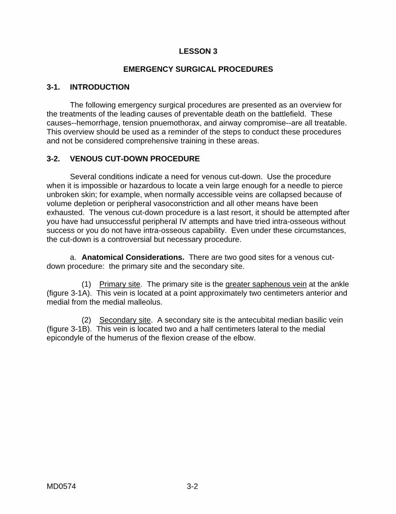
MD0574 3-2
LESSON 3
EMERGENCY SURGICAL PROCEDURES 3-1. INTRODUCTION The following emergency surgical procedures are presented as an overview for the treatments of the leading causes of preventable death on the battlefield. These causes--hemorrhage, tension pnuemothorax, and airway compromise--are all treatable. This overview should be used as a reminder of the steps to conduct these procedures and not be considered comprehensive training in these areas. 3-2. VENOUS CUT-DOWN PROCEDURE Several conditions indicate a need for venous cut-down. Use the procedure when it is impossible or hazardous to locate a vein large enough for a needle to pierce unbroken skin; for example, when normally accessible veins are collapsed because of volume depletion or peripheral vasoconstriction and all other means have been exhausted. The venous cut-down procedure is a last resort, it should be attempted after you have had unsuccessful peripheral IV attempts and have tried intra-osseous without success or you do not have intra-osseous capability. Even under these circumstances, the cut-down is a controversial but necessary procedure. a. Anatomical Considerations. There are two good sites for a venous cut-down procedure: the primary site and the secondary site. (1) Primary site. The primary site is the greater saphenous vein at the ankle (figure 3-1A). This vein is located at a point approximately two centimeters anterior and medial from the medial malleolus. (2) Secondary site. A secondary site is the antecubital median basilic vein (figure 3-1B). This vein is located two and a half centimeters lateral to the medial epicondyle of the humerus of the flexion crease of the elbow.
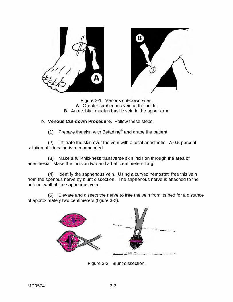
MD0574 3-3
Figure 3-1. Venous cut-down sites. A. Greater saphenous vein at the ankle.
B. Antecubital median basilic vein in the upper arm.
b. Venous Cut-down Procedure. Follow these steps. (1) Prepare the skin with Betadine® and drape the patient. (2) Infiltrate the skin over the vein with a local anesthetic. A 0.5 percent solution of lidocaine is recommended. (3) Make a full-thickness transverse skin incision through the area of anesthesia. Make the incision two and a half centimeters long. (4) Identify the saphenous vein. Using a curved hemostat, free this vein from the spenous nerve by blunt dissection. The saphenous nerve is attached to the anterior wall of the saphenous vein. (5) Elevate and dissect the nerve to free the vein from its bed for a distance of approximately two centimeters (figure 3-2).
Figure 3-2. Blunt dissection.
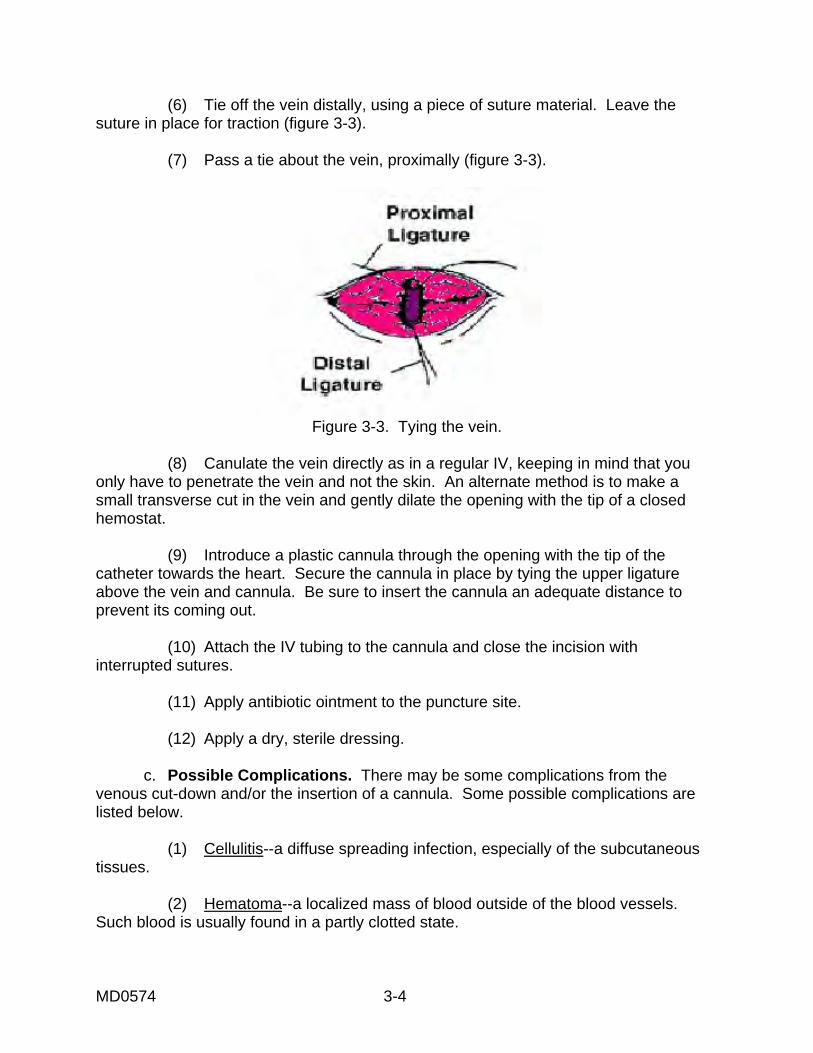
MD0574 3-4
(6) Tie off the vein distally, using a piece of suture material. Leave the suture in place for traction (figure 3-3). (7) Pass a tie about the vein, proximally (figure 3-3).
Figure 3-3. Tying the vein.
(8) Canulate the vein directly as in a regular IV, keeping in mind that you only have to penetrate the vein and not the skin. An alternate method is to make a small transverse cut in the vein and gently dilate the opening with the tip of a closed hemostat. (9) Introduce a plastic cannula through the opening with the tip of the catheter towards the heart. Secure the cannula in place by tying the upper ligature above the vein and cannula. Be sure to insert the cannula an adequate distance to prevent its coming out. (10) Attach the IV tubing to the cannula and close the incision with interrupted sutures. (11) Apply antibiotic ointment to the puncture site. (12) Apply a dry, sterile dressing. c. Possible Complications. There may be some complications from the venous cut-down and/or the insertion of a cannula. Some possible complications are listed below. (1) Cellulitis--a diffuse spreading infection, especially of the subcutaneous tissues. (2) Hematoma--a localized mass of blood outside of the blood vessels. Such blood is usually found in a partly clotted state.
MD0574 3-5
(3) Phlebitis--inflammation of a vein. (4) Perforation of the posterior wall of a vein--a back wall of a vein is pierced. (5) Venous thrombosis--a blood clot in a vein. (6) Nerve transections--cutting across a nerve. (7) Arterial transection--cutting across an artery. NOTE: Fewer complications occur when aseptic technique is used and proper care is taken when the cannula is placed. 3-3. NEEDLE DECOMPRESSION (THORACENTESIS) Needle decompression is the removal of air and fluid from the chest cavity by puncture. a. Indications for Needle Decompression. Indications for needle depression Include the following. (1) Under combat conditions, if a casualty has unilateral chest trauma and an increase in respiratory effort, these are the only conditions needed to indicate the need for immediate chest needle decompression. (2) Under more controlled conditions, the following signs and symptoms should be observed prior to chest needle decompression. (a) Severe respiratory distress. (b) Jugular vein distention. (c) Hyper resonance on the affected side. (d) Absent or diminished breath sounds on the affected side. (e) Pulseless paradox (the radial pulse fades or disappears upon inspiration). (f) Tracheal deviation may be seen, but is considered a very late and grave sign.
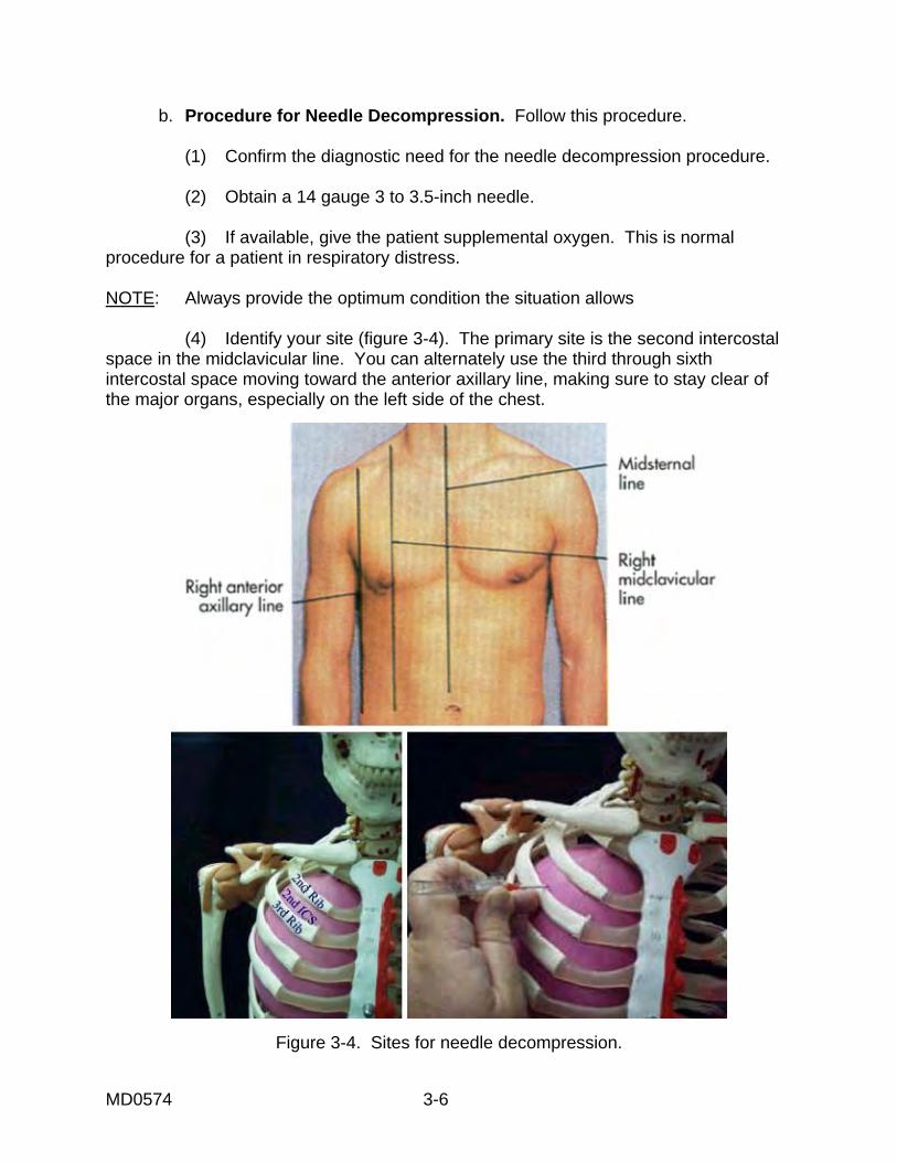
MD0574 3-6
b. Procedure for Needle Decompression. Follow this procedure. (1) Confirm the diagnostic need for the needle decompression procedure. (2) Obtain a 14 gauge 3 to 3.5-inch needle. (3) If available, give the patient supplemental oxygen. This is normal procedure for a patient in respiratory distress. NOTE: Always provide the optimum condition the situation allows (4) Identify your site (figure 3-4). The primary site is the second intercostal space in the midclavicular line. You can alternately use the third through sixth intercostal space moving toward the anterior axillary line, making sure to stay clear of the major organs, especially on the left side of the chest.
Figure 3-4. Sites for needle decompression.
MD0574 3-7
(5) Anesthetize the area locally if time permits. Remember, this is a life-saving emergency procedure. The patient’s airway is compromised and the chest needle decompression is the fix. Do not waste time getting the airway open. (6) Position the patient. You may position him in an upright position if you have determined that he does not have a cervical spine fracture. (7) Insert the thoracentesis needle into the skin. Direct the needle just over the rib into the intercostal space. (8) Puncture the parietal pleural space. (9) Aspirate as much air as necessary to relieve the patient's acute symptoms. (10) Leave the plastic catheter in place and apply a bandage or a small dressing. 3-4. CHEST TUBE INSERTION Chest tubes are placed so that air and blood can be removed from the intrathoracic cavity. Place the chest tube in the appropriate space so that air is removed and the condition is relieved. Chest tubes are placed so that gravity aids suction in fluid removal. a. Indications for Chest Tube Insertion. Three conditions commonly require the insertion of a tube into the chest. The conditions are hemothorax, pneumothorax, and tension pneumothorax. Hemothorax is the accumulation of blood in the pleural cavity. Pneumothorax is the accumulation of air in the pleural space. The amount of trapped air or gas determines the degree of lung collapse. In tension pneumothorax, air in the pleural space is under higher pressure than air in the adjacent lung and the vascular structures. A patient with tension pneumothorax must have prompt treatment or he will suffer fatal pulmonary and circulatory impairment. b. Equipment for Chest Tube Insertion. Equipment needed includes the following: (1) Betadine® or another solution in preparing the site for chest tube insertion. (2) Sponges for preparation of the site. (3) 2 large, curved, Kelly clamps. (4) 6 x 10 millimeter syringes with number 18 and number 25 gauge needles.
MD0574 3-8
(5) Scalpel and blade. (6) One percent lidocaine. (7) Suture (to secure the chest tube). (8) Suture (for wound approximation). (9) Appropriate chest tube size. (10) Chest drainage collection device. (11) Sterile surgical gloves. (12) Occlusive dressing material. (13) Sterile drape. (14) Needle holder. (15) Tape. c. Procedure for Chest Tube Insertion. Monitor the patient throughout the procedure. Perform the following steps. (1) Put on the gloves and mask. (2) The site is about nipple level (the fifth or sixth intercostal space) on his affected side on the mid to anterior axillary plane. See figure 3-5. (3) Prepare and drape the patient. (4) Locally, anesthetize the skin and rib periosteum if the patient's condition and time permit. (5) Incise the skin, creating a tunnel. Using a scalpel and blade, make a two to three centimeter transverse incision through the subcutaneous tissues, just over the top of the rib. Create a tunnel by spreading the clamp. (6) Using the clamp, make a hole into the pleural space. Grip the clamp along its shaft to prevent plunging too deeply into the chest. Make the hole at the top edge of the patient's rib. Spread the clamp to enlarge the hole and remove the clamp.
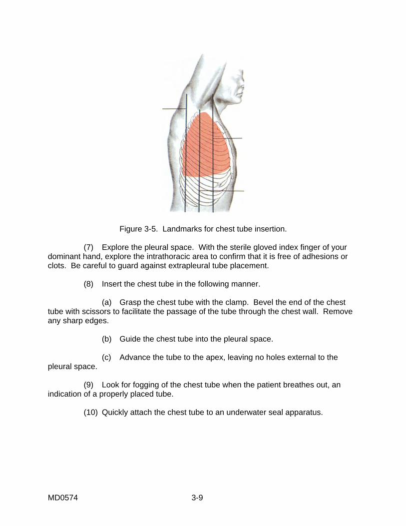
MD0574 3-9
Figure 3-5. Landmarks for chest tube insertion.
(7) Explore the pleural space. With the sterile gloved index finger of your dominant hand, explore the intrathoracic area to confirm that it is free of adhesions or clots. Be careful to guard against extrapleural tube placement. (8) Insert the chest tube in the following manner. (a) Grasp the chest tube with the clamp. Bevel the end of the chest tube with scissors to facilitate the passage of the tube through the chest wall. Remove any sharp edges. (b) Guide the chest tube into the pleural space. (c) Advance the tube to the apex, leaving no holes external to the pleural space. (9) Look for fogging of the chest tube when the patient breathes out, an indication of a properly placed tube. (10) Quickly attach the chest tube to an underwater seal apparatus.
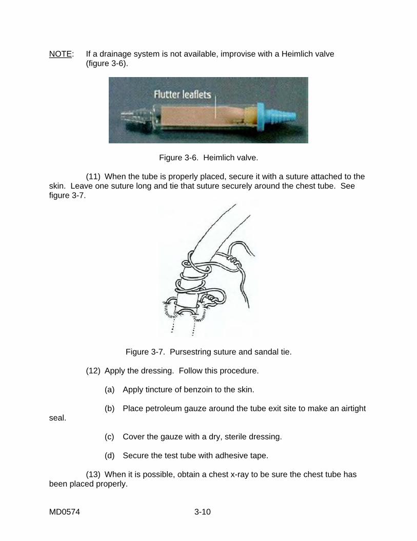
MD0574 3-10
NOTE: If a drainage system is not available, improvise with a Heimlich valve (figure 3-6).
Figure 3-6. Heimlich valve. (11) When the tube is properly placed, secure it with a suture attached to the skin. Leave one suture long and tie that suture securely around the chest tube. See figure 3-7.
Figure 3-7. Pursestring suture and sandal tie. (12) Apply the dressing. Follow this procedure. (a) Apply tincture of benzoin to the skin. (b) Place petroleum gauze around the tube exit site to make an airtight seal. (c) Cover the gauze with a dry, sterile dressing. (d) Secure the test tube with adhesive tape. (13) When it is possible, obtain a chest x-ray to be sure the chest tube has been placed properly.
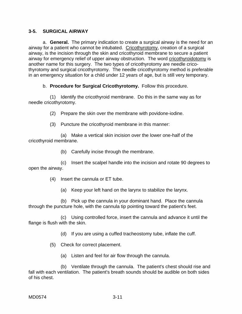
MD0574 3-11
3-5. SURGICAL AIRWAY a. General. The primary indication to create a surgical airway is the need for an airway for a patient who cannot be intubated. Cricothyrotomy, creation of a surgical airway, is the incision through the skin and cricothyroid membrane to secure a patient airway for emergency relief of upper airway obstruction. The word cricothyroidotomy is another name for this surgery. The two types of cricothyrotomy are needle crico-thyrotomy and surgical cricothyrotomy. The needle cricothyrotomy method is preferable in an emergency situation for a child under 12 years of age, but is still very temporary. b. Procedure for Surgical Cricothyrotomy. Follow this procedure. (1) Identify the cricothyroid membrane. Do this in the same way as for needle cricothyrotomy. (2) Prepare the skin over the membrane with povidone-iodine. (3) Puncture the cricothyroid membrane in this manner: (a) Make a vertical skin incision over the lower one-half of the cricothyroid membrane. (b) Carefully incise through the membrane. (c) Insert the scalpel handle into the incision and rotate 90 degrees to open the airway. (4) Insert the cannula or ET tube. (a) Keep your left hand on the larynx to stabilize the larynx. (b) Pick up the cannula in your dominant hand. Place the cannula through the puncture hole, with the cannula tip pointing toward the patient's feet. (c) Using controlled force, insert the cannula and advance it until the flange is flush with the skin. (d) If you are using a cuffed tracheostomy tube, inflate the cuff. (5) Check for correct placement. (a) Listen and feel for air flow through the cannula. (b) Ventilate through the cannula. The patient's chest should rise and fall with each ventilation. The patient's breath sounds should be audible on both sides of his chest.
MD0574 3-12
(6) Secure the cannula with tape. NOTE: Due to its potential hazards, cricothyrotomy should be performed ONLY by personnel who have been thoroughly trained in this method and who have been approved by their physician directors to use this method. 3-6. CLOSING Trauma strikes all ages. Generally, trauma is swift in onset and slow in recovery, with many problems along the way for those trying to manage its course. Trauma can have a devastating effect on the members of our society. Prevention is paramount. Where prevention fails, we must be knowledgeable and prepared to meet the patient's immediate needs.
Continue with Exercises
MD0574 3-13
EXERCISES, LESSON 3 INSTRUCTIONS. The following exercises are to be answered by writing the answer in the space provided or by matching terms as directed. After you have completed all the exercises, turn to "Solutions to Exercises, Lesson 3" at the end of the exercises and check your answers. 1. A venous cut-down procedure is being performed. The primary site for this procedure is _______________________________, which is located at the. _____________________. 2. The secondary site for a venous cut-down procedure is the ________________, located close to the ___________________. 3. List three possible complications of a venous cut-down. a. _______________________________________________. b. _______________________________________________. c. _______________________________________________. 4. Needle decompression is ___________________________________________ ________________________________________________________________. 5. List three indications that needle decompression should be performed under controlled conditions. a. _______________________________________________. b. _______________________________________________. c. _______________________________________________.
MD0574 3-14
6. List three indications that the patient needs a chest tube insertion. a. _______________________________________________. b. _______________________________________________. c. _______________________________________________ 7. What is the primary indication for creating a surgical airway? ________________________________________________________________ ________________________________________________________________ 8. The two qualifications a 68W Medical NCO must have to perform a surgical cricothyrotomy are: a. _______________________________________________. b. _______________________________________________. 9. To insert the cannula properly when you are performing a surgical cricothyrotomy, you should: a. Keep your left hand on the ___________________ to stabilize it. b. Pick up the cannula in your __________________ hand. c. Place the cannula through the puncture hole, with the cannula tip pointing toward the patient's _________________________. d. Using controlled force, advance the cannula until the _______________ is flush with the skin. e. If you are using a cuffed tracheostomy tube, __________________ the cuff.
MD0574 3-15
10. To check for correct placement of the cannula in a patient who has had a surgical cricothyrotomy, you should: a. ________________________________________________. b. _______________________________________________. 11. Listed below are the major steps in the procedure of inserting a chest tube. Place the steps in the order in which they would be performed.
a. _____ b. _____ c. _____ d. _____ e. _____ f. _____ g. _____ h. _____
S. Apply a dressing. T. Explore the pleural space. U. Suture the skin, securing the chest tube. V. Insert the chest tube. W. Locally, anesthetize the skin and periosteum if there is time. X. Prep and drape the patient. Y. Perforate into the pleural space. Z. Incise the skin and create a tunnel.
Check Your Answers on Next Page
MD0574 3-16
SOLUTIONS TO EXERCISES, LESSON 3 1. The greater saphenous vein; ankle. (para 3-2a(1)) 2. The antecubital median basilic vein; elbow. (para 3-2a(2)) 3. You are correct if you listed any three of the following: Cellulitis. Hematoma. Phlebitis. Perforation of the posterior wall of the vein. Venous thrombosis. Nerve transection. Arterial transection. (para 3-2c(1) through (7)) 4. Needle decompression is the removal of air and fluid from the chest cavity by puncture. (para 3-3) 5. You are correct if you listed any three of the following: Severe respiratory distress. Jugular vein distention. Hyper resonance on the affected side. Absent or diminished breath sounds on the affected side. Pulseless paradox (the radial pulse fades or disappears upon inspiration). Tracheal deviation may be seen but is considered a very late and grave sign.
(para 3-3a(2)(a)-(f)) 6. The patient has hemothorax. The patient has pneumothorax. The patient has tension pneumothorax. (para 3-4a) 7. The primary indication to create a surgical airway is the need for an airway for a patient who cannot be intubated. (para 3-6a) 8. Thoroughly trained in the procedure. Approved by the physician directors to perform a cricothyrotomy. (para 3-5b, NOTE) 9. a. Larynx. b. Dominant. c. Feet. d. Flange. e. Inflate. (para 3-5(b)(4)(a)-(e)) 10. a. Listen and feel for air flow through the cannula b. His chest rises and falls with each ventilation. (para 3-5b(5)(a), (b)

MD0574 3-17
11. a. X_ b. W_ c. Z_ d. Y_ e. T_ f. V_ g. U_ h. S (para 3-4c(3)-(12))
End of Lesson 3