Embed Size (px)
Citation preview
Surgical Options for the Treatment of
Presbyopia
Lance J. Kugler, MDWang Vision Institute
Nashville, TN

Presbyopia in the Emmetrope –the most difficult group to satisfy
• Patients with good uncorrected distance vision are uncompromising to any changes in distance vision
• Post-LASIK emmetropes have added difficulty with refractive lens exchange due to IOL power determination

Surgical Correction of PresbyopiaSTATIC CORRECTION
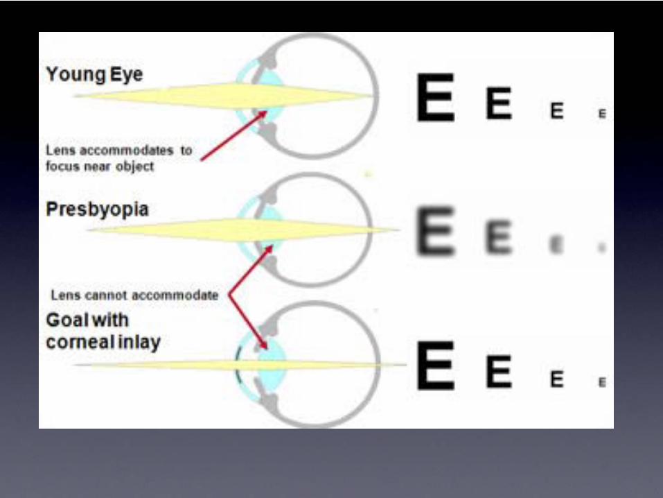
• Cornea Related:• Monovision• Multifocality• Pinhole Implant
(Karma implant)
• Lens Related:• Exchange the lens• Multifocal lens
implant
DYNAMIC CORRECTION
• Lens Related:• Exchange the lens• Accommodating lens -
Crystalens
• Scleral Related:• Improve the natural lens’
focusing power• Scleral Spacing Procedure
(“SSP”)
SSP for Presbyopia in the Emmetrope
• SSP alters the configuration of the sclera around the lens equator in four oblique quadrants.
• SSP does not involve surgery on the visual axis.
• SSP is designed to correct presbyopia with a ciliary muscle / zonule / natural lens approach.
• The PSI (implants) are removable, thus SSP is reversible.
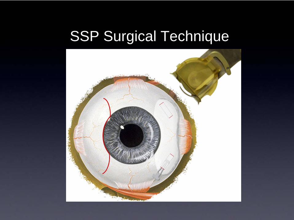
SSP Surgical Technique
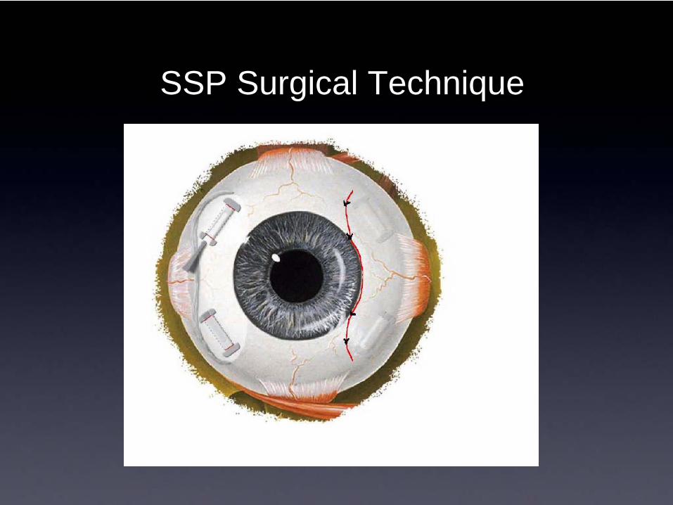
SSP Surgical Technique
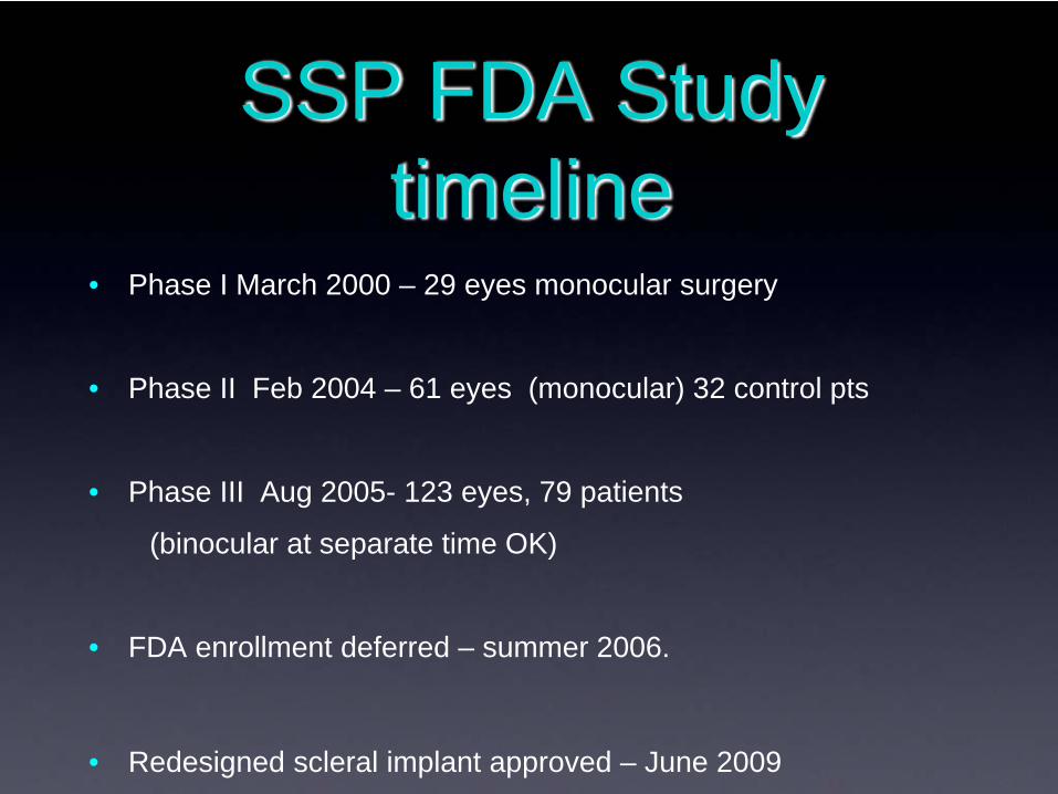
SSP FDA Study timeline
• Phase I March 2000 – 29 eyes monocular surgery
• Phase II Feb 2004 – 61 eyes (monocular) 32 control pts
• Phase III Aug 2005- 123 eyes, 79 patients
(binocular at separate time OK)
• FDA enrollment deferred – summer 2006.
• Redesigned scleral implant approved – June 2009
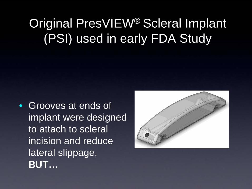
Original PresVIEW® Scleral Implant (PSI) used in early FDA Study
• Grooves at ends of implant were designed to attach to scleral incision and reduce lateral slippage, BUT…
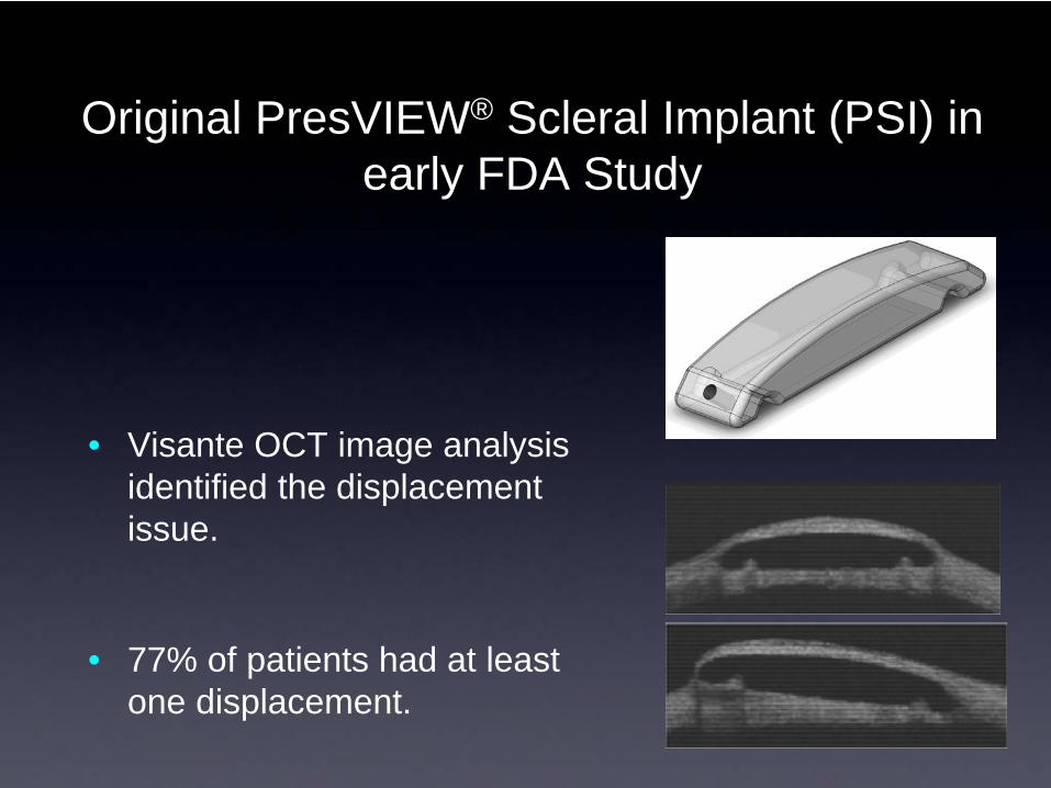
Original PresVIEW® Scleral Implant (PSI) in early FDA Study
• Visante OCT image analysis identified the displacement issue.
• 77% of patients had at least one displacement.
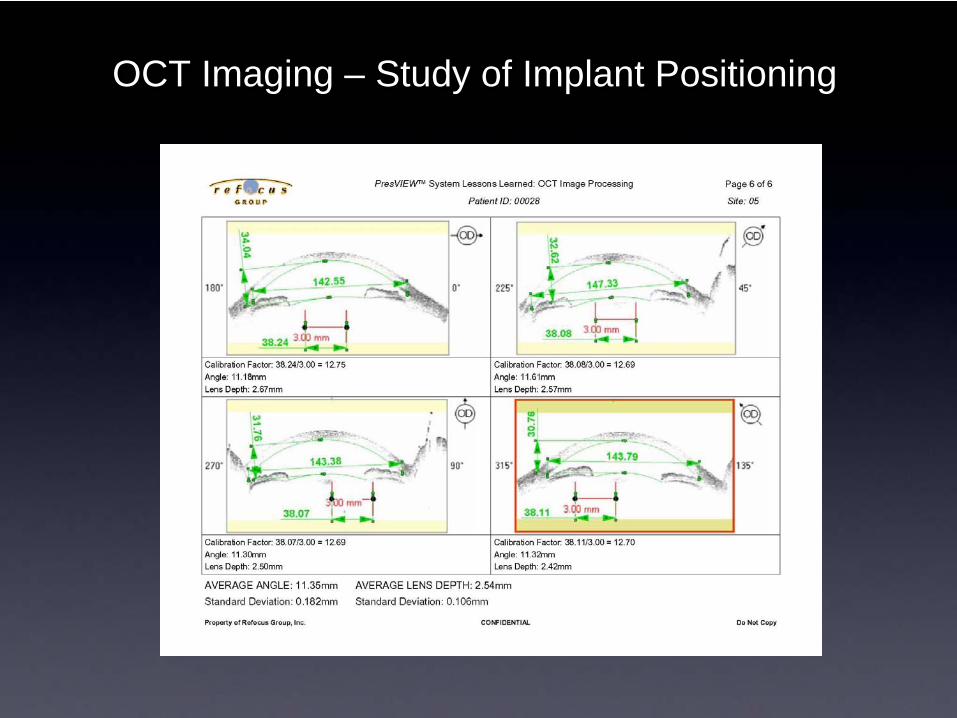
OCT Imaging – Study of Implant Positioning
SSP Surgical Technique and Design Issues
• Implant displacement.
• Location of implants relative to limbus varied widely.
• Depth of surgical incisions varied widely.
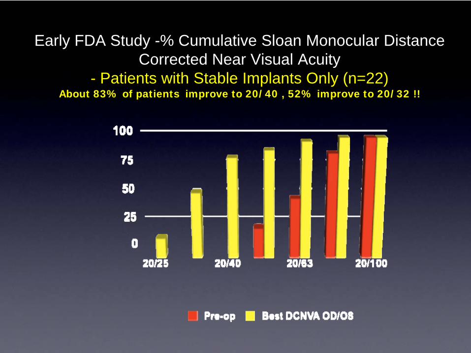
Early FDA Study -% Cumulative Sloan Monocular Distance Corrected Near Visual Acuity
- Patients with Stable Implants Only (n=22)About 83% of patients improve to 20/40 , 52% improve to 20/32 !!
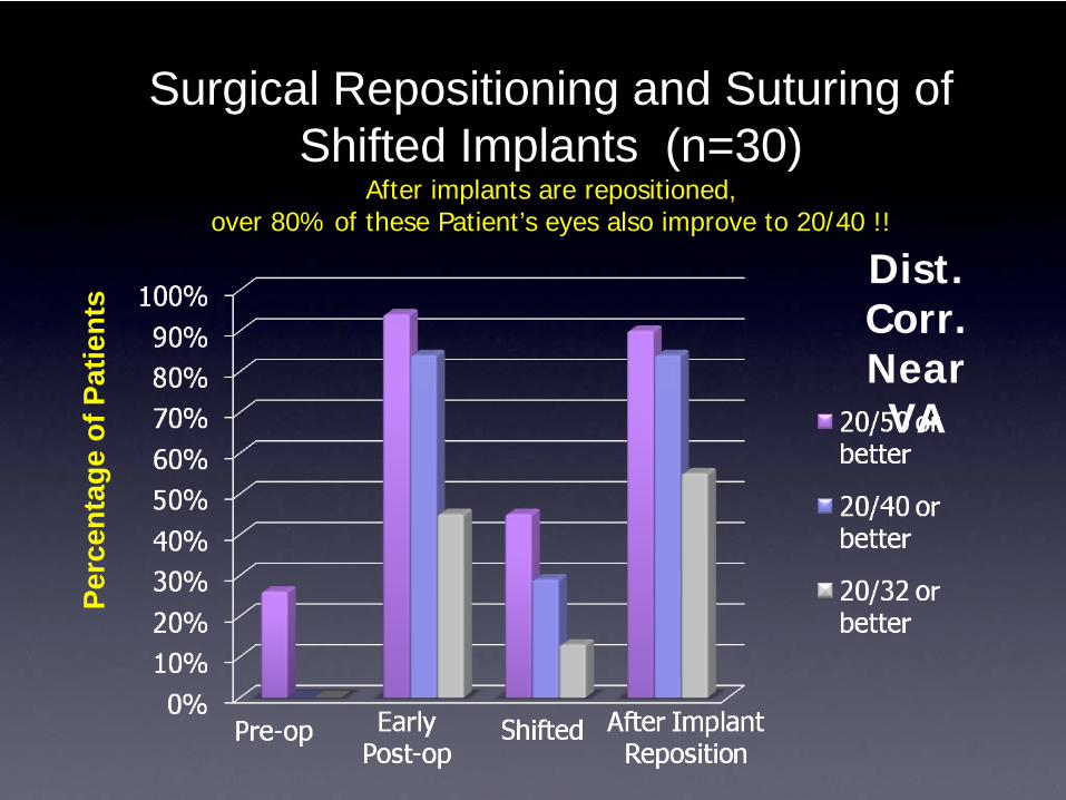
Surgical Repositioning and Suturing of Shifted Implants (n=30)
After implants are repositioned, over 80% of these Patient’s eyes also improve to 20/40 !!
Perc
enta
ge o
f Pat
ient
s
Dist. Corr. Near VA
Second Generation Implantand Improved Surgical Instrumentation
• Third party research engineering firm enlisted
- second quarter 2006.
• New stable implant design identified, manufactured, validation testing - early 2007.
• Initial test surgeries - summer 2007.
• Extensive clinical testing – 2007 & 2008.
• Better surgical instrumentation
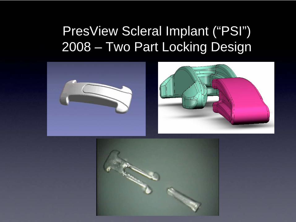
PresView Scleral Implant (“PSI”)2008 – Two Part Locking Design
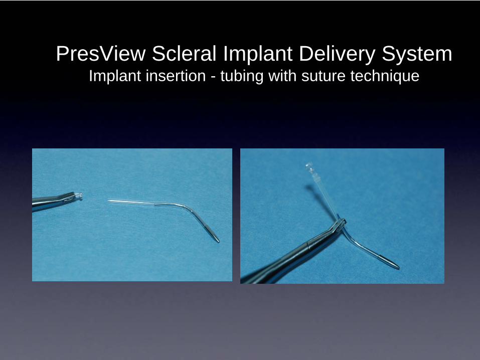
PresView Scleral Implant Delivery SystemImplant insertion - tubing with suture technique
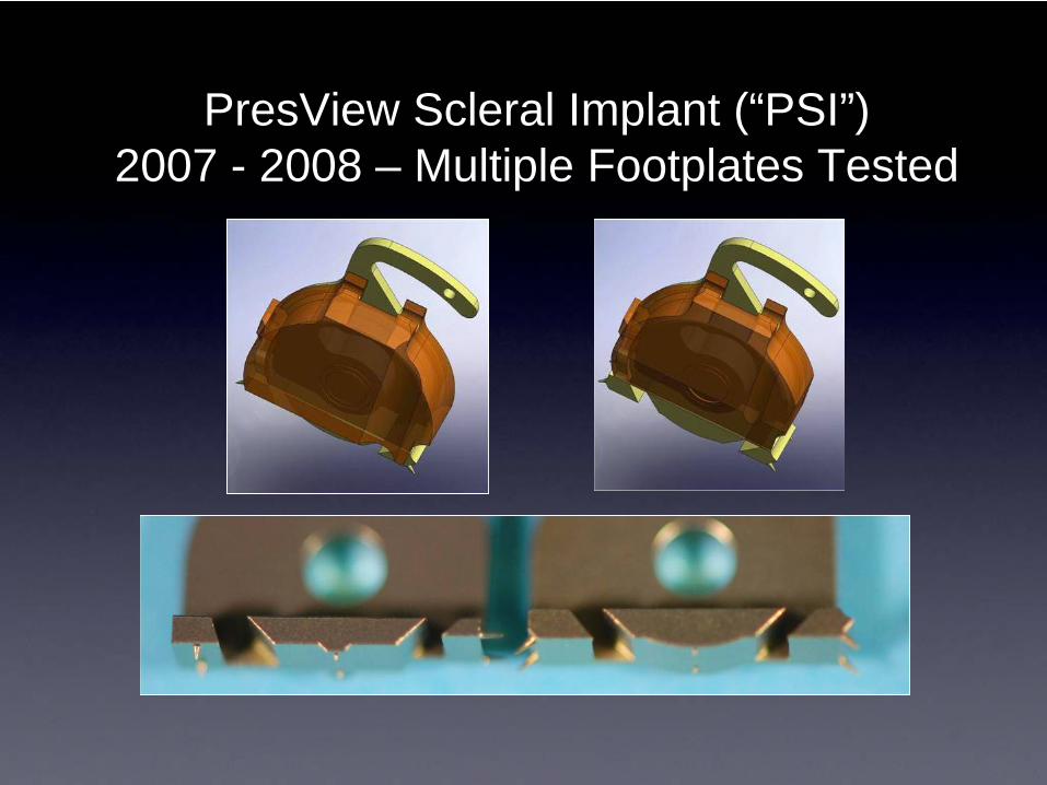
PresView Scleral Implant (“PSI”)2007 - 2008 – Multiple Footplates Tested

PresView Scleral Implant (“PSI”)2007 - 2008 – Sharper Blade Tested

PresView Scleral Implant (“PSI”)2007 - 2008 – Marking Enhancements
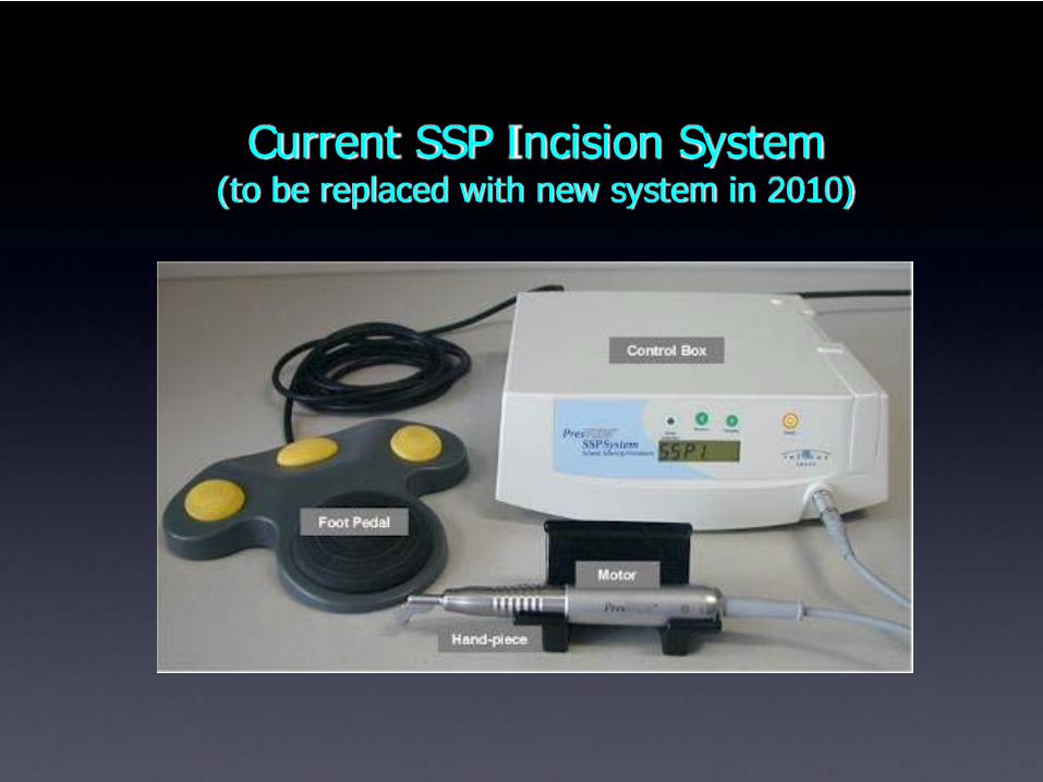
Current SSP Incision System(to be replaced with new system in 2010)
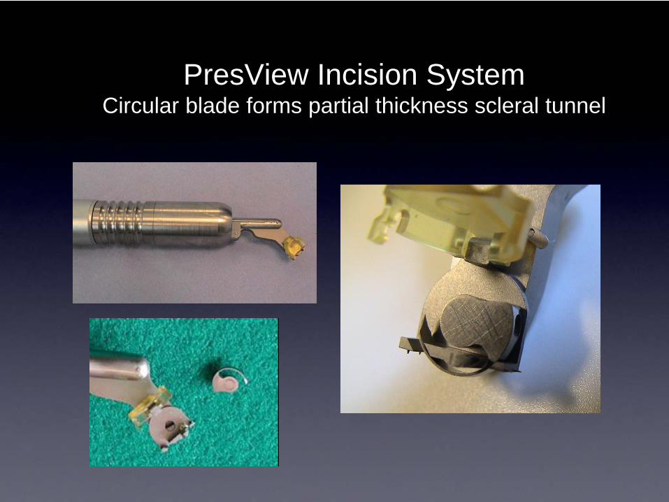
PresView Incision SystemCircular blade forms partial thickness scleral tunnel
Central American Clinical SiteRedesigned Implant, System and
Approach
• Larger, Longer Two-Part Implant
More Surface Area At Ends – Greater Vaulting
• Applied Tear Film Therapy
• Applied Vision Exercise

Central American Clinical Data% Cumulative Sloan Monocular Distance Corrected Near Visual Acuity
Two-Part Implant Design
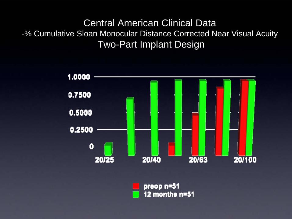
Central American Clinical Data -% Cumulative Sloan Monocular Distance Corrected Near Visual Acuity
Two-Part Implant Design

Scleral Spacing Procedure –Mechanism of Action
SSP – Mechanism of ActionTriad of Accommodation
• Both eyes converge.
• Pupils experience miosis (constriction).
• Ciliary muscles contract
SSP – Patient SelectionKey to Success
• Patient understanding and cooperation.
• Muscle rehabilitation required.
• Commitment to near vision activities.
• Use of reading glasses prevents rehabilitation.
Scleral Spacing Procedure –Additional Development Activities
• Lightweight spring powered incision device.
• Improved device for fixation of the eye.
• Ultimately - docking of the incision device to the fixation device.
• Objective – shorter, more repeatable surgery.
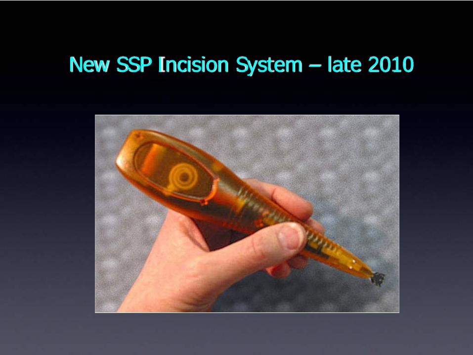
New SSP Incision System – late 2010
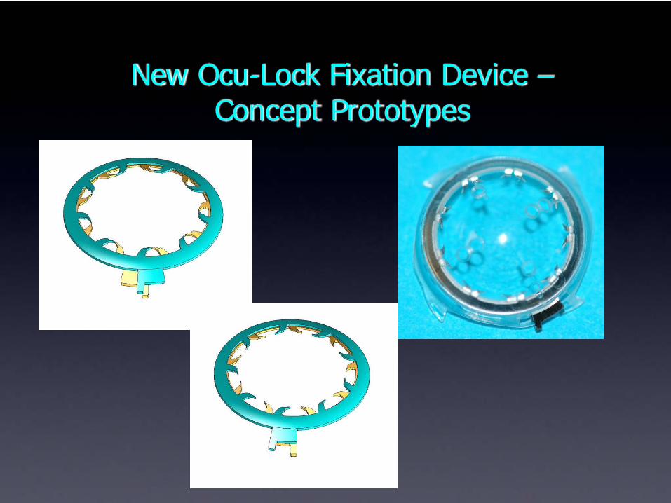
New Ocu-Lock Fixation Device –Concept Prototypes
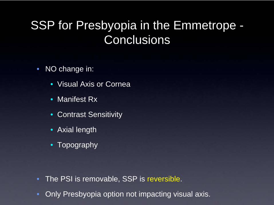
SSP for Presbyopia in the Emmetrope -Conclusions
• NO change in:
• Visual Axis or Cornea
• Manifest Rx
• Contrast Sensitivity
• Axial length
• Topography
• The PSI is removable, SSP is reversible.
• Only Presbyopia option not impacting visual axis.
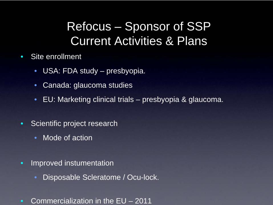
Refocus – Sponsor of SSPCurrent Activities & Plans
• Site enrollment
• USA: FDA study – presbyopia.
• Canada: glaucoma studies
• EU: Marketing clinical trials – presbyopia & glaucoma.
• Scientific project research
• Mode of action
• Improved instumentation
• Disposable Scleratome / Ocu-lock.
• Commercialization in the EU – 2011
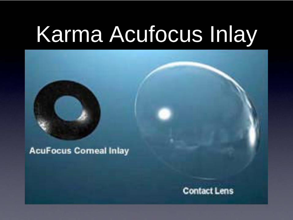
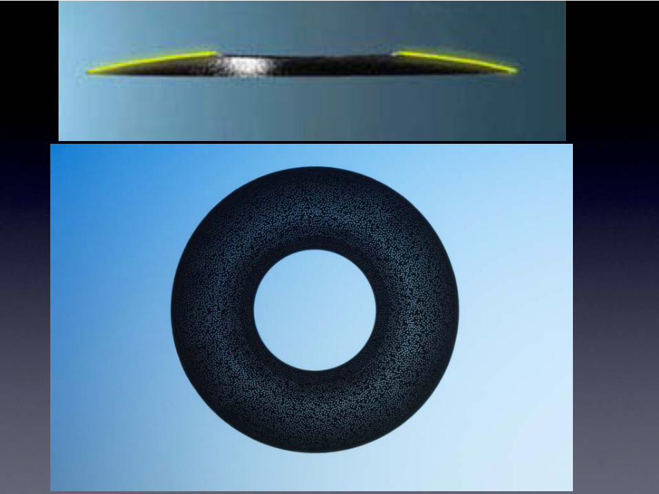
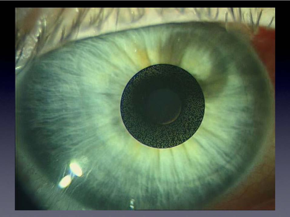
Karma Acufocus Inlay


![Corneal Remodeling to Correct Presbyopia: Mechanism of A ... · in accommodation [1]. Surgical treatment options include LASIK monovision, multifocal LASIK, accommodative, multifocal,](https://img.pdfslide.net/doc/110x75/613e8be969193359046d2fc9/corneal-remodeling-to-correct-presbyopia-mechanism-of-a-in-accommodation-1.jpg)