Embed Size (px)
Citation preview
J. Comp. Path. 2014, Vol. 150, 345e349 Available online at www.sciencedirect.com
ScienceDirect
www.elsevier.com/locate/jcpa
DISEASE IN WILDLIFE OR EXOTIC SPECIES
T-lymphocyte-rich Thymoma and MyastheniaGravis in a Siberian Tiger (Panthera tigris altaica)*
*T
Com
wh
any
Cor
002
http
K. Allan*, N. Masters†, S. Rivers‡, K. Berryx, A. Routh† and C. Lamm*
*School of Veterinary Medicine, University of Glasgow, Bearsden Road, Glasgow G61 1QH, †Zoological Society of London,
Regent’s Park, London NW1 4RY, ‡Abbey Veterinary Services, 89 Queen Street, Newton Abbot, Devon TQ12 2BG andxZSL Whipsnade Zoo, Dunstable, Bedfordshire LU6 2LF, UK
his i
mo
ich
me
resp
1-99
://d
Summary
A 10-year-old captive male Siberian tiger (Panthera tigris altaica) presented with acute onset collapse, vomit-ing and dyspnoea, preceded by a 6-month period of progressive muscle wasting. Following humane destruc-tion, post-mortem examination revealed a large multilobulated mass in the cranial mediastinum, which wasdiagnosed as a T-lymphocyte-rich thymoma with the aid of immunohistochemistry. Retrospective serologyfor acetylcholine receptor antibodies (titre 3.90 nmol/l) confirmed a diagnosis of thymoma-associated myas-thenia gravis. Thymomas are reported rarely in wild carnivores, but when detected they appear to be similarin morphology to those seen in domestic carnivores and may also be accompanied by paraneoplastic syn-dromes. The clinical signs of myasthenia gravis in the tiger were consistent with those reported in catsand dogs and the condition is proposed as an important differential diagnosis for generalized weakness incaptive Felidae.
� 2013 The Authors. Published by Elsevier Ltd. All rights reserved.
Keywords: myasthenia gravis; Panthera tigris altaica; Siberian tiger; thymoma
Thymoma is an uncommon neoplasm of the cranialmediastinum, composed of neoplastic thymic epithe-lium usually accompanied by various degrees of lym-phocytic infiltration (Jacobs et al., 2008). In people,thymomas are organised into five categories (A, AB,B1, B2 and B3) using a World Health Organization(WHO) classification system that aims to predictthe clinical behaviour and prognosis of this neoplasm(Travis et al., 2004; Suster and Moran, 2006). Inanimals thymomas are classified on the basis of thepredominant cell population within the mass andmay be lymphocyte predominant, epithelial cellpredominant or of an intermediate lympho-epithelial subtype. Immunohistochemistry (IHC) isoften required to confirm the presence of neoplasticthymic epithelial cells in lymphocyte-rich thymomas
s an open-access article distributed under the terms of the Creative
ns Attribution-NonCommercial-No Derivative Works License,
permits non-commercial use, distribution, and reproduction in
dium, provided the original author and source are credited.
ondence to: K. Allan (e-mail: [email protected]).
75/$ - see front matter
x.doi.org/10.1016/j.jcpa.2013.11.204
and to differentiate thymomas from mediastinal lym-phomas, which occur more commonly in most ani-mals (Jacobs et al., 2008). Spontaneously-occurringthymomas have been reported in a range of domesticanimals including dogs and cats (Day, 2008), cattle(Ecco et al., 2006), rabbits (Kunzel et al., 2012) andgoats (Hadlow, 1978) as well as in various laboratoryprimates and rodents (Walsh and Poteracki, 1994;Brandes et al., 2004; Schwartz et al., 2011).However, in wildlife species reports of thymomas arescarce. Here we describe the morphological andparaneoplastic features of a thymoma diagnosed atpost-mortem examination in a captive Siberian tiger(Panthera tigris altaica).
A 10-year-old, male neutered Siberian tiger, bornand housed at the Zoological Society of London(ZSL) Whipsnade collection, presented on 27thOctober 2011 to resident veterinary staff in sternal re-cumbency with acute onset vomiting, depression anda right-sided head tilt. The tiger had been treated forprogressive muscle wasting and suspected renal insuf-ficiency with oral benazepril hydrochloride (0.5 mg/
� 2013 The Authors. Published by Elsevier Ltd. All rights reserved.
346 K. Allan et al.
kg q24h; Fortekor Flavour 20 mg for dogs; Novartis,Camberley, Surrey, UK) for 6 months prior to acutepresentation. Following collapse, general anaesthesiawas induced to facilitate clinical examination using480 mg ketamine (ketamine 1 g powder for reconsti-tution; Kyron Laboratories, Benrose, Johannesburg,South Africa) and 6 mg medetomidine (Zalopine10 mg/ml; Orion Pharma, Newbury, Berkshire,UK) administered by remote intramuscular injec-tion. Endotracheal intubation was performed andanaesthesia was maintained with oxygen and isoflur-ane (Isoflurane-Vet 100% w/w inhalation vapour;Merial Animal Health, Harlow, Essex, UK). Onphysical examination, the tiger was tachycardicwith poor peripheral circulation. An abnormal respi-ratory pattern, characterized by inspiratory stridoraccompanied by irregular periods of apnoea, wasobserved and intermittent positive pressure ventila-tion was initiated. Venous blood samples were ob-tained, but standard haematological andbiochemical parameters were within published refer-ence values for this species (ISIS, 2002). On welfaregrounds, the tiger was humanely destroyed and sub-mitted for pathological examination.
Post-mortem examination revealed a large, 1.5 kg,18 � 15 � 10 cm, well-demarcated, multilobulated,mediastinal mass within the cranial thorax (Fig. 1).The pleural and peritoneal cavities both containedsmall amounts of serosanguineous fluid. There wasgeneralized depletion of subcutaneous fat stores inaddition to moderate atrophy of skeletal muscleover the hindquarters.
Representative tissue samples were collected andfixed in 10% neutral buffered formalin and submitted
Fig. 1. Intrathoracicmass after reflection of the ventral thorax andsternum. Awell-demarcated, multilobulatedmass expandsthe cranial mediastinum. Bar, 3 cm.
to Abbey Veterinary Services, Newton Abbott, UK,for examination. Tissue samples were processedroutinely and sections (4 mm) were stained with hae-matoxylin and eosin (HE). Subsequently, samplesfrom the mediastinal mass were transported to theUniversity of GlasgowVeterinary Diagnostic Servicesfor IHC. Sections were labelled with a panel of pri-mary antibodies including mouse anti-vimentin(Clone V9, Dako, Ely, UK; dilution 1 in 50), whichdid not require antigen retrieval, and mouse anti-human cytokeratin (Clone MNF116, Dako; dilution1 in 100), which required enzymatic antigen retrievalwith proteinase K. Heat-induced epitope retrieval us-ing sodium citrate buffer (pH 6.0) was required forthe following antibodies: mouse anti-humanCD79acy (Clone HM57, Dako; dilution 1 in 100);mouse anti-human B cell-specific activator protein(Clone DAK-Pax 5, Dako; dilution 1 in 100) and rab-bit anti-human CD3 (Dako, dilution 1 in 100). IHCwas performed using a Dako Autostainer. Tissue sec-tions were also stained with Astra blue (SigmaeAldrich, Gillingham, UK) as previously described(Blaies and Williams, 1981).
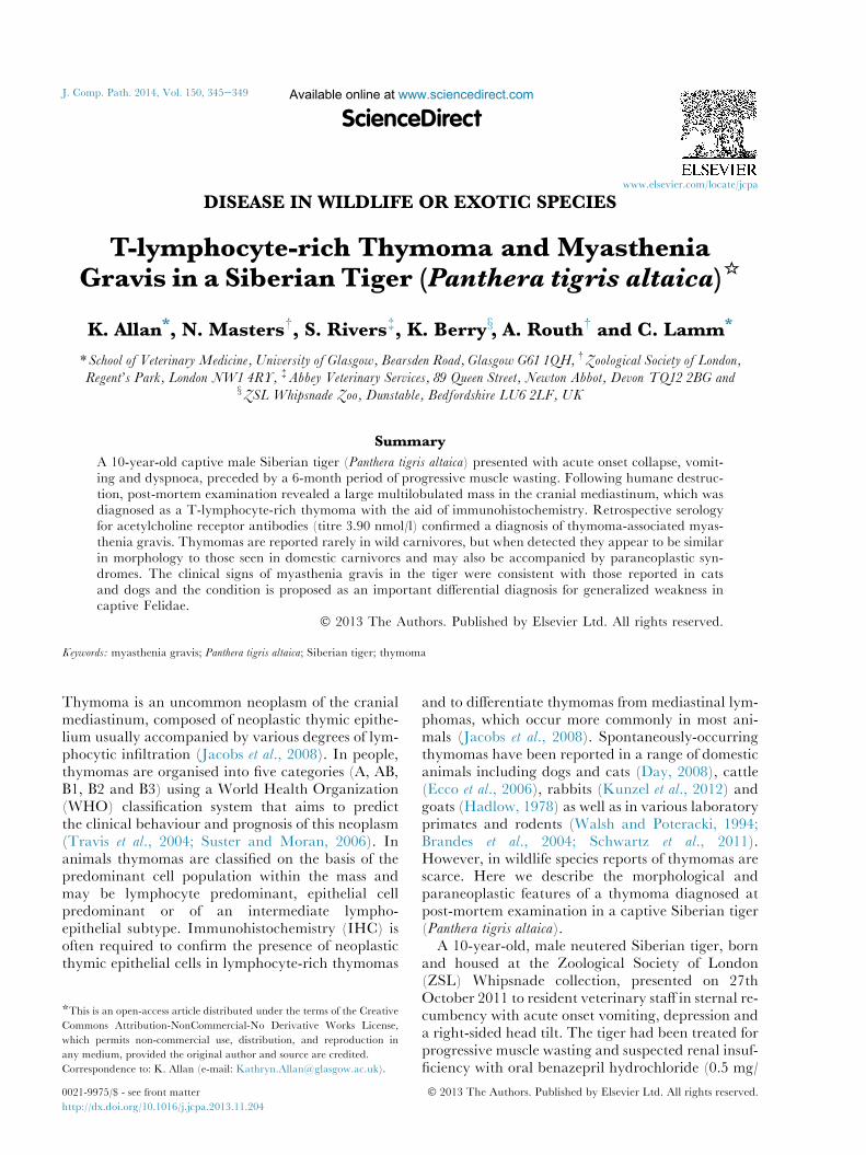
Microscopically, the mass was a large, multilobu-lated, densely cellular and poorly demarcatedtumour, composed of abundant, densely packed andwell-differentiated small to medium sized lympho-cytes arranged in sheets that frequently obscured aless numerous background population of polygonalto stellate neoplastic cells. The neoplastic cells werearranged in variably packed cords and supported bya well-vascularized collagenous and adipose connec-tive tissue stoma. The neoplastic cells were large(15e20 mm in diameter) and poorly differentiatedwith moderate amounts of finely granular eosino-philic cytoplasm and indistinct cell borders (Fig. 2).The nuclei were oval, centrally located, often with asingle prominent magenta nucleolus and peripheral-ized chromatin. There was moderate anisocytosisand anisokaryosis, but mitoses were rare, with <1mitotic figure per 10 high-power (�400) fields.Throughout the tumour were numerous, multifocalaggregates of eosinophils admixed with smallernumbers of plasma cells and mast cells.
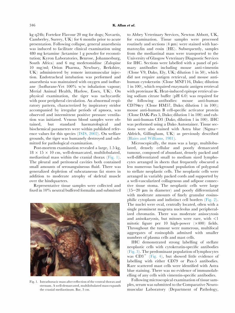
IHC demonstrated strong labelling of stellateneoplastic cells with cytokeratin-specific antibodies(Fig. 3). The predominant population of lymphocyteswas CD3+ (Fig. 4), but showed little evidence oflabelling with either CD79 or Pax-5 antibodies.Rare scattered mast cells were identified with Astrablue staining. There was no evidence of immunolab-elling of any cells with vimentin-specific antibodies.
Followingmicroscopical examination of tissue sam-ples, serumwas submitted to the Comparative Neuro-muscular Laboratory (Department of Pathology,
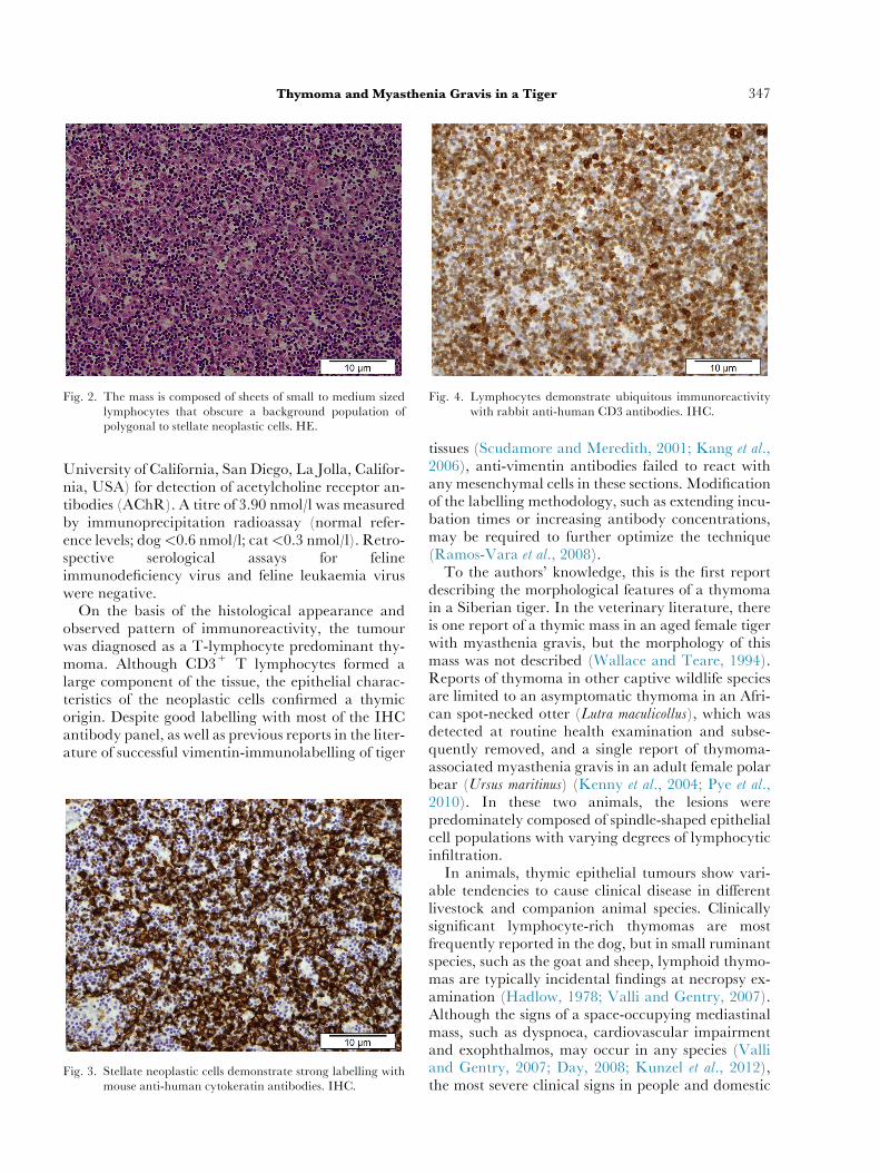
Fig. 2. The mass is composed of sheets of small to medium sizedlymphocytes that obscure a background population ofpolygonal to stellate neoplastic cells. HE.
Fig. 4. Lymphocytes demonstrate ubiquitous immunoreactivitywith rabbit anti-human CD3 antibodies. IHC.
Thymoma and Myasthenia Gravis in a Tiger 347
University of California, San Diego, La Jolla, Califor-nia, USA) for detection of acetylcholine receptor an-tibodies (AChR). A titre of 3.90 nmol/l was measuredby immunoprecipitation radioassay (normal refer-ence levels; dog<0.6 nmol/l; cat<0.3 nmol/l). Retro-spective serological assays for felineimmunodeficiency virus and feline leukaemia viruswere negative.
On the basis of the histological appearance andobserved pattern of immunoreactivity, the tumourwas diagnosed as a T-lymphocyte predominant thy-moma. Although CD3+ T lymphocytes formed alarge component of the tissue, the epithelial charac-teristics of the neoplastic cells confirmed a thymicorigin. Despite good labelling with most of the IHCantibody panel, as well as previous reports in the liter-ature of successful vimentin-immunolabelling of tiger
Fig. 3. Stellate neoplastic cells demonstrate strong labelling withmouse anti-human cytokeratin antibodies. IHC.
tissues (Scudamore and Meredith, 2001; Kang et al.,2006), anti-vimentin antibodies failed to react withany mesenchymal cells in these sections. Modificationof the labelling methodology, such as extending incu-bation times or increasing antibody concentrations,may be required to further optimize the technique(Ramos-Vara et al., 2008).
To the authors’ knowledge, this is the first reportdescribing the morphological features of a thymomain a Siberian tiger. In the veterinary literature, thereis one report of a thymic mass in an aged female tigerwith myasthenia gravis, but the morphology of thismass was not described (Wallace and Teare, 1994).Reports of thymoma in other captive wildlife speciesare limited to an asymptomatic thymoma in an Afri-can spot-necked otter (Lutra maculicollus), which wasdetected at routine health examination and subse-quently removed, and a single report of thymoma-associated myasthenia gravis in an adult female polarbear (Ursus maritinus) (Kenny et al., 2004; Pye et al.,2010). In these two animals, the lesions werepredominately composed of spindle-shaped epithelialcell populations with varying degrees of lymphocyticinfiltration.
In animals, thymic epithelial tumours show vari-able tendencies to cause clinical disease in differentlivestock and companion animal species. Clinicallysignificant lymphocyte-rich thymomas are mostfrequently reported in the dog, but in small ruminantspecies, such as the goat and sheep, lymphoid thymo-mas are typically incidental findings at necropsy ex-amination (Hadlow, 1978; Valli and Gentry, 2007).Although the signs of a space-occupying mediastinalmass, such as dyspnoea, cardiovascular impairmentand exophthalmos, may occur in any species (Valliand Gentry, 2007; Day, 2008; Kunzel et al., 2012),the most severe clinical signs in people and domestic
348 K. Allan et al.
carnivores are usually associated with the presence ofthymoma-associated paraneoplastic disease, mostcommonly myasthenia gravis.
Myasthenia gravis is an autoimmune disorder thatcauses focal or generalized muscle weakness as aconsequence of autoantibody-mediated destructionof acetylcholine receptors within neuromuscular syn-apses (Shelton, 2002). In people, myasthenia gravis isdiagnosed in 30e44% of cases of thymoma and itsoccurrence is usually associated with widespread infil-tration of the thymoma with numerous T lympho-cytes in various stages of maturation (thymopoiesis)(Marx et al., 2010). In domestic animals, the syn-drome is less well described, but paraneoplastic myas-thenia gravis can be seen at a similar frequency indogs with thymoma (Aronsohn et al., 1984; Jacobset al., 2008) and the presence of follicle-like aggregatesof lymphocytes within canine and feline thymomas isthought to be positively associated with its occurrence(Valli and Gentry, 2007).
In human patients, myasthenia gravis is character-ized by abnormal limb muscle fatigue and weakness,which worsens with exercise. Localized symptoms,such as oculomotor weakness or difficulties chewingand swallowing, may also be seen (Allen and Lueck,2002). Clinical signs in affected dogs also range fromlocalized deficits, such as dysphagia and regurgita-tion, to generalized muscle weakness or acute, fulmi-nating disease with rapid onset tetraparesis andrespiratory failure (Dewey et al., 1997; Shelton et al.,2000; Shelton, 2002). In cats, generalized muscleweakness is the most common presentation (Sheltonet al., 2000).
In exotic Felidae and other species, diagnosis ofmyasthenia gravis relies on demonstrating circulatingAChR antibodies with cross-reactivity to fetal canineand feline muscle antigen (Shelton, 2002; Kennyet al., 2004). Demonstration of AChR antibodiesusing the immunoprecipitation radioassay haspreviously been used to diagnose myasthenia gravisin a captive Siberian tiger from Milwaukee CountryZoo, using antibody titres from three healthy tigers(<0.1 nmol/l) to provide a baseline value for thisspecies (Wallace and Teare, 1994). Cross-reactingAChR antibodies were substantially elevated in ourcase in comparison to this baseline, as well as to refer-ence values used for domestic carnivores. Clinicalsigns described in Siberian tigers with elevatedAChR antibody levels are consistent with those re-ported in dogs and cats with myasthenia gravis. Inparticular, similarities were noted between the severefulminating form of myasthenia gravis in dogs and theacute and severe nature of the end-stage disease pre-sentation in this case (Dewey et al., 1997; King andVite, 1998; Kenny et al., 2004).
In conclusion, this paper presents the first morpho-logical description of a T-lymphocyte-rich thymomain a Siberian tiger, as well as the second laboratory-confirmed case report of myasthenia gravis in this spe-cies. The histological and paraneoplastic features ofthis tumour are consistent with thymomas in domesticcarnivores and good cross-reactivity of tiger AChRantibodies with feline and canine antigens is demon-strated. Thymoma-associated myasthenia gravisshould be considered as an important differential incaptive tigers and other carnivores presenting withgeneralized or focal muscle weakness, dysphagiaand regurgitation or with acute collapse and respira-tory compromise.
Acknowledgments
The authors thank L. Stevenson at the University ofGlasgow Veterinary Diagnostic Service for invalu-able technical assistance with immunohistochemistryand staining and D. Shelton at the ComparativeNeuromuscular Laboratory, University of California,SanDiego, for acetylcholine receptor antibody testingand advice on its interpretation. K. Allan is supportedby the Wellcome Trust, UK (Grant number 096400/Z/11/Z).
Conflict of interest statement
The authors report no conflict of interest.
References
Allen CMC, Lueck CJ (2002) Neurological disease. In:Da-
vidson’s Principles and Practice of Medicine, C Haslett,ER Chilvers, NA Boon, NR Colledge, JAA Hunter,Eds., Churchill Livingstone, Edinburgh, pp.1183e1185.
Aronsohn MG, Schunk KL, Carpenter JL, King NW(1984) Clinical and pathologic features of thymoma in15 dogs. Journal of the American Veterinary Medical Associa-
tion, 184, 1355e1362.Blaies DM, Williams JF (1981) A simplified method for
staining mast cells with Astra blue. Stain Technology,56, 91e94.
Brandes K, Fend F, Monecke S, Teifke J, Breuer W et al.(2004) Comparative morphologic and immunohisto-chemical investigation of spontaneously occurring thy-momas in a colony of European hamsters. Veterinary
Pathology, 41, 346e352.Day M (2008) Review of thymic pathology in 30 cats and
36 dogs. Journal of Small Animal Practice, 38, 393e403.Dewey CW, Bailey CS, Shelton GD, Kass PH (1997) Clin-
ical forms of acquired myasthenia gravis in dogs: 25cases (1988e1995). Journal of Veterinary Internal Medicine,11, 50e57.
Thymoma and Myasthenia Gravis in a Tiger 349
Ecco R, Langohr IM, Tury E, Santos Junior HL,Jacobina GC (2006) Mixed thymoma in a cow. Journalof Veterinary Diagnostic Investigation, 18, 503e507.
Hadlow WJ (1978) High prevalence of thymoma in thedairy goat. Report of seventeen cases. Veterinary Pathol-ogy, 15, 153e169.
ISIS (2002) International Species Information System. http://www.ivis.org accessed 27th October, 2011.
Jacobs R, Messick J, Valli V (2008) Tumors of the hemo-lymphatic system. In: Tumors in Domestic Animals, 4th
Edit., DJ Meuten, Ed., Blackwell Publishing, Iowa,pp. 119e198.
Kang MS, Park MS, Kwon SW, Ma SA, Cho DY et al.(2006) Amyloid-producing odontogenic tumour (calci-fying epithelial odontogenic tumour) in the mandibleof a Bengal tiger (Panthera tigris tigris). Journal of Compar-ative Pathology, 134, 236e240.
Kenny DE, Baier J, Knightly F, Steinheimer D, Getzy DMet al. (2004) Myasthenia gravis in a polar bear (Ursus
maritimus). Journal of Zoo and Wildlife Medicine, 35,409e411.
King L, Vite C (1998) Acute fulminating myastheniagravis in five dogs. Journal of the American Veterinary Med-
ical Association, 212, 830e834.Kunzel F, Hittmair KM, Hassan J, Dupre G, Russold E
et al. (2012) Thymomas in rabbits: clinical evaluation,diagnosis, and treatment. Journal of the American Animal
Hospital Association, 48, 97e104.Marx A,Willcox N, LeiteMI, ChuangWY, Schalke B et al.
(2010) Thymoma and paraneoplastic myastheniagravis. Autoimmunity, 43, 413e427.
Pye GW, White A, Robbins PK, Burns RE, Rideout BA(2010) Preventive medicine success: thymoma removalin an African spot-necked otter (Lutra maculicollis). Jour-nal of Zoo and Wildlife Medicine, 41, 732e734.
Ramos-Vara JA, Kiupel M, Baszler T, Bliven L,Brodersen B et al. (2008) Suggested guidelines for immu-nohistochemical techniques in veterinary diagnostic lab-
oratories. Journal of Veterinary Diagnostic Investigation, 20,393e413.
Schwartz JA, Solomon JA, Henkelman K, Leininger JR,IversonWO (2011) Spontaneous thymoma in a juvenilecynomolgus macaque (Macaca fascicularis). Toxicologic
Pathology, 39, 706e710.Scudamore C, Meredith A (2001) Sertoli cell tumour in an
Amur tiger. Journal of Comparative Pathology, 124, 79e82.Shelton GD (2002) Myasthenia gravis and disorders of
neuromuscular transmission. Veterinary Clinics of North
America: Small Animal Practice, 32, 189e206.Shelton GD, Ho M, Kass PH (2000) Risk factors for ac-
quired myasthenia gravis in cats: 105 cases(1986e1998). Journal of the American Veterinary Medical
Association, 216, 55e57.Suster S, Moran CA (2006) Thymoma classification: cur-
rent status and future trends. American Journal of ClinicalPathology, 125, 542e554.
Travis W, Brambilla E, Muller-Hermelink H, Harris C(2004) World Health Organization Classification of Tumors:
Pathology and Genetics of Tumors of the Lung, Pleura, Thymus
and Heart. IARC Press, Lyon, pp. 152e166.Valli VE, Gentry PA (2007) Hematopoietic system. In:
Jubb, Kennedy and Palmer’s Pathology of Domestic Animals,Vol. 3, MG Maxie, Ed., Elsevier Saunders, New York,pp. 272e273.
Wallace RS, Teare JA (1994) Myasthenia gravis in a Sibe-rian tiger. Proceedings of the Meeting of the American Associ-
ation of Zoo Veterinarians and American Association of
Reptilian and Amphibian Veterinarians, pp. 154e156.Walsh KM, Poteracki J (1994) Spontaneous neoplasms in
control Wistar rats. Fundamental and Applied Toxicology,22, 65e72.
½ R
A
eceived, August 19th, 2013
ccepted, November 19th, 2013
�
![Parathyroid Adenoma/Thymoma Case Reportadenoma and thymoma without mention of sestamibi uptake by the thymoma (whether such imaging was performed or not). Byrne et al. [13] demonstrated](https://img.pdfslide.net/doc/110x75/5e2f040ac0577556e1278f0b/parathyroid-adenomathymoma-case-adenoma-and-thymoma-without-mention-of-sestamibi.jpg)