Embed Size (px)
Citation preview
ORIGINAL ARTICLE
TDP-43 Proteinopathy and Motor Neuron Diseasein Chronic Traumatic Encephalopathy
Ann C. McKee, MD, Brandon E. Gavett, PhD, Robert A. Stern, PhD, Christopher J. Nowinski, AB,Robert C. Cantu, MD, Neil W. Kowall, MD, Daniel P. Perl, MD, E. Tessa Hedley-Whyte, MD,
Bruce Price, MD, Chris Sullivan, Peter Morin, MD, PhD, Hyo-Soon Lee, MD, Caroline A. Kubilus,Daniel H. Daneshvar, MA, Megan Wulff, MPH, and Andrew E. Budson, MD
AbstractEpidemiological evidence suggests that the incidence of amyo-
trophic lateral sclerosis is increased in association with head injury.Repetitive head injury is also associated with the development ofchronic traumatic encephalopathy (CTE), a tauopathy characterizedby neurofibrillary tangles throughout the brain in the relative absenceof A-amyloid deposits. We examined 12 cases of CTE and, in 10,found a widespread TAR DNA-binding protein of approximately43 kd (TDP-43) proteinopathy affecting the frontal and temporalcortices, medial temporal lobe, basal ganglia, diencephalon, andbrainstem. Three athletes with CTE also developed a progressivemotor neuron disease with profound weakness, atrophy, spasticity,and fasciculations several years before death. In these 3 cases, therewere abundant TDP-43Ypositive inclusions and neurites in the spinal
cord in addition to tau neurofibrillary changes, motor neuron loss,and corticospinal tract degeneration. The TDP-43 proteinopathyassociated with CTE is similar to that found in frontotemporal lobardegeneration with TDP-43 inclusions, in that widespread regions ofthe brain are affected. Akin to frontotemporal lobar degenerationwith TDP-43 inclusions, in some individuals with CTE, the TDP-43proteinopathy extends to involve the spinal cord and is associatedwith motor neuron disease. This is the first pathological evidence thatrepetitive head trauma experienced in collision sports might beassociated with the development of a motor neuron disease.
Key Words: Amyotrophic lateral sclerosis, Chronic brain injury,Motor neuron disease, Sports, Tau proteins, TDP-43.
INTRODUCTIONAmyotrophic lateral sclerosis (ALS) is a fatal progressive
degeneration of motor neurons in the brain and spinal cord.Whereas 90% to 95% of ALS cases are sporadic, gene muta-tions in copper/zinc superoxide dismutase 1, senataxin, dynac-tin, angiogenic, and TAR-DBP (the gene for transactiveresponse DNA-binding protein of 43 kd [TDP-43] on chromo-some 1) account for some familial forms of the disease (1, 2).ALS is pathologically characterized by motor neuron loss andcorticospinal tract degeneration. In addition, remaining motorneurons in sporadic ALS often have ubiquitin- and TDP-43Yimmunoreactive inclusion bodies that appear either as roundedhyaline inclusions or as skeinlike inclusions (3Y6).
Although the etiology of sporadic ALS is unknown, it haslong been suspected to involve a complex interaction betweenmultiple genetic and environmental risk factors. Many envi-ronmental risk factors have been considered as possible trig-gers of the neurodegenerative cascade in ALS, including ahistory of trauma to the brain and spinal cord (7Y13), a historyof participation in varsity athletics and a slim physique (14),strenuous physical activity (14Y17), cigarette smoking (18Y20),and exposure to heavy metals (21Y28), radiation, electricalshocks (12, 29), and pesticides (8, 24, 30, 31). Yet, of all theputative environmental risk factors, trauma to the CNS emergesas one of the strongest and most consistent contenders for ini-tiating the molecular cascades that result in ALS (9, 32), as wellas other neurodegenerative processes, such as Alzheimer dis-ease (AD) (33Y39) and Parkinson disease (40, 41).
J Neuropathol Exp Neurol � Volume 69, Number 9, September 2010918
J Neuropathol Exp NeurolCopyright � 2010 by the American Association of Neuropathologists, Inc.
Vol. 69, No. 9September 2010
pp. 918Y929
From the Geriatric Research Education Clinical Center (ACM, CS, PM,AEB), Bedford Veterans Administration Hospital, Bedford; and Centerfor the Study of Traumatic Encephalopathy (ACM, BEG, RAS, CJN,RCC, NWK, DHD, MW) and Departments of Neurology (ACM, BEG,RAS, CS, PM, H-SL, CAK, AEB) and Pathology (ACM, NWK), BostonUniversity School of Medicine, Boston, Massachusetts; National VA ALSBiorepository (ACM), Tucson, Arizona; Sports Legacy Institute (CJN,RCC), Waltham; Department of Neurosurgery (RCC), Boston UniversitySchool of Medicine, Boston; Department of Neurosurgery (RCC),Emerson Hospital, Concord; and Geriatric Research Education ClinicalCenter (NWK), Jamaica Plain Veterans Administration Medical Center,Boston, Massachusetts; Department of Pathology (Neuropathology)(DPP), Mount Sinai School of Medicine, New York, New York; and CSKubik Laboratory for Neuropathology and Departments of Pathology(ETH-W) and Neurology (BP), Massachusetts General Hospital, HarvardMedical School, Boston; and Department of Neurology (BP), McLeanHospital, Belmont, Massachusetts.
Send correspondence and reprint requests to: Ann C. McKee, MD, BedfordVeterans Administration Medical Center, 182-B, 200 Springs Rd, Bedford,MA 01730; E-mail: [email protected]
This study was supported by the Boston University Alzheimer’s DiseaseCenter NIA P30 AG13846, supplement 0572063345-5; the MountSinai Alzheimer’s Disease Research Center Grants P50AG05138 andP01AG02219; the Massachusetts Alzheimer’s Disease Research CenterGrant P50AG05134; the VA Biorepository (CSP 501), funded by theDepartment of Veterans Affairs; the Sports Legacy Institute; the NationalOperating Committee on Standards for Athletic Equipment; and by an un-restricted gift from the National Football League. The authors acknowledgethe use of resources and facilities at the Edith Nourse Rogers MemorialVeterans Hospital in Bedford, Massachusetts.
Supplemental digital content is available for this article. Direct URL citationsappear in the printed text and are provided in the HTML and PDF versionsof this article on the journal’s Web site (www.jneuropath.com).
That mechanical or traumatic injury to the head, neck, orspine might be etiologically related to ALS has been suggestedfor more than 100 years (42, 43). More recent literature pointstoward a trend not only between CNS trauma and the devel-opment of ALS but also between a smaller number of yearsbetween the last injury and ALS diagnosis, and older age at thelast injury and the development of ALS (9, 32). In a case-control study of 109 cases of ALS and 255 controls, Chen et al(9) found that having experienced repeated head injuries orhaving been injured within the 10 years before diagnosis wasassociated with a more than 3-fold higher risk of ALS (oddsratio [OR], 3.1; 95% confidence interval [95% CI], 1.2Y8.1;and OR, 3.2; 95% CI, 1.0Y10.2, respectively), with a slightlyelevated risk for the interval 11 to 30 years. The authors furtherperformed a meta-analysis of 8 previous ALS studies andestimated a pooled OR of 1.7 (95% CI, 1.3Y2.2) for at least 1
previous head injury. Another case-control study, which wasnot included in the meta-analysis, reported an increased risk ofALS when the last head injury occurred at an older age andcloser to the time of diagnosis (10). ALS incidence and mor-tality are also reported to be unusually high among professionalsoccer players in Italy (16, 17, 44). ALS mortality for Italianprofessional soccer players was increased 12-fold, whereasmortality from other causes was generally lower or comparableto that of the general population (17). Moreover, an incidencestudy involving 7,325 Italian professional soccer playersshowed that the incidence of ALS was 6.5 times higher thanexpected (16). An increased incidence of ALS has also beenreported in American football players (45), and clusters of ALShave been found in Canadian and National Football Leagueplayers, including 3 members of the 1970s San Francisco49ers (46).
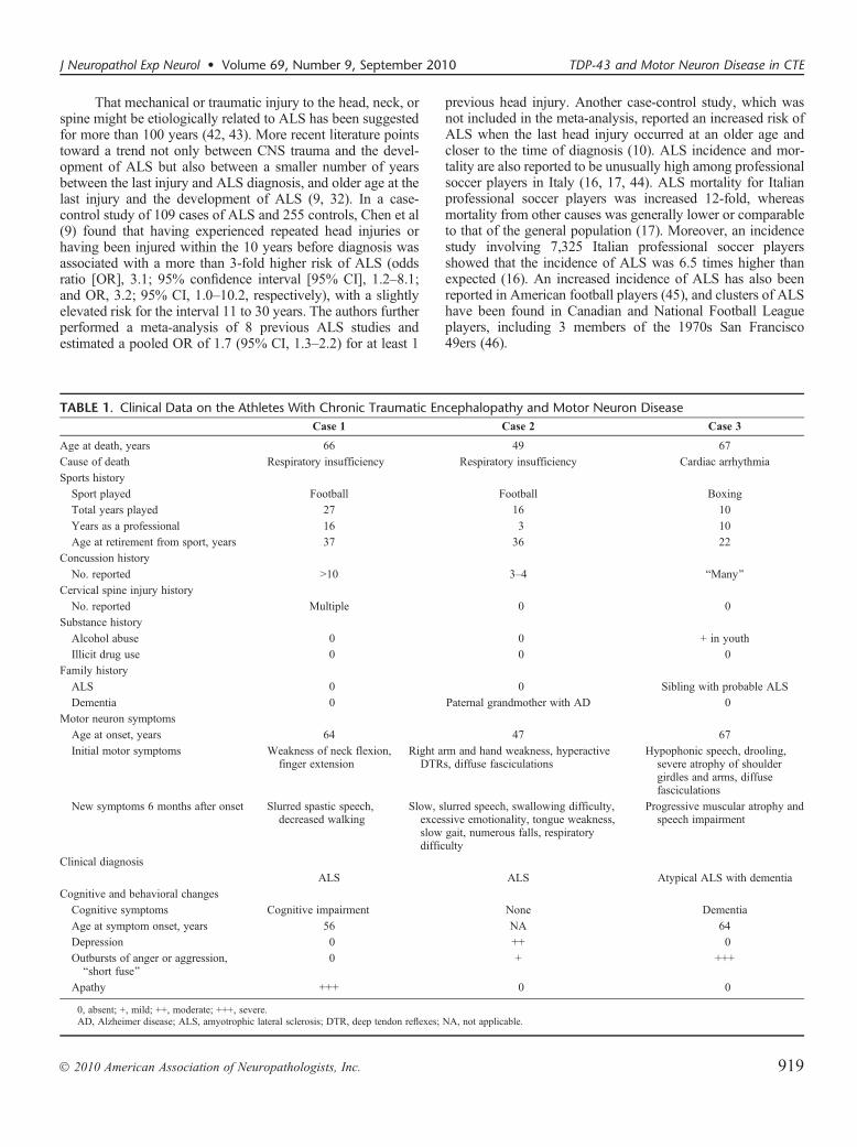
TABLE 1. Clinical Data on the Athletes With Chronic Traumatic Encephalopathy and Motor Neuron DiseaseCase 1 Case 2 Case 3
Age at death, years 66 49 67
Cause of death Respiratory insufficiency Respiratory insufficiency Cardiac arrhythmia
Sports history
Sport played Football Football Boxing
Total years played 27 16 10
Years as a professional 16 3 10
Age at retirement from sport, years 37 36 22
Concussion history
No. reported 910 3Y4 BMany[
Cervical spine injury history
No. reported Multiple 0 0
Substance history
Alcohol abuse 0 0 + in youth
Illicit drug use 0 0 0
Family history
ALS 0 0 Sibling with probable ALS
Dementia 0 Paternal grandmother with AD 0
Motor neuron symptoms
Age at onset, years 64 47 67
Initial motor symptoms Weakness of neck flexion,finger extension
Right arm and hand weakness, hyperactiveDTRs, diffuse fasciculations
Hypophonic speech, drooling,severe atrophy of shouldergirdles and arms, diffusefasciculations
New symptoms 6 months after onset Slurred spastic speech,decreased walking
Slow, slurred speech, swallowing difficulty,excessive emotionality, tongue weakness,slow gait, numerous falls, respiratorydifficulty
Progressive muscular atrophy andspeech impairment
Clinical diagnosis
ALS ALS Atypical ALS with dementia
Cognitive and behavioral changes
Cognitive symptoms Cognitive impairment None Dementia
Age at symptom onset, years 56 NA 64
Depression 0 ++ 0
Outbursts of anger or aggression,Bshort fuse[
0 + +++
Apathy +++ 0 0
0, absent; +, mild; ++, moderate; +++, severe.AD, Alzheimer disease; ALS, amyotrophic lateral sclerosis; DTR, deep tendon reflexes; NA, not applicable.
J Neuropathol Exp Neurol � Volume 69, Number 9, September 2010 TDP-43 and Motor Neuron Disease in CTE
� 2010 American Association of Neuropathologists, Inc. 919
The risk of ALS was reported to be increased approx-imately 2-fold among veterans of the 1991 Gulf War dur-ing the 10 years after the war (47Y49). This elevated riskwas evident among deployed military personnel who wereon active duty, with statistically significant elevations espe-cially notable among those in the Air Force and Army. Asecond independent study involving only Gulf War veteransyounger than 45 years also found an elevated risk of ALS inthis population (50). Recently, Schmidt et al (32) found thatmilitary veterans who had experienced head injuries duringthe last 15 years had an adjusted OR for the development ofALS of 2.33 (95% CI, 1.18Y4.61) relative to veterans with-out any head injuries, and that this association was strongestin APOE-?4 carriers. Several specific attributes of the at-riskmilitary veterans contributed to their higher prevalence ofhead injuries, including combat-related injuries during de-ployment to major conflicts and participation in competitivesports.
Repetitive head injury is also associated with the de-velopment of chronic traumatic encephalopathy (CTE), a pro-gressive tauopathy clinically associated with behavioral andpersonality changes, parkinsonism, and dementia (51). Re-cently, TDP-43 immunoreactivity was found in the cere-bral cortex in 3 cases of CTE associated with boxing (52).Although TDP-43 was originally thought to be a specificmarker for ALS and frontotemporal lobar degeneration (FTLD)with tau-negative ubiquitin-positive TDP-43Ypositive inclu-sions (FTLD-U, recently renamed FTLD-TDP) (5, 53Y57),TDP-43Ypositive inclusions have now been found in a varietyof other neurodegenerative disorders.
Through the brain donation program for Center for theStudy of Traumatic Encephalopathy at Boston UniversitySchool of Medicine and the Bedford VA Medical Center,we analyzed the brains and spinal cords of 12 former athletes
with CTE; 3 of the athletes with CTE also had signs andsymptoms of motor neuron disease (MND). We compared thenature and distribution of tau and TDP-43 immunoreactivity inthe brain and spinal cord of the 9 athletes with CTE withoutMND (CTEYnoMND)with those of the 3 with CTEwithMND(CTE + MND) and with the findings in the spinal cords of12 normal controls and 12 individuals with sporadic ALS. Wewanted to establish the extent of the TDP-43 proteinopathy inCTE, the relationship of TDP-43 immunoreactivity to symp-toms of MND, and whether the TDP-43Yimmunoreactivepathology colocalized with tau pathology.
MATERIALS AND METHODS
SubjectsThe brain and spinal cords from 11 of the 12 athletes
with pathologically verified CTE were received through thebrain donation program of the Boston University Alzheimer’sDisease Center (ADC) and the Center for the Study of Trau-matic Encephalopathy. Brain and spinal cord tissues from theremaining athlete (Case 3) were received from the Massa-chusetts Alzheimer’s Disease Research Center Brain Bank.Samples of paraffin-embedded brain and spinal cord sectionsfrom 12 age- and sex-matched subjects with pathologicallyverified ALS and 6 of the 12 normal controls were receivedfrom the National VA ALS biorepository. Samples of formalin-fixed spinal cord from the remaining 6 age- and sex-matchedcontrol subjects who were neurologically intact at time of deathwere received from the Mount Sinai ADC.
For the athletes, the concussion history, injury history,history of cognitive, behavioral, and neurological abnormal-ities, motor symptoms, neurological examinations, and clinicalstatus at the time of death were determined through review of
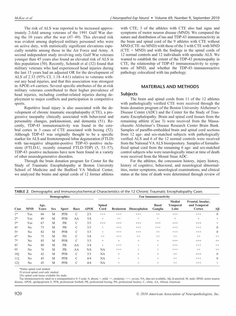
TABLE 2. Demographic and Immunocytochemical Characteristics of the 12 Chronic Traumatic Encephalopathy Cases
Case
Demographics Tau Immunoreactivity
AAMNDAge,Years Sex Sport Race APOE
SpinalCord Brainstem Diencephalon
BasalGanglia
MedialTemporal
Lobe
Frontal, Insular,and Temporal
Cortex
1* Yes 66 M PFB C 2/3 +++ +++ +++ ++ +++ +++ 0
2* Yes 49 M PFB AA 3/4 + ++ + + + + +
3* Yes 67 M PB C 3/3 +++ +++ +++ +++ +++ +++ +
4† No 73 M PB C 3/3 + +++ +++ +++ +++ +++ 0
5† No 82 M PFB C 3/3 + +++ +++ ++ +++ +++ 0
6* No 75 M PH C 3/4 ++ +++ ++ ++ ++ ++ ++
7* No 85 M PFB C 3/3 + + + + +++ +++ ++
8† No 80 M PB AA 3/4 + +++ + + +++ +++ ++
9† No 76 M PB AA NA NA +++ + + +++ ++ ++
10‡ No 42 M PFB C 3/3 NA + + + ++ +++ 0
11‡ No 45 M PFB C 4/4 NA + + + ++ +++ 0
12‡ No 45 M PFB C 4/4 NA + + + ++ +++ +
*Entire spinal cord studied.†Cervical spinal cord only studied.‡No spinal cord tissue available for study.Tau immunoreactivity rated on a semiquantitative 0Y3 scale: 0, absent; +, mild; ++, moderate; +++, severe; NA, data not available; AA, A-amyloid; M, male; MND, motor neuron
disease; APOE, apolipoprotein E; PFB, professional football; PB, professional boxing; PH, professional hockey; C, white; AA, African American.
McKee et al J Neuropathol Exp Neurol � Volume 69, Number 9, September 2010
� 2010 American Association of Neuropathologists, Inc.920

medical records and interviews with the players’ next of kin.When available, additional informants were interviewed toobtain confirmation or the information provided. Interviewswere conducted by a neuropsychologist (Robert Stern) who wasblinded to the results of the neuropathologic examination at thetime of the interview. Informants were interviewed beforereceiving information concerning the results of the neuro-pathologic examination. Medical records review was also con-ducted by a neurologist (Neil Kowall) and a neuropathologist(Ann McKee).
Neuropathologic ExaminationThe neuropathologic processing followed the proce-
dures previously established for the Boston University ADC,Mount Sinai ADC, and Massachusetts Alzheimer’s DiseaseResearch Center Brain Bank; these include a comprehen-sive analysis of all neurodegenerative conditions (58). Braintissues from 3 athletes were received in fragments fixed informalin after processing by medical examiners; brain andspinal cord tissues from the other 9 were received either freshor fixed in formalin. Paraffin-embedded sections from at least25 brain regions were stained with Luxol fast blue, hematox-ylin and eosin, Bielschowsky silver, AT8 (a mouse monoclonalantibody directed against phosphoserine 202 and phospho-threonine 205 of PHF-tau, 1:2000; Pierce Endogen, Rockford,IL), PHF-1, a monoclonal antibody against phosphoserine 396and phosphoserine 404 of hyperphosphorylated tau (1:1000;courtesy of Peter Davies), TDP-43 (rabbit polyclonal toTAR DNA-binding protein, 1:1000; Abcam, Cambridge, MA),LN3 (HLA-DR Class II [major histocompatibility complex],1:2000; Zymed, San Francisco, CA), ubiquitin (rabbit poly-clonal, 1:2000; Dako North America Inc, Carpinteria, CA), >-synuclein (rabbit polyclonal, 1:15,000; Chemicon, Temecula,CA), and A-amyloid (AA, mouse monoclonal, 1:2000, formicacid pretreatment; Dako North America Inc). In addition,multiple large coronal fragments were cut at 50 Km on a sledgemicrotome and stained as free-floating sections using CP13,a monoclonal antibody directed against phosphoserine 202of tau, considered to be the initial site of tau phosphorylationin neurofibrillary tangle (NFT) formation (1:200; courtesy ofPeter Davies), glial fibrillary acidic protein (mouse mono-clonal, 1:2000; Chemicon), TDP-43, PHF-1, ubiquitin, AA,and LN3, all counterstained with cresyl violet as previouslydescribed (51). For the control and ALS subjects, 10-Km-thicksections were prepared from paraffin blocks and stained withLuxol fast blue, hematoxylin and eosin, AT8, and TDP-43.
Double immunofluorescence staining was carried outto assess colocalization by confocal imaging using a LeicaSP5 laser scanning confocal microscope. Paraffin-embeddedtissue sections were deparaffinized in xylene and rehydratedthrough alcohol before being subjected to antigen retrieval informic acid. Sections were blocked in Super Block buffer(ScyTek, Logan, UT) containing 5% serum before avidin/biotin blocking and incubation overnight at 4-C with theinitial primary antibody. Sections were then incubated for1 hour at room temperature with a biotinylated secondaryantibody followed by avidin conjugated with either AlexaFluor 488 or Alexa Fluor 555 (Invitrogen). This protocol
(beginning with blocking procedures) was repeated for sec-ondary antigen detection.
Apolipoprotein E genotyping was conducted on 11 ofthe 12 athletes using restriction isotyping for determiningapolipoprotein E isoforms based on brain tissue samples.
RESULTS
Clinical DataAt death, the 12 athletes who developed CTE ranged in
age from 42 to 85 years (mean, 65.4 years; SD, 15.9 years)and included 7 former football players (6 of whom playedprofessionally and 1 in college), 4 retired professional boxers,and 1 professional hockey player (Tables 1 and 2). The 3athletes who developed clinically diagnosed MND included 2retired professional football players and a former professionalboxer. The players with CTE + MND did not differ from the 9athletes with CTEYno MND with respect to age at death, totalyears of participation in the sport, age at retirement from thesport, or concussion histories (data not shown).
The 3 athletes with CTE + MND had clinical pre-sentations characterized by profound muscle weakness, mus-cle atrophy, spasticity, and diffuse fasciculations (Table 1).Two developed motor symptoms after the development ofcognitive impairment or dementia and behavioral changes;one developed parkinsonism in addition to dementia, behav-ioral changes, and MND; and one developed prominentsymptoms of MND in the presence of behavioral changes anddepression but without evidence of cognitive decline or par-kinsonism. In all 3, the MND began with bilateral involvementof the shoulder girdles, neck, and arms, involved the bulbarmusculature early in the disease course, and resulted in deathwithin 2 to 3 years (see data, Supplemental Digital Content 1,http://links.lww.com/NEN/A161 for case summaries).
Pathological AspectsAthletes With CTE Without MND
Microscopically, the brains of all 9 athletes with CTEYnoMND showed the pathological changes of CTE, consisting ofnumerous tau-positive NFTs, neuropil neurites, and astrocytictangles in the frontal, temporal, and insular cortices, dien-cephalon, basal ganglia, and brainstem (Fig. 1). Of the 5 caseswith spinal cord tissue available for study, all 5 demonstratedoccasional tau-immunoreactive neurites and NFTs in the pos-terior, lateral, and/or anterior horns of the spinal cord; 7 of the 9also showed extensive TDP-43 immunoreactivity. The TDP-43Ypositive short threadlike and ring-shaped neurites (RNs),filamentous neuronal inclusions (FNIs), and ring-shaped glialinclusions (RGIs) were found in the frontal and temporal cortexand insula (Table 3, Figs. 1Y3). The TDP-43Ypositive RNsand RGIs were frequent in the subcortical white matter, andTDP-43Ypositive FNIs and RNs were common in the brain-stem, including the substantia nigra pars compacta, oculo-motor, inferior olivary, dorsal medullary, and hypoglossalnuclei (Fig. 3). The TDP-43Ypositive neurites were found inthe amygdala, hippocampus, caudate, putamen, thalamus,and hypothalamus (not shown). Double immunostaining andconfocal microscopy showed that the great majority of the
J Neuropathol Exp Neurol � Volume 69, Number 9, September 2010 TDP-43 and Motor Neuron Disease in CTE
� 2010 American Association of Neuropathologists, Inc. 921
McKee et al J Neuropathol Exp Neurol � Volume 69, Number 9, September 2010
� 2010 American Association of Neuropathologists, Inc.922
TDP-43Ypositive neurites and inclusions were not tau positive(Figs. 1, 2, 4). In the brain that showed the least TDP-43immunoreactivity (Case 10), RNs were limited to the substantianigra. Ubiquitin-positive or TDP-43Ypositive inclusions werenot found in the dentate gyrus in any case of CTE; a few caseshad rare TDP-43Ypositive neurites in the dentate gyrus(Table 3).
Athletes With CTE and MNDThe macroscopic features of the brains and spinal cords
of the athletes with CTE + MND did not differ from thosewith CTEYno MND, except for atrophy of the ventral roots ofthe spinal cord. All 3 brains showed the characteristicmicroscopic findings of CTE (Fig. 1). The 2 athletes withcognitive impairment or dementia and MND (Cases 1 and 3)had advanced CTE with dense NFTs throughout the medialtemporal lobe structures, neocortex, olfactory bulb, substantianigra, locus caeruleus, mammillary body, hypothalamus, andperiventricular regions. Betz cells in the precentral gyrus alsobore tau-immunoreactive NFTs. Case 2 displayed milderCTE, with involvement primarily limited to the frontal cortex,diencephalon, and brainstem. Abundant TDP-43YpositiveRNs, FNIs, and RGIs were found in widespread regions of the
brain and spinal cord in all 3 CTE + MND brains, generally ina far greater density than that found in the CTEYno MNDbrains. In the cortex, TDP-43Ypositive pathology was foundthroughout all cortical laminae and was most prominent in themotor cortex (Fig. 1).
In Cases 1 and 2 (both of whom developed end-stageMND with severe respiratory insufficiency and muscularweakness in the final months of their lives), the medullarypyramids and lateral corticospinal tracts throughout the spinalcord showed marked myelin and axonal loss with astrocy-tosis. Loss of anterior horn cells was profound, and there wasmarked atrophy and gliosis of ventral roots (Fig. 2). In Case 3(whose motor neuron symptoms were not as severe), therewas only mild degeneration of the lateral corticospinal tractsand ventral root atrophy. Staining with LN-3 showed numer-ous activated microglia and macrophages throughout the brainand spinal cord, most intense in the lateral corticospinal tractsof the cord. Anterior horn cells were markedly reduced innumber in Cases 1 and 2 and moderately reduced in numberin Case 3. Tau-positive neurites, astrocytic tangles, and NFTswere found in the posterior, lateral, and ventral horns, mostfrequently in Cases 1 and 3 where numerous astrocytic tanglessurrounded degenerating anterior horn cells (Fig. 2; Table 3).
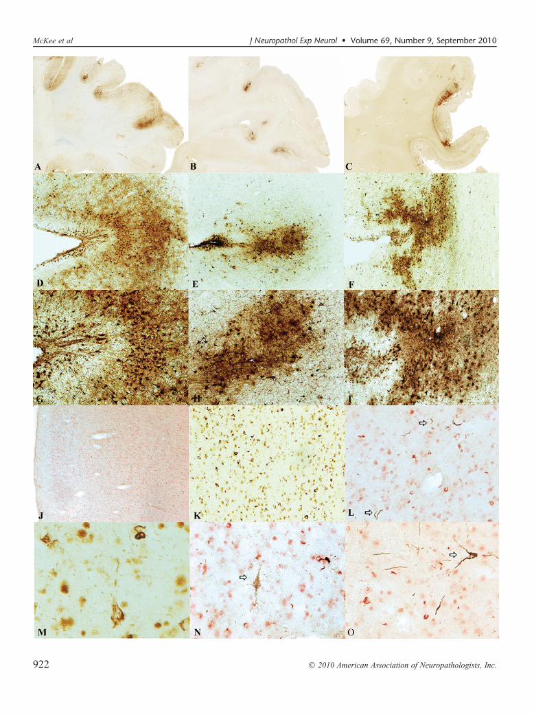
FIGURE 1. Cases of chronic traumatic encephalopathy (CTE) with widespread TAR DNA-binding protein of approximately 43 kd(TDP-43) immunoreactivity and motor neuron disease (CTE + MND). (AYC) Tau-immunoreactive neurofibrillary degeneration inthe frontal cortex of the 3 cases of CTE + MND (whole-mount 50-Km sections immunostained for CP13, original magnification:1�). (DYI) Tau-positive neurofibrillary tangles, glial tangles, and neuropil neurites are particularly dense at the depth of thecortical sulci, CP13 immunostain, original magnification: (DYF) 50�; (GYI) 100�. (J) TDP-43 immunostaining reveals abundantTDP-positive pathology in all cortical layers in the frontal cortex of Case 1, original magnification: 50�. (K) Numerous TDP-43Ypositive ring-shaped neurites (RNs) and ring-shaped glial inclusions (RGIs) in the frontal cortex of Case 2, original magnification:200�. (L)Double immunostaining shows that most TDP-43Ypositive RNs and RGIs (red) are not colocalized with tau-positive neurites(PHF-1 brown, arrows), original magnification: 400�. (M) TDP-43Ypositive filamentous neuronal inclusions, original magnifica-tion: 600�. (N) Double immunostaining shows a tau-positive pretangle (PHF-1 brown, arrow) that is not associated with TDP-43immunoreactivity (red), original magnification: 400�. (O) Double immunostaining shows a tau-positive tangle (PHF-1, arrow)that is not associated with TDP-43 immunoreactivity (red), original magnification: 400�.
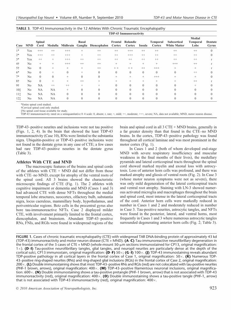
TABLE 3. TDP-43 Immunoreactivity in the 12 Athletes With Chronic Traumatic Encephalopathy
Case MND
TDP-43 Immunoreactivity
SpinalCord Medulla Midbrain
BasalGanglia Diencephalon
FrontalCortex
RolandicCortex Insula
TemporalCortex
SubcorticalWhite Matter
MedialTemporal
LobeDentateGyrus
1* Yes +++ ++ +++ + ++ ++ +++ ++ ++ ++ ++ 0
2* Yes +++ ++ +++ + ++ ++ +++ ++ ++ ++ ++ 0
3* Yes ++ + +++ ++ ++ ++ ++ ++ ++ ++ ++ r
4† No + + +++ ++ ++ + + + + +++ ++ r
5† No 0 + + + ++ + + + + ++ +++ + + r
6* No 0 + 0 + 0 0 0 0 0 0 ++ 0
7* No 0 0 + 0 0 + + + +++ + + 0
8† No 0 + + ++ + + + + + + ++ r
9† No NA ++ ++ + + 0 0 0 0 0 + 0
10‡ No NA NA + 0 0 0 0 0 0 0 0 0
11‡ No NA NA 0 0 0 0 0 0 0 0 0 0
12‡ No NA NA 0 0 0 0 0 0 0 0 0 0
*Entire spinal cord studied.†Cervical spinal cord only studied.‡No spinal cord tissue available for study.TDP-43 immunoreactivity rated on a semiquantitative 0Y4 scale: 0, absent; r, rare; +, mild; ++, moderate; +++, severe; NA, data not available; MND, motor neuron disease.
J Neuropathol Exp Neurol � Volume 69, Number 9, September 2010 TDP-43 and Motor Neuron Disease in CTE
� 2010 American Association of Neuropathologists, Inc. 923
McKee et al J Neuropathol Exp Neurol � Volume 69, Number 9, September 2010
� 2010 American Association of Neuropathologists, Inc.924

In the remaining anterior horn cells in all 3 cases, there werefrequent TDP-43Ypositive tau-negative FNIs, RNs, and RGIs(Figs. 2 and 4).
Normal Control CasesSections from the cervical, thoracic, and lumbar spinal
cord of 12 neurologically normal controls ranging in age from53 to 84 years (mean, 66.0 years; SD, 10.6 years), 9 men and3 women, were examined. Rare tau-positive neurites were
found in the ventral horn of 4 cases, primarily in the cervicalcord; no tau-positive NFTs or astrocytic tangles were observed.There was no TDP-43 immunoreactivity in any case.
Sporadic ALS CasesSections from cervical, thoracic, and lumbar spinal cord
in 12 age-matched men with sporadic ALS, ranging in agefrom 45 to 77 years (mean, 64.3 years; SD, 9.5 years) wereexamined. Rare tau-positive neurites were found in 1 of
FIGURE 3. TAR DNA-binding protein of approximately 43 kd (TDP-43) immunoreactivity in chronic traumatic encephalopathy(CTE). TDP-43 immunoreactivity is found as glial cytoplasmic inclusions (GCIs) and neuropil neurites in multiple brainstem nucleiincluding hypoglossal nucleus (A), oculomotor nucleus (B), substantia nigra (C) (original magnification: 200�). TDP-43 immu-noreactivity in the medial temporal lobe structures consists primarily of dotlike neurites. (D) Hippocampus, CA1 (original magni-fication: 200�). TDP-43Ypositive dystrophic neurites and GCIs are also found in white matter. (E) Subcortical frontal white matter(original magnification: 200�). No ubiquitinated or TDP-43Ypositive inclusions are found in the dentate gyrus of the hippocampus.(F) Dentate gyrus of the hippocampus (original magnification: 400�).
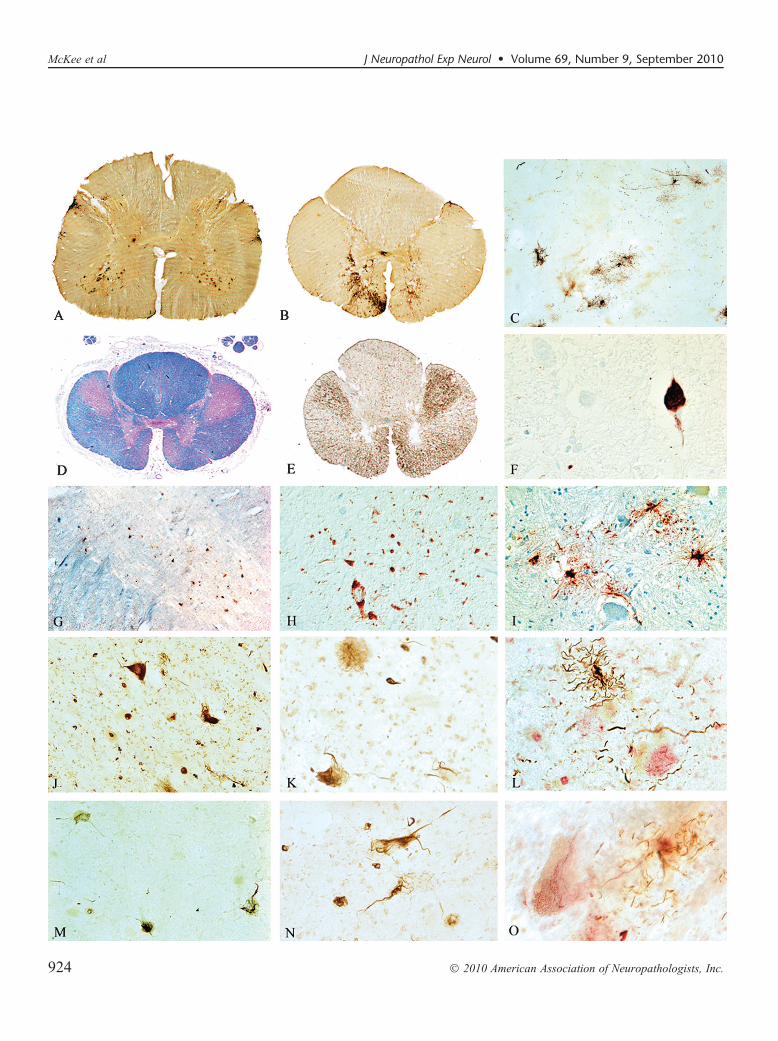
FIGURE 2. Spinal cord pathology in chronic traumatic encephalopathy (CTE) with TAR DNA-binding protein of approximately 43 kd(TDP-43) proteinopathy, tauopathy, and motor neuron disease (CTE + MND). (A, B) Whole-mount 50-Km sections of lumbar andthoracic spinal cord immunostained with antibody AT8 showing abundant tau immunostaining in the ventral horns. (C) Tau-positiveastrocytic tangles in the ventral horn of Case 1 (AT8 immunostain, original magnification: 100�). (D) Whole-mount 10-Km sectionthrough high thoracic spinal cord showing marked myelin and axonal loss in the lateral and ventral corticospinal tracts. Atrophicventral roots are not visible. Luxol fast blue hematoxylin and eosin stain, original magnification: 1�. (E)Whole-mount 50-Km sectionof high thoracic cord showing intense immunoreactivity for activated microglia and macrophages in lateral and corticospinal tracts(LN-3 immunostain, original magnification: 1�). (F) Tau-positive neurofibrillary tangles in ventral horn of Case 3, AT8 immunostain,original magnification: 600�. (G) TDP-43 immunoreactivity in ventral horn, original magnification: 50�. (H) TDP-43 immunor-eactive filamentous neuronal inclusions (FNIs), ring-shaped glial inclusions (RGIs), and ring-shaped neurites (RNs) in the ventral hornsof the lumbar spinal cord in Case 3, original magnification: 200�. (I) Tau-positive astrocytes and their processes surroundingdegenerating anterior horn cells in the thoracic spinal cord (AT8 immunostain, original magnification: 350�). (J) TDP-43YpositiveFNIs, RGIs, and RNs in the ventral horns of the lumbar spinal cord in Case 2, original magnification: 200�. (K) TDP-43Ypositive FNI inthe anterior horn, original magnification: 400�.(L) Double immunostained sections show tau-positive astrocytes (brown) and theirprocesses surrounding anterior horn neurons containing TDP-43Ypositive filamentous inclusions (red), PHF-1 and TDP-43 immu-nostains, original magnification: 400�. (M) TDP-43Ypositive FNIs, RGIs, and RNs in the lumbar ventral horns in Case 1, originalmagnification: 200�. (N) TDP-43Ypositive FNIs in the anterior horn, original magnification: 400�. (O) Double immunostainedsections showing tau-positive astrocytes (brown) and their processes surrounding anterior horn neurons containing TDP-43YpositiveFNIs (red), PHF-1 and TDP-43 immunostains, original magnification: 600�.
J Neuropathol Exp Neurol � Volume 69, Number 9, September 2010 TDP-43 and Motor Neuron Disease in CTE
� 2010 American Association of Neuropathologists, Inc. 925
12 cases of sporadic ALS. Immunoreactivity for TDP-43 waspresent in every ALS case and ranged from TDP-43Ypositivethreadlike and curved neurites to abundant short skeinlikeinclusions or punctate neuronal cytoplasmic immunoreac-tivity within the remaining anterior horn cells.
DISCUSSIONWe report a widespread TDP-43 proteinopathy in more
than 80% of our cases of CTE that involved the brainstem,basal ganglia, diencephalon, medial temporal lobe, frontal,temporal, and insular cortices, and subcortical white matter inmost cases. Moreover, in 3 athletes with CTE who developeda progressive MND several years before death, there wereextensive TDP-43Ypositive FNIs, RGIs, and RNs in theanterior horns of the spinal cord in addition to tau-positiveastrocytic tangles, neurites, and occasional NFTs. Thesefindings suggest that a distinctive widespread TDP-43 pro-teinopathy is associated with CTE, and that in some individ-uals, the TDP-43 proteinopathy extends to involve the spinalcord and is clinically manifest as MND.
Widespread TDP-43 immunoreactivity has been pre-viously reported in CTE (52). The shared presence of 2aggregated phosphorylated proteins associated with neuro-degeneration in the great majority of cases argues against thecoincidental occurrence of CTE and sporadic ALS, suggest-ing instead that a common stimulus provokes the pathologicalaccumulation of both proteins. In CTE cases with mild TDP-43 immunoreactivity, TDP-43Yimmunoreactive RGIs andRNs were found in the midbrain, medulla, basal ganglia, andmedial temporal lobe; and in the least affected case of CTE,TDP-43Ypositive RNs were limited to the substantia nigra.
Occasional TDP-43Ypositive RNs were found in the spinalcord of Case 4, a 73-year-old former professional boxer withsevere end-stage CTE but with no recognized symptoms ofmotor neuron dysfunction, and Case 8, a former professionalhockey player with advanced CTE.
Frontotemporal lobar degeneration is also associatedwith MND and TDP-43 immunoreactivity, although FTLD-TDP 43/MND is characterized by neuronal inclusions that areimmunoreactive for ubiquitin but not tau (FTLD with tau-negative ubiquitin-positive inclusions), and there are no tau-immunoreactive NFTs, astrocytic tangles, or neurites (59).Conversely, the TDP-43 positivity found in CTE is associatedwith an extensive tauopathy, and none of the CTE cases boreany ubiquitin or TDP-43Ypositive inclusions in the dentategyrus. Moreover, the morphology and cortical distribution ofTDP-43Ypositive RNs, FGIs, and RGIs in CTE seem to bedistinct.
In CTE +MND, tau-positive NFTs, astrocytic tangles, andneurites are also found in the spinal cord. No tau-immunoreactiveNFTs were found in our cases of sporadic ALS, and none havebeen described in the literature (60, 61). Rare tau NFTs havebeen described in the spinal cord from individuals with spora-dic AD, but we found none in the spinal cord from age-matched controls (60, 61).
The TDP-43 is a highly conserved protein that is widelyexpressed in many tissues, including the CNS. Its physio-logical functions are diverse and incompletely understood butlikely involve the regulation of multiple biological processesvia TDP-43 binding to DNA, RNA, and other proteins.Originally, TDP-43 was thought to be specific to FTLD-TDP,ALS-D/FTLD-MND, and ALS, but it is now recognized in a
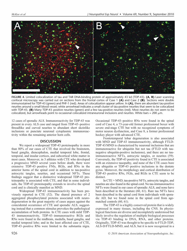
FIGURE 4. Limited colocalization of tau and TAR DNA-binding protein of approximately 43 kd (TDP-43). (A, B) Laser scanningconfocal microscopy was carried out on sections from the frontal cortex of Case 1 (A) and Case 2 (B). Sections were doubleimmunostained for TDP-43 (green) and PHF-1 (red). Areas of colocalization appear yellow. In (A), there are abundant tau-positiveneurites around a small blood vessel; white arrowhead indicates a small cluster of tau-positive neurites that seem to be colocalizedwith TDP-43. (B) Many TDP-43Ypositive neurites (green) and a few tau-positive neurites (red). Most neurites do not seem to becolocalized, but arrowheads point to occasional colocalized intraneuronal inclusions and neurites. White bars = 200 Km.
McKee et al J Neuropathol Exp Neurol � Volume 69, Number 9, September 2010
� 2010 American Association of Neuropathologists, Inc.926

variety of other disorders in which it is present to a lesserdegree and considered a secondary pathology. The majorTDP-43 proteinopathies, FTLD-TDP, FTLD-MND/ALS-D,and ALS, once thought to be discrete clinical and patho-logical subtypes, have more recently been considered torepresent different points on a continuous spectrum of amultisystem degeneration. The FTLD-TDP, FTLD-MND/ALS-D, and ALS all show widespread CNS TDP-43 path-ology; however, the presence of MND or ALS is associatedwith a higher burden of inclusions in lower motor neurons,and cognitive dysfunction is related to a higher degree ofcortical pathology (6, 62).
Diseases with secondary TDP-43 pathology include ADand hippocampal sclerosis (63), Guam parkinsonism-dementiacomplex (PDC) (64), Pick disease, corticobasal degeneration,argyrophilic grain disease, and Lewy body disease (5, 28, 65).CTE is a 3R/4R tauopathy; when TDP-43 has been reported inother 3R/4R tauopathies, such as AD and Guam PDC, it maybe seen in neurons with NFTs. We also found a minor degreeof colocalization of TDP-43 and tau; however, the majority ofTDP-43Ypositive inclusions were in discrete structures. In AD,TDP-43 immunoreactivity is limited primarily to the medialtemporal lobe, except in advanced stages (i.e. Braak V or VI) inwhich it may be found in the neocortex and basal ganglia (52).
Neuropathologically, the tau neurofibrillary degener-ation of CTE most resembles that in Guam PDC (60),although the patchy irregular involvement of the cortex andthe perivascular distribution of dense NFTs distinguish CTEfrom Guam PDC. In Guam PDC, TDP-43Ypositive neuritesare found in the frontal subcortical white matter and cortex,and neuronal cytoplasmic inclusions are found in the dentategyrus of the medial temporal lobe (64). Although the patho-genesis of Guam PDC is unknown, environmental factors,such as toxins in cycad seeds, minerals in the soil or drinkingwater, and genetic susceptibility have been implicated (66).Guam PDC might be another example of an environmentallyacquired tauopathy and TDP-43 proteinopathy that is asso-ciated with cognitive impairment, parkinsonism, and, in someinstances, MND.
Recent work using in vitro and in vivo animal modelsindicates that overexpression of wild-type human TDP-43 andits dislocation from the neuronal nucleus to the cytoplasm areassociated with neurodegeneration and cell death (67). Injec-tions of viral wild-type human TDP-43 constructs in the sub-stantia nigra of rats caused loss of substantia nigra neurons,gliosis, and altered behavior (68). Transgenic Drosophilaoverexpressing human TDP-43 in motor neurons developedaxonal swelling, reduction in axonal branches, and motorneuron loss. Similarly, transgenic mice overexpressing TDP-43in spinal and cortical motor neurons were reported to developa dose-dependent degeneration of cortical and spinal motorneurons, nonmotor cortical and subcortical neurons, and spasticquadriplegia reminiscent of ALS (69).
During a traumatic brain injury, the brain and spinal cordundergo shear deformation, producing a transient elongation orstretch of axons. Traumatic axonal injury also perturbs thecytoskeleton, causing dissolution of microtubules and neuro-filaments and pathological reorganization of neurofilamentproteins (70). By virtue of its capacity to bind to neurofilament
mRNA and stabilize the mRNA transcript (71), TDP -43 playsa critical role in mediating the response of the neuronal cyto-skeleton to axonal injury. Models of traumatic brain injury inrodents show the accumulation of many key proteins that formpathological aggregates in human neurodegenerative diseases,including neurofilament proteins, amyloid precursor protein, >-synuclein, and tau (72). The TDP-43 is intrinsically prone toaggregation, and TDP-43 expression is upregulated afterexperimental axotomy in spinal motor neurons of the mouse(73). Conceivably, traumatic axonal injury may also accelerateTDP-43 accumulation, aggregation, and dislocation to thecytoplasm and thereby enhance its neurotoxicity.
There are multiple other biological mechanisms bywhich repetitive head injury trauma may trigger the molecularpathways leading to neuronal degeneration in CTE and CTE +MND, including inflammation, glutamate excitotoxicity, andoxidative stress. Their contributions to the molecular cascadesin CTE remain to be explored. Additional studies are clearlynecessary to understand the specific additional risk factors(including genetic), pathogenesis, and potential for ther-apeutic intervention in CTE and CTE + MND. The existenceof a long period of latency between the traumatic injury andthe onset of CTE or MND increases the window for imple-menting therapeutic measures to dampen or block the neuro-degenerative cascade triggered by such injuries.
This report suggests that the play of contact sports,including boxing, football, and hockey, might be associatedwith a widespread TDP-43 proteinopathy that, in some indi-viduals, is manifest as MND. The accumulation of TDP-43 aswell as tau in the neocortex, medal temporal lobes, and deeperbrain structures likely contributes to the overall clinical mani-festations of cognitive and memory loss, behavioral changes,and parkinsonism. Whether repetitive head trauma alone pro-vokes these neurodegenerative cascades or only in associationwith certain genetic constellations remains to be determined.
ACKNOWLEDGMENTSThe authors thank the families for their generous par-
ticipation in this study.
REFERENCES1. Bruijn LI, Miller TM, Cleveland DW. Unraveling the mechanisms
involved in motor neuron degeneration in ALS. Annu Rev Neurosci2004;27:723Y49
2. Mulder DW, Kurland LT, Offord KP, et al. Familial adult motor neurondisease: Amyotrophic lateral sclerosis. Neurology 1986;36:511Y17
3. Okamoto K, Murakami N, Kusaka H, et al. Ubiquitin-positive intra-neuronal inclusions in the extramotor cortices of presenile dementiapatients with motor neuron disease. J Neurol 1992;239:426Y30
4. Wightman G, Anderson VE, Martin J, et al. Hippocampal and neocorticalubiquitin-immunoreactive inclusions in amyotrophic lateral sclerosis withdementia. Neurosci Lett 1992;139:269Y74
5. Dickson DW. Neuropathology of non-Alzheimer degenerative disorders.Int J Clin Exp Pathol 2009;3:1Y23
6. Geser F, Martinez-Lage M, Kwong LK, et al. Amyotrophic lateral scle-rosis, frontotemporal dementia and beyond: The TDP-43 diseases.J Neurol 2009;256:1205Y14
7. Kondo K, Tsubaki T. Case-control studies of motor neuron disease:Association with mechanical injuries. Arch Neurol 1981;38:220Y26
8. Deapen DM, Henderson BE. A case-control study of amyotrophic lateralsclerosis. Am J Epidemiol 1986;123:790Y99
9. Chen H, Richard M, Sandler DP, et al. Head injury and amyotrophiclateral sclerosis. Am J Epidemiol 2007;166:810Y16
J Neuropathol Exp Neurol � Volume 69, Number 9, September 2010 TDP-43 and Motor Neuron Disease in CTE
� 2010 American Association of Neuropathologists, Inc. 927
10. Binazzi A, Belli S, Uccelli R, et al. An exploratory case-control study onspinal and bulbar forms of amyotrophic lateral sclerosis in the province ofRome. Amyotroph Lateral Scler 2009;10:361Y69
11. Kurtzke JF, Beebe GW. Epidemiology of amyotrophic lateral sclerosis: 1.A case-control comparison based on ALS deaths. Neurology 1980;30:453Y62
12. Gawel M, Zaiwalla Z, Rose FC. Antecedent events in motor neurondisease. J Neurol Neurosurg Psychiatry 1983;46:1041Y43
13. Strickland D, Smith SA, Dolliff G, et al. Physical activity, trauma, andALS: A case-control study. Acta Neurol Scand 1996;94:45Y50
14. Scarmeas N, Shih T, Stern Y, et al. Premorbid weight, body mass, andvarsity athletics in ALS. Neurology 2002;59:773Y75
15. Longstreth WT, McGuire V, Koepsell TD, et al. Risk of amyotrophiclateral sclerosis and history of physical activity: A population-basedcase-control study. Arch Neurol 1998;55:201Y6
16. Chio A, Benzi G, Dossena M, et al. Severely increased risk of amyo-trophic lateral sclerosis among Italian professional football players. Brain2005;128:472Y76
17. Belli S, Vanacore N. Proportionate mortality of Italian soccer players: Isamyotrophic lateral sclerosis an occupational disease? Eur J Epidemiol2005;20:237Y42
18. Kamel F, Umbach DM, Munsat TL, et al. Association of cigarettesmoking with amyotrophic lateral sclerosis. Neuroepidemiology 1999;18:194Y202
19. Nelson LM, McGuire V, Longstreth WT, et al. Population-based case-control study of amyotrophic lateral sclerosis in western Washingtonstate. I. Cigarette smoking and alcohol consumption. Am J Epidemiol2000;151:156Y63
20. Weisskopf MG, McCullough ML, Calle EE, et al. Prospective study ofcigarette smoking and amyotrophic lateral sclerosis. Am J Epidemiol2004;160:26Y33
21. Campbell AM, Williams ER, Barltrop D. Motor neurone disease andexposure to lead. J Neurol Neurosurg Psychiatry 1970;33:877Y85
22. Armon C, Kurland LT, Daube JR, et al. Epidemiologic correlates ofsporadic amyotrophic lateral sclerosis. Neurology 1991;41:1077Y84
23. Chancellor AM, Slattery JM, Fraser H, et al. Risk factors for motorneuron disease: A case-control study based on patients from the ScottishMotor Neuron Disease Register. J Neurol Neurosurg Psychiatry 1993;56:1200Y6
24. McGuire V, Longstreth WT, Nelson LM, et al. Occupational exposuresand amyotrophic lateral sclerosis. A population-based case-control study.Am J Epidemiol 1997;145:1076Y88
25. Kamel F, Umbach DM, Munsat TL, et al. Lead exposure and amyo-trophic lateral sclerosis. Epidemiology 2002;13:311Y19
26. Felmus MT, Patten BM, Swanke L. Antecedent events in amyotrophiclateral sclerosis. Neurology 1976;26:167Y72
27. Pierce-Ruhland R, Patten BM. Repeat study of antecedent events inmotor neuron disease. Ann Clin Res 1981;13:102Y7
28. Gresham LS, Molgaard CA, Golbeck AL, et al. Amyotrophic lateralsclerosis and occupational heavy metal exposure: A case-control study.Neuroepidemiology 1986;5:29Y38
29. Haynal A, Regli F. Amyotrophic lateral sclerosis associated with accu-mulated electric injury [In German]. Confin Neurol 1964;24:189Y98
30. Savettieri G, Salemi G, Arcara A, et al. A case-control study of amyo-trophic lateral sclerosis. Neuroepidemiology 1991;10:242Y45
31. Majoor-Krakauer D, Willems PJ, Hofman A. Genetic epidemiology ofamyotrophic lateral sclerosis. Clin Genet 2003;63:83Y101
32. Schmidt S, Kwee LC, Allen KD, et al. Association of ALS with headinjury, cigarette smoking and APOE genotypes. J Neurol Sci 2010;291:22Y29
33. Fleminger S, Oliver DL, Lovestone S, et al. Head injury as a risk factorfor Alzheimer’s disease: The evidence 10 years on; a partial replication. JNeurol Neurosurg Psychiatry 2003;74:857Y62
34. Mortimer JA, French LR, Hutton JT, et al. Head injury as a risk factor forAlzheimer’s disease. Neurology 1985;35:264Y67
35. O’Meara ES, Kukull WA, Sheppard L, et al. Head injury and risk ofAlzheimer’s disease by apolipoprotein E genotype. Am J Epidemiol1997;146:373Y84
36. Mehta KM, Ott A, Kalmijn S, et al. Head trauma and risk of dementia andAlzheimer’s disease: The Rotterdam Study. Neurology 1999;53:1959Y62
37. Katzman R, Galasko DR, Saitoh T, et al. Apolipoprotein-epsilon4 andhead trauma: Synergistic or additive risks? Neurology 1996;46:889Y91
38. Mayeux R, Ottman R, Maestre G, et al. Synergistic effects of traumatichead injury and apolipoprotein-epsilon 4 in patients with Alzheimer’sdisease. Neurology 1995;45:555Y57
39. Plassman BL, Havlik RJ, Steffens DC, et al. Documented head injury inearly adulthood and risk of Alzheimer’s disease and other dementias.Neurology 2000;55:1158Y66
40. Bower JH, Maraganore DM, Peterson BJ, et al. Head trauma precedingPD: A case-control study. Neurology 2003;60:1610Y15
41. Goldman SM, Tanner CM, Oakes D, et al. Head injury and Parkinson’sdisease risk in twins. Ann Neurol 2006;60:65Y72
42. Erb W. Zur Casuistik der bulbaren Lahmungen: Uber einen neuenwahrscheinlich bulbaren Lahmungscomplex. Eur Arch Psychiatry ClinNeurosci 1879;9:325Y50
43. Hanisch R, Dworsky RL, Henderson BE. Letter: A search for clues to thecause of amyotrophic lateral sclerosis. Arch Neurol 1976;33:456Y57
44. Al-Chalabi A, Leigh PN. Trouble on the pitch: Are professional footballplayers at increased risk of developing amyotrophic lateral sclerosis?Brain 2005;128:451Y53
45. Abel EL. Football increases the risk for Lou Gehrig’s disease, amyo-trophic lateral sclerosis. Percept Mot Skills 2007;104:1251Y54
46. Wallis C, Dorfman A. Medicine: Probing AMysterious Cluster. TIME.com[Web site]. February 23, 1987. Available at: http://www.time.com/time/magazine/article/0,9171,963607-1,00.html. Accessed September 28, 2008
47. Horner RD, Kamins KG, Feussner JR, et al. Occurrence of amyotrophiclateral sclerosis among Gulf War veterans. Neurology 2003;61:742Y49
48. Coffman CJ, Horner RD, Grambow SC, et al. Estimating the occurrenceof amyotrophic lateral sclerosis among Gulf War (1990Y1991) veteransusing capture-recapture methods. Neuroepidemiology 2005;24:141Y50
49. Weisskopf MG, O’Reilly EJ, McCullough ML, et al. Prospective study ofmilitary service and mortality from ALS. Neurology 2005;64:32Y37
50. Haley RW. Excess incidence of ALS in young Gulf War veterans. Neu-rology 2003;61:750Y56
51. McKee A, Cantu R, Nowinski C, et al. Chronic traumatic encephalopathyin athletes: Progressive tauopathy after repetitive head injury. J Neuro-pathol Exp Neurol 2009;68:709Y35
52. King A, Sweeney F, Bodi I, et al. Abnormal TDP-43 expression is identi-fied in the neocortex in cases of dementia pugilistica, but is mainly con-fined to the limbic system when identified in high and moderate stages ofAlzheimer’s disease. Neuropathology 2010 [Epub ahead of print]
53. Arai T, Hasegawa M, Akiyama H, et al. TDP-43 is a component ofubiquitin-positive tau-negative inclusions in frontotemporal lobar degener-ation and amyotrophic lateral sclerosis. Biochem Biophys Res Commun2006;351:602Y11
54. Neumann M, Sampathu DM, Kwong LK, et al. Ubiquitinated TDP-43 infrontotemporal lobar degeneration and amyotrophic lateral sclerosis.Science 2006;314:130Y33
55. Hu WT, Josephs KA, Knopman DS, et al. Temporal lobar predominanceof TDP-43 neuronal cytoplasmic inclusions in Alzheimer disease. ActaNeuropathol 2008;116:215Y20
56. Mackenzie IR, Baborie A, Pickering-Brown S, et al. Heterogeneity ofubiquitin pathology in frontotemporal lobar degeneration. Acta Neuro-pathol 2006;112:539Y49
57. Sampathu DM, Neumann M, Kwong LK, et al. Pathological hetero-geneity of frontotemporal lobar degeneration with ubiquitin-positiveinclusions delineated by ubiquitin immunohistochemistry and novelmonoclonal antibodies. Am J Pathol 2006;169:1343Y52
58. Vonsattel JP, Aizawa H, Ge P, et al. An improved approach to preparehuman brains for research. J Neuropathol Exp Neurol 1995;54:42Y56
59. Cairns NJ, Bigio EH, Mackenzie IRA, et al. Neuropathologic diagnosticand nosologic criteria for frontotemporal lobar degeneration: Consensusof the Consortium for Frontotemporal Lobar Degeneration. Acta Neuro-pathol 2007;114:5Y22
60. Schmidt ML, Zhukareva V, Perl DP, et al. Spinal cord neurofibrillarypathology in Alzheimer disease and Guam parkinsonism-dementia com-plex. J Neuropathol Exp Neurol 2001;60:1075Y86
61. Umahara T, Hirano A, Kato S, et al. Demonstration of neurofibrillarytangles and neuropil thread-like structures in spinal cord white matter inparkinsonism-dementia complex on Guam and in Guamanian amyo-trophic lateral sclerosis. Acta Neuropathol 1994;88:180Y84
62. Geser F, Martinez-Lage M, Robinson J, et al. Clinical and pathologicalcontinuum of multisystem TDP-43 proteinopathies. Arch Neurol 2009;66:180Y89
McKee et al J Neuropathol Exp Neurol � Volume 69, Number 9, September 2010
� 2010 American Association of Neuropathologists, Inc.928
63. Amador-Ortiz C, Lin WL, Ahmed Z, et al. TDP-43 immunoreactivity inhippocampal sclerosis and Alzheimer’s disease. Ann Neurol 2007;61:435Y45
64. Hasegawa M, Arai T, Akiyama H, et al. TDP-43 is deposited in the Guamparkinsonism-dementia complex brains. Brain 2007;130:1386Y94
65. Nakashima-Yasuda H, Uryu K, Robinson J, et al. Co-morbidity ofTDP-43 proteinopathy in Lewy bodyYrelated diseases. Acta Neuropathol2007;114:221Y29
66. Sieh W, Choi Y, Chapman NH, et al. Identification of novel susceptibilityloci for Guam neurodegenerative disease: Challenges of genome scans ingenetic isolates. Hum Mol Genet 2009;18:3725Y38
67. Barmada SJ, Skibinski G, Korb E, et al. Cytoplasmic mislocalization ofTDP-43 is toxic to neurons and enhanced by a mutation associated withfamilial amyotrophic lateral sclerosis. J Neurosci 2010;30:639Y49
68. Tatom JB, Wang D, Dayton R, et al. Mimicking aspects of fronto-temporal lobar degeneration and Lou Gehrig’s disease in rats via TDP-43overexpression. Mol Ther 2009;17:607Y13
69. Wils H, Kleinberger G, Janssens J, et al. TDP-43 transgenic mice developspastic paralysis and neuronal inclusions characteristic of ALS andfrontotemporal lobar degeneration. Proc Natl Acad Sci U S A 2010;107:3858Y63
70. Serbest G, Burkhardt M, Siman R, et al. Temporal profiles of cytoskeletalprotein loss following traumatic axonal injury in mice. Neurochem Res2007;32:2006Y14
71. Strong MJ, Volkening K, Hammond R, et al. TDP43 is a human lowmolecular weight neurofilament (hNFL) mRNA-binding protein. MolCell Neurosci 2007;35:320Y27
72. Uryu K, Chen XH, Martinez D, et al. Multiple proteins implicated inneurodegenerative diseases accumulate in axons after brain trauma inhumans. Exp Neurol 2007;208:185Y92
73. Moisse K, Mepham J, Volkening K, et al. Cytosolic TDP-43 expressionfollowing axotomy is associated with caspase 3 activation in NFL-/- mice:Support for a role for TDP-43 in the physiological response to neuronalinjury. Brain Res 2009;1296:176Y86
J Neuropathol Exp Neurol � Volume 69, Number 9, September 2010 TDP-43 and Motor Neuron Disease in CTE
� 2010 American Association of Neuropathologists, Inc. 929