Embed Size (px)
Citation preview
TEMPORAL SELECTION ENHANCES V1 ACTIVITY
1
Running Head: TEMPORAL SELECTION ENHANCES V1 ACTIVITY 1 2 3 4 5 6 7 8 9
The Selection of Events in Time Enhances Activity Throughout Early Visual Cortex 10 11
Khena M. Swallow 12 Tal Makovski 13
Yuhong V. Jiang 14 15
Department of Psychology and Center for Cognitive Sciences 16 University of Minnesota 17
18 19 20 21 22 23 24 25 26 27 28 Address Correspondence to: 29 Khena M. Swallow 30 Department of Psychology 31 University of Minnesota 32 N218 Elliott Hall 33 75 East River Road 34 Minneapolis, MN 55455 35 [email protected] 36 Office: (612) 626-3577 37 Fax: (612) 626-2079 38
39
40
41
TEMPORAL SELECTION ENHANCES V1 ACTIVITY
2
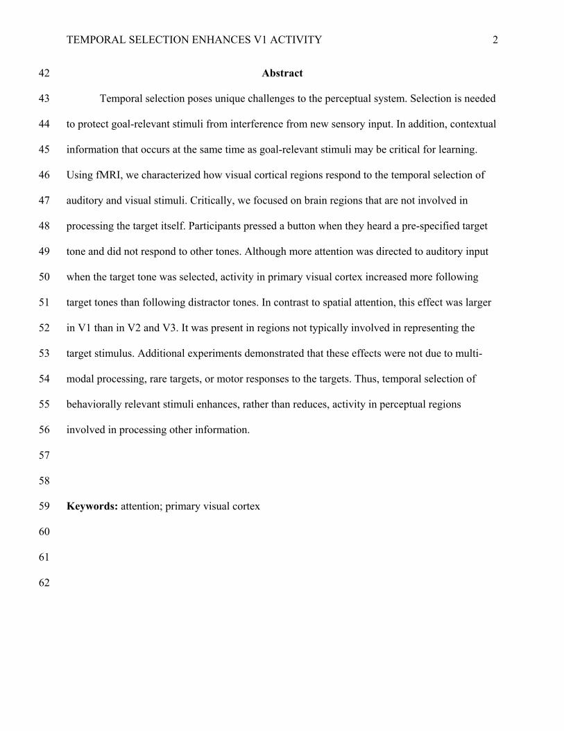
Abstract 42
Temporal selection poses unique challenges to the perceptual system. Selection is needed 43
to protect goal-relevant stimuli from interference from new sensory input. In addition, contextual 44
information that occurs at the same time as goal-relevant stimuli may be critical for learning. 45
Using fMRI, we characterized how visual cortical regions respond to the temporal selection of 46
auditory and visual stimuli. Critically, we focused on brain regions that are not involved in 47
processing the target itself. Participants pressed a button when they heard a pre-specified target 48
tone and did not respond to other tones. Although more attention was directed to auditory input 49
when the target tone was selected, activity in primary visual cortex increased more following 50
target tones than following distractor tones. In contrast to spatial attention, this effect was larger 51
in V1 than in V2 and V3. It was present in regions not typically involved in representing the 52
target stimulus. Additional experiments demonstrated that these effects were not due to multi-53
modal processing, rare targets, or motor responses to the targets. Thus, temporal selection of 54
behaviorally relevant stimuli enhances, rather than reduces, activity in perceptual regions 55
involved in processing other information. 56
57
58
Keywords: attention; primary visual cortex 59
60
61
62
TEMPORAL SELECTION ENHANCES V1 ACTIVITY
3
Although the natural environment is usually stable over time, changes in sensory input 63
occur with the appearance of new objects and navigation through the environment. Some of 64
these changes may be more relevant to a person’s goals than others. Adaptive perception requires 65
attentional selection over time (Chun & Potter, 1995; Pashler, 1994; Neisser, 1976). Previous 66
studies have characterized temporal selection as a late process that facilitates encoding into 67
working memory (Bowman & Wyble, 2007; Chun & Potter, 1995; Olivers & Meeter, 2008). 68
However, its impact on early visual cortical activity is poorly understood. In this study, we use 69
functional Magnetic Resonance Imaging (fMRI) to examine how the temporal selection of brief 70
auditory and visual stimuli affects activity in early visual cortical regions that are not involved in 71
coding them. 72
One way temporal selection may affect early visual activity is by recruiting spatial 73
selection mechanisms for a brief period of time. Spatial selection prioritizes the processing of 74
selected locations. It ensures that objects in those locations successfully compete for neural 75
representation within a neuron’s receptive field (Desimone & Duncan, 1995; Reynolds & 76
Chelazzi, 2004). The resulting bias manifests as increased activity in regions representing the 77
attended location, and decreased activity in regions representing nearby locations (Desimone & 78
Duncan, 1995; Reynolds & Heeger, 2009). This modulation is greater in later visual areas that 79
have larger receptive fields (Kastner, de Weerd, Desimone, & Ungerleider, 1998). Selecting 80
information in time, however, poses a distinct set of computational challenges. Unlike 81
simultaneously presented stimuli, sequentially presented stimuli do not strongly compete within 82
a neuron’s receptive field (Kastner et al., 1998; Luck, Chelazzi, Hillyard, & Desimone, 1997). 83
Rather, competition in time results from the need to accumulate sensory information over time 84
(Gold & Shadlen, 2007; Ploran, 2007) and the fact that new sensory input tends to override older 85
TEMPORAL SELECTION ENHANCES V1 ACTIVITY
4
sensory input (Becker, Pashler, & Anstis, 2000; Breitmeyer & Ganz, 1976; Enns & Di Lollo, 86
2003). Temporal selection therefore must ensure that relevant sensory input from one moment in 87
time is sufficiently available for later processing before new input is encountered. The different 88
computational challenges facing temporal and spatial selection make it unlikely that temporal 89
selection is just the brief application of spatial selection. 90
The perceptual context of behaviorally relevant stimuli may be critical for representing 91
and responding to them (Davenport & Potter, 2004; Shinoda, Hayhoe & Shrivastava, 2001; Oliva 92
& Torralba, 2007), and for learning when and where to anticipate them (Brockmole, Castelhano, 93
& Henderson, 2006; Chun & Jiang, 1998). Because sensory input can change rapidly, temporal 94
selection may need to influence perceptual processing in a temporally constrained manner that is 95
not necessarily restricted to the selected input. 96
This study investigated the impact of temporal selection on visual cortical activity. 97
Participants selected auditory or visual targets from a stream of distractors. Extensive studies 98
have shown that regions involved in processing these stimuli respond more strongly to attended 99
than unattended stimuli (Hon, Thompson, Sigala, & Duncan, 2009; Jäncke, Mirzazade, & Joni 100
Shah, 1999; Reynolds & Chelazzi, 2004). Our study is unique in that, rather than examining how 101
temporal selection affects processing of the selected targets, we ask how temporal selection 102
influences activity in regions that are not involved in processing them. 103
One possibility is that temporal selection of a target interferes with activity in regions 104
representing other perceptual information. Interference is predicted based on the idea that 105
attention is competitive both within and across modalities (Desimone & Duncan, 1995; Johnson 106
& Zatorre, 2006; Shomstein & Yantis, 2004; Spence & Driver, 1997). Indeed, attending to, 107
rather than ignoring, auditory stimuli reduces early visual cortical responses to simultaneously 108
TEMPORAL SELECTION ENHANCES V1 ACTIVITY
5
presented visual stimuli, and vice versa (Johnson & Zatorre, 2005; 2006). Likewise, within the 109
visual modality, directing attention to one location reduces cortical responses to stimuli at other 110
locations (Brefczynski & DeYoe, 1999; Luck et al., 1997; Schwartz, Vuilleumier, Hutton, 111
Maravita, Dolan, & Driver, 2005; Silver, Ress, & Heeger, 2007). Because selecting targets in 112
time exerts greater attentional demands than rejecting distractors (cf. the attentional blink; Chun 113
& Potter, 1995; Raymond, Shapiro, & Arnell et al., 1992), detecting auditory targets could 114
reduce activity in the visual cortex, and detecting centrally presented visual targets could reduce 115
activity in the peripheral visual cortex. 116
The second possibility is that temporal selection could result in increased (rather than 117
decreased) activity in visual cortical areas that are not involved in processing the selected 118
stimuli. The appearance of a target in a temporal stream constitutes a goal-relevant change in the 119
environment. This change may trigger cognitive processes that update representations of the 120
current context in memory. Target detection produces a late positive deflection in the event-121
related potential (P3) in electrophysiological studies, which may reflect the updating of mental 122
models of the current context (Donchin & Coles, 1988). Several theories propose that people 123
update representations of goals and context in active memory at behaviorally relevant moments 124
in time (Bouret & Sara, 2005; O’Reilly, Braver, & Cohen, 1999; Zacks, Speer, Swallow, Braver, 125
& Reynolds, 2007). Consistent with these theories, information that coincides with changes in 126
observed events is better remembered than information presented at other moments (Swallow, 127
Zacks, & Abrams, 2009). In addition, target detection itself can enhance memory for and 128
learning of concurrent stimuli. In the attentional boost effect, visual images presented at the same 129
time as a visual or auditory target are better encoded into memory than those that coincide with 130
distractors (Lin, Pype, Murray, & Boynton, 2010; Swallow & Jiang, 2010). In addition, 131
TEMPORAL SELECTION ENHANCES V1 ACTIVITY
6
perceptual sensitivity to a subliminally presented motion direction increases after it has been 132
repeatedly paired with centrally presented targets rather than distractors (Seitz & Watanabe, 133
2003). 134
To examine these divergent predictions, in three experiments participants monitored a 135
series of tones and pressed a button whenever they heard a target tone. We examined how the 136
detection of auditory targets influenced blood oxygen level dependent (BOLD) activity in the 137
visual cortex. A fourth experiment presented visual targets and distractors at fixation, and 138
examined whether detecting visual targets enhances activity in regions of visual cortex 139
representing the periphery. If temporal selection exhibits stimulus and spatial specificity, then 140
activity in early visual cortex should decrease or remain unchanged when an auditory (or visual) 141
target is presented. In contrast, if the effects of temporal selection are not spatially and modality 142
specific, then activity in early visual cortex may increase when an auditory (or visual) target is 143
presented. 144
Although the main purpose of these experiments was to examine how temporal selection 145
influences early visual cortical activity, we also tested whether its effects interact with the 146
presence or absence of concurrent, task-relevant visual input. Instead of attending to one 147
modality (Johnson & Zatorre, 2005, 2006; Shomstein & Yantis, 2004), in bimodal conditions 148
participants attended to both visual and auditory stimuli. Our data provide the first clear evidence 149
that temporal selection of a stimulus, even an auditory one, enhances, rather than reduces, visual 150
cortical activity in regions that do not typically represent it. In addition, the pattern of modulation 151
differs qualitatively from spatial selection. 152
153
Methods 154
TEMPORAL SELECTION ENHANCES V1 ACTIVITY
7
Overview of Experiments 155
We performed five fMRI experiments (Table 1). For most experiments participants 156
monitored a stream of auditory (Experiments 1, 3, and 4) or visual (Experiment 2) stimuli for a 157
pre-specified target. They pressed a button as quickly as possible whenever a target occurred. 158
For example, in the auditory task participants pressed the button whenever they heard a high-159
pitched tone rather than a low-pitched tone. Tone timing and status as a target or distractor were 160
irregular and unpredictable, preventing hemodynamic and oscillatory effects associated with 161
stimulus entrainment and expectation from influencing the data (Lakatos, Karmos, Mehta, 162
Ulbert, & Schroeder, 2008; Sirotin & Das, 2009). These experiments contrasted the response of 163
early visual cortical areas to stimuli that required temporal selection (target) with their response 164
to stimuli that did not require selection (distractor). On some scans, images of faces and scenes 165
were presented during the detection task to evaluate whether its effects interact with visual 166
processing. 167
Experiment 1 established that the temporal selection of auditory targets is associated with 168
increased activity in early visual cortex. Subsequent experiments tested whether these effects can 169
be attributed to multi-modal processing (Experiment 2), and occur when targets are as common 170
as distractors (Experiment 3). Finally, the potential contributions of eye movements (Experiment 171
4) and manual button presses (Experiment 5) were evaluated. 172
173
--------------INSERT TABLE 1 HERE-------------- 174
175
Participants 176

TEMPORAL SELECTION ENHANCES V1 ACTIVITY
8
Participants were healthy volunteers 18-36 years old with normal or corrected-to-normal 177
visual acuity and hearing. There were 10 volunteers in Experiment 1, 9 volunteers in Experiment 178
2, 8 volunteers in Experiments 3 and 5, and 10 volunteers in Experiment 4. The same 179
participants were tested in Experiments 3 and 5, and three of these also completed Experiment 1. 180
All participants provided informed consent and were compensated for their time. The University 181
of Minnesota IRB approved all experimental procedures. 182
183
MRI Image Acquisition and Pre-Processing 184
Experiments 1-5 were performed in a Siemens 3T MRI Scanner with a standard 12-185
channel head coil at the University of Minnesota Center for Magnetic Resonance Research. A 186
high-resolution T1-weighted MPRAGE (1x1x1 mm) anatomical scan was acquired for each 187
participant. This scan was used for cortical reconstruction in Freesurfer (Fischl, Sereno, & Dale, 188
1999). A standard T2*-weighted EPI sequence measured the BOLD signal during the functional 189
scans. BOLD data for the main tasks were collected in 32 contiguous transverse slices (4 mm 190
thick, 3.4 mm isotropic voxels; TR = 2 s, TE = 30 ms, Flip angle = 75º; for Experiment 4 there 191
were 34, 3.5 mm thick slices), providing full brain coverage except for the base of the 192
cerebellum. For retinotopic mapping, BOLD data were acquired in 16 contiguous coronal slices 193
oriented perpendicular to the calcarine sulcus (4 mm thick, 3 mm isotropic voxels; TR = 1 s, TE 194
= 30 ms, Flip angle = 60º). Functional data were motion corrected, smoothed with a 6 mm 195
FWHM Gaussian filter and aligned to the reconstructed surface. For whole-brain analyses, 196
structural data were aligned to the MNI305 atlas. 197
198
Experimental Design and Procedure 199
TEMPORAL SELECTION ENHANCES V1 ACTIVITY
9
Experiment 1: Auditory Detection Task. To test the effect of temporal selection on 200
activity in early visual cortex, participants were asked to monitor intermittently presented 201
auditory tones (650 Hz for high-pitched tones; 350 Hz for low-pitched tones; 45 ms duration plus 202
1955 ms blank) for a tone of a pre-specified pitch (target; Figure 1). They pressed a button as 203
soon they heard a target tone but made no response to tones of a different pitch (distractor). The 204
pitch of the target tone was counterbalanced across scans. There were 211 2 s long trials per 205
scan. The first 3 and last 8 trials were fixation periods. The remaining 200 trials included 50 no-206
tone baseline trials, 30 target tone trials, and 120 distractor tone trials. Tones were presented at 207
the beginning of a volume acquisition and no more than once every 2 s. To optimize estimation 208
efficiency, the trial sequence was determined with Freesurfer’s optseq2 algorithm. 209
The presence of visual images during the detection task was manipulated across scans. In 210
the two no-image (blank) scans the only visual stimulus was a red fixation cross (0.26°x0.26° 211
viewing angle) on a gray background. In four image scans1 visual images (4.5°x4.5° viewing 212
angle) were presented in the central visual field during the detection task. On each trial a face, 213
scene, or scrambled image onset at the same time as a target or distractor tone. The image was 214
presented for 500 ms and then masked with a scrambled version of itself for 1500 ms. A red 215
fixation cross appeared in the center of the screen at all times. In addition to responding to the 216
target tones, participants were instructed to remember the faces and scenes for a later memory 217
test. Faces and scenes were acquired through online sources and scrambled images were 218
generated from the face and scene images. Faces, scenes, and scrambled images were evenly and 219
randomly divided among target and distractor trials for each participant. Scrambled images were 220
presented on the no-tone fixation trials. Each image was presented twice, each time with the 221

TEMPORAL SELECTION ENHANCES V1 ACTIVITY
10
same type of tone (e.g., a target or distractor). A demo can be viewed online at 222
http://jianglab.psych.umn.edu/targetdetection/targetdetection.htm. 223
After scanning was complete participants performed a two-alternative forced choice 224
recognition test on the faces and scenes. One old and one new image were presented on the left 225
and right side of the screen on each trial. Participants selected the image they believed was 226
shown to them during the continuous detection task. Tests of faces and scenes were randomly 227
intermixed. 228
229
Experiment 2: Visual Detection Task (with images). Experiment 2 investigated the effect 230
of temporal selection of visual stimuli on activity in non-stimulated visual regions. For the visual 231
detection task participants monitored a stream of intermittently presented black or white squares 232
(2s/item; 0.34°x0.34° viewing angle) that appeared for 80 ms at fixation. Participants pressed a 233
key as quickly as possible whenever the square was white (target) and made no response when 234
the square was black (distractor). On each trial the square onset at the same time as the image 235
(500 ms duration), which was then masked for 1500 ms. Other than replacing the auditory tones 236
with the squares, Experiment 2 was the same as the image scans in Experiment 1. We did not 237
include no-image scans. 238
239
Experiment 3: Equal Frequency Targets and Distractors (no-image). Experiment 3 240
equated the proportion of target and distractor tones. Participants performed the same auditory 241
detection task used in Experiment 1, but with 30 target trials, 30 distractor trials, and 30 no-tone 242
fixation trials per scan. No visual images were presented. Other than the target to distractor ratio 243
TEMPORAL SELECTION ENHANCES V1 ACTIVITY
11
and the total number of trials, Experiment 3 was the same as the no-image scans in Experiment 244
1. 245
246
Experiment 4: Eye Tracking During the Auditory Detection Task (with images). 247
Experiment 4 was similar to the image scans in Experiment 1, except that target and distractor 248
tones were equally likely to occur and eye gaze position was measured. There were 60 target 249
tone trials, 60 distractor tone trials, and 40 no-tone trials per scan. Target and distractor tone 250
trials were evenly divided across face, scene, and scrambled images. No-tone trials were 251
presented with scrambled images only. 252
During scanning eye gaze position was measured with an MRI compatible ASL LRO-6 253
eye-tracker (60 Hz sampling rate). The x and y coordinates of gaze position and pupil diameter 254
of one eye were recorded. Linear interpolation was used to estimate gaze position during periods 255
of signal loss due to blinks or noise. The data were smoothed with a normal filter (bandwidth = 5 256
samples) and resampled to 12 data points per second. Four participants were excluded due to the 257
poor quality of their eye data (more than 80% of the eye data samples were acquired during a 258
signal loss; for the other six participants fewer than 30% of the samples were acquired during a 259
signal loss). 260
261
Experiment 5: Self-Generated Button-Press Task. Participants in Experiment 3 also 262
performed a self-generated button press task in two additional scans2. In each 202 s long scan, 263
participants were instructed to press a button at any time they wanted. Prior to scanning 264
participants practiced the task to ensure that button presses were not too frequent or infrequent. 265
The mean interval between button presses was 5.67 s (SD = 1.21; mean min and max = 2.43-14.2 266
TEMPORAL SELECTION ENHANCES V1 ACTIVITY
12
s), similar to that between targets in Experiment 3 (mean = 6 s, SD = 0.1; mean min and max = 267
2-21 s), t(7) = -0.78, p = .46. Throughout the scan participants fixated a cross (0.26°x0.26° 268
viewing angle) in the center of a gray background. Other than cues to start and end the task, no 269
other visual or auditory stimuli were presented. 270
271
Functional Data Analysis 272
Region of Interest (ROI) and whole-brain analyses of the functional data were performed 273
in a standard two-step analysis in Freesurfer using the general linear model (GLM; Friston et al., 274
1995). Linear drift and autocorrelated noise (20s window) were removed for all analyses. 275
For the whole-brain analysis the shape of the hemodynamic response was modeled as a 276
gamma function (delta = 2.25, tau = 1.25) at each voxel, resulting in one regressor per voxel per 277
condition. For each voxel, beta-weights for the response to distractors were subtracted from 278
those for targets and submitted to a t-test. The resulting statistical parametric maps were 279
thresholded at an uncorrected p-value of .001 (t > 3.1) for cortical regions and a p-value of .0001 280
(t > 3.7) for subcortical regions. Thresholds all resulted in a False Discovery Rate (FDR) of < 281
0.05 (Genovese, Lazar, & Nichols, 2002). Correction for multiple comparisons was performed 282
during cluster identification. Clusters were defined as a set of activated voxels whose area was 283
greater than would be expected by chance. Chance was determined in a Monte-Carlo simulation 284
in which the size of clusters of activated voxels under the null hypothesis was determined over 285
10,000 permutations separately for the left and right hemispheres and for subcortical structures. 286
Only clusters with a brain-wise p-value < .05 are reported. 287
ROI analyses estimated the hemodynamic response to the different types of events using 288
the finite impulse response approach. For Experiments 1-4, the hemodynamic response was 289
TEMPORAL SELECTION ENHANCES V1 ACTIVITY
13
modeled over a 22 s peristimulus window beginning 4 s before the onset of the event. This 290
analysis produced eleven regressors per condition, one for each time point in the peristimulus 291
window. For Experiment 5 a 26 s long peristimulus window that began 8 s before the button 292
press was used, resulting in thirteen regressors. Beta values were used to calculate signal 293
intensity, which was averaged across all voxels within an ROI for each individual, each time 294
point, and each condition. 295
Random-effects analyses on the ROI data were performed using Analysis of Variance 296
(ANOVA). To simplify these analyses, the peak response to events of each condition was 297
estimated for Experiments 1-4. Peak signal change was defined as the difference between the 298
mean pre-stimulus signal and the maximum signal observed 2-6 s after stimulus presentation 299
(units are in percent signal change from the pre-stimulus baseline). For Experiment 1 these 300
values were submitted to an ANOVA with tone status (target/distractor), image presence 301
(blank/image), region eccentricity (central/periphery), and area (V1/V2/V3) as factors. For 302
Experiment 2 square status and eccentricity were included as factors (only image scans were 303
included in that experiment and ROIs were only available for the pericalcarine cortex; see 304
below). For Experiments 3 and 4, tone status, eccentricity, and area were included as factors. 305
Analyses of the FFA and PPA for Experiments 1 and 2 included only detection stimulus status 306
(target/distractor) and image type (face/scene/scrambled) as factors. For Experiment 5, an 307
ANOVA with timepoint (13 levels), area (V1/V2/V3), and eccentricity (central/periphery) was 308
performed to determine whether early visual cortex responded to self-generated button presses. 309
310
Region Localization 311
TEMPORAL SELECTION ENHANCES V1 ACTIVITY
14
FFA and PPA Localizer. To localize visual regions selectively involved in processing 312
faces (the fusiform face area; FFA) and scenes (the parahippocampal place area; PPA), 313
participants completed two scans of a standard blocked design localizer task (Yovel & 314
Kanwisher, 2004). Participants monitored a series of images of faces, scenes, objects, and 315
scrambled images for immediate image repetitions. For each participant the FFA was defined as 316
the portion of cortex in and around the mid-fusiform gyrus whose activity was greater when 317
faces were presented than when objects were presented (t>2.7). The PPA was defined as the 318
portion of cortex in and around parahippocampal gyrus that was more active when scenes were 319
presented than when scrambled images were presented (t>2.7). 320
Retinotopic Mapping. Early visual cortical areas were identified using a standard 321
travelling wave retinotopic mapping procedure that included two polar angle and two 322
eccentricity mapping scans (Engel, Glover, & Wandell, 1997; Schira,Tyler, Breakspear, & 323
Spehar, 2009; Sereno, Dale, Reppas, Kwong, Belliveau, et al., 1995). We identified the 324
boundaries between V1, V2, and V3 based on shared horizontal and vertical meridian maps. 325
These areas were then separated into regions representing the central and peripheral visual fields 326
using data from the eccentricity and localizer scans. Central regions included all voxels activated 327
by images in the localizer scans (6.1° wide), exceeding the region activated by images in the 328
continuous detection task (4.5° wide). Peripheral regions were approximately the same length as 329
the central regions. Because clear boundaries between V1, V2, and V3 could not be discerned 330
from the retinotopic data in Experiment 2, V1 was anatomically defined in Freesurfer as 331
pericalcarine cortex (Desikan, Ségonne, Fischl, Quinn, Dickerson, et al., 2006). To avoid 332
overlap, voxels were included in the retinotopically defined ROIs only if at least 50% of their 333
volume was contained within the boundaries for that ROI. 334
TEMPORAL SELECTION ENHANCES V1 ACTIVITY
15
Primary Auditory Cortex. To examine its response to auditory and visual stimuli, primary 335
auditory cortex (A1), corresponding to the transverse temporal gyrus (Howard, Volkov, Mirsky, 336
Garell, Noh, et al., 2000), was defined for each participant in Experiments 1 and 2 using 337
Freesurfer’s cortical parcellation (Desikan et al., 2006). 338
339
Results 340
Behavioral Data from Experiments 1-4 341
Participants accurately followed the detection task instructions (Table 2). They responded 342
quickly to the targets and made few responses to distractors. Two participants (one each in 343
Experiments 1 and 2) for whom equipment problems prevented recording behavioral data were 344
excluded from these analyses. The experimenter verified correct performance of the task for 345
these two participants during scanning. 346
347
--------------INSERT TABLE 2 HERE-------------- 348
349
Recognition memory for the images was also examined. Data from Experiments 1, 2, and 350
4 were analyzed in a single ANOVA with detection stimulus status (target/distractor), image type 351
(face/scene), and Experiment as factors (Table 3). Although the effect was small (2.8%) relative 352
to previous reports (cf. Swallow & Jiang, 2010), images that were presented with a target were 353
better recognized than those presented with a distractor, resulting in a main effect of detection 354
stimulus status, F(1,20) = 4.42, p = .048, ηp2 = .181. In addition, faces were better recognized 355
than scenes, main effect of image type, F(1,20) = 72.3, p < .001, ηp2 = .783. No other effects or 356
interactions, including those involving experiment, were significant, F’s < 1.58, p’s > .23. 357
TEMPORAL SELECTION ENHANCES V1 ACTIVITY
16
358
--------------INSERT TABLE 3 HERE-------------- 359
360
Experiment 1: Whole-Brain Analysis of Temporal Selection of Auditory Targets 361
To confirm that the tones activated auditory cortex, whole-brain and ROI analyses 362
examined activity following tones relative to fixation periods (see Methods). A cluster of reliably 363
activated voxels (t > 2.3, p < .01, false positives controlled for by cluster size, see Methods) was 364
identified in the right superior temporal sulcus and middle temporal gyrus, (peak: [61,-35,-5]). In 365
addition, the estimated response of anatomically defined A1 to tones was submitted to an 366
ANOVA with time, tone status, and image presence as factors. A main effect of time indicated 367
that it was activated by tones, F(10,90) = 20.7, p < .001, ηp2 = .696. It also responded more 368
strongly to target than distractor tones, as indicated by a reliable time x tone status interaction, 369
F(10,90) = 8.3, p < .001, ηp2 = .48. Thus, auditory cortex was reliably activated by the tones, and 370
this response was modulated by temporal selection. 371
372
--------------INSERT FIGURE 1 HERE-------------- 373
374
Voxels whose response to target and distractor tones reliably differed were also identified 375
(see Methods). Regions that responded more strongly to target than distractor tones included 376
those typically activated in attentional selection tasks (Figure 1; Table 4): the anterior insula, the 377
anterior cingulate, the intraparietal sulcus, and the supramarginal gyrus3 (Bledowski, Prvulovic, 378
Goebel, Zanella, & Linden, 2004; Corbetta, Patel, & Shulman, 2008; Duncan, 2010; Hon et al., 379
2009). In addition, the pericalcarine cortex, right middle temporal gyrus, precuneus, basal 380
TEMPORAL SELECTION ENHANCES V1 ACTIVITY
17
ganglia, thalamus, cerebellum, and the posterior brain stem in the vicinity of the locus coeruleus 381
were more active following target than following distractor tones. 382
383
--------------INSERT TABLE 4 HERE-------------- 384
385
The Effect of Temporal Selection on Ventral Visual Areas 386
If the effects of temporal selection on brain activity are not specific to processing the 387
relevant stimulus itself, then it should affect activity in visual cortical areas. To test this, we first 388
contrasted the response of early visual cortex to target and distractor tones in retinotopically 389
defined regions of V1, V2, and V3 representing the central and peripheral visual fields (Figure 390
2). Peak signal changes to events in each condition (see Methods) were analyzed with an 391
ANOVA that included tone status, image presence, eccentricity, and area as factors. The results 392
of this analysis are presented in two parts. 393
394
--------------INSERT FIGURE 2 HERE-------------- 395
396
Despite greater attentional demands when target tones were detected, early visual cortex 397
responded more strongly to target tones than to distractor tones, resulting in a reliable main effect 398
of tone status on peak percent signal change, F(1,9) = 23.4, p < .001, ηp2 = .722. In addition, 399
temporal selection enhanced activity throughout early visual cortex, though its effects decreased 400
from early to late visual areas. The effect of tone status was similar in central and peripheral 401
ROIs, as there were no reliable interactions between tone status and region eccentricity, largest 402
F(2,18) = 1.17, p’s > .333. However, tone status more strongly modulated activity in V1 than in 403
TEMPORAL SELECTION ENHANCES V1 ACTIVITY
18
V2 and V3, leading to a reliable interaction between tone status and area, F(2,18) = 15.4, p < 404
.001, ηp2 = .631. The overall effect of tones on early visual cortical activity decreased from V1 to 405
V3, particularly in the peripheral eccentricities, as indicated by an area x eccentricity interaction, 406
F(2,18) = 3.75, p = .043, ηp2 = .294, a main effect of area F(2,18) = 13.9, p < .001, ηp
2 = .607), 407
and a marginal main effect of eccentricity, F(1,9) = 4.48, p = .063, ηp2 = .332. The decrease in 408
the magnitude of the effect of target tones through the visual processing stream is readily 409
apparent in Figure 2c, which plots peak signal change for target and distractor tones presented 410
with and without images in each region. 411
Early visual cortical regions responded more strongly to target tones, which required 412
selection, than to distractor tones, which did not. Thus, temporal selection of auditory stimuli 413
appears to elicit increased activity in visual cortical areas. Surprisingly, the effects of temporal 414
selection were not spatially or modality specific and appeared to decrease along the ventral 415
visual processing stream. These data are in stark contrast to those of spatial selection. In addition 416
to increasing activity in perceptual regions involved in processing the selected stimulus (Luck et 417
al., 1997; Silver et al., 2007; Tootell, Hadjikhani, Hall, Marrett, Vanduffel, et al., 1998), spatial 418
selection follows a reverse hierarchy, more strongly modulating activity in later than in early 419
visual areas (Buffalo, Fries, Landman, Liang, & Desimone, 2010; Hochstein & Ahissar, 2002; 420
Kastner et al., 1998). 421
422
The Interaction of Temporal Selection and Early Visual Stimulus Processing 423
A second goal of Experiment 1 was to examine the interaction of the temporal selection 424
of a behaviorally relevant stimulus (the target tone) and the processing of separate, concurrent 425
TEMPORAL SELECTION ENHANCES V1 ACTIVITY
19
stimuli. Responses to target and distractor tones were therefore evaluated when the auditory 426
stimuli were or were not presented during a visual encoding task. 427
Surprisingly, the effect of temporal selection of auditory tones on early visual cortical 428
activity was not affected by a concurrent visual task (there were no interactions involving tone 429
status and image presence, largest F(1,9) = 1.23, p = .295). Furthermore, the progression of the 430
effect of target tones from V1 to V2 to V3 did not change when an image was presented (there 431
were no reliable interactions involving image presence and area, including the three- and four-432
way interactions with eccentricity, largest F(2,18) = 2.65, p = .098 for the image presence x area 433
x eccentricity interaction). Image presence increased activity in the central, but not peripheral, 434
visual fields, resulting in a reliable interaction between image presence and eccentricity, F(1,9) = 435
16.4, p = .003, ηp2 = .645. This finding confirms that these regions distinguished between 436
stimulated and nonstimulated regions of the visual field. However, a concurrent image encoding 437
task does not appear to influence the persistence or distribution of the effect of auditory targets 438
on early visual cortical activity. 439
Additional analyses were performed to determine if face and scene selective visual areas 440
are differentially modulated by temporal selection when their preferred stimuli are presented 441
(Figure 3). The FFA and PPA were identified in seven participants using anatomical criteria and 442
functional data from a separate localizer task. For both regions, peak signal change estimates 443
were submitted to an ANOVA with tone status and image type (face/scene/scrambled) as factors. 444
Main effects of image type indicated that the FFA responded most strongly to faces, F(1,6) = 445
52.8, p < .001, ηp2 = .898, and the PPA responded most strongly to scenes, F(1,6) = 59.6, p < 446
.001, ηp2 = .908. However, there was little evidence of an effect of auditory targets on activity in 447
either region, particularly for their preferred stimuli (FFA: no main effect of tone status, F(1,6) = 448

TEMPORAL SELECTION ENHANCES V1 ACTIVITY
20
2.95, p = .136, no tone status x image type interaction, F(2,12) = 1.68, p = .228; PPA: no main 449
effect of tone status, F(1,6) = 3.17, p = .125; and no tone status x image type interaction, F(2,12) 450
= 0.58, p = .575). Thus, the effect of auditory targets was absent in the FFA and PPA. 451
452
--------------INSERT FIGURE 3 HERE-------------- 453
454
The data from Experiment 1 demonstrated a clear and robust effect of auditory target 455
stimuli on activity in early visual cortex. This response was present in both central and peripheral 456
regions, and was stronger in V1 than in V2 and V3 and absent in the FFA and PPA. Moreover, it 457
appeared to interact minimally with the presence of attended and easily perceived visual stimuli. 458
Its lack of specificity, its decrease through ventral visual cortex, and its insensitivity to 459
the presence of competing stimuli clearly distinguish the effect of temporal selection from those 460
of visuo-spatial attention, visual imagery, arousal, and alerting. The modulatory effects of visuo-461
spatial attention and imagery on visual cortex are spatially constrained and larger in late than in 462
early visual areas (Buffalo et al., 2010; Cichy, Heinzle, & Haynes, 2011; Kastner, et al., 1998; 463
Reynolds & Heeger, 2009; Slotnick, Thompson, & Kosslyn, 2005). In addition, enhanced 464
activity in the fusiform gyrus, but not early visual cortex, is often observed in response to 465
arousing stimuli and alerting signals (Anderson, Christoff, Panitz, Rossa, & Gabrieli, 2003; Fan, 466
McCandliss, Fossella, Flombaum, & Posner, 2005; Jiang & He, 2006; Thiel, Zilles, & Fink, 467
2004). 468
The data from Experiment 1 also stand in contrast to previous reports on the effects of 469
directing attention to a single modality. Typically, selective attention to a single modality results 470
in decreased activity in regions processing the nonselected modality (Johnson & Zatorre, 2005; 471
TEMPORAL SELECTION ENHANCES V1 ACTIVITY
21
2006; Shomstein & Yantis, 2004). In those studies, visual and auditory stimuli were presented to 472
participants who were instructed to attend to either the visual or auditory modality at different 473
times. When sustained attention was directed to the auditory modality activity in visual cortex 474
decreased. The data from Experiment 1 suggest that transient attention to auditory stimuli has a 475
markedly different effect on activity in visual perceptual areas, both when attention is also 476
directed to visual stimuli (as in the image scans) and when it is not (as in the no image scans). 477
Despite the unusual distribution of the effect of temporal selection on early visual cortical 478
activity, these data are not without precedent. One other study has reported non-perceptual 479
enhancements of visual cortical activity in response to task-relevant events that marked 480
transitions in the task (Jack, Shulman, Snyder, McAvoy, & Corbetta, 2006). In that study, 481
activity in early visual cortex, particularly in peripheral regions of V1, increased in response to a 482
variety of task-relevant events. The non-perceptual modulation of activity in early visual cortex 483
was dissociated from spatial selection both in terms of its cortical distribution and by its 484
occurrence in regions that did not contain visual stimuli. 485
The data from Experiment 1 provide substantial support in favor of the idea that non-486
perceptual factors can modulate activity in early visual cortex. However, they begin to provide 487
greater insight into when these modulations are likely to occur by linking them to temporal 488
selection. They also begin to investigate how these modulations may interact with visual 489
stimulus processing. Because target detection appears to increase the amplitude of the response 490
of early visual cortex to auditory tones, we refer to the effect of targets on early visual cortical 491
activity as the target-mediated boost. 492
Although target tones required temporal selection, there were other potentially relevant 493
differences between target and distractor tones in Experiment 1 that could have produced the 494
TEMPORAL SELECTION ENHANCES V1 ACTIVITY
22
target-mediated boost. These were addressed in the next set of experiments, which examined 495
whether the target-mediated boost occurs for visual targets, frequent targets, and self-paced 496
button presses. An additional experiment examined the role of eye movements. 497
498
The Role of Multi-Modal Processing in the Target-Mediated Boost: Visual Targets 499
Efferent projections to early visual cortex, particularly peripheral V1, originate in part 500
from auditory cortex, including the superior temporal sulcus (Doty, 1983; Falchier, Clavagnier, 501
Barone, & Kennedy, 2002; Rockland & Ojima, 2003). These projections raise the possibility that 502
the target-mediated boost observed in Experiment 1 reflects audiovisual integration. Indeed, the 503
literature on multi-modal processing questions the degree to which early sensory areas are 504
unisensory in nature (Brosch, Selezneva, & Scheich, 2005; Driver & Noesselt, 2008; Ghazanfar 505
& Schroeder, 2006). Auditory stimuli appear to facilitate the processing of low-threshold visual 506
stimuli (Noesselt, Tyll, Boehler, Budinger, Heinze, et al., 2010) and enhance early visual cortical 507
responses to visual stimuli (Molholm, Ritter, Murray, Javitt, Schroeder, et al., 2002; Naue, Rach, 508
Strüber, Huster, Zaehle, et al., 2011), particularly when auditory and visual stimuli are 509
predictably associated (Baier, Kleinschmidt, & Müller, 2006). 510
Experiment 2 was performed for two reasons. The first was to determine whether the 511
target-mediated boost was specific to the temporal selection of auditory stimuli. The second was 512
to more strongly produce competitive interactions between the selected stimulus and concurrent 513
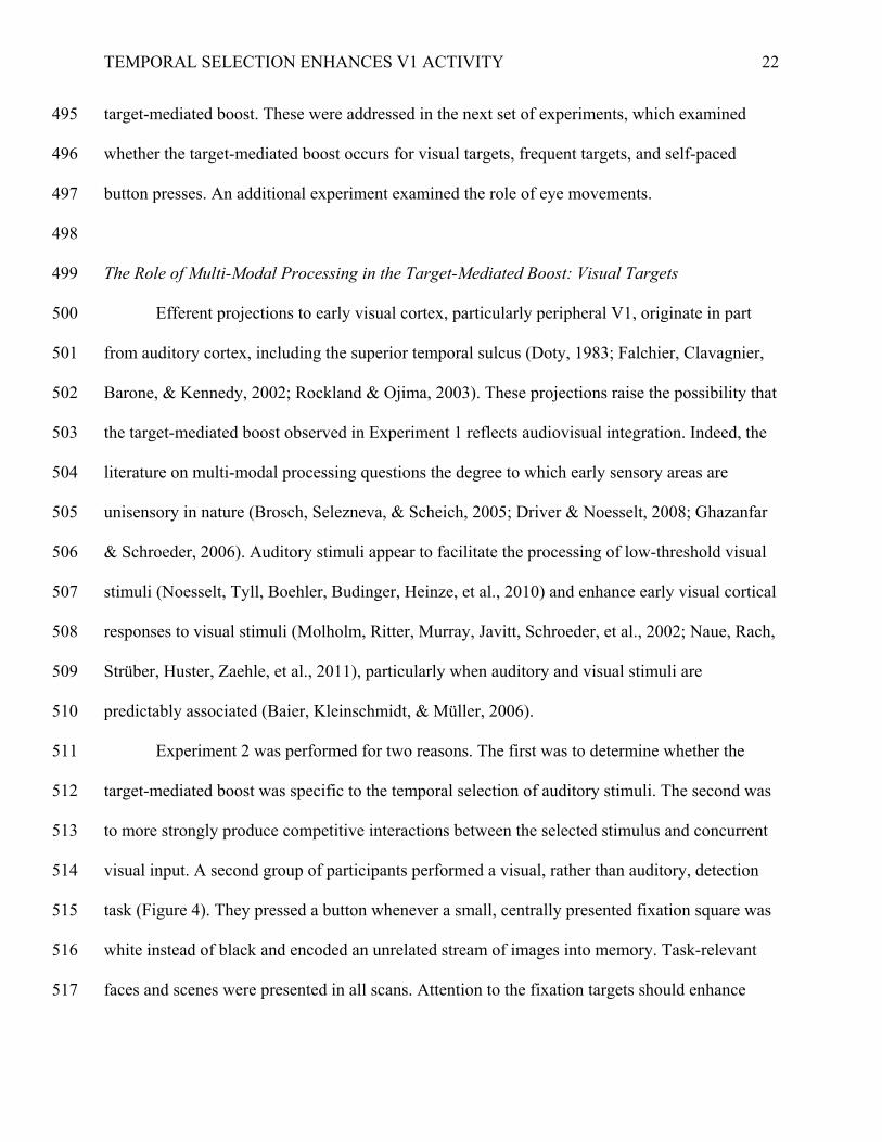
visual input. A second group of participants performed a visual, rather than auditory, detection 514
task (Figure 4). They pressed a button whenever a small, centrally presented fixation square was 515
white instead of black and encoded an unrelated stream of images into memory. Task-relevant 516
faces and scenes were presented in all scans. Attention to the fixation targets should enhance 517
TEMPORAL SELECTION ENHANCES V1 ACTIVITY
23
activity in the central visual field. It may also decrease activity in regions representing other 518
spatial locations (Reynolds & Heeger, 2009; Silver et al., 2007; Tootell et al., 1998). Of critical 519
interest is how the appearance of a centrally presented target modulates activity in visual regions 520
representing the peripheral visual field. If the target-mediated boost reflects temporal selection of 521
a behaviorally relevant stimulus, regardless of its modality, then it should occur for visual as well 522
as auditory targets, even in regions that are not stimulated. If it instead reflects audiovisual 523
integration, then the target-mediated boost should not occur in Experiment 2. 524
525
--------------INSERT FIGURE 4 HERE-------------- 526
527
Figure 4 illustrates the regions of interest and their response to centrally presented target 528
and distractor squares during the visual detection task. V1 was anatomically defined as 529
pericalcarine cortex and divided into central and periphery regions using the localizer data. Peak 530
signal change in the resulting ROIs was then submitted to an ANOVA with square status and 531
eccentricity as factors. Responses were stronger in central V1 than in periphery V1, resulting in a 532
main effect of eccentricity F(1,8) = 7.62, p = .025, ηp2 = .488. More importantly, activity in both 533
the central and periphery regions of V1 was greater when a fixation target was presented than 534
when a distractor was presented (main effect of square status, F(1,8) = 15.4, p = .004, ηp2 = .658, 535
and no interaction between square status and eccentricity, F(1,8) = 0.05, p = .829). The target-536
mediated boost was present throughout V1, even in regions that were not stimulated and that did 537
not contain the selected target. 538
The progression of the target-mediated boost through visual cortex, and its interaction 539
with cortical responses to concurrent images was also examined for the FFA and PPA. Peak 540
TEMPORAL SELECTION ENHANCES V1 ACTIVITY
24
signal change from these regions was submitted to an ANOVA with square status and image 541
type as factors. Reliable main effects of image type in both the FFA, F(2,16) = 62.6, p < .001, 542
ηp2 = .887, and the PPA, F(2,14) = 22.1, p < .001, ηp
2 = .76, confirmed that these regions were 543
selectively activated by faces and scenes respectively. Although selecting a visual target might 544
increase activity in these regions, an interaction of the target-mediated boost with image 545
processing should produce effects that are specific to the type of stimulus preferred by these 546
regions. However, although the FFA increased more in activity for target squares than distractor 547
squares, leading to a main effect of square status, F(1,8) = 6.18, p = .038, ηp2 = .436, this 548
response was not reliably greater for faces than for scenes or scrambled images, as indicated by a 549
nonsignificant interaction between square status and image type, F(2,16) = 0.25, p = .783. 550
Similarly, the marginal main effect of square status in the PPA, F(1,7) = 3.55, p = .101, did not 551
depend on whether the concurrent image was a scene or another type of image, nonsignificant 552
interaction between square status and image type, F(2,14) = 0.17, p = .844. 553
Thus, the FFA and PPA showed weaker effects of targets than did V1 (0.02 for the FFA, 554
0.03 for the PPA, and 0.1 and 0.09 for central and periphery V1), and these effects were not 555
specific to their preferred stimuli. Just as with auditory target tones, the boost elicited by visual 556
target squares diminishes in a feed-forward manner through the ventral visual processing stream, 557
and does not depend on the type of stimulus presented. 558
Finally, because Experiment 2 utilized visual targets and distractors, it was possible to 559
examine activity in primary auditory cortex (A1). A t-test indicated that this region showed a 560
reliably larger peak response to visual targets than to distractors (Figure 4d), t(8) = 2.42, p = 561
.042, d = 0.753, suggesting that the target-mediated boost may not be confined to visual 562
perceptual areas. 563

TEMPORAL SELECTION ENHANCES V1 ACTIVITY
25
The data from Experiment 2 demonstrate that the target-mediated boost in visual cortex is 564
not specific to the selection of auditory stimuli. It is therefore unlikely that the target-mediated 565
boost reflects either multi-modal processing or feedback from auditory perceptual regions such 566
as STS to V1. This conclusion is consistent with the finding that the target-mediated boost was 567
not stronger in periphery V1, which receives more projections from auditory cortex than does 568
central V1 (Falchier et al., 2002). Rather, the boost appears to reflect processes that are triggered 569
by the temporal selection of behaviorally relevant stimuli. 570
571
Early Visual Cortical Responses to Common Auditory Targets 572
In the previous two experiments the detection stimuli (tones and centrally presented 573
squares) were three-times more likely to be distractors than targets. Targets therefore may have 574
triggered processes associated with rare, or unexpected stimuli, including novelty processing, 575
expectancy violations, and the orienting response (Donchin & Coles, 1988; Polich, 2007; 576
Shulman, Astafiev, Franke, Pope, Snyder, et al., 2009; Sokolov, Nezlina, Polyanskii, & Evtikhin, 577
2002). To determine whether the target-mediated boost reflects processes associated with rare 578
stimuli, a third experiment was run with auditory tones that were equally likely to be targets and 579
distractors. Distractors were as novel and unexpected as targets. If rare or novel stimuli are 580
necessary for the target-mediated boost, then it should be absent in Experiment 3. In contrast, if 581
the target-mediated boost reflects temporal selection, it should occur when targets are frequent as 582
well as when they are rare. 583
As can be seen in Figure 5, early visual cortical responses to tones were greater when 584
they were targets than when they were distractors, even when they were equally frequent. Peak 585
signal change for the retinotopically defined ROIs were submitted to an ANOVA with tone 586
TEMPORAL SELECTION ENHANCES V1 ACTIVITY
26
status, area, and eccentricity as factors. Across all regions the main effect of tone status was 587
marginal, F(1,7) = 5.11, p = .058, ηp2 = .422, with the magnitude of the effect decreasing from 588
V1 to V3, producing a reliable interaction between tone status and area, F(2,14) = 11.3, p = .001, 589
ηp2 = .618. A follow up ANOVA only on V1 confirmed that it was more active following a 590
target tone than a distractor tone, main effect of tone status F(1,7) = 7.52, p = .029, ηp2 = .518. 591
Overall, the main effect of area indicated that responses to tones were larger in V1 than in V2 or 592
V3, F(2,14) = 11.6, p = .001, ηp2 = .623 (see Figure 5). There were no reliable effects of 593
eccentricity, p’s > .474. These data replicate the target-mediated boost in a task in which images 594
were never presented and were never task relevant. More importantly, the target-mediated boost 595
was present when target tones were as frequent as distractor tones. Hence, rare target stimuli are 596
not necessary for the target-mediated boost. 597
598
--------------INSERT FIGURE 5 HERE-------------- 599
600
Testing the Contributions of Eye-Movements and Button Presses to the Target-Mediated Boost 601
One consideration was the potential role of the motor response in the target-mediated 602
boost. Although participants were instructed to fixate on the center of the screen, they may have 603
moved their eyes or blinked more following a target than a distractor. In addition, targets, but not 604
distractors, required a manual response. Although a manual response is not necessary for the 605
nonperceptual activity produced by task transitions (Jack et al., 2006), it remains possible that 606
the act of pressing a button could increase its magnitude. 607
To investigate the relationship between eye movements and the target-mediated boost, 608
eye gaze position, blinks (defined as eye data signal losses), and BOLD data were 609
TEMPORAL SELECTION ENHANCES V1 ACTIVITY
27
simultaneously measured in Experiment 4. A new group of participants performed the auditory 610
detection task with equally frequent targets and distractors as they encoded background images 611
into memory. 612
Analyses of the eye data indicated that there were no reliable differences in blinks or eye 613
movements following targets and distractors, t(5) = -0.5, p = .64 for blinks and t(5) = 1.12, p = 614
.315 for distance. Importantly, a target-mediated boost was observed (Figure 6). Peak changes in 615
BOLD signal were submitted to an ANOVA with area, eccentricity, and tone status as factors. A 616
reliable interaction between area and tone status indicated that peak activity in early visual cortex 617
was greater following target tones than following distractor tones, but that this effect decreased 618
from V1 to V3, F(2,10) = 4.54, p = .04, ηp2 = .476. Main effects of area and eccentricity 619
indicated that responses to tones decreased from V1 to V3, F(2,10) = 7.51, p = .01, ηp2 = .6, and 620
were larger in central than in peripheral eccentricities, F(1,5) = 37.6, p = .002, ηp2 = .882. No 621
other effects or interactions were significant, F’s < 3.69, p’s > .11. 622
An additional eye-tracking experiment replicated Experiment 1 outside the scanner. This 623
experiment had a larger sample size (N = 9) and included more trials than Experiment 4. Its 624
findings were consistent with the conclusion that participants move their eyes a similar amount 625
following target and distractor tones. There were only small deviations in eye position from 626
fixation, and no differences in the amount the eyes moved or blinked across target and distractor 627
trials, regardless of whether an image was presented (no reliable effects of tone status or images: 628
eye movements, largest F(1,8) = 1.27, p = .292; blinks, largest F(1,8) = 1.61, p = .24). Thus, the 629
eye movement data indicated that the target-mediated boost occurs even when there are no 630
apparent differences in eye movements or blinks across target and distractor trials. 631
632
TEMPORAL SELECTION ENHANCES V1 ACTIVITY
28
--------------INSERT FIGURE 6 HERE-------------- 633
634
A final experiment examined the relationship between the target-mediated boost and 635
button presses. For Experiment 5, participants who completed the auditory detection task in 636
Experiment 3 also completed a self-generated button-press task. If the target-mediated boost in 637
Experiment 3 was due to the button press response to targets, then activity in central and 638
periphery V1 in these same participants should increase following a self-generated button press. 639
Rather than leading to a widespread and immediate enhancement of activity in V1, 640
however, self-generated button presses produced an initial decrease in activity followed 641
approximately 12 s later by an increase in activity (Figure 7). These effects were confined to 642
central V1. An ANOVA with time, area, and eccentricity as factors indicated that central V1 643
showed a stronger response around button presses than did the other regions, resulting in reliable 644
interactions between time, area, and eccentricity, F(24,168) = 2.24, p = .002, ηp2 = .243, and 645
time and area, F(24,168) = 2.26, p = .001, ηp2 = .244, and a trend for an interaction between area 646
and eccentricity, F(2,14) = 2.23, p = .093. No other effects or interactions were reliable, F’s < 647
1.5, p’s > .256. In contrast, the response to targets was observed in both central and periphery 648
regions and followed a more or less standard hemodynamic response function, peaking 649
approximately 4 s after the onset of the tone (Figure 2). Thus, the same group of participants who 650
showed a target-mediated boost in Experiment 3 showed a different response to self-generated 651
button presses in Experiment 5. 652
653
--------------INSERT FIGURE 7 HERE-------------- 654
655
TEMPORAL SELECTION ENHANCES V1 ACTIVITY
29
656
Discussion 657
Attentional selection is typically considered to be a process that enhances neural 658
responses to the selected stimuli. However, the computational demands of a mechanism that 659
selects stimuli in time suggest that its effects may need to be brief and spatially unconstrained. 660
This study investigated whether temporal selection influences activity in perceptual regions that 661
are not typically involved in processing the selected stimulus. Previous data suggest that 662
temporal selection could either increase or decrease activity in these regions. Whereas some 663
behavioral studies show better encoding of stimuli that coincide with goal-relevant events (Lin et 664
al., 2010; Seitz & Watanabe, 2003; Swallow & Jiang, 2010), other neuroimaging studies suggest 665
that increasing attention to one stimulus should reduce activity in regions not involved in 666
processing them (Brefczynski & DeYoe, 1999; Johnson & Zatorre, 2005; 2006; Luck et al., 667
1997; Schwartz et al., 2005; Shomstein & Yantis, 2004; Silver et al., 2007). The data reported 668
here clearly showed that temporal selection of goal-relevant stimuli is associated with a 669
nonspecific increase in activity in early visual cortical regions. 670
Most neuroscience research on attentional selection has focused on selection in space. 671
Spatial selection results in the modulation of neural activity in visual areas of the brain 672
(Reynolds & Heeger, 2009; Tootell et al., 1998). Modulatory or biasing signals are generated in 673
dorsal and ventral attentional networks that include inferior parietal sulcus, angular gyrus, the 674
frontal eye fields, and right middle frontal gyrus (Culham, Cavanagh, & Kanwisher, 2001; 675
Corbetta, Patel, & Shulman, 2008). These networks bias activity in visual regions towards the 676
representation of salient or behaviorally relevant spatial locations or visual features (Desimone & 677
Duncan, 1995). Although the exact nature of these modulations is unclear (cf. Reynolds & 678
TEMPORAL SELECTION ENHANCES V1 ACTIVITY
30
Heeger, 2009), spatial selection proceeds in the opposite direction in the visual processing stream 679
than does perceptual processing (Hochstein & Ahissar, 2002). Spatial selection tends to produce 680
stronger and earlier modulatory effects in late visual regions such as V4 than in early visual 681
regions such as V1 (Buffalo et al, 2010; Kastner et al., 1998). Moreover, spatial selection 682
enhances neural processing in regions representing the attended region of space (Luck et al., 683
1997) and can reduce activity in regions representing other spatial locations (Brefczynski & 684
DeYoe, 1999; Silver, et al, 2007). Thus, spatial selection involves the interaction of neural 685
systems that orient attention to goal-relevant or salient regions in space with regions involved in 686
processing sensory information at those and other locations. 687
In contrast to visuo-spatial attention, in the present study temporal selection was 688
associated with spatially diffuse increases in BOLD activity that were stronger in early than in 689
late visual cortex. Several experiments demonstrated that this target-mediated boost in early 690
visual cortex was due to temporal selection rather than to audio-visual integration, differences in 691
the novelty or expectancy of target and distractor stimuli, or to hand or eye movements in 692
response to targets. Rather, the data suggest a strong relationship between the temporal selection 693
of behaviorally relevant stimuli and spatially non-selective increases in activity in early 694
perceptual cortical regions. 695
These effects diverge from earlier studies showing that sustained attention to an auditory 696
or visual stimulus reduces activity in regions that are not involved in its representation. Other 697
work has shown that directing attention to the auditory rather than visual modality reduces 698
activity in visual cortex (Johnson & Zatorre, 2005; 2006; Shomstein & Yantis, 2004). In 699
addition, sustained attention to one spatial location reduces activity in regions representing 700
nonattended spatial locations (Brefczynski & DeYoe, 1999; Silver et al., 2007). These and 701
TEMPORAL SELECTION ENHANCES V1 ACTIVITY
31
similar data support the suggestion of a push-pull relationship in selective attention: Increasing 702
attention to one modality or spatial location reduces attention to other modalities and locations 703
(Pinsk, Doniger, & Kastner, 2003; Shomstein & Yantis, 2004). The observation that transient 704
attention to a stimulus presented in one modality (auditory or visual) or spatial location enhances 705
activity in perceptual regions that are not involved in its processing is a striking contrast to these 706
previous data. However, the critical manipulation in Experiments 1-4 was whether a briefly 707
presented stimulus was a target, rather than which modality or spatial location should be 708
attended. The outcome of these experiments underscores the distinctive computational 709
challenges that face a temporal selection mechanism, suggesting that temporal selection is more 710
than a temporally constrained application of spatial selection. 711
Although the pattern of activity in early visual cortex reported in this study is unusual in 712
studies of attentional selection, a similar pattern has been reported for task transitions (Jack et al., 713
2006). In that study, participants performed a simple discrimination task on visual or auditory 714
stimuli. Activity in early visual cortex, particularly in peripheral regions of V1, increased in 715
response to auditory events that signaled the beginning of a trial and that signaled that a response 716
should be made or cancelled. 717
The experiments reported here represent a substantial extension of these findings to a 718
markedly different paradigm – one that required participants to be nearly continuously engaged 719
in a task with no clear trial structure or task transitions. More importantly, they offer new insight 720
into which factors may be important for generating these modulations. In the previous study, all 721
events in a trial were associated with increased activity in peripheral V1 (Jack et al., 2006). 722
Experiments 1-5 constrain accounts of the V1 and target-mediated boost. They demonstrate that 723
the early visual cortical boost does not depend on stimulus novelty or expectation, that it is 724
TEMPORAL SELECTION ENHANCES V1 ACTIVITY
32
weaker for auditory and visual stimuli that do not require a response, and that it does not occur 725
for self-generated button presses. Rather than occurring for all sensory or motor events that could 726
structure a task over time, non-perceptual boosts of visual cortical activity appear to be specific 727
to events that require temporal selection. Moreover, the present study suggests that these non-728
perceptual modulations may be more general than previously understood. They occur in early 729
auditory cortex and when visuo-spatial attention is directed to concurrent images or central 730
visual targets. 731
The target-mediated boost may be related to findings from a single-unit and multi-unit 732
recording study on non-human primates (Brosch, et al., 2005). For that study, macaques were 733
trained to release a bar when the pitch of a tone sequence decreased. The firing rate of neurons in 734
auditory cortex increased in response to visual and behavioral events that occurred as part of the 735
task. Other neuroimaging work in humans has also found that activity in extrastriate visual areas 736
increases when a target sound previously associated with a visual image is expected (Bueti & 737
Macaluso, 2010). The data from Experiments 1-4 suggest that similar modulations occur in 738
humans and in early visual and auditory cortex. However, unlike earlier data, the target-mediated 739
boost was observed in participants with little previous experience in the detection task. In 740
addition, the effect occurred in visual cortex when the task was purely auditory (Experiment 3) 741
and in auditory cortex when the task was purely visual (Experiment 2). It is therefore unlikely 742
that the target-mediated boost reflects a learned association between visual, auditory, and 743
behavioral events. 744
745
Potential Cognitive and Neural Sources of the Target-Mediated Boost 746
TEMPORAL SELECTION ENHANCES V1 ACTIVITY
33
Temporal selection has been conceptualized as a gate that increases the likelihood that 747
the selected input enters working memory (Bowman & Wyble, 2007; Chun & Potter, 1995; 748
Olivers & Meeter, 2008). However, in these models temporal selection’s facilitory effects are 749
constrained to the selected item and to later perceptual areas. The data presented here suggest 750
that, at the very least, current models of temporal selection are incomplete. Because temporal 751
selection diffusely enhances activity in early visual areas, whatever mechanism underlies it must 752
have effects that extend beyond late perceptual regions representing the selected stimulus. 753
Rather, the fact that increased activity in early visual cortical areas is associated with temporal 754
selection and changes in task structure (Jack et al., 2006) is consistent with a different set of 755
models: those that describe how the cognitive system represents goals and external events. In 756
these models, changes in context or the completion of a goal can trigger a gating mechanism that 757
updates neural representations to better reflect the new situation (Aston-Jones & Cohen, 2005; 758
Bouret & Sara, 2005; Frank, Loughry, & O’Reilly, 2001; O’Reilly et al., 1999; Zacks et al., 759
2007). The broad early visual cortical activity corresponding to these moments in time reported 760
here is consistent with such an updating mechanism (cf. Jack et al., 2006). 761
The fact that non-perceptual modulations of activity in early visual cortex are strongest in 762
V1 suggests that they do not arise from indirect feedback from late visual or frontoparietal 763
attentional regions. Rather, the relationship between the early visual cortical boost and task 764
structure and attention suggests two potential subcortical sources. The first is the dopamine based 765
gating system in the basal ganglia. According to one model, the basal ganglia act as a gate that 766
protects representations of goals and context from disruption by new input from other cortical 767
regions. When goals are completed or the context changes, the gating mechanism is triggered to 768
allow active memory updating and to initiate motor actions (Frank et al., 2001; O’Reilly et al., 769
TEMPORAL SELECTION ENHANCES V1 ACTIVITY
34
1999). The release of dopamine from the basal ganglia is also associated with expectancy 770
violations, facilitating reinforcement learning by signaling unexpected rewards (Schultz & 771
Dickinson, 2000). 772
A second potential source of the boost is the phasic release of norepinephrine (NE) from 773
the locus-coeruleus (LC), which has been characterized as a temporal attentional filter 774
(Nieuwenhuis, Aston-Jones, & Cohen, 2005). The LC-NE response is thought to facilitate the 775
updating of neuronal representations in response to external cues by enhancing their responsivity 776
to new input (Aston-Jones & Cohen, 2005; Bouret & Sara, 2005). It has been proposed that the 777
LC-NE response to targets in continuous detection tasks like those used here may give rise to the 778
P3b (Nieuwenhuis et al., 2005), which is positively correlated with activity in pericalcarine 779
cortex (Mantini, Corbetta, Perrucci, Romani, & Del Gratta, 2009, supplementary material). 780
781
Functional Consequences 782
Although the timing and nature of the target-mediated boost are consistent with a role in 783
context updating, there was little evidence in the current study that it interacted with the visual 784
processing of attended, supra-threshold images. Moreover, the target-mediated boost was present 785
in the periphery even when visuo-spatial attention was allocated to centrally presented visual 786
stimuli. Directing attention to a central visual stimulus neither limited the boost to regions 787
representing the stimulus nor increased the magnitude of the boost in later visual areas. Although 788
the data demonstrate an effect of temporal selection on early visual cortical activity, they provide 789
no clear answers regarding the functional consequences of this activity. 790
The recognition data from these experiments were unusual in that they showed a 791
relatively small memory advantage for images presented at the same time as targets relative to 792
TEMPORAL SELECTION ENHANCES V1 ACTIVITY
35
those presented with distractors (Lin, et al, 2010; Swallow & Jiang, 2010). We can only 793
speculate as to why this attentional boost effect was small in these experiments. However, an 794
obvious difference is that the stimuli appeared at regular and predictable intervals in previous 795
behavioral studies. In contrast, the present study used no-tone intervals to jitter the detection 796
stimuli. The regular presentation of the detection stimuli and images in previous studies may 797
have facilitated discrimination of the targets and distractors by making the stimuli more 798
predictable. Unpredictable stimuli could induce a less efficient mode of attention than the 799
rhythmic and predictable stimuli used in earlier experiments (Schroeder & Lakatos, 2009). 800
On the surface, these data suggest that non-perceptual modulations of V1 activity may be 801
epiphenomenal, having no effect on visual processing. The small memory effect as well as the 802
fact that the FFA and PPA showed similar responses to targets and distractors, even for their 803
preferred stimuli, are consistent with this possibility. However, in addition to the tenuousness of 804
conclusions based on null effects, the conclusion that the non-perceptual V1 modulations are 805
epiphenomenal is premature for several reasons. First, the present studies used stimuli that were 806
ideal for examining the effects of temporal selection on activity in category selective visual 807
regions (the FFA and the PPA). However, because the boost was strongest in V1, its effects on 808
perceptual processing might be strongest for visual features that are represented in V1. Second, it 809
is possible that temporal selection has its greatest effects on visual processing when the visual 810
input is degraded (cf Noesselt et al., 2010). In addition, the functional consequences of the boost 811
on perceptual processing may not be immediately observable. Indeed, it is possible that the 812
target-mediated boost facilitates perceptual learning of visual features that coincide with goal-813
relevant stimuli, as in task irrelevant perceptual learning (Seitz & Watanabe, 2003). 814
TEMPORAL SELECTION ENHANCES V1 ACTIVITY
36
A final possibility is that the target-mediated boost does not directly enhance perceptual 815
processing. Rather it could act as an entrainment signal to synchronize periodic fluctuations in 816
the neuronal sensitivity of perceptual regions representing various visual features, modalities, 817
and stimulus locations (Engel & Singer, 2001; Lakatos et al., 2008; Schroeder & Lakatos, 2009). 818
Although the stimuli used in our experiments were presented at variable and unpredictable 819
intervals, each instance of a behaviorally relevant stimulus could produce a signal that entrains 820
neural processing when it occurs with sufficient regularity. 821
822
Conclusion 823
Spatial selection and increased attentional demands tend to increase cortical responses in 824
regions that represent the selected stimuli, while decreasing activity in regions that do not (Luck 825
et al., 1997; Silver et al., 2007). In contrast, the data presented here demonstrate that temporal 826
selection of auditory and visual stimuli increases activity in early visual cortical regions. 827
Nonvisual stimuli were found to enhance activity in early visual cortical areas when they were 828
selected. These modulations diverge from those of spatial selection in two critical ways: they are 829
not constrained to the spatial location or modality of the target, and they decrease, rather than 830
increase, along the ventral visual processing stream. These differences underscore the divergent 831
computational demands of spatial and temporal selection. They also join a growing body of 832
evidence that suggests that current models and understanding of temporal selection need to be 833
extended to account for its effects on context processing. 834
835
836
Acknowledgements 837
TEMPORAL SELECTION ENHANCES V1 ACTIVITY
37
We thank Steve Engel for comments on the manuscript, Min Bao, Steve Engel, Mark 838
Schira, and Bo-Yeong Won for help with retinotopy, and Phil Burton, Andrea Grant, Pete 839
Kollasch, and Gail Rosenbaum for help with eye tracking. Tal Makovski is now at the 840
Department of Psychology, College of Management Academic Studies, Rishon LeZion, Israel. 841
842
Grants 843
This research was funded in part by ARO 60343-LS-II and by a CLA brain imaging fund 844
from the University of Minnesota. 845
846
Author Contributions 847
Khena Swallow designed the studies, collected the data, analyzed the data, and wrote the 848
paper. Tal Makovski collected the data and wrote the paper. Yuhong Jiang designed the studies, 849
collected the data, analyzed the data, and wrote the paper. 850
851
852
853
854
TEMPORAL SELECTION ENHANCES V1 ACTIVITY
38
References 855
Anderson AK, Christoff K, Panitz D, de Rosa E, Gabrieli JDE. Neural correlates of the 856
automatic processing of threat facial signals. J Neurosci 23: 5627-5633, 2003. 857
Aston-Jones G, Cohen JD. An integrative theory of locus coeruleus-norepinephrine function: 858
Adaptive gain and optimal performance. Annu Rev Neurosci 28: 403-450, 2005. 859
Baier B, Kleinschmidt A, Müller NG. Cross-modal processing in early visual and auditory 860
cortices depends on expected statistical relationship of multisensory information. J 861
Neurosci 26: 12260-12265, 2006. 862
Becker MW, Pashler H, Anstis SM. The role of iconic memory in change-detection tasks. 863
Perception 29: 273-286, 2000. 864
Bledowski C, Prvulovic D, Goebel R, Zanella FE, Linden DE. Attentional systems in target and 865
distractor processing: a combined ERP and fMRI study. Neuroimage 22: 530-540, 2004. 866
Bouret S, Sara SJ. Network reset: a simplified overarching theory of locus coeruleus 867
noradrenaline function. Trends Neurosci 28: 574-582, 2005. 868
Bowman H, Wyble B. The simultaneous type, serial token model of temporal attention and 869
working memory. Psychol Rev 114: 38-40, 2007. 870
Brefczynski JA, DeYoe EA. A physiological correlate of the “spotlight” of visual attention. Nat 871
Neurosci 2: 370-374, 1999. 872
Breitmeyer BG, Ganz L. Implications of sustained and transient channels for theories of visual 873
pattern masking, saccadic suppression, and information processing. Psychol Rev 83: 1-36, 874
1976. 875
Brockmole JR, Castelhano MS, Henderson JM. Contextual cuing in naturalistic scenes: Global 876
and local contexts. J Exp Psychol Learn Mem Cogn 32: 699-706, 2006. 877
TEMPORAL SELECTION ENHANCES V1 ACTIVITY
39
Brosch M, Selezneva E, Scheich H. Nonauditory events of a behavioral procedure activate 878
auditory cortex of highly trained monkeys. J Neurosci 25: 6797-6806, 2005. 879
Bueti D, Macaluso E. Auditory temporal expectations modulate activity in visual cortex. 880
Neuroimage 51: 1168-1183. 881
Buffalo EA, Fries P, Landman R, Liang H, Desimone R. A backward progression of attentional 882
effects in the ventral stream. Proc Natl Acad Sci 107: 361-365, 2010. 883
Chun MM, Potter MC. A two-stage model for multiple target detection in rapid serial visual 884
presentation. J Exp Psychol Hum Percept Perform 21: 109-127, 1995. 885
Chun MM, Jiang YV. Contextual cueing: Implicit learning and memory of visual context guides 886
spatial attention. Cogn Psychol 36: 28-71, 1998. 887
Cichy RM, Heinzle J, Haynes JD. Imagery and perception share cortical representations of 888
content and location. Cereb Cortex 22: 372-380, 2012. 889
Corbetta M, Patel G, Shulman GL. The reorienting system of the human brain: from 890
environment to theory of mind. Neuron 58: 306-324, 2008. 891
Culham JC, Cavanagh P, Kanwisher NG. Attention response functions: Characterizing brain 892
areas using fMRI activation during parametric variations of attentional load. Neuron 32: 893
737-745, 2001. 894
Davenport JL, Potter MC. Scene consistency in object and background perception. Psychol Sci 895
15: 559-564, 2004. 896
Desikan RS, Ségonne F, Fischl B, Quinn BT, Dickerson BC, Blacker D, Buckner RL, Dale AM, 897
Maguire RP, Hyman BT, Albert MS, Killiany RT. An automated labeling system for 898
subdividing the human cerebral cortex on MRI scans into gyral based regions of interest. 899
Neuroimage 31: 968-980, 2006. 900
TEMPORAL SELECTION ENHANCES V1 ACTIVITY
40
Desimone R, Duncan J. Neural mechanisms of selective visual attention. Annu Rev Neurosci. 18: 901
193-222, 1995. 902
Donchin E, Coles MGH. Is the P300 component a manifestation of context updating? Behav 903
Brain Sci 11: 357-374, 1988. 904
Doty RW. Nongeniculate afferents to striate cortex in macaques. J Comp Neurol. 218: 159-173, 905
1983. 906
Driver J, Noesselt T. Multisensory interplay reveals cross-modal influences on `sensory-specific’ 907
brain regions, neural responses, and judgments. Neuron 57: 11-23, 2008. 908
Duncan J. The multiple-demand (MD) system of the primate brain: mental programs for 909
intelligent behaviour. Trends Cogn Sci 14: 172-179, 2010. 910
Engel AK, Singer W. Temporal binding and the neural correlates of sensory awareness. Trends 911
Cogn Sci 5: 16-25, 2001. 912
Engel SA, Glover GH, Wandell BA. Retinotopic organization in human visual cortex and the 913
spatial precision of functional MRI. Cereb Cortex 7: 181-192, 1997. 914
Enns J, Di Lollo V. What’s new in visual masking? Trends Cogn Sci 4:342-352, 2003. 915
Falchier A, Clavagnier S, Barone P, Kennedy H. Anatomical evidence of multimodal integration 916
in primate striate cortex. J Neurosci 22: 5749 -5759, 2002. 917
Fan J, McCandliss BD, Fossella J, Flombaum JI, Posner M I. The activation of attentional 918
networks. Neuroimage 26: 471-479, 2005. 919
Fischl B, Sereno MI, Dale AM. Cortical surface-based analysis: II: Inflation, flattening, and a 920
surface-based coordinate system. Neuroimage 9: 195-207, 1999. 921
TEMPORAL SELECTION ENHANCES V1 ACTIVITY
41
Frank MJ, Loughry B, O’Reilly RC. Interactions between frontal cortex and basal ganglia in 922
working memory: A computational model. Cogn Affect Behav Neurosci 1: 137-160, 923
2001. 924
Friston KJ, Holmes AP, Worsley KJ, Poline JP, Frith CD, Frackowiak RSJ. Statistical parametric 925
maps in functional imaging: A general linear approach. Hum Brain Mapp 2: 189-210, 926
1995. 927
Genovese C, Lazar NA, Nichols T. Thresholding of statistical maps in functional neuroimaging 928
using the false discovery rate. Neuroimage 15:8 870-878, 2002. 929
Ghazanfar AA, Schroeder CE. Is neocortex essentially multisensory? Trends Cogn Sci 10: 278-930
285, 2006. 931
Gold JI, Shadlen MN. The neural basis of decision making. Annu Rev Neurosci 30: 535-574, 932
2007. 933
Hochstein S, Ahissar M. View from the top: Hierarchies and reverse hierarchies in the visual 934
system. Neuron 36: 791-804, 2002. 935
Hon N, Thompson R, Sigala N, Duncan J. Evidence for long-range feedback in target detection: 936
Detection of semantic targets modulates activity in early visual areas. Neuropsychologia 937
47: 1721-1727, 2009. 938
Howard MA, Volkov IO, Mirsky R, Garell PC, Noh MD, Granner M, Damasio H, 939
Steinschneider M, Reale RA, Hind JE, Brugge JF. Auditory cortex on the human 940
posterior superior temporal gyrus. J Comp Neurol 416: 79-92, 2000. 941
Jack AI, Shulman GL, Snyder AZ, McAvoy MP, Corbetta M. Separate modulations of human 942
V1 associated with spatial attention and task structure. Neuron 51: 135-147, 2006. 943
TEMPORAL SELECTION ENHANCES V1 ACTIVITY
42
Jäncke L, Mirzazade S, Shah NJ. Attention modulates activity in the primary and secondary 944
auditory cortex: a functional magnetic resonance imaging study in human subjects. 945
Neurosci Lett 266: 125-128, 1999. 946
Jiang Y, He S. Cortical responses to invisible faces: Dissociating subsystems for facial-947
information processing. Curr Biol 16: 2023-2029, 2006. 948
Johnson JA, Zatorre RJ. Attention to simultaneous unrelated auditory and visual events: 949
behavioral and neural correlates. Cereb Cortex 15: 1609-1620, 2005. 950
Johnson JA, Zatorre RJ. Neural substrates for dividing and focusing attention between 951
simultaneous auditory and visual events. Neuroimage 31: 1673-1681, 2006. 952
Kastner S, de Weerd P, Desimone R, Ungerleider LG. Mechanisms of directed attention in the 953
human extrastriate cortex as revealed by function MRI. Science 282: 108-111, 1998. 954
Lakatos P, Karmos G, Mehta AD, Ulbert I, Schroeder CE. Entrainment of neuronal oscillations 955
as a mechanism of attentional selection. Science 320: 110 -113, 2008. 956
Lin JY, Pype AD, Murray SO, Boynton GM. Enhanced memory for scenes presented at 957
behaviorally relevant points in time. PLoS Biol 8:e1000337, 2010. 958
Luck SJ, Chelazzi L, Hillyard SA, Desimone R. Neural mechanisms of selective attention in 959
areas V1, V2, and V4 in macaque visual cortex. J Neurophysiol 77: 24-42, 1997. 960
Mantini D, Corbetta M, Perrucci MG, Romani GL, Del Gratta C. Large-scale brain networks 961
account for sustained and transient activity during target detection. Neuroimage 44: 265-962
274, 2009. 963
Molholm S, Ritter W, Murray MM, Javitt DC, Schroeder CE, Foxe JJ. Multisensory auditory-964
visual interactions during early sensory processing in humans: a high-density electrical 965
mapping study. Cogn Brain Res 14:115-128, 2002. 966
TEMPORAL SELECTION ENHANCES V1 ACTIVITY
43
Naue N, Rach S, Strüber D, Huster RJ, Zaehle T, Körner U, Herrmann CS. Auditory event-967
related response in visual cortex modulates subsequent visual responses in humans. J 968
Neurosci 31: 7729 -7736, 2011. 969
Neisser U. Cognition and Reality: Principles and Implications of Cognitive Psychology. New 970
York, NY. W H Freeman, 1976. 971
Nieuwenhuis S, Aston-Jones G, Cohen JD. Decision making, the P3, and the locus coeruleus--972
norepinephrine system. Psychol Bull 131: 510-532, 2005. 973
Noesselt T, Tyll S, Boehler CN, Budinger E, Heinze HJ, Driver J. Sound-induced enhancement 974
of low-intensity vision: Multisensory influences on human sensory-specific cortices and 975
thalamic bodies relate to perceptual enhancement of visual detection sensitivity. J 976
Neurosci 30: 13609 -13623, 2010. 977
Oliva A, Torralba A. The role of context in object recognition. Trends Cogn Sci, 11: 520-527, 978
2007. 979
Olivers CNL, Meeter M. A boost and bounce theory of temporal attention. Psychol Rev 115: 980
836-863, 2008. 981
O’Reilly RC, Braver TS, Cohen JD. A biologically based computational model of working 982
memory. In A. Miyake & P. Shah (Eds.). Models of Working Memory: Mechanisms of 983
Active Maintenance and Executive Control. Cambridge: Cambridge University Press. p 984
375-411, 1999. 985
Pashler H. Dual-task interference in simple tasks: data and theory. Psychol Bull 116: 220-244, 986
1994. 987
Pinsk MA, Doniger GM, Kastner S. Push-pull mechanism of selective attention in human 988
extrastriate cortex. J Neurophysiol 92: 622-629, 2004. 989
TEMPORAL SELECTION ENHANCES V1 ACTIVITY
44
Ploran, EJ, Nelson, SM, Velanova, K, Donaldson, DI, Peterson, SE, Wheeler, ME. Evidence 990
accumulation and the moment of recognition: dissociating perceptual recognition 991
processing using fMRI. J Neurosci 27: 11912-11924, 2007. 992
Polich J. Updating P300: An integrative theory of P3a and P3b. Clin Neurophysiol 118: 2128-993
2148, 2007. 994
Raymond JE, Shapiro KL, Arnell KM. Temporary suppression of visual processing in an RSVP 995
task: An attentional blink? J Exp Psychol Hum Percept Perform 18: 849-860, 1992. 996
Reynolds JH, Chelazzi L. Attentional modulation of visual processing. Annu Rev Neurosci, 27: 997
611-647, 2004. 998
Reynolds JH, Heeger DJ. The normalization model of attention. Neuron 61: 168-185, 2009. 999
Rockland KS, Ojima H. Multisensory convergence in calcarine visual areas in macaque monkey. 1000
Int J of Psychophysiol 50: 19-26, 2003. 1001
Schroeder CE, Lakatos P. Low-frequency neural oscillations as instruments of sensory selection. 1002
Trends Neurosci 32: 9-18, 2009. 1003
Schira MM, Tyler CW, Breakspear M, Spehar B. The foveal confluence in human visual cortex. 1004
J Neurosci 29: 9050-9058, 2009. 1005
Schultz W, Dickinson A. Neuronal coding of prediction errors. Annu Rev of Neurosci 23: 473-1006
500, 2000. 1007
Schwartz S, Vuilleumier P, Hutton C, Maravita A, Dolan RJ, Driver J. Attentional load and 1008
sensory competition in human vision: Modulation of fMRI responses by load at fixation 1009
during task-irrelevant stimulation in the peripheral visual field. Cereb Cortex 15: 770-1010
786, 2005. 1011
Seitz AR, Watanabe T. Is subliminal learning really passive? Nature 422: 36, 2003. 1012
TEMPORAL SELECTION ENHANCES V1 ACTIVITY
45
Sereno MI, Dale AM, Reppas JB, Kwong KK, Belliveau JW, Brady TJ, Rosen BR, Tootell 1013
RBH. Borders of multiple visual areas in humans revealed by functional magnetic 1014
resonance imaging. Science 268: 889-893, 1995. 1015
Shinoda H, Hayhoe MM, Shrivastava A. What controls attention in natural environments? Vision 1016
Research 41: 3535-3545, 2001. 1017
Shomstein S, Yantis S. Control of attention shifts between vision and audition in human cortex. J 1018
Neurosci 24: 10702-10706, 2004. 1019
Shulman GL, Astafiev SV, Franke D, Pope DLW, Snyder AZ, McAvoy MP, Corbetta M. 1020
Interaction of stimulus-driven reorienting and expectation in ventral and dorsal 1021
frontoparietal and basal ganglia-cortical networks. J Neurosci 29: 4392-4407, 2009. 1022
Silver MA, Ress D, Heeger DJ. Neural correlates of sustained spatial attention in human early 1023
visual cortex. J Neurophysiol 97: 229-237, 2007. 1024
Sirotin YB, Das A. Anticipatory haemodynamic signals in sensory cortex not predicted by local 1025
neuronal activity. Nature 457: 475-479, 2009. 1026
Slotnick SD, Thompson WL, Kosslyn SM. Visual mental imagery induces retinotopically 1027
organized activation of early visual areas. Cereb Cortex 15: 1570-1583, 2005. 1028
Sokolov EN, Nezlina NI, Polyanskii VB, Evtikhin DV. The orientating reflex: The “targeting 1029
reaction” and “searchlight of attention.” Neurosci Behav Physiol 32: 347-362, 2002. 1030
Spence C, Driver J. On measuring selective attention to an expected sensory modality. 1031
Perception & Psychophysics 59: 389-403, 1997. 1032
Swallow KM, Jiang YV. The attentional boost effect: Transient increases in attention to one task 1033
enhance performance in a second task. Cognition 115: 118-132, 2010. 1034
TEMPORAL SELECTION ENHANCES V1 ACTIVITY
46
Swallow KM, Zacks JM, Abrams RA. Event boundaries in perception affect memory encoding 1035
and updating. J Exp Psychol Gen 138: 236-257, 2009. 1036
Thiel CM, Zilles K, Fink GR. Cerebral correlates of alerting, orienting and reorienting of 1037
visuospatial attention: an event-related fMRI study. Neuroimage 21: 318-328, 2004. 1038
Tootell RBH, Hadjikhani N, Hall EK, Marrett S, Vanduffel W, Vaughan JT, Dale AM. The 1039
retinotopy of visual spatial attention. Neuron 21: 1409-1422, 1998. 1040
Yovel G, Kanwisher N. Face perception: Domain specific, not process specific. Neuron 44: 889-1041
898, 2004. 1042
Zacks JM, Speer NK, Swallow KM, Braver TS, Reynolds JR. Event perception: A mind/brain 1043
perspective. Psychol Bull 133: 273-293, 2007. 1044
1045

TEMPORAL SELECTION ENHANCES V1 ACTIVITY
47
Endnotes 1046
1. One participant completed 6 image scans. 1047
2. One participant completed four scans in the self-generated button press task. 1048
3. Voxels in bilateral intraparietal cortex were reliably activated by target tones relative to 1049
distractor tones (t > 3.1, p < .001, FDR < .05) prior to correction for multiple comparisons based 1050
on cluster size. The cluster that included activated voxels in the right supramarginal gyrus 1051
extended into the middle temporal lobe. 1052
1053
TEMPORAL SELECTION ENHANCES V1 ACTIVITY
48
Figure Captions 1054
Figure 1. Design and group level data from Experiment 1. a) For the auditory detection task 1055
participants monitored a series of tones and pressed a button whenever the tone was a pre-1056
specified pitch (a target; green note). They made no response to other tones (distractor; red note). 1057
Variable intervals of time in which no tones were presented (blue) separated the tones. In some 1058
scans participants viewed a gray screen throughout the task. In other scans visual images were 1059
also presented. b) Regions whose activity was greater following target than distractor tones (t > 1060
3.1, p < .001, FDR < .05) on the cortical surface and in subcortical regions. 1061
1062
Figure 2. Definition of ROIs and their response to auditory target and distractor tones in 1063
Experiment 1. a) For each individual, central and periphery V1, V2, and V3 ROIs were defined 1064
using polar angle mapping data and localizer data. Data are shown for one individual (on the 1065
flattened occipital lobe for retinotopy and on the cortical surface for the remaining contrasts). 1066
The FFA and PPA were also identified from the localizer data. All voxels in an ROI were used to 1067
estimate the hemodynamic response to the different types of trials. b) The mean timecourse of 1068
the response of early visual cortical areas to target (solid lines) and distractor tones (dashed lines) 1069
presented with images (blue lines) and without images (red lines). c) Peak signal change 1070
following target tones (solid lines) and distractor tones (dashed lines) presented with and without 1071
visual images in early visual regions of interest. Error bars represent ±one standard error around 1072
the mean in all panels. 1073
1074
TEMPORAL SELECTION ENHANCES V1 ACTIVITY
49
Figure 3. Timecourse of the response of the fusiform face area (FFA) and parahippocampal place 1075
area (PPA), to (a) auditory targets and distractors in Experiment 1, and (b) fixation targets and 1076
distractors in Experiment 2. Error bars represent ±1 standard error of the mean. 1077
1078
Figure 4. Design, regions of interest, and data from Experiment 2. a) The detection task was 1079
similar to the image scans in Experiment 1, except that the detection stimuli were visual. 1080
Participants monitored centrally presented squares (80 ms duration) that appeared in front of the 1081
background images. They pressed a button when the square was white rather than black and 1082
encoded the background images (500 ms duration) for a later memory test. Images were 1083
followed by a scrambled image masked for 1500 ms. A central fixation cross (not drawn to 1084
scale) was presented after the square was removed from the screen. b) V1 was anatomically 1085
localized to the pericalcarine cortex for each participant (colored map shows data from one 1086
participant). Localizer data were used to define the boundaries between central and periphery 1087
regions as in Experiment 1. All voxels in an ROI were used to estimate the hemodynamic 1088
response to the target and distractor squares. Thresholds and color maps are as in Figure 2. c) 1089
The timecourse of the response of central and periphery V1 to centrally presented visual target 1090
and distractor squares. d) The timecourse of the response of primary auditory cortex to centrally 1091
presented target and distractor squares. Error bars in all panels represent ±1 standard error of the 1092
mean. 1093
1094
Figure 5. Timecourse of the hemodynamic response of early visual regions to auditory target and 1095
distractor tones that occurred with equal frequency (Experiment 3). No visual images were 1096
presented in these scans. Error bars represent ±1 standard error of the mean. 1097
TEMPORAL SELECTION ENHANCES V1 ACTIVITY
50
1098
Figure 6. The target-mediated flash and eye movement data in Experiment 4. a) Peak percent 1099
signal change in retinotopically defined early visual areas V1, V2, and V3 following target and 1100
distractor tones. Responses were separately estimated for regions representing the central and 1101
peripheral visual fields. b) Distance of gaze position from fixation coordinates during the 2s 1102
period following target and distractor tones in X and Y coordinates. c) Total distance that the 1103
eyes moved, in degrees, during the 2s period that followed target and distractor tones. Note that a 1104
shift in gaze position of one degree, and then back, would result in a total distance of 2 degrees. 1105
d) Proportion of samples that were flagged as a signal loss in the 2s period following target and 1106
distractor tones. Error bars represent ±1 standard error of the mean. 1107
1108
Figure 7. Timecourse of the response of retinotopically defined early visual regions to self-1109
generated button presses in Experiment 5. Note that the pre-event time is longer than in previous 1110
experiments. Error bars represent ±1 standard error of the mean. 1111
1112
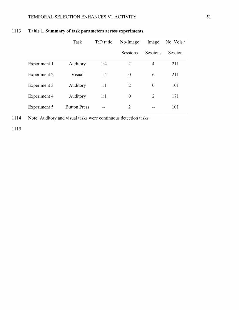
TEMPORAL SELECTION ENHANCES V1 ACTIVITY
51
Table 1. Summary of task parameters across experiments. 1113
Task T:D ratio No-Image
Sessions
Image
Sessions
No. Vols./
Session
Experiment 1 Auditory 1:4 2 4 211
Experiment 2 Visual 1:4 0 6 211
Experiment 3 Auditory 1:1 2 0 101
Experiment 4 Auditory 1:1 0 2 171
Experiment 5 Button Press -- 2 -- 101
Note: Auditory and visual tasks were continuous detection tasks. 1114
1115
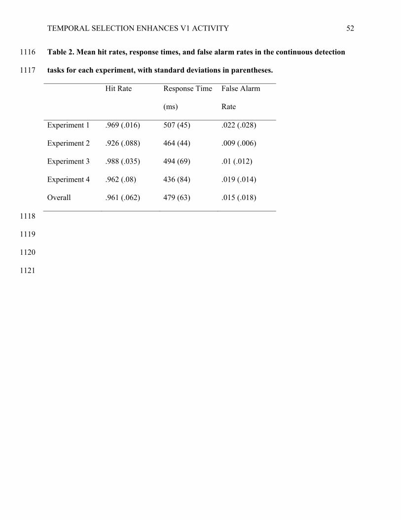
TEMPORAL SELECTION ENHANCES V1 ACTIVITY
52
Table 2. Mean hit rates, response times, and false alarm rates in the continuous detection 1116
tasks for each experiment, with standard deviations in parentheses. 1117
Hit Rate Response Time
(ms)
False Alarm
Rate
Experiment 1 .969 (.016) 507 (45) .022 (.028)
Experiment 2 .926 (.088) 464 (44) .009 (.006)
Experiment 3 .988 (.035) 494 (69) .01 (.012)
Experiment 4 .962 (.08) 436 (84) .019 (.014)
Overall .961 (.062) 479 (63) .015 (.018)
1118
1119
1120
1121
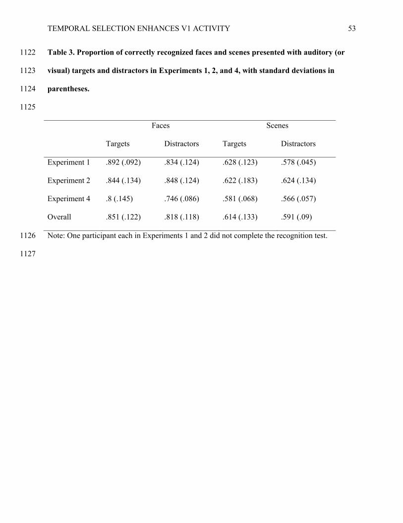
TEMPORAL SELECTION ENHANCES V1 ACTIVITY
53
Table 3. Proportion of correctly recognized faces and scenes presented with auditory (or 1122
visual) targets and distractors in Experiments 1, 2, and 4, with standard deviations in 1123
parentheses. 1124
1125
Faces Scenes
Targets Distractors Targets Distractors
Experiment 1 .892 (.092) .834 (.124) .628 (.123) .578 (.045)
Experiment 2 .844 (.134) .848 (.124) .622 (.183) .624 (.134)
Experiment 4 .8 (.145) .746 (.086) .581 (.068) .566 (.057)
Overall .851 (.122) .818 (.118) .614 (.133) .591 (.09)
Note: One participant each in Experiments 1 and 2 did not complete the recognition test. 1126
1127
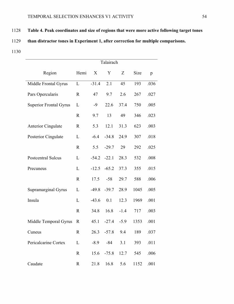
TEMPORAL SELECTION ENHANCES V1 ACTIVITY
54
Table 4. Peak coordinates and size of regions that were more active following target tones 1128
than distractor tones in Experiment 1, after correction for multiple comparisons. 1129
1130
Talairach
Region Hemi X Y Z Size p
Middle Frontal Gyrus L -31.4 2.1 45 193 .036
Pars Opercularis R 47 9.7 2.6 267 .027
Superior Frontal Gyrus L -9 22.6 37.4 750 .005
R 9.7 13 49 346 .023
Anterior Cingulate R 5.3 12.1 31.3 623 .003
Posterior Cingulate L -6.4 -34.8 24.9 307 .018
R 5.5 -29.7 29 292 .025
Postcentral Sulcus L -54.2 -22.1 28.3 532 .008
Precuneus L -12.5 -65.2 37.3 355 .015
R 17.5 -58 29.7 588 .006
Supramarginal Gyrus L -49.8 -39.7 28.9 1045 .005
Insula L -43.6 0.1 12.3 1969 .001
R 34.8 16.8 -1.4 717 .003
Middle Temporal Gyrus R 45.1 -27.4 -5.9 1353 .001
Cuneus R 26.3 -57.8 9.4 189 .037
Pericalcarine Cortex L -8.9 -84 3.1 393 .011
R 15.6 -75.8 12.7 545 .006
Caudate R 21.8 16.8 5.6 1152 .001
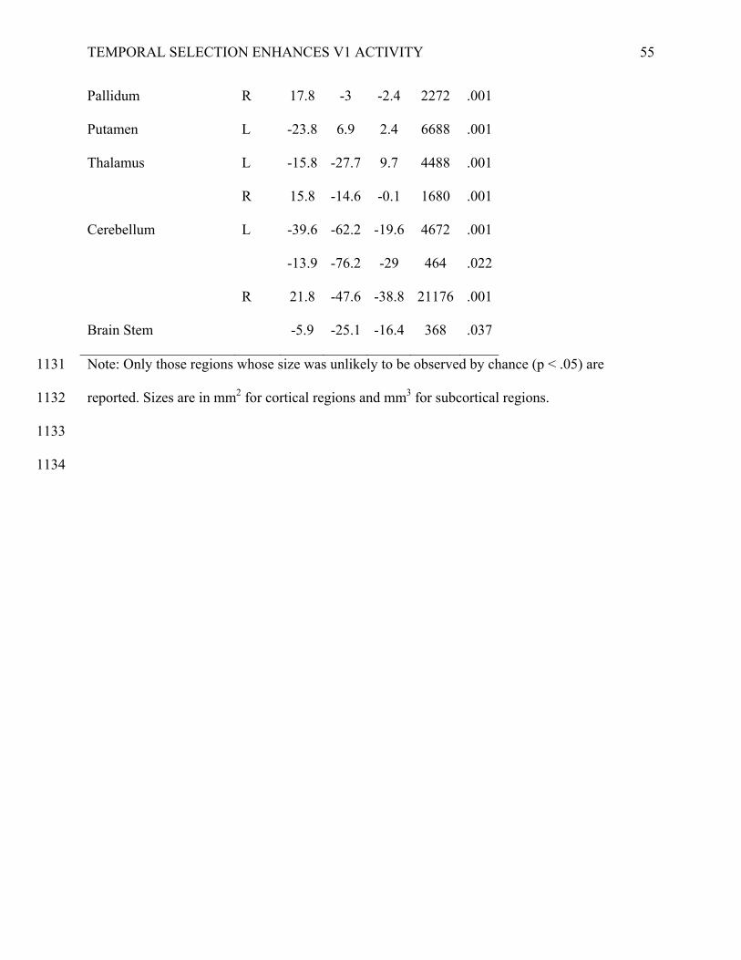
TEMPORAL SELECTION ENHANCES V1 ACTIVITY
55
Pallidum R 17.8 -3 -2.4 2272 .001
Putamen L -23.8 6.9 2.4 6688 .001
Thalamus L -15.8 -27.7 9.7 4488 .001
R 15.8 -14.6 -0.1 1680 .001
Cerebellum L -39.6 -62.2 -19.6 4672 .001
-13.9 -76.2 -29 464 .022
R 21.8 -47.6 -38.8 21176 .001
Brain Stem
-5.9 -25.1 -16.4 368 .037
Note: Only those regions whose size was unlikely to be observed by chance (p < .05) are 1131
reported. Sizes are in mm2 for cortical regions and mm3 for subcortical regions. 1132
1133
1134
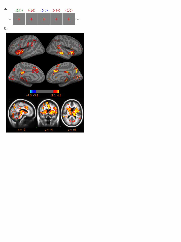
a.
b.
z = +9 x = -‐9 y = +4
-‐4.3 -‐3.1 4.3 3.1
((♪))
+ … … ((♪))
+ ((…))
+ + ((♪))
+ ((♪))
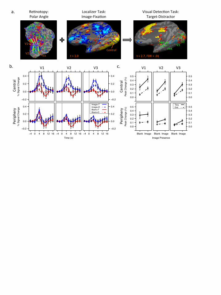
a. Visual Detec-on Task: Target-‐Distractor
V1 V2
V3 FFA
PPA
t > 2.7, FDR < .01
b. c.
Localizer Task: Image-‐Fixa-on
t > 1.0
Periphery
Central
Re-notopy: Polar Angle
V1
V2 V3
Time (s)
% S
igna
l Cha
nge
−0.2
0.0
0.2
0.4
−4 0 4 8 12 16
● ● ●
●●
●●
●● ● ●
●● ●
●
●
●
● ●
●● ●
●● ●
●
●
● ●
●● ●
●
●● ●
●
●
●
● ●
●
●●
−4 0 4 8 12 16
● ● ●●
●
●●
●● ● ●
●● ●
●
●
●
●●
●● ●●
● ●●
●
● ●● ● ● ●
●●
●
●
●
●
● ●●
●●
−4 0 4 8 12 16
−0.2
0.0
0.2
0.4
● ● ●● ●
● ●● ● ● ●● ● ● ●
●
●●
●
●● ●
● ●●
●
●
● ●● ● ● ●
●●
●
●
●
●●
●●
● ●
Image+TImage+DBlank+TBlank+D
Time (s)
% S
igna
l Cha
nge
−0.2
0.0
0.2
0.4
● ● ●●
●
●●
●●
● ●● ●
●
●
●
●
●●
●
● ●
●●
●
●
●●
●
● ●●
●●
●
●
●
●●
●
●● ●
●● ● ●
●●
●●
●● ● ●● ● ●
●
●
●
●●
●
●●
●● ●
●
●●
●
● ●●
●● ●
●
●
●●
●
●● ●
●
−0.2
0.0
0.2
0.4
● ● ●● ●
●●
●● ● ●● ● ●
●●
●
● ●
●
● ●●
● ●
●
● ●
●● ● ● ●●
●●
●
● ●
●
● ● ● ●Central
Perip
hery
V1 V2 V3
Image Presence
Peak
Cha
nge
(%)
0.00.10.20.30.40.5
●
●●
●
●
●
●
●
0.00.10.20.30.40.5
●
●
●
●
Image Presence
Peak
Cha
nge
(%)
0.00.10.20.30.40.5
Blank Image
●●
●●
Blank Image
●●
●
●
Blank Image
0.00.10.20.30.40.5
●●
●
●
TargDist
Central
Perip
hery
V1 V2 V3

Time (s)
% S
igna
l Cha
nge
0.0
0.2
0.4
● ● ●
●
●
●
●● ● ● ●● ● ●
●
●
●
●
● ● ● ●● ● ● ●
●●
● ● ● ● ●● ● ●●
●●
● ● ● ●● 0.0
0.2
0.4
● ● ● ●● ● ● ● ● ● ●● ●
●●
●●
● ● ● ● ●
Time (s)
% S
igna
l Cha
nge
0.0
0.2
0.4
● ● ●
●
●
●
●
● ● ● ●●
●●
●
●
●
●
● ●● ●
● ● ● ●●
●● ● ● ● ●● ● ●
●●
●
● ●● ● ● 0.0
0.2
0.4
● ● ● ● ● ● ● ● ● ● ●● ● ● ●● ●
●● ●
● ●
Time (s)
% S
igna
l Cha
nge
0.0
0.2
0.4
−4 0 4 8 12 16
● ● ● ●●
●● ● ● ● ●● ●
●●
●
●● ● ● ●
●
−4 0 4 8 12 16
● ● ●
●
●
●
● ● ● ● ●● ● ●
●
●
●
● ● ● ●●
−4 0 4 8 12 16
0.0
0.2
0.4
● ● ● ● ●● ● ● ● ● ●● ●
● ●●
● ● ● ● ● ●
TargetDistractor
FFA
PPA
Face Scene Scrambled
Time (s)
% S
igna
l Cha
nge
0.0
0.2
0.4
−4 0 4 8 12 16
● ● ● ●●
● ● ● ● ● ●● ● ●
●
●
●● ●
●● ●
−4 0 4 8 12 16
● ● ●
●
●
●
● ● ● ● ●● ● ●
●
●
●
● ● ● ● ●
−4 0 4 8 12 16
0.0
0.2
0.4
● ● ● ● ● ● ● ● ● ● ●● ● ● ●●
● ● ● ●● ●
TargetDistractor
FFA
PPA
Face Scene Scrambled
a.
b.
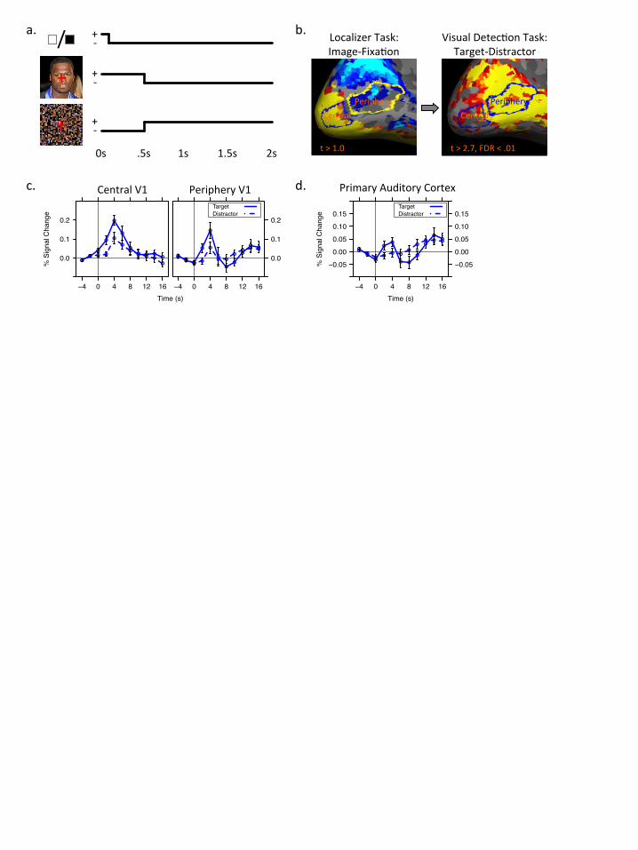
Localizer Task: Image-‐Fixa5on
t > 1.0
Central Periphery
Visual Detec5on Task: Target-‐Distractor
t > 2.7, FDR < .01
Central Periphery
a.
c. d.
b.
Time (s)
% S
igna
l Cha
nge
0.0
0.1
0.2
−4 0 4 8 12 16
●● ● ●
●
●
●●
● ●
●●
●
●
●
●
●
●
● ● ●●
−4 0 4 8 12 16
0.0
0.1
0.2
●●
●●
●
● ●
●●
● ●
●
●●
●
●
●
●●
●
● ●
TargetDistractor
Central V1 Periphery V1
+
/ + -‐
0s .5s 1s 2s 1.5s
+ -‐
+ -‐
+
Time (s)
% S
igna
l Cha
nge
−0.050.000.050.100.15
−4 0 4 8 12 16
−0.050.000.050.100.15
●●
● ●● ●
●
●● ● ●
●
●
●
●●
● ●
●
●
●●
TargetDistractor
Primary Auditory Cortex

Time (s)
% S
igna
l Cha
nge
−0.1
0.0
0.1
0.2
●
●●
● ●
●●
●
●
● ●
● ●
●
●
●
●
●●
●
● ●
●● ●
●● ● ●
●●
● ●
● ●●
●
●●
●●
●
● ●
−0.1
0.0
0.1
0.2
●● ●
●●
● ●●
●● ●
● ●● ● ●
● ● ●
●
● ●
TargetDistractor
Time (s)
% S
igna
l Cha
nge
−0.1
0.0
0.1
0.2
−4 0 4 8 12 16
●● ●
●●
● ● ●●
●●
●●
●
●
●
●
●●
●
●
●
−4 0 4 8 12 16
● ● ●● ●
●● ●
●● ●
●● ●
●
●
●
●●
●
●●
−4 0 4 8 12 16
−0.1
0.0
0.1
0.2
● ● ● ● ●●
● ●
● ● ●● ●
●● ●
●●
●
●●
●
Central
Perip
hery
V1 V2 V3
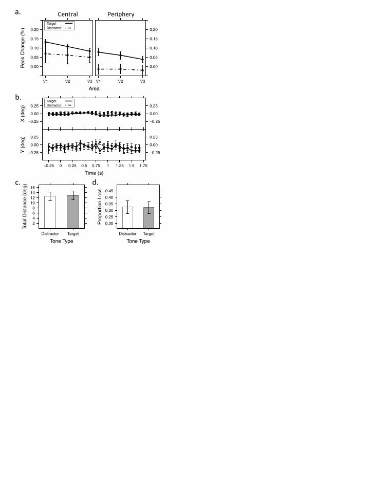
Time (s)
X (d
eg)
−0.25
0.00
0.25
−0.25 0 0.25 0.5 0.75 1 1.25 1.5 1.75
−0.25
0.00
0.25
●● ● ● ● ● ● ● ● ● ● ● ● ● ● ● ● ● ●
● ● ● ●●● ● ● ● ● ● ● ● ● ● ● ● ● ● ● ● ● ● ● ● ● ● ● ●
TargetDistractor
Time (s)
Y (d
eg)
−0.25
0.00
0.25
−0.25 0 0.25 0.5 0.75 1 1.25 1.5 1.75
−0.25
0.00
0.25
●● ● ●
●●
●● ●
● ●●
●
●
●● ● ●
● ● ● ● ● ●●
●● ●
● ● ●●
●●
●●
●
●
● ●●
●●
● ●● ● ●
Tone Type
Prop
ortio
n Lo
ss
0.200.250.300.350.400.45
Distractor Target
Tone Type
Tota
l Dis
tanc
e (d
eg)
2468
10121416
Distractor Target
Area
Peak
Cha
nge
(%)
0.00
0.05
0.10
0.15
0.20
V1 V2 V3
●●
●
●
●
●
V1 V2 V3
0.00
0.05
0.10
0.15
0.20
● ●●
●
●
●
TargetDistractor
a.
b.
d.
Central Periphery
c.

Time (s)
% S
igna
l Cha
nge
−0.1
0.0
0.1
0.2
−0.1
0.0
0.1
0.2
Time (s)
% S
igna
l Cha
nge
−0.1
0.0
0.1
0.2
−8 −4 0 4 8 12 16 −8 −4 0 4 8 12 16 −8 −4 0 4 8 12 16
−0.1
0.0
0.1
0.2
Central
Perip
hery
V1 V2 V3