Embed Size (px)
Citation preview

Global Salm-Surv
AA gglloobbaall SSaallmmoonneellllaa
ssuurrvveeiillllaannccee aanndd llaabboorraattoorryy ssuuppppoorrtt pprroojjeecctt of the World Health Organization
of the World Health Organization
th
EDITED BY: RENE S. HENDRIKSEN (DFVF), JAAP AGENAAR (ASG), MARCEL VAN BERGEN (ASG)
Laboratory Protocols
Level 2 Training Course
Identification of thermotolerant Campylobacter
5 Ed. March. 2003 W

Contents:
. Introduction to identification of thermotolerant Campylobacter from food, faeces or water 3
. Identification of thermotolerant Campylobacter from food, faeces or water .........................5
. Composition and preparation of culture media and reagents..................................................8
ecord sheet: Isolation and identification of Campylobacter from faeces, food or water ….. 11
ppendix 1. esult sheet for identification of Campylobacter ................................................14
ppendix 2. Photographs of pos. and neg. reactions of biochemical tests on Campylobacter.15
Page 1 2 3 R A R A
2
1. Identification of thermotolerant Campylobacter from food, faeces or water Introduction The following procedures will guide you through the steps that are necessary to carry out a biochemical identification of Campylobacter. Campylobacter are generally identified by: • Slender helical or curved gram-negative rods. • Highly motile by means of a single polar flagellum. • Optimal oxygen concentration for growth 5-10%. • Do not ferment or oxidize sugars. • Do not produce indole (mind the different with hydrolysis of indoxyl acetate!). According to ISO 10272 (Microbiology of food and animal feeding stuffs – Horizontal method for detection of thermotolerant Campylobacter) Campylobacter is identified by the following characteristics: • morphology and motility • morphology in Gram staining • oxidase • glucose • lactose • sucrose • gas In this course identification and differentiation of strains is performed by: • morphology and motility • morphology in Gram staining • katalase • oxidase • hippurate hydrolysis • hydrolysis of indoxyl acetate Campylobacter from faeces, food or water Campylobacter food poisoning occurs in most cases sporadically affecting individuals. Outbreaks due to Campylobacter infections are rare. Outbreaks due to contaminated milk and drinking water are described more often than food borne outbreaks. Campylobacter jejuni is the most common cause of human bacterial enteritis but Campylobacter coli may also be responsible. Campylobacter jejuni is commonly isolated from chicken and cattle, and chicken is expected to be one of the major sources of infection for humans. Pigs commonly carry Campylobacter coli In some countries where large quantities of pork are consumed Campylobacter coli infections frequently occur.
3

Campylobacter may also be present in faeces or food in low numbers and they may be injured. To diminish the risk of obtaining false negative results, selective enrichment of a large food sample can be performed: • Enrichment in selective enrichment broth (e.g. Preston). • Selective plating on CCD-agar plates. References 1. Nachamkin I. and M. J. Blaser (eds) (2000). Campylobacter 2nd ed. ASM Press, Washington, D.C.
4
2. Identification of thermotolerant Campylobacter from food, faeces or water Materials Equipment • Disposable inoculation loops (1 µl and 10 µl) • Incubators at 37°C/42oC • Microscope • Slides • Cover glass • Mineral oil • Paper disc 6 mm • Pipettes for 0.2 ml (e.g. 1 ml pipettes) • 200 ml flask • Forceps • Eppendorf tubes, 1.5 ml • Drop counters Media • Sterile water • 3%-H2O2 • 1%-hippurate solution • 3.5%-ninhydrin solution • 10%-indoxyl acetat solution • Oxidase sticks • Gram staining reagents • Crystal violet • Gram’s iodine • Ethanol (95%) • Carbol fuchsine
Bacterial strains: Campylobacter.lari ATCC 35221 Campylobacter.coli ATCC 33559 Campylobacter.jejuni ATCC 700819 Pseudomonas aeruginasa ATCC 27853 Enterococcus faecalis ATCC 29212 Staphylococcus aureus ATCC 29213 Safety Carry out all procedures in accordance with the local codes of safe practice.
5

Procedure Theory / comments Identification
A striking character of Campylobacter is their helical or curved shape. Long spiral forms can resemble spirochaetes superficially, but campylobacters have flagella, usually single, at one or both poles and are highly motile, spinning around their long axes and frequently reversing direction.
Microscopy (morphology and motility) One drop of sterile saline is placed on a slide. Colonies from the CCD agar plates are mixed with the saline. Place cover glass above the colonies, and place the slide in the microscope.
Gram staining (morphology) Gram negative bacteria (like Campylobacter) are decolourised and stained red by the counter-stain (Carbol fuchsine). Campylobacter are curved or gull shaped forms. Old cultures may contain coccoid bacteria.
One small drop of saline is placed on a slide. Colonies from the CCD agar plates are mixed with saline and smeared over the surface of the slide. The smears are allowed to dry thoroughly. The smears are fixed by passing the slide, smear up, quickly through the Bunsen flame three times. After cooling the smears can be stained. Between each staining reagent the smear is washed under a gently running tap, excess of ater tipped off before the next reagent is added.
1. Crystal violet (60 sec) 2. Gram’s iodine (60 sec) 3. Ethanol (decolouriser) (60 sec) 4. Carbol fuchsine (60 sec)
Test for catalase The catalase-enzyme cleaves the hydrogen peroxide H2O2 + H2O2 => O2 + 2H2O The peroxidase is only able to reduce H2O2 if an organic substrate is present at the same time and serves as a donor for hydrogen.
Put a colony at a small spot on a slide (do NOT not make a suspension; just dry). Put one drop of 3%-H2O2 on the spot with the bacterial material. Examine immediately for evolution of gas, which indicates catalase activity.
Test for oxidase Transfer one colony to a filter paper. Soak the filter in a oxidase solution. Appearance of a blue color within 10 sec indicates a positive result.
The method is based on the principle that certain phenylen-diamin-derivatives are oxidised by cytochrom C to produce a bluish indophenol. Commercial kits are available.
6

Hippurate hydrolysis Hydrolysis of hippuracid releases benzoeacid. Hippuracid is soluble in excess of an acidic solution of ferrichloride while benzoeacid precipitates.
Suspend a loopful of a growth from an 18-24 hour columbia agar plate containing 5% cattle blood culture in 400 µl of a 1%-hippurate solution (take care not to incorporate agar!). Incubate at
1%-hippurate solution: freshly prepared or stored at -20 oC for about 6 month.
37oC for 2 hours. Then slowly add 200 µl 3.5%-ninhydrin solution to the side of
3.5%-ninhydrin solution: Stable for about one month. Stored at room temperature in a dark bottle.
the tube to form an overlay. Reincubate at 37oC for 10 min, and read the reaction. Positive reaction: dark purple/blue.Negative reaction: clear or gray.
Identification Hydrolysis of indoxyl acetate
The bacterial enzyme esterase releases indoxyl from indoxyl acetate which spontaneously forms indigo in the presence of oxygen.
Add 50 µl of a 10% (w/v) solution of indoxyl acetate in acetone to an absorbent paper disc 6 mm in diameter and allow to dry in air. Apply growth from a Campylobacter colony directly to disc and then wet with a drop of sterile distilled water. Appearance of a blue-green color within 5-10 minutes indicates a positive result.
Dried discs are stable for at least 12 months if stored at 4ºC in a dark glass bottle with silica gel. Discs should not be used if the color has changed from white, or if the expiration date has passed Appendix 1. Result sheet Appendix 2. Photographs of pos. and neg.
reactions of biochemical tests on Campylobacter
7
3. Composition and preparation of culture media and reagents The media and reagents are available from companies like Oxoid, Merck and Difco. The composition of the dehydrated media given below is an example and may vary a little among the different manufacturers. Also the media should be prepared according to the manufacturers description if it differs from the description given here. Saline solution Sodium chloride 8.5 g Water 1000 ml Preparation: Dissolve the sodium chloride in the water, by heating if necessary. Adjust pH ∼ 7.0 after sterilisation. Dispense the solution into tubes so 4 ml is obtained after autoclaving at 121oC for 20 min. 3,5 % Ninhydrin solution Ninhydrin (C9H6O4) 3,5 g Acetone (C3H6O) 50 ml Butanol (C4H10O) 50 ml Dissolve the chemical in the solutions. Stored at + 5oC in dark bottles of 20 ml. 1% Hippurate solution Natriumhippurat (C9H8NNaO3) 1 g PBS 99 ml
Dissolve the chemical with the solutions. Stored at -20oC in tubes of 15 ml. Gram-staining Crystal violet Crystal violet 2.0 Ethanol 95% (vol/vol) 20.0 ml Ammonium oxalate 0.8 g Distilled water 80.0 ml The crystal violet is first dissolved in the ethanol, then the ammonium oxalate is dissolved in the distilled water. The two solutions are added together. To aid the dissolving process, both mixtures are agitated in a bath of hot water.
8

Gram’s iodine Iodine crystals 1.0 g Potassium iodide 2.0 g Distilled water 200 ml
The iodine crystals and the potassium iodine are ground together in a mortar and the distilled water is added slowly. If necessary the mixture can be agitated in a bath of hot water to aid dissolution.
Decolourizer Ethanol 95% (vol/vol)
Carbol fuchsine (counterstain) Concentrated carbol fuchsine 10.0 ml Distilled water 90.0 ml 10% (wt/vol) Indoxylacetate solution Indoxylacetat (C10H9NO2) 10 g Acetone (C3H6O) 90 ml Dissolve the chemical in acetone. Stored at +4oC in a dark bottle. Oxidase solution L(+)-Ascorbicacid 0,03 g N,N,N´,N´- Tetramethyl-p-Phenylendiamine Dihydrochloride (C10H16N2 • 2HCl) 0,03 g Sterile water 30 ml Dissolve the chemicals in water, and store the solution in a dark bottle at +5 oC for 3 weeks. References 1. BARROW & FELTHAM (eds.): Cowan and Steel´s Manual for the Identification of Medical
Bacteria, 3 rd edn. 2. NMKL method no. 119, 2nd ed, Campylobacter Jejuni/Coli detection in foods. Nordic
committee on food analysis.
9

Record sheet: Quality Control / Batch Control Date:____________________ Init.:________________________ Biochemical tests QC-Strain
C.jejuni ATCC 700819
C.coli ATCC 33559
C.lari ATCC 35221
E. faecalis ATCC 29212
S. aureus ATCC 29213
P. aeruginosa ATCC 27853
Gram staining
Test for catalase
Test for oxidase
Hippurate hydrolysis
Hydrolysis of indoxyl acetate
11
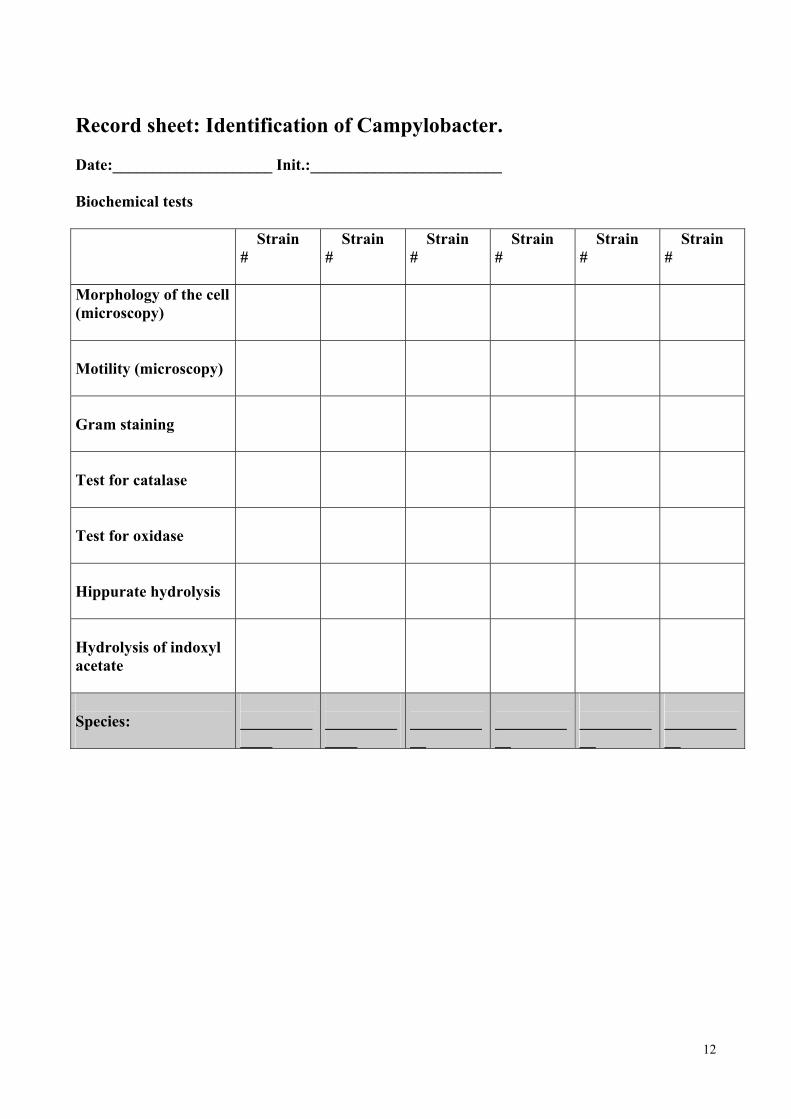
Record sheet: Identification of Campylobacter. Date:____________________ Init.:________________________ Biochemical tests
Strain #
Strain #
Strain #
Strain #
Strain #
Strain #
Morphology of the cell (microscopy)
Motility (microscopy)
Gram staining
Test for catalase
Test for oxidase
Hippurate hydrolysis
Hydrolysis of indoxyl acetate
Species:
_____________
_____________
___________
___________
___________
___________
12

Record sheet: Isolation and identification of Campylobacter from faeces, food or water. Date:____________________ Init.:________________________ Biochemical tests Faeces-
sample 1
Faeces- sample 2
Food-sample 1
Food-sample 2
Water-sample 1
Water-sample 2
Morphology of the cell (microscopy)
Motility (microscopy)
Gram staining
Test for catalase
Test for oxidase
Hippurate hydrolysis
Hydrolysis of indoxyl acetate
Species:
_____________
_____________
___________
___________
___________
___________
13
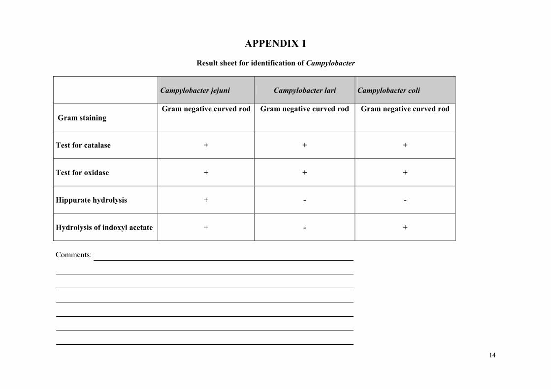
APPENDIX 1
Result sheet for identification of Campylobacter
Campylobacter jejuni Campylobacter lari
Campylobacter coli
Gram staining
Gram negative curved rod Gram negative curved rod Gram negative curved rod
Test for catalase
+ +
+
Test for oxidase
+ +
+
Hippurate hydrolysis
+ -
-
Hydrolysis of indoxyl acetate
+ -
+
Comments:
14
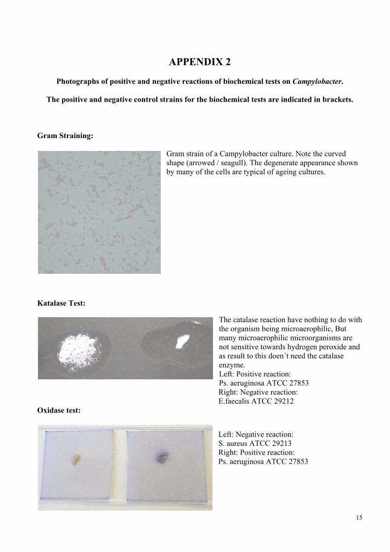
APPENDIX 2
Photographs of positive and negative reactions of biochemical tests on Campylobacter.
The positive and negative control strains for the biochemical tests are indicated in brackets.
Gram Straining:
Gram strain of a Campylobacter culture. Note the curved shape (arrowed / seagull). The degenerate appearance shown by many of the cells are typical of ageing cultures.
Katalase Test:
The catalase reaction have nothing to do with the organism being microaerophilic, But many microaerophilic microorganisms are not sensitive towards hydrogen peroxide and as result to this doen´t need the catalase enzyme. Left: Positive reaction: Ps. aeruginosa ATCC 27853 Right: Negative reaction: E.faecalis ATCC 29212
Oxidase test:
Left: Negative reaction: S. aureus ATCC 29213 Right: Positive reaction: Ps. aeruginosa ATCC 27853
15

16
Hippurate hydrolysis:
Left: Negative reaction: C.coli ATCC 33559 Right: Positive reaction: C. jejuni ATCC 700819
Hydrolysis of Indoxyl acetate:
Left: Negative reaction: C.lari ATCC 35221 Right: Positive reaction: C. jejuni ATCC 700819
Reference: 1. Food-borne Pathogens. Monograph Number 3 Campylobacter, Oxoid Standards. 2. © Institut for Veterinær Mikrobiologi, Den Kgl. Veterinær- og Landbohøjskole, 2000.