Embed Size (px)
Citation preview
THE AMERICAN JOURNAL OF CANCER
A Continuation of The Journal of Cancer Research
~ ’ O L U M E XXVI MAR.CH, 1936 NUMBER 3 _-
INTRATHORACIC SYMPATHOBLASTOMA
PRODUCING THE SYMPTOMATOLOGY OF A SUPERIOR PULMONARY SULCUS TUMOR ( PANCOAST)
THOMAS T. FROST, M.D., A i m SIDNEY E. WOLPAW, M D
(From the Departments of Pathology and Medicine, Cleveland City Hospital)
In 1932 Pancoast (1) described a tumor occurring at the thoracic inlet and characterized clinically by ( a ) pain about the shoulder, high in the axilla, or down the inner side of the arm or the ulnar side of the forearm, ( b ) Horner’s syndrome, ( c ) loss of power and wasting of the muscles of the hand, ( d ) x-ray evidence of a small, homogeneous shadow at the apex of the lung with destruc- tion of the posterior parts of one or more ribs and often of the adjacent vertebrae. To the growth presenting such clinical and roentgenographic fea- tures Pancoast applied the name superior pulmonary sulcus tumor since “this term implies its approximate location, and a lack of origin from the lungs, pleura, ribs, or mediastinum.”
Pancoast further concluded that tumors presenting this characteristic pic- ture formed a definite pathological entity, the tumor being epithelial in its histopathology, although of uncertain origin. An origin from an embryonal epithelial rest, such as a fifth pharyngeal pouch, was suggested. Of the seven cases reported, biopsies were obtained in two. Case No. 1 was first thought to be an endothelioma of the pleura, but upon later consideration the diagnosis was changed to carcinoma spino-cellulare “ with groups of prickle cells.” Case No. 2 was diagnosed simply as carcinoma. Two other patients-cases 6 and 7-each had a previous history of carcinoma of the cervix, one four years and the other two years previously, treated by radiation, and showing local disappearance of the tumor without recurrence. Both were considered to have primary superior pulmonary sulcus tumors. No pathological studies were made, however, so that the possibility that these were secondary growths cannot be eliminated. No biopsies or autopsies were secured in the remain- ing three cases, so that the nature of the tumors is not known.
To the group of cases presenting the clinical features enumerated by Pancoast we wish to add the following because of the unusual type of tumor.
4x3
484 THOMAS T. FROST A N D SIDNEY E. WOLPAW
CASE REPORT
J. R., a thirty-eight-year-old white man, was admitted to the Dermatological Service of the Cleveland City Hospital on Feb. 13, 1934, complaining of pain in the right arm of four months’ duration.
The past history was unimportant except for a chancre in 1914, which received local therapy. The patient had been married for twenty-two years, and his wife and one daughter were living and well.
The patient had been in good health until his present illness. In October 1933 he noticed the onset of a peculiar numbness confined to the volar surface of the right arm and forearm. This was soon followed by pain about the right elbow with frequent radiation to the fingers and into the shoulder. The pain was severe and knife-like and required the use of strong sedatives. Hyperesthesia finally became so marked that the patient was forced t o carry the arm well wrapped and in a sling position. H e had lost thirty-six pounds in weight. Slight difficulty in swallowing and breathing had occurred, but were not outstanding symptoms. Because of the previous history of syphilis the patient was referred to the hospital for antisyphilitic therapy.
Physical examination revealed the following relevant facts. There was marked plosis of the right lid with constriction of the right pupil. Both pupils reacted to light. The right supraclavicular fossa appeared full but there was no palpable mass. The superficial veins of the right side of the neck and chest were dilated. The trachea was deviated to the left. Examination of the heart and lungs revealed no abnormalities. The blood-pressure in the left arm was 120/60; in the right arm 114/78. The fingers were clubbed. The extremities were thin, corresponding to the patient’s weight loss, but the right forearm and hand showed definite atrophy. There were no fibrillary twitchings. Tendon reflexes were present in both extremities. There was hyperesthesia of the ulnar surface of the forearm near the elbow but no other sensory disturbance. The neurological examination otherwise showed no abnormal findings.
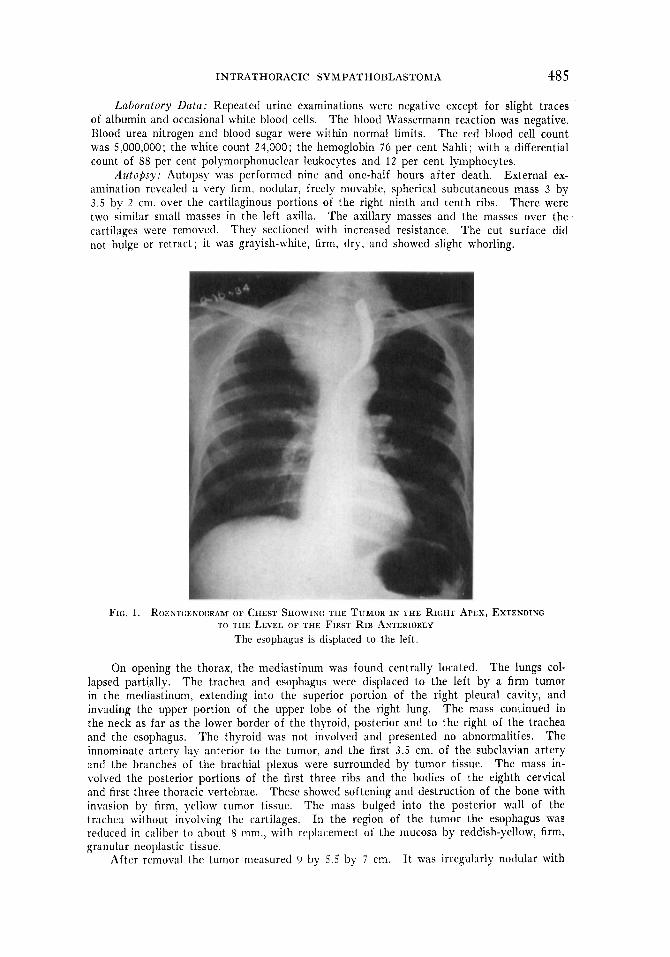
Fluoroscopic and x-ray examination of the chest revealed a shadow of increased density in the right apex, measuring about 8 cm. in diameter. The esophagus and trachea were displaced to the left and anteriorly. The heart was of normal size. The aorta was not enlarged but the aortic knob was slightly displaced downwards. The mass did not pulsate. There was no evidence of invasion of the ribs or vertebrae, although no special views of these areas were taken.
Cozirx: During his four and one-half weeks hospital stay the patient’s pain continued unabated. Dyspnea and dysphagia, which were minor symptoms on admission, became greatly aggravated. The apical mass was considered to be either a non-pulsating innominate aneurysm or a mediastinal tumor with invasion of the brachial plexus. Because of the possibility that the latter lesion might be radiosensitive, deep x-ray therapy was begun (treatment to the right anterior clavicular area; distance 50 cm.; field 1 2 by 20 cm.; filters 0.5 mm. Cu, 1 mm. Al; kv. 200; ma. 5 ; 200 r ; 15 minutes). This was abandoned after two treatments because of the patient’s extreinely poor condition.
Four weeks after admission to the hospital the patient developed acute urinary retention and rapidly increasing paralysis of both lower extremities. Neurological examination re- vealed the ptosis of the right lid and constriction of the right pupil previously described. There was hypesthesia to pin-prick and cotton below the level of the first thoracic segment on each side of the body. There was sensory impairment over both upper extremities cor- responding to the cutaneous distribution of the first and second thoracic segments. On the right there was a trigger zone along the ulnar surface of the forearm near the elbow. Severe, aching pain was elicited by stroking the area, with radiation of the pain down the arm. Tendon reflexes were elicited on the left upper extremity but not on the right. There was marked atrophy of the right arm, forearm, and hand; and to a slight degree of the left. There was complete flaccid paralysis of both lower extremities. Tendon reflexes were elicited with difficulty in the left lower extremity and none on the right.
Following the onset of bladder symptoms treatment was symptomatic and the patient died five days later, on the thirty-first hospital day, March 16, 1934. The clinical diagnosis submitted was (1) mediastinal tumor with invasion of the brachial plexus on the right and extension into the spinal canal, ( 2 ) tertiary syphilis.
There had been no miscarriages.
INTRATHORACIC SYMPATHOBLASTOMA 48 5
Laboratory Data; Repeated urine examinations were negative except for slight traces of albumin and occasional white blood cells. The blood Wassermann reaction was negative. Blood urea nitrogen and blood sugar were within normal limits. The red blood cell count was 5,000,000; the white count 21,000; the hemoglobin 76 per cent Sahli; with a differential count of 88 per cent polymorphonuclear leukocytes and 12 per cent lymphocytes.
Autopsy: Autopsy was performed nine and one-half hours after death. External ex- amination revealed a very firm, nodular, freely movable, spherical subcutaneous mass 3 by 3 5 by 2 cm. over the cartilaginous portions of the right ninth and tenth ribs. There were two similar small masses in the left axilla. The axillary masses and the masses over the cartilages were removed. The cut surface did not bulge or retract; it was grayish-white, firm, dry. and showed slight whorling.
They sectioned with increased resistance.
FIG. 1. ROENTGENOGRAM O F CHEST SHOWINC TlrE TUMOR I N THE RIGHT APEX, EXTENDING TO THE LEVEL O F THE FTRST RIB ANTERIORLY
The esophagus is displaced to the left.
On opening the thorax, the mediastinum was found centrally located. The lungs col- lapsed partially. The trachea and esophagus were displaced to the left by a firm tumor in the mediastinum, extending into the superior portion of the right pleural cavity, and invading the upper portion of the upper lobe of the right lung. The mass continued in the neck as far as the lower border of the thyroid, posterior and to the right of the trachea and the esophagus. The innominate artery lay anterior to the tumor, and the first 3.5 cm. of the subclavian artery and the branches of the brachial plexus were surrounded by tumor tissue. The mass in- volved the posterior portions of the first three ribs and the bodies of the eighth cervical and first three thoracic vertebrae. These showed softening and destruction of the bone with invasion by firm, yellow tumor tissue. The mass bulged into the posterior wall of the trachea without involving the cartilages. I n the region of the tumor the esophagus was reduced in cdiber to about 8 mm., with replacement of the mucosa by reddish-yellow, firm, granular neoplastic tissue.
I t was irregularly nodular with
The thyroid was not involved and presented no abnormalities.
After removal the tumor measured 9 by 5.5 by 7 cm.

486 THOMAS T. FROST AND SIDNEY E. WOLPAW
small spicules of bone adherent to its posterior portion. It sectioned with moderately in- creased resistance. The cut surface was firm, neither bulging nor retracting, and was dry except for a few small areas where liquefaction necrosis had occurred. The tumor was composed of bands of dense gray tissue arranged in irregular strands with some tendency towards whorl formation. Between these strands were softer, friable, pale yellow areas of irregular shape, varying from 1 mm. to 1 cm. in diameter. Along its advancing edge the tumor extended into the pulmonary alveoli, leaving the al- veolar outlines well marked. The broncho- plmonary, peribronchial, bifurcation, and parntracheal lymph nodes were slightly enlarged and soft.
There was no capsule visible.
This portion of the tumor was soft and necrotic.
On section they revealed a moist, friable, grayish-black surface.
The right kidney and adrenal were in their normal positions, with the normal relation- ship to each other. The adrenal was much enlarged and was invaded hy an irregular nodular mass which extended into the upper third of the kidney. On section the same type of tumor tissue was found as in the mediastinul mass. The left adrenal was large but on section presented no tumor tissue.
Small nodular masses similar to those already descrihed were found in the line of mesenteric attachment of the upper portion of the jejunum, where several coils of intestine were bound together by dense adhesions.
In the region of the eighth cervical and first and second thoracic vertebrae the dura was densely adherent to the underlying vertebral bodies. The dura in this area was hyperemic, rough, and granular, hut not adherent to the cord. The underlying bone was soft, friable, and infiltrated with grayish-yellow tumor tissue which bulged into the spinal canal beneath the dura, compressing the cord. On section of the cord no definite areas of degeneration were seen.
The remaining lesions found in the body will be indicated in the final anatomical diagnosis.
The spinal cord was removed.
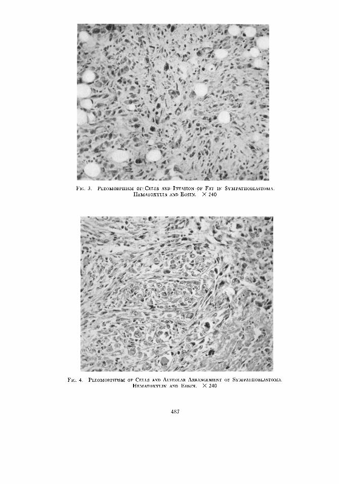
FIG. 3 . PLEOMORPKISM OF1 CELLS 4ND INVASION OF FAT IN SYMPATHOBLASTOMA HEXIATOXYLIN AND EOSIN. x 240
FIG. 4. PLEOMClRPIilSM OF CELLS AND ALVEOLAR hRANGEMENT OF SYMPATIIOBLASTOMA HEMATOXYLIN AXD EOSIN. x 240
487
488 THOMAS T. FROST AND SIDNEY E. WOLPAW
Microscopic Examinatiorz: The tumor is composed of large, pleomorphic cells, Ire- quently arranged to form alveoli, supported on varying amounts of relatively avascular connective tissue. I n the adrenal the tumor follows the arrangement of the cortical cells, replacing them and appearing in colunps and solid cords. The cells are large, polyhedral, and vary markedly in size ancl sh:ipe, The cytoplasm is vesicular, basophilic, and is frequently elongated to form one or more processes. These vary in length, are occasionally branched, and appear to contain fihrillae which frequently unite to form a complicated plexus. Some of these cells are in close proximity to this plexus; others are separate and united to it by their cytoplasmic processes. The nuclei are eccentrically placed, large, vesicular, with a single eccentrically placed, prominent nucleolus and irregular masses of chromatin concentrated at the periphery. The nuclear membrane is distinct. Mitotic figures, frequently atypical, are numerous. Multinucleated cells and giant nuclei are frequent.
In the kidney this is prominent and rich in collagen.
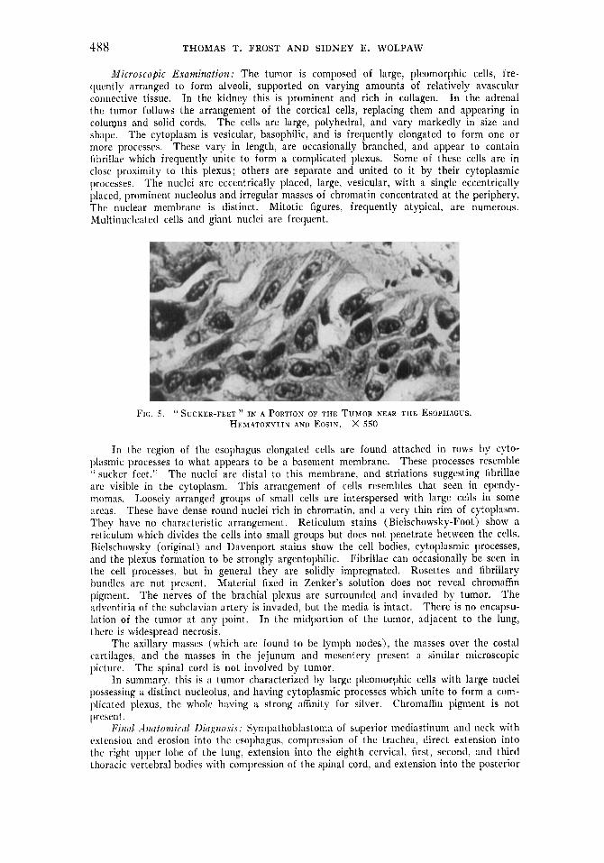
FIG. 5 . SUCKER-FEET ” I N A PORTION OF THE TUMOR NEAR T H E ESOPIIACUS. HEMATOXYLIN AND EOSIN. X 550
In the region of the esophagus elongated cells are found attached in rows by cyto- plasmic processes to what appears to be a hasement membrane. These processes resemble “ sucker feet.” The nuclei are distal to this membrane, and striations suggesting fibrillae are visible in the cytoplasm. This arrangement of cells resembles that seen in ependy- momas. Loosely arranged groups of small cells are interspersed with large cells in some areas. These have dense round nuclei rich in chromatin, and ;t very thin rim of cytoplasm. They have no characteristic arrangement. Reticulum stains (Bielschowsky-Foot) show a reticulum which divides the cells into small groups but does not penetrate between the cells. Bielschowsky (original) and Davenport stains show the cell bodies, cytoplasmic processes, and the plexus formation to be strongly argentophilic. Fibrillae can occasionally be seen in the cell processes, but in general they are solidly impregnated. Rosettes and fibrillary bundles are not present. Material fixed in Zenker’s solution does not reveal chromaffin pigment. The adventitia of the subclavian artery is invaded, hut the media is intact. There is no encapsu- lation of the tumor ;it any point. In the midportion of the tumor, adjacent to the lung, there is widespread necrosis.
The axillary masses (which are found to be lymph nodes), the masses over the costiil cartilages, and the masses in the jejunum and mesentery present a similar microscopic picture.
In summary, this is a tumor characterized by large pleomorphic cells with large nuclei possessing a distinct nucleolus, and having cytoplasmic processes which unite to form a com- plicatctl plexus, the wholc h‘iving ii strong affinity for silver. Chromaffin pigment is not present.
Sq mpathoblastonia of superior nicdiustinum ancl neck with extension and erosion into the esophagus, compression of the trachea, direct extension inio the right upper lobe of the lung. extension into the eighth cervical, first, second, and third thoracic vertebral hodies with compression of the spinal cord, and extension into the posterior
The nerves of the brachial plexus are surrounded and invaded by tumor.
The spinal cord is not involved by tumor.
F i i i i i l . liiiitoiriiral Uiagiio.
INTRATHORACIC SYMPATHOBLASTOMA 489
portions of the right first, second, and third ribs; metastases to the right adrenal and kidney, the jejunum, left axillary nodes, and subcutaneous tissue of the chest; pulmonary congestion and edema; bronchopneumonia with abscess formation; congestion and cloudy swelling of all internal organs ; chronic cystitis, ureteritis, and pyelitis (ascending) ; distention of blad- der; brown atrophy of the heart; stellate scar, left lobe of liver; mild arteriosclerosis.
DISCUSSION
A variety of tumors arising at the thoracic inlet have been considered in the pathologic diagnosis.
Carcinoma of the Bronchus: Four types of bronchial carcinoma occur- the squamous epithelioma, small-cell carcinoma, adenocarcinoma, and carci-
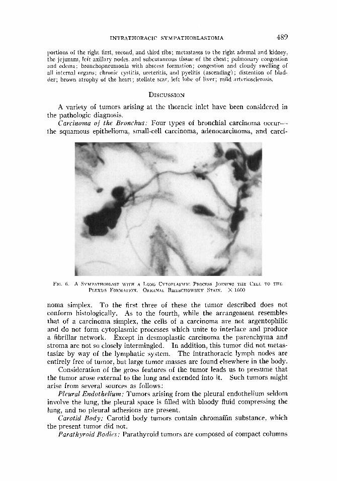
FIG. 6. A SYMPATHOBLAST W l l H A LONG CYTOPLASMIC PROCESS JOINING TIIE CELL TO TIIE
PLEXUS FORMATION. ORIGIXAL BIELSCHOWSKY STAIX. X 1600
noma simplex. To the first three of these the tumor described does not conform histologically. As to the fourth, while the arrangement resembles that of a carcinoma simplex, the cells of a carcinoma are not argentophilic and do not form cytoplasmic processes which unite to interlace and produce a fibrillar network. Except in desmoplastic carcinoma the parenchyma and stroma are not so closely intermingled. In addition, this tumor did not metas- tasize by way of the lymphatic system. The intrathoracic lymph nodes are entirely free of tumor, but large tumor masses are found elsewhere in the body.
Consideration of the gross features of the tumor leads us to presume that the tumor arose external to the lung and extended into it. Such tumors might arise from several sources as follows:
Pleural Endothelium .- Tumors arising from the pleural endothelium seldom involve the lung, the pleural space is filled with bloody fluid compressing the lung, and no pleural adhesions are present.
Carotid Body: Carotid body tumors contain chromaffin substance, which the present tumor did not.
Parathyroid Bodies: Parathyroid tumors are composed of compact columns
490 THOMAS T. FROST A N D SIDNEY E. WOLPAW
of opaque epithelium rich in glycogen, often in palisade arrangement, and without cytoplasmic processes.
Thyroid Gland: This organ was normal. Rranchial Clcfts: Tumors of branchial cleft origin are squamous epithe-
liomas; ciliated columnar epithelium and lymphoid tissue are frequently present.
Sarcomus: The reticulum present in this tumor does not have the arrange- ment characteristic of sarcoma.
Sympathetic Cervical Ganglia: Max Bielschowsky ( 2 ) has classified tu- mors of Sympathetic origin according to their embryogenesis. The primi- tive formative cells of the sympathetic nervous system are termed sympatho- gonia. These cells are of a lymphocyte-like character. They possess nuclei rich in chromatin and surrounded by a small, hardly visible ring of cytoplasm. They have an alveolar arrangement, thus forming nests, rosettes, and cylin- drical cords. These cells normally develop in two directions. In the one direction they form sympathoblasts, which further mature into sympathetic ganglion cells. In the other, they develop into pheochronioblasts, with a strong affinity for chrome salts. These develop into pheochromocytes whose mature stage is best seen in the large epithelial cells of the adrenal medulla. The sympathoblast is a larger cell than the sympathogonium with a clearer, almost vesicular nucleus and a larger protoplasmic body. I t produces axons and axon bundles which may be traced a short distance and which may be demonstrated by the silver method. A central nucleolus is frequently present in the nucleus. In tumors of the sympathetic system which reach this stage of differentiation, cells similar to those just described are found, and their axons unite to form a complicated plexus. Multinucleated giant cells and mitotic figures are present. These tumors are designated sympathoblastonias. They are found (in addition to the adrenal) in the sympathetic chain with a lateral localization in the neck, and close to the spine in the thoracic and lumbar regions.
The tumor found in this case fulfills the criteria for a tumor arising from the sympathetic nervous system, and the degree of differentiation justifies the diagnosis of sympathoblastoma. I t is thought to have arisen from the inferior cervical sympathetic ganglion. No tuniors described in the literature have the exact histological characters of this tumor, because they have occurred in purer form, the sympathogonioma, the sympathoblastoma, the pheochromo- cytoma, and the ganglioneuroma. The situation near the neck is that next in order of frequency to the adrenal. The moderate degree of malignancy is in harmony with the approach to the more mature type of cell.
Clinically this case presents the characteristics enumerated by Pancoast for a superior pulmonary sulcus tumor. Horner’s syndrome and invasion of the brachial plexus with arm pain and muscular weakness were both present. A small, dense, homogeneous mass was found at the right apex. Although the available roentgen films showed no destruction of ribs or spine, their involvement was revealed at autopsy. It may be noted that in three of Pancoast’s cases-Nos. 2 , 3 , and 7-the apical tumor preceded by months the roentgen evidence of invasion of bone.
INTRATHORACIC SYMPATHOBLASTOMA 49 1
The symptomatology produced by invasion of the brachial plexus, sympa- thetic chain, ribs, and vertebrae, either singly or in combination, is well known and has been described during the course of various apical lesions, particularly primary and secondary pulmonary neoplasms, apical tuberculosis, cervical tumors, and aneurysms. Analysis of the criteria enumerated by Pancoast for superior pulmonary sulcus tumors, however, permits the inclusion only of those growths which involve all of the above organs during the course of the disease. Such cases as the bronchogenic carcinoma described by Da Rin (3) or the cases of associated carcinoma and tuberculosis of Fried (4) , and Courcoux and Lereboullet ( 5 ) do not fall into this category since they lack either x-ray or autopsy evidence of rib involvement. Since Pancoast’s report in 1932, however, several autopsied cases have appeared in the literature which fulfill the stated requirements.
Jacox’ ( 6 ) autopsied case was a mucin-forming adenocarcinoma. In the opinion of Dr. C. V. Weller, (‘ both by direct evidence and by exclusion one is forced to the conclusion that this (tumor) is of bronchiogenic origin.”
Steiner and Francis ( 7 ) have reported two autopsied cases, one of which is open to question because of incomplete clinical and pathological data. Case No. 1, which is described in detail, was a mucin-secreting adenocarcinoma. The authors state that, while the tumor from its histopathological appearance might well have originated in the lung, there was nothing in its gross or microscopic characteristics to indicate that it arose from remnants of branchial pouches.
An origin from either a bronchus or an embryonal rest such as a fifth pharyngeal pouch could not be eliminated.
Fried’s first case (9) was a primary carcinoma of the bronchus. His second case (10) was a squamous epithelial carcinoma also of bronchial origin.
Of the cases reported by Browder and De Veer ( l l ) , Nos. 2 and 4 were autopsied and fulfill the necessary requirements for a superior pulmonary sulcus tumor. Both were squamous-cell carcinomas arising in the right upper lobe of the lung.
The autopsied material thus shows the clinical entity to which Pancoast has called attention occurring in one epidermoid carcinoma of uncertain origin, six carcinomas of bronchogenic origin, and one intrathoracic sympatho- blastoma. Consideration of these cases indicates that apical lesions other than epithelial carcinomas arising from embryonal branchiogenic rests may also produce the clinical picture of a superior pulmonary sulcus tumor. Neither the superior pulmonary sulcus tumors described by Pancoast nor the syndrome said to be characteristic of them can, therefore, be attributed to a new specific pathological entity among intrathoracic neoplasms. This opinion has also been expressed by Weller ( 1 2 ) , Fried (4, lo ) , and Browder and De Veer (11).
SUMMARY
Clarke’s (8) case was an epidermoid carcinoma.
1. A case fulfilling the clinical criteria for a superior pulmonary sulcus tumor is presented.
3.02 THOMAS T. FROST AND SIDNEY E. WOLPAW
2 . The tumor histologically is a sympathoblastoma, probably arising from the inferior cervical sympathetic ganglion.
3 . The autopsied material in the literature is reviewed and indicates that the clinical syndrome which has been described under the name of superior pulmonary sulcus tumor cannot be attributed to a specific pathological entity but may be caused by various tumors arising near the thoracic inlet.
NOTE: We are indebted to Dr. H. N. Cole of the Dermatology Department for the privilege of reporting this case, and to Dr. H. T. Karsner for the photomicrographs repro- duced in Figures 3 and 4 and for valuable help and criticism. This case was mentioned in the discussion by Dr. Karsner at the 1934 meeting of the American Association of Patholo- gists and Bacteriologists (13). The roentgen study was made by Dr. Eugene Freedman, to whom we are indebted for the roentgenogram.
BIBLIOGRAPHY
1 . I’ANCOA~T, H. K.: J. A. M. A. 99: 1391, 1932. 2. BIELSCHOWSKY, M.: Cytology and Cellular Pathology of the Nervous System by
3. DA RIN, C.: Semana med. 2 : 81, 1031. 4. FRIED, B. M.: Am. J. Cancer 23: 247, 1935. 5. COURCOUX, A., A N D LEREBOULLET, J.: Arch. mCd.-chir. de l’app. respir. 6: 569, 1931. 6. JACOX, H. W.: J. A. M. A. 103. 84, 1934. 7. STEINER, P. E., AND FRANCIS, I?.: Am. J. Cancer 2 2 : 776, 1934. 8. CLARKE, B. E.: Am. J. Path. 10: 693. 1931.
Wilder Penfield, Paul B. Hoeber, Inc., New York, 1032.
9. I;RIED, B. M. : Primary Carcinoma of the Lung, Williams and Wilkins Co., Baltimore, 1932, p. 226.
10. FRIED, B: M.: Am. J. Cancer 20: 791, 1034. 11. BROWDER, J., A N D DE VEER, J. A.: Am. J . Cancer 24: 507, 1935 12. WELLER, C. V.: Quoted by Jacox (6). 13. KARSNER, H. T . : In discussion of the paper by Clarke (8).


![A Cancer Journal for Clinicians [ Cancer Statistics, 2010]](https://img.pdfslide.net/doc/110x75/5447f486b1af9f65618b46dc/a-cancer-journal-for-clinicians-cancer-statistics-2010.jpg)