Embed Size (px)
Citation preview
THE CELL CYCLE
The Key Roles of Cell Division
1. Cell division functions in
reproduction, growth, and
repair
2. Cell division distributes
identical sets of chromosomes
to daughter cells. (somatic cells
only)
Review of microtubules
• The ability of organisms to reproduce their kind is one characteristic that best distinguishes living things from nonliving matter.
• The continuity of life from one cell to another is based on the reproduction of cells via cell division.cell division.
• This division process occurs as part of the cell cycle, the life of a cell from its origin in the division of a parent cell until its own division into two.
IntroductionIntroduction
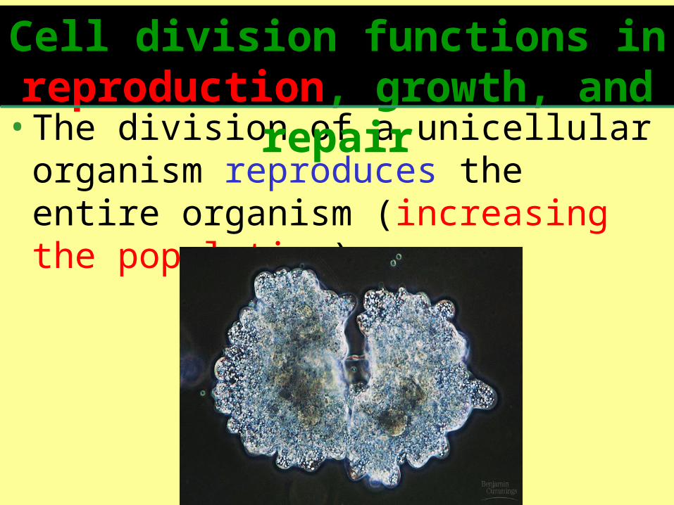
•The division of a unicellular organism reproduces the entire organism (increasing the population).
Cell division functions in reproduction, growth, and
repair

• Cell division is also central to the development of a multicellular organism that begins as a fertilized egg or zygote.
• Multicellular organisms also use cell division to repair and renew cells that die from normal wear and tear or accidents.
Cell division functions in reproduction, growth, and repair
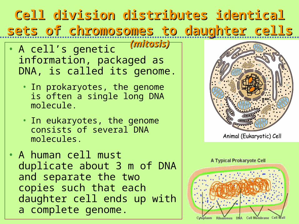
• A cell’s genetic information, packaged as DNA, is called its genome.
• In prokaryotes, the genome is often a single long DNA molecule.
• In eukaryotes, the genome consists of several DNA molecules.
• A human cell must duplicate about 3 m of DNA and separate the two copies such that each daughter cell ends up with a complete genome.
Cell division distributes identical sets of Cell division distributes identical sets of chromosomes to daughter cells chromosomes to daughter cells (mitosis)(mitosis)
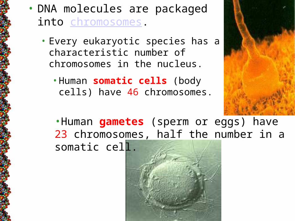
• DNA molecules are packaged into chromosomes.
• Every eukaryotic species has a characteristic number of chromosomes in the nucleus.
• Human somatic cells (body cells) have 46 chromosomes.
•Human gametes (sperm or eggs) have 23 chromosomes, half the number in a somatic cell.
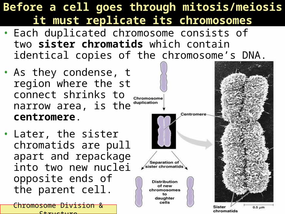
• Each duplicated chromosome consists of two sister chromatids which contain identical copies of the chromosome’s DNA.
• As they condense, the region where the strands connect shrinks to a narrow area, is the centromere.
• Later, the sister chromatids are pulled apart and repackaged into two new nuclei at opposite ends of the parent cell.
Before a cell goes through mitosis/meiosis it must replicate its chromosomes
Chromosome Division & Structure
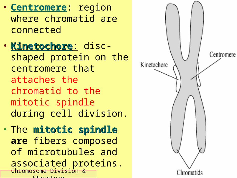
• Centromere: region where chromatid are connected
• KinetochoreKinetochore: disc-shaped protein on the centromere that attaches the chromatid to the mitotic spindle during cell division.
• The mitotic spindlemitotic spindle are fibers composed of microtubules and associated proteins.Chromosome Division &
Structure

Chromosome Division & Structure
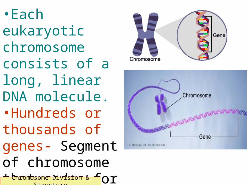
•Each eukaryotic chromosome consists of a long, linear DNA molecule.•Hundreds or thousands of genes- Segment of chromosome that codes for a protein.
•Humans: ~20,000 genes
Chromosome Division & Structure

• Associated with DNA: Proteins that maintain its structure and help control gene activity.
• This DNA-protein complex, chromatin, is organized into a long thin fiber.
• After the DNA duplication, chromatin condenses, coiling and folding to make a smaller package.
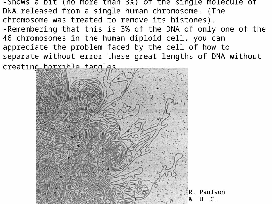
-Shows a bit (no more than 3%) of the single molecule of DNA released from a single human chromosome. (The chromosome was treated to remove its histones). -Remembering that this is 3% of the DNA of only one of the 46 chromosomes in the human diploid cell, you can appreciate the problem faced by the cell of how to separate without error these great lengths of DNA without creating horrible tangles.
R. Paulson & U. C. Laemmli

• Cell division requires the distribution of identical genetic material - DNA - to two daughter cells. (Mitosis/Somatic cells)
• Mitosis: A dividing cell duplicates its DNA, allocates the two copies to opposite ends of the cell, and then splits into two daughter cells. (DNA replication)
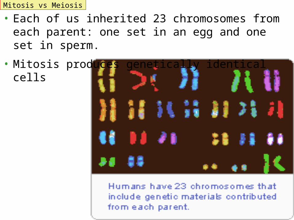
• Each of us inherited 23 chromosomes from each parent: one set in an egg and one set in sperm.
• Mitosis produces genetically identical cells
Mitosis vs Meiosis

• In contrast, gametes (eggs or sperm) are produced only in gonads (ovaries or testes).
•In the gonads, cells undergo a variation of cell division, meiosis, which yields four daughter cells, each with half the chromosomes of the parent.
•In humans, meiosis reduces the number of chromosomes from 46 to 23.
•Fertilization fuses two gametes together and doubles the number of chromosomes to 46 again.
Mitosis vs Meiosis
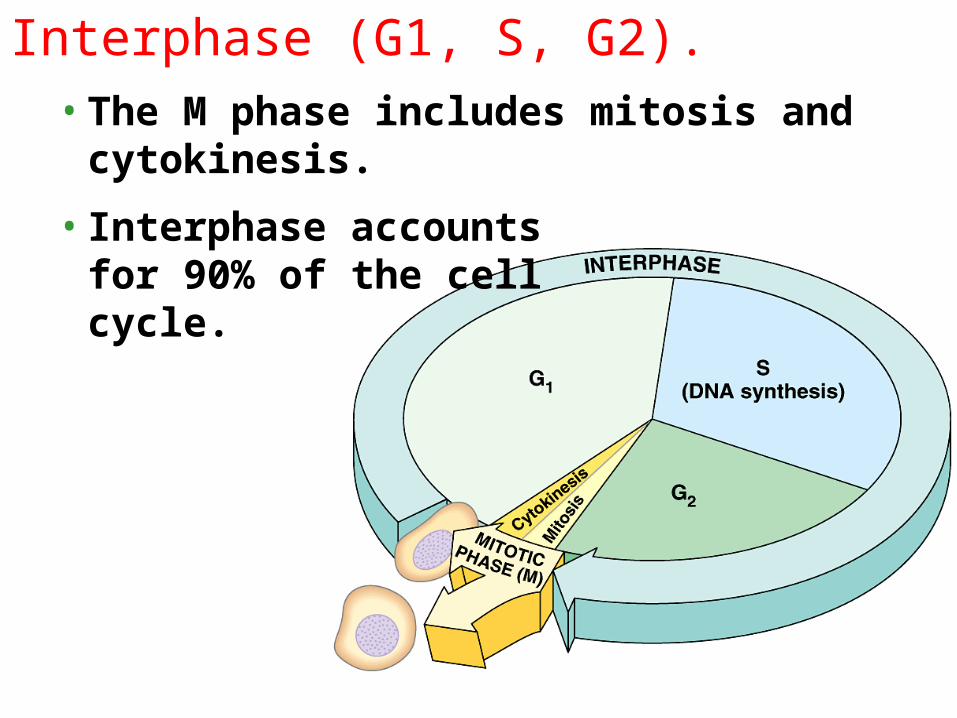
Interphase (G1, S, G2).• The M phase includes mitosis and
cytokinesis.
• Interphase accounts for 90% of the cell cycle.

• During interphase the cell cell growsgrows by producing proteins and cytoplasmic organelles, copies its chromosomes, and prepares for cell division.
• Interphase has three subphases:
Copyright © 2002 Pearson Education, Inc., publishing as Benjamin Cummings
–The G1 phase (“first gap”) centered on growth.
–The S phase (“synthesis”) when the chromosomes are copied.
–The G2 phase (“second gap”) where the cell completes preparations for cell division.–And then the cell divides (M).
•The daughter cells may then repeat the cycle.

Mitosis is the division of the Mitosis is the division of the nucleusnucleus.
Continuous process, but divided into 4 major phases.
1.prophase
2.metaphase
3.anaphase
4.telophase.

• By late interphase, the chromosomes have been duplicated but are loosely packed.
• The centrosomes have been duplicated and begin to organize microtubules into an aster (“star”).
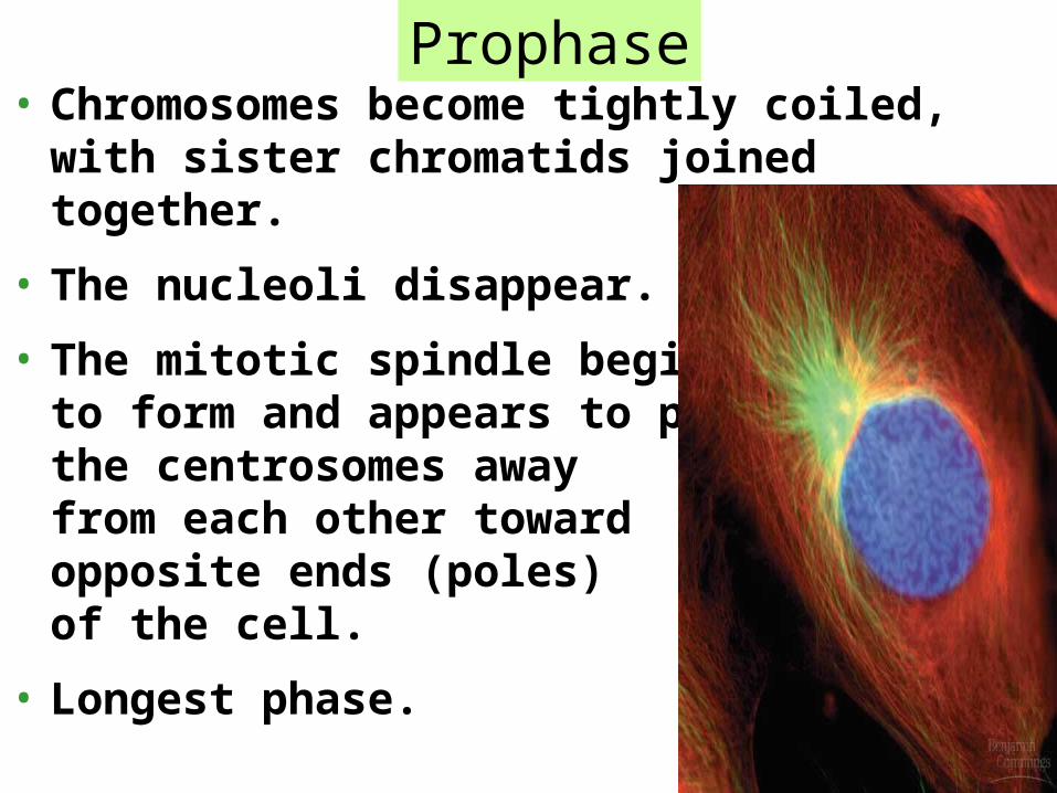
• Chromosomes become tightly coiled, with sister chromatids joined together.
• The nucleoli disappear.
• The mitotic spindle begins to form and appears to push the centrosomes away from each other toward opposite ends (poles) of the cell.
• Longest phase.
Prophase

• During prometaphase, the nuclear envelope fragments and microtubules from the spindle interact with the chromosomes.
• Microtubules from one pole attach to one of two kinetochores, while microtubules from the other pole attach to the other kinetochore.
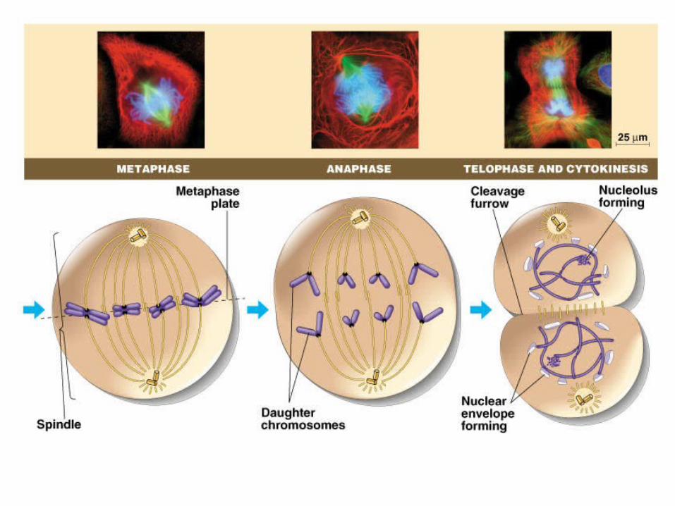
Metaphase

• The spindle fibers push the sister chromatids until they are all arranged at the metaphase plate, an imaginary plane equidistant between the poles, defining metaphase.
• The spindle fibers elongate by incorporating more subunits of the protein tubulin.
Metaphase
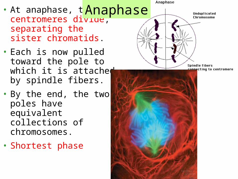
• At anaphase, the centromeres divide, separating the sister chromatids.
• Each is now pulled toward the pole to which it is attached by spindle fibers.
• By the end, the two poles have equivalent collections of chromosomes.
• Shortest phase
Anaphase

• Cell continues to elongate as free spindle fibers from each centrosome push off each other.
• Two nuclei begin to form, surrounded by the fragments of the parent’s nuclear envelope.
• Chromatin becomes less tightly coiled.
• Cytokinesis, division of thecytoplasm, begins.
Telophase
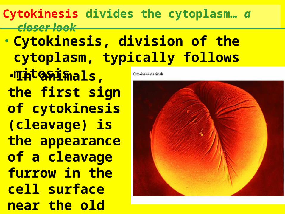
• Cytokinesis, division of the cytoplasm, typically follows mitosis.
Cytokinesis divides the cytoplasm… a closer look
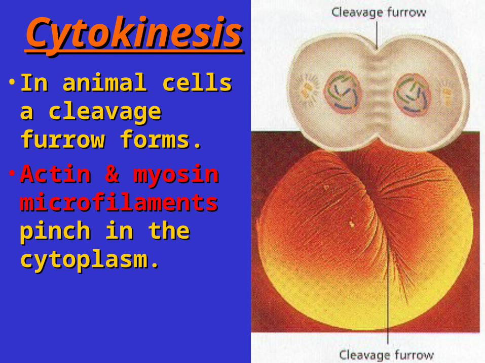
•In animals, the first sign of cytokinesis (cleavage) is the appearance of a cleavage furrow in the cell surface near the old metaphase plate.
CytokinesiCytokinesiss
•In animal cells In animal cells a cleavage a cleavage furrow forms.furrow forms.
•Actin & myosin Actin & myosin microfilamentsmicrofilaments pinch in the pinch in the cytoplasm. cytoplasm.
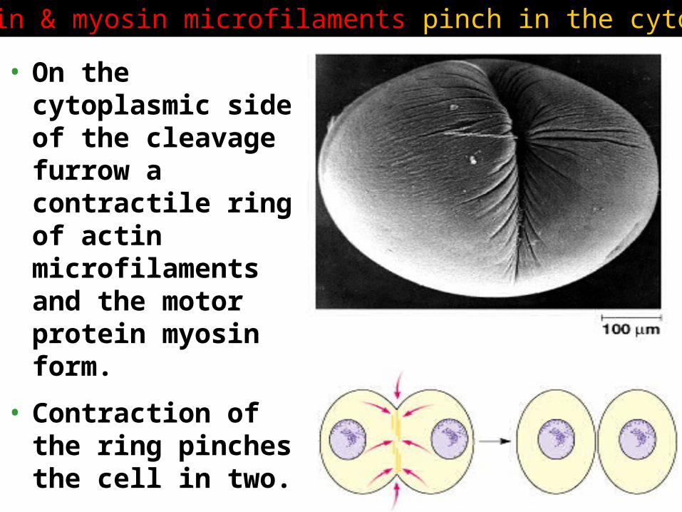
• On the cytoplasmic side of the cleavage furrow a contractile ring of actin microfilaments and the motor protein myosin form.
• Contraction of the ring pinches the cell in two.
Actin & myosin microfilaments pinch in the cytoplasm
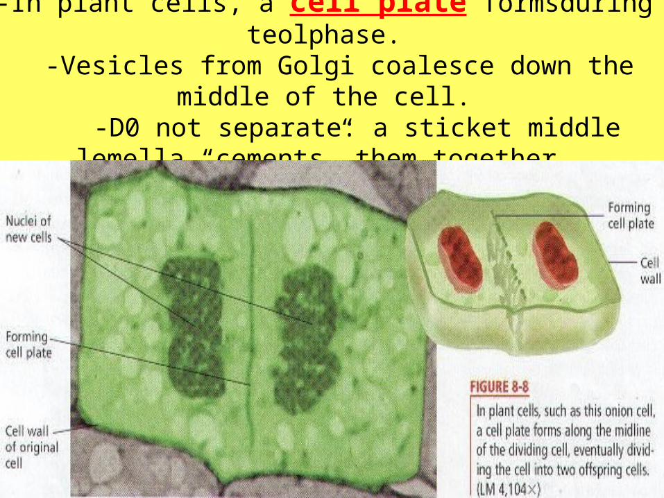
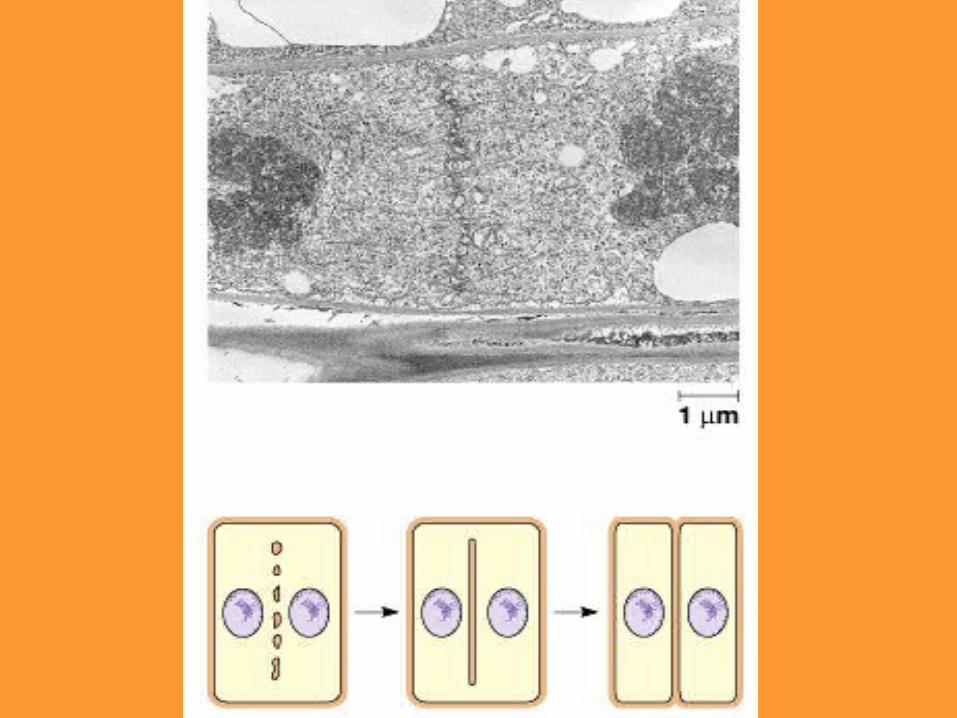
-In plant cells, a cell plate formsduring teolphase.
-Vesicles from Golgi coalesce down the middle of the cell.
-D0 not separate: a sticket middle lemella “cements” them together.

•The plate enlarges until its membranes fuse with the plasma membrane at the perimeter, with the contents of the vesicles forming new wall material in between.

REVIEW

• Assembly of the spindle microtubules starts in the centrosome.• The centrosome
(microtubule-organizing center) of animals has a pair of centrioles at the center, but the function of the centrioles is somewhat undefined.
The mitotic spindle …a closer look
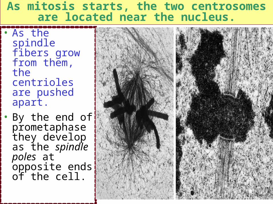
• As the spindle fibers grow from them, the centrioles are pushed apart.
• By the end of prometaphase they develop as the spindle poles at opposite ends of the cell.
As mitosis starts, the two centrosomes are located near the nucleus.
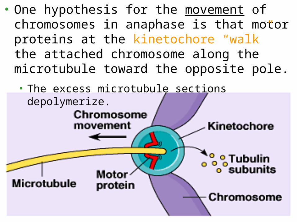
• One hypothesis for the movement of chromosomes in anaphase is that motor proteins at the kinetochore “walk” the attached chromosome along the microtubule toward the opposite pole.
• The excess microtubule sections depolymerize.


http://www.hhmi.org/research/investigators/nogales.html
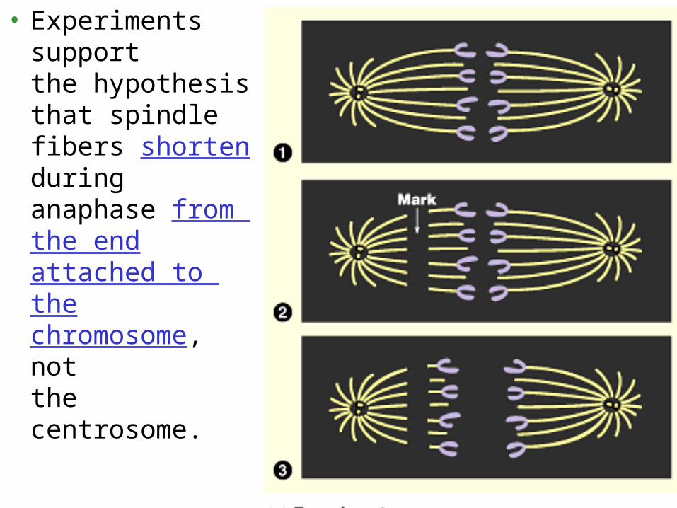
• Experiments support the hypothesis that spindle fibers shorten during anaphase from the end attached to the chromosome, not the centrosome.

Mitosis in eukaryotes Mitosis in eukaryotes
may have evolved may have evolved
from from binary fissionbinary fission in in
bacteriabacteria
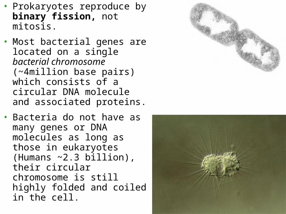
• Prokaryotes reproduce by binary fission, not mitosis.
• Most bacterial genes are located on a single bacterial chromosome (~4million base pairs) which consists of a circular DNA molecule and associated proteins.
• Bacteria do not have as many genes or DNA molecules as long as those in eukaryotes (Humans ~2.3 billion), their circular chromosome is still highly folded and coiled in the cell.
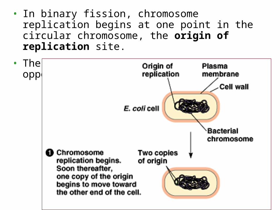
• In binary fission, chromosome replication begins at one point in the circular chromosome, the origin of replication site.
• These copied regions begin to move to opposite ends of the cell.
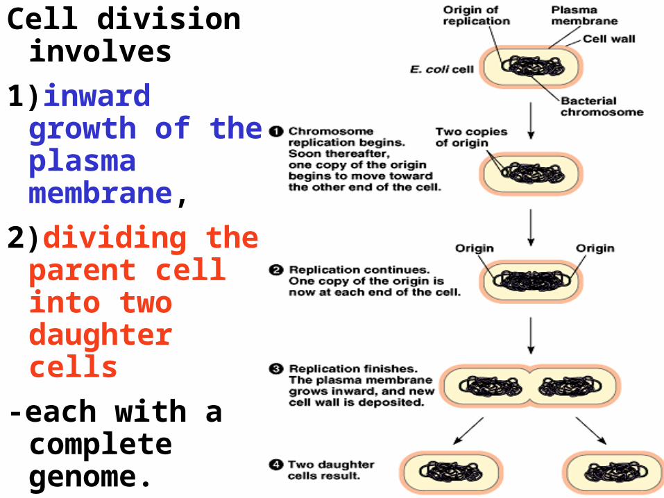
Cell division involves
1)inward growth of the plasma membrane,
2)dividing the parent cell into two daughter cells
-each with a complete genome.

Regulation of the Cell Cycle
1. There is a molecular control system
drives the cell cycle
2. Internal and external cues help regulate
the cell cycle
3. Cancer cells have escaped from cell cycle
controls

Overview:• A cell cycle control system regulates the rate at which cells
divide.
• Cells also have a predetermined life span.
• Several checkpoints act as built-in stop signs that halt the cell until the are overridden by go-ahead signals.
• Three checkpoints exist in G1, G2, and M.
• G1=restriction point (most important in mammals.)
• Frequency of cell division varies with cell type.
• Some human cells divide frequently throughout life (skin cells), others have the ability to divide, but keep it in reserve (liver cells)
• Bone marrow are always dividing in order to produce new read and white blood cells.
Timing is controlled by 2 kinds of molecules:
• Cyclins
• Cyclin-depend kinases (Cdks)-(proteins)
• MPF (M-phase promoting factor)

• The cell cycle appears to be driven by specific chemical signals in the cytoplasm.
• Fusion of an S phase cell and a G1 phase cell induces the G1 nucleus to start S phase.
• Fusion of a cell in mitosis with one in interphase induces the second cell to enter mitosis.
A molecular control system drives the cell A molecular control system drives the cell cyclecycle
Cell cycle control systemCell cycle control system::
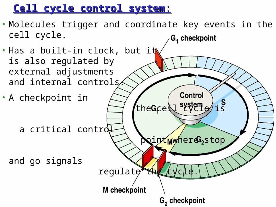
• Molecules trigger and coordinate key events in the cell cycle.
• Has a built-in clock, but it is also regulated by external adjustments and internal controls.
• A checkpoint in the cell cycle is a critical control point where stop and go signals regulate the cycle.
G1 checkpoint
G1G1
G0
(a) If a cell receives a go-ahead signal at the G1 checkpoint, the cell continues on in the cell cycle.
(b) If a cell does not receive a go-ahead signal at the G1checkpoint, the cell exits the cell cycle and goes into G0, a nondividing state.
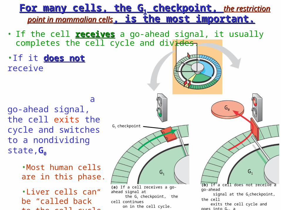
For many cells, the GFor many cells, the G11 checkpoint, checkpoint, the restriction the restriction
point in mammalian cellspoint in mammalian cells, is the most important., is the most important.
• If the cell receivesreceives a go-ahead signal, it usually completes the cell cycle and divides.
•If it does notdoes not receive a go-ahead signal, the cell exits the cycle and switches to a nondividing state,G0
•Most human cells are in this phase.
•Liver cells can be “called back” to the cell cycle by external cues (growth factors), but highly specialized nerve and muscle cells never divide.

The Cell Cycle Clock: Cyclins & Cyclin-Dependent Kinases
1.Two types of regulatory proteins are involved in cell
cycle control
• Cyclins
• Cyclin-dependent kinases (Cdks)A kinase enzyme that modifies other proteins by chemically adding
phosphate groups to them (phosphorylation).
Overview Clip 2
Overview clip 1
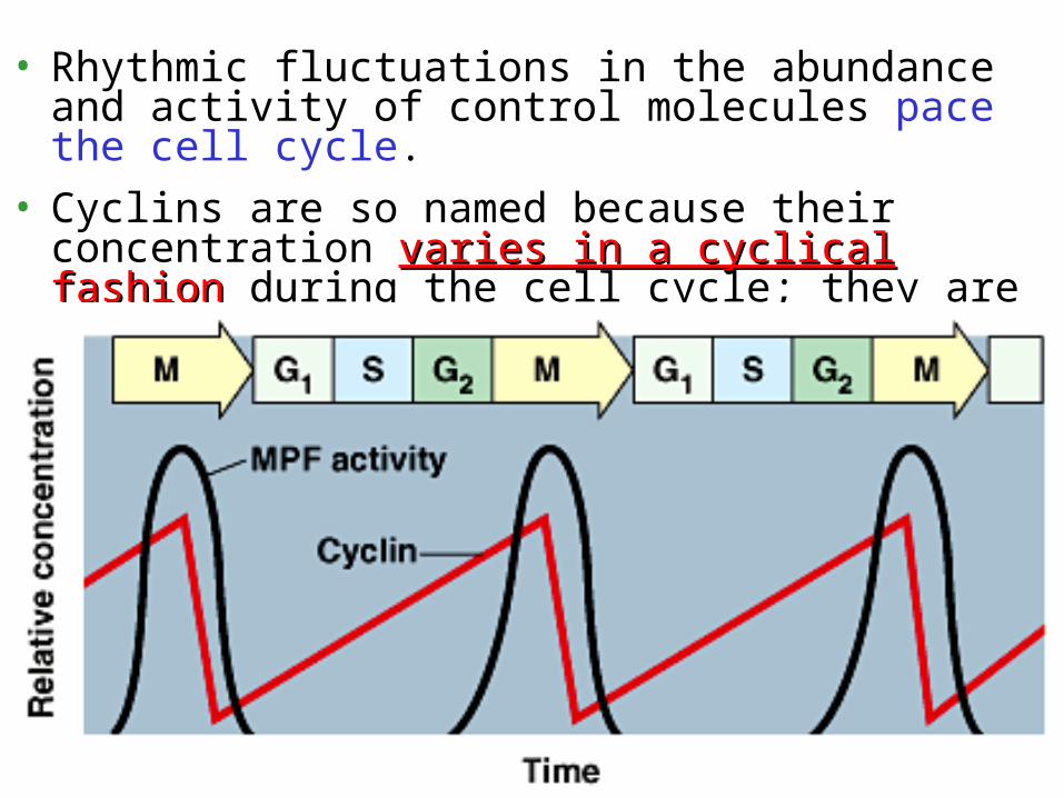
• Rhythmic fluctuations in the abundance and activity of control molecules pace the cell cycle.
• Cyclins are so named because their concentration varies in a cyclical fashionvaries in a cyclical fashion during the cell cycle; they are produced or degraded as needed in order to drive the cell through the different stages of the cell cycle

• Cyclin levels rise sharply throughout interphase, then fall abruptly during mitosis.
• Peaks in the activity of MPF (“M-phase-promoting-factor”) correspond to peaks in cyclin concentration.
MPF-1st Cdk discovered
Another Protein
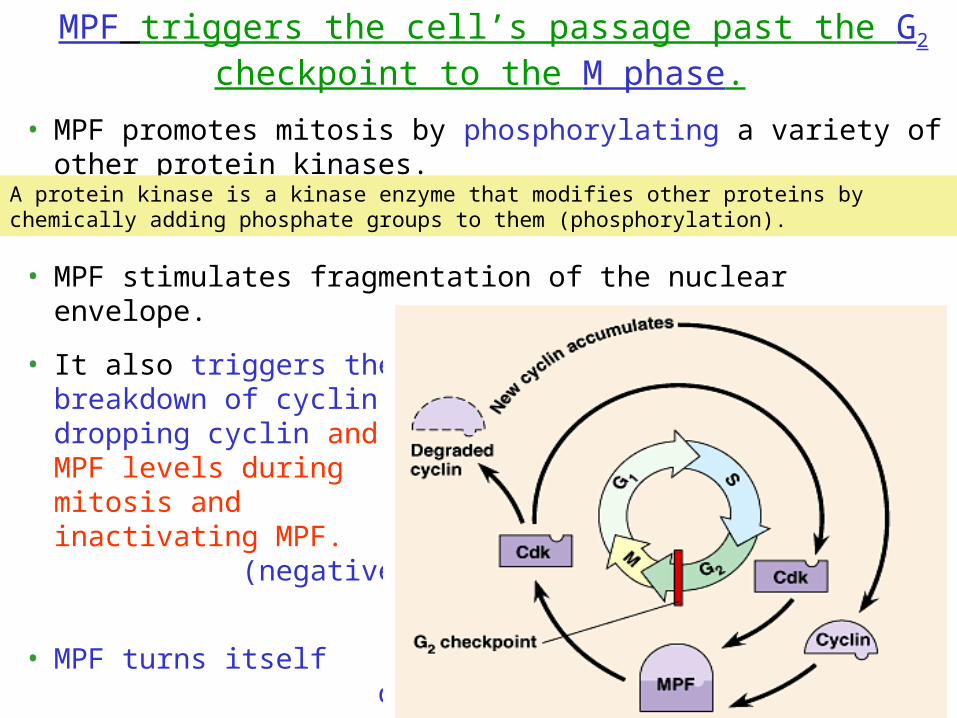
MPF triggers the cell’s passage past the G2 checkpoint to the M phase.
• MPF promotes mitosis by phosphorylating a variety of other protein kinases.
• MPF stimulates fragmentation of the nuclear envelope.
• It also triggers the breakdown of cyclin, dropping cyclin and MPF levels during mitosis and inactivating MPF. (negative feedback loop)
• MPF turns itself off.
A protein kinase is a kinase enzyme that modifies other proteins by chemically adding phosphate groups to them (phosphorylation).

• Some signals originate inside the cell, others outside. A variety of external chemical and physical factors can influence cell division.
• Particularly important for mammalian cells are growth factorsgrowth factors, proteins released by one group of cells that stimulate other cells to divide.
• This triggers a signal-transduction pathway that leads to cell division.
• Each cell type probably responds specifically to a certain growth factor or combination of factors.
Internal and external cues help regulate the cell cycle
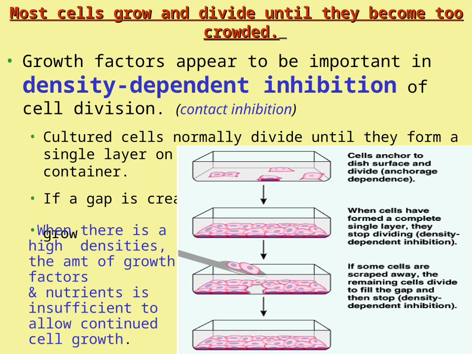
Most cells grow and divide until they become too Most cells grow and divide until they become too crowded.crowded.
• Growth factors appear to be important in
density-dependent inhibition of cell division. (contact inhibition)
• Cultured cells normally divide until they form a single layer on the inner surface of the culture container.
• If a gap is created, the cells will grow to fill the gap.
•When there is a high densities, the amt of growth factors & nutrients is insufficient to allow continued cell growth.

• Most animal cells also exhibit anchorage anchorage dependencedependence for cell division.
• To divide they must be anchored to a substratum, typically the extracellular matrix of a tissue.
• Control appears to be mediated by connections between the extracellular matrix and plasma membrane proteins and cytoskeletal elements.
• Cancer cells are free of both density-dependent inhibition and anchorage dependence.
• Cancer cells divide excessively and invade other tissues because they are free of the body’s control mechanisms. • Cancer cells do not stop dividing when growth
factors are depleted either because they manufacture their own, have an abnormality in the signaling pathway, or have a problem in the cell cycle control system.
• If and when cancer cells stop dividing, they do so at random points, not at the normal checkpoints in the cell cycle.
Cancer cells have escaped from cell cycle controls

• Cancer cell may divide indefinitely if they have a continual supply of nutrients.
• In contrast, nearly all mammalian cells divide 20 to 50 times under culture conditions before they stop, age, and die.
• Cancer cells may be “immortal”.
• Cells (HeLa) from a tumor removed from a woman (Henrietta Lacks) in 1951 are still reproducing in culture.
Copyright © 2002 Pearson Education, Inc., publishing as Benjamin Cummings
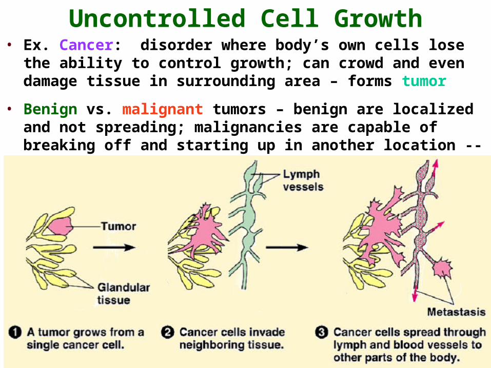
Uncontrolled Cell Growth• Ex. Cancer: disorder where body’s own cells lose the
ability to control growth; can crowd and even damage tissue in surrounding area – forms tumor
• Benign vs. malignant tumors – benign are localized and not spreading; malignancies are capable of breaking off and starting up in another location -- metastasis
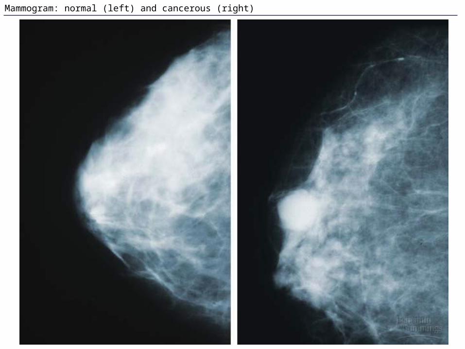
Mammogram: normal (left) and cancerous (right)