Embed Size (px)
Citation preview
RESEARCH ARTICLE Open Access
The design evolution of interbody cages inanterior cervical discectomy and fusion:a systematic reviewElizabeth Chong1,2, Matthew H Pelletier2, Ralph J Mobbs1,3,4* and William R Walsh2
Abstract
Background: Anterior cervical discectomy with fusion is a common surgical procedure for patients experiencing painand/or neurological deficits due to cervical spondylosis. Although iliac crest bone graft remains the gold standardtoday, the associated morbidity has inspired the search for alternatives, including allograft, synthetic and factor/cell-based grafts; and has further led to a focus on cage fusion technology. Compared to their graft counterparts,cage interbody implants have enhanced biomechanical properties, with designs constantly improving to maximisebiocompatibility and osseointegration. We present a systematic review examining the historical progress of implantdesigns and performance, as well as an update on the currently available designs, and the potential future of cervicalinterbody implants.
Methods: We performed a systematic review using the keywords “cervical fusion implant design”, with no limitson year of publication. Databases used were PubMed, Medline, Embase and Cochrane. In addition, the search wasextended to the reference lists of selected articles.
Results: 180 articles were reviewed and 64 articles were eligible for inclusion. Exclusion criteria were based aroundstudy design, implant information and patient cohorts. The evolution of cage implant design has been shaped byimproved understanding of ideal anatomy, progress in materials research and continuing experimentation of structuraldesign. Originally, designs varied primarily in their choice of structure, however long-term studies have displayed theoverall advantages of non-threaded, wedge shaped cages in complementing healthy anatomical profiles, and thusfocus has shifted to refining material utilisation and streamlining anterior fixation.
Conclusions: Evolution of design has been dramatic over the past decades; however an ideal cage design has yetto be realised. Current research is focusing on the promotion of osseointegration through bioactiviation of surfacematerials, as well as streamlining anterior fixation with the introduction of integrated screws and zero profiledesigns. Future designs will benefit from a combination of these advances in order to achieve ideal disc heights,cervical alignments and fusions.
Keywords: Anterior cervical discectomy fusion, Review, ACDF, Interbody, Cage, Design, Evolution
* Correspondence: [email protected] of New South Wales, Randwick, NSW 2031, Australia3Neurospine Clinic, Randwick, NSW 2031, AustraliaFull list of author information is available at the end of the article
© 2015 Chong et al.; licensee BioMed Central. This is an Open Access article distributed under the terms of the CreativeCommons Attribution License (http://creativecommons.org/licenses/by/4.0), which permits unrestricted use, distribution, andreproduction in any medium, provided the original work is properly credited. The Creative Commons Public DomainDedication waiver (http://creativecommons.org/publicdomain/zero/1.0/) applies to the data made available in this article,unless otherwise stated.
Chong et al. BMC Musculoskeletal DisordersDOI 10.1186/s12891-015-0546-x
BackgroundAge-related degeneration of the cervical spine is evidentin over 50% of the middle-aged population and is themost common cause of neural dysfunction [1]. Althoughthe majority of cases are asymptomatic, changes such asdisc herniation, osteophyte formation and hypertrophiedligaments may compress the cervical neuraxis to resultin cervical pain, radiculopathy, or myelopathy [2]. Firstline treatment is conservative; however surgery is indi-cated in symptomatic patients who are unresponsive toconservative management.Anterior cervical decompression and fusion (ACDF) is
one of the most widely used surgical treatments for pa-tients with cervical spondylosis [3]. It is also an indicatedtreatment in cases of cervical realignment, trauma andneoplasm [4]. ACDF achieves stabilisation and solid arth-rodesis with good-to-excellent clinical outcomes and min-imal surgical risks. The anterior approach to cervicaldecompression was first described by Cloward [5], andRobinson and Smith [6] in the 1950s. Both described ananterior approach via a longitudinal incision along theanterior border of the sternocleidomastoid muscle toallow for soft tissue dissection and annular incision. Fol-lowing discectomy and removal of any compressive struc-tures, fusion was then achieved using an autogenous graft.Although technical modifications have been made overthe years, this procedure is still standard today, leaving im-provements in fusion rates and clinical outcomes to begenerated through changes in implant design and material[7]. Initially, market available cage materials and designsvaried dramatically, with a selection between ceramic andalloy materials in threaded and non-threaded designs. Thishas shifted dramatically through the years, with moderndesigns conforming to a non-threaded, wedge shaped pro-file, and a choice between titanium alloy and the newer,polyetheretherketone (PEEK) materials. This article re-views the evolution of cervical interbody implant designsand assesses future research directions.
MethodsAfter performing initial, non-systematic searches using theterms “Anterior Cervical Discectomy Fusion”, “ACDF”and “Cervical Fusion” in conjunction with the terms“cage”, “design” and “implant”, we performed a systematicreview of the literature using the following protocol: wesearched the databases PubMed, Medline, Embase andCochrane using the keywords “cervical fusion implant de-sign” for articles available in the English language pub-lished up to March 2014. There were no limits or species(See Additional file 1: PRISMA Flow Diagram).
Results180 abstracts were searched for relevance and, of these,64 articles were selected for analysis. Articles were
selected based on their detail and relevance to the topicof cage design; both clinical and laboratory studies wereincluded. Laboratory studies comparing cage designsand materials that were controlled and reliable were uti-lised to inform theoretical advantages of specific designs.The inclusion criteria of clinical studies were prospectiveand retrospective designss with patients requiring ACDFin the treatment of degenerative cervical disease, patientcohorts larger than 30 individual pati, implanting cagesfilled with allograft with or without anterior plating.Exclusion criteria included studies with non-degenerativedisease cohorts, using additional proteins to promotefusion and ossification, or those that did not report onfusion rates, clinical outcomes and/or complication rates.In addition, the search was extended by manually search-ing the reference sections of relevant articles; this added12 publications (See Additional file 2: Table S1).
DiscussionHistorical evolution: bone grafts to fusion devicesThe first anterior cervical interbody techniques were intro-duced by Cloward, and Robinson and Smith in the 1950s.Cloward’s procedure involved insertion of a dowel graftfollowing decompression. Required bone was harvestedfrom the iliac crest of the patients or via allograft bonebank and was then pre-cut into a graft sized slightly widerand shorter than the drill hole. Insertion was achievedthrough distraction and force [5]. Robinson and Smith’sapproach utilised a similar initial distraction and anteriordecompression, however achieved fusion with the insertionof a horseshoe graft harvested from the patient’s iliac crestwithout the need for extensive graft-site modification.Autograft interbody designs have since evolved to
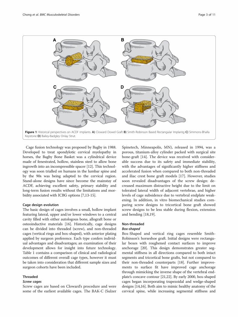
improve stability and distraction. In 1969, Simmons andBhalla [8] described the benefits of a keystone graft,which increased distraction height by locking into a pre-pared defect, thereby improving stability and fusion. In1960, Bailey and Badgley expanded the usage of ACDFto treat neoplasm and instability through the usageof onlay strut grafts. A technique which later evolvedinto anterior cervical corpectomy [9]. Limitations of theautogenous graft are an important consideration in all ofthese procedures. Iliac crest bone graft (ICBG) harvest-ing is associated with high levels of short and long-termmorbidity at the harvest site, including pain, wounddrainage, infection, haemtomas, nerve injury and iliaccrest fractures or deformity [10]. Initially, alternativegraft materials were sought as a method of circum-venting donor site limitations. However, not only didautograft remain superior in fusion, subsidence and ex-trusion rates but each alternative involved its own limi-tations [2,11]. As a result, the focus has switchedtowards cage implants as a graft substitutes as viablealternatives to autograft (Figure 1).
Chong et al. BMC Musculoskeletal Disorders Page 2 of 11
Cage fusion technology was proposed by Bagby in 1988.Developed to treat spondylotic cervical myelopathy inhorses, the Bagby Bone Basket was a cylindrical devicemade of fenestrated, hollow, stainless steel to allow boneingrowth into an incompressible spacer [12]. This technol-ogy was soon trialled on humans in the lumbar spine andby the 90s was being adapted to the cervical region.Stand-alone designs have since become the mainstay ofACDF, achieving excellent safety, primary stability andlong-term fusion results without the limitations and mor-bidity associated with ICBG options [7,13-15].
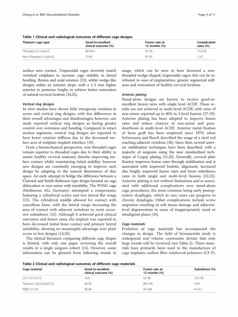
Cage design evolutionThe basic design of cages involves a small, hollow implantfeaturing lateral, upper and/or lower windows to a centralcavity filled with either autologous bone, allograft bone orosteoinductive materials [16]. Historically, cage designscan be divided into threaded (screw), and non-threadedcages (vertical rings and box-shaped), with anterior platingapplied by surgeon preference. Each type confers individ-ual advantages and disadvantages; an examination of theirdevelopment allows for insight into future technology.Table 1 contains a comparison of clinical and radiologicaloutcomes of different overall cage types, however it mustbe taken into consideration that different sample sizes andsurgeon cohorts have been included.
ThreadedScrew cagesScrew cages are based on Cloward’s procedure and weresome of the earliest available cages. The BAK-C (Sulzer
Spinetech, Minneapolis, MN), released in 1994, was aporous, titanium-alloy cylinder packed with surgical sitebone-graft [14]. The device was received with consider-able success due to its safety and immediate stability,with the advantages of significantly higher stiffness andaccelerated fusion when compared to both non-threadedand iliac crest bone graft models [17]. However, studiessoon revealed disadvantages of the screw design: de-creased maximum distractive height due to the limit ontolerated lateral width of adjacent vertebrae, and higherlevels of cage subsidence due to vertebral endplate weak-ening. In addition, in vitro biomechanical studies com-paring screw designs to tricortical bone graft showedscrew designs to be less stable during flexion, extensionand bending [18,19].
Non-threadedBox-shapedBox-Shaped and vertical ring cages resemble Smith-Robinson’s horseshoe graft. Initial designs were rectangu-lar boxes with roughened contact surfaces to improveanchorage [20]. This design demonstrates greater seg-mental stiffness in all directions compared to both intactsegments and tricortical bone grafts, but not compared totheir non-threaded counterparts [18]. Further improve-ments to surface fit have improved cage anchoragethrough mimicking the inverse shape of the vertebral end-plate’s concave contour [21,22]. By early 2000, box-shapedcages began incorporating trapezoidal and wedge-shapeddesigns [14,16]. Both aim to mimic healthy anatomy of thecervical spine, while increasing segmental stiffness and
Figure 1 Historical perspectives on ACDF implants. A) Cloward Dowel Graft B) Smith-Robinson Based Rectangular Implantg C) Simmons-BhallaKeystone D) Bailey-Badgley Onlay Strut.
Chong et al. BMC Musculoskeletal Disorders Page 3 of 11
surface area contact. Trapezoidal cages inversely matchvertebral endplates to increase cage stability in lateralbending, flexion and axial rotation [23], whilst wedge-likedesigns utilise an anterior slope, with a 1-2 mm higheranterior to posterior height, to achieve better restorationof natural cervical lordosis [24,25].
Vertical ring designsIn vitro studies have shown little intragroup variation inscrew and vertical ring designs, with few differences intheir overall advantages and disadvantages; however, onestudy reported vertical ring designs as having greatercontrol over extension and bending. Compared to intactmotion segments, vertical ring designs are reported tohave lower rotation stiffness due to the decreased sur-face area of endplate-implant interface [18].From a biomechanical perspective, non-threaded cages
remain superior to threaded cages due to their ability tomimic healthy cervical anatomy, thereby improving sur-face contact whilst maintaining initial stability; howevernew designs are constantly emerging to improve eachdesign by adapting to the natural dimensions of discspace. An early attempt to bridge the difference between aCloward and Smith-Robinson type design focused on cagedislocation or non-union with instability. The WING cage(Medinorm AG, Germany) attempted a compromise,featuring a cylindrical centre and two lateral flat wings[23]. The cylindrical middle allowed for contact withcancellous bone, with the lateral wings increasing thearea of contact with adjacent vertebrae to resist exces-sive subsidence [23]. Although it achieved good clinicaloutcomes and fusion rates, the implant was reported tohave decreased initial bone contact and primary lateralinstability, showing no meaningful advantage over plainscrew or box designs [14,26].The clinical literature comparing different cage shapes
is limited, with only one paper reviewing the overallresults in a single surgeon cohort [14]. However, someinformation can be gleaned from following trends in
usage, which can be seen to have favoured a non-threaded wedge-shaped, trapezoidal cages; this can be at-tributed to ease of implantation, greater segmental stiff-ness and restoration of healthy cervical lordosis.
Anterior platingStand-alone designs are known to receive good-to-excellent fusion rates with single level ACDF. These re-sults are not achieved in multi-level ACDF, with rates ofnon-union reported up to 40% in 3-level fusions [27-29].Anterior plating has been adopted to improve fusionrates and reduce chances of non-union and pseu-doarthosis in multi-level ACDF. Anterior metal fixationof bone graft has been employed since 1970, whenSchurmann and Busch described the usage of a steel rodreaching adjacent vertebrae [30]. Since then, several anter-ior stabilisation techniques have been described, with amajority of surgeons using the now standardised tech-nique of Caspar plating [31,32]. Generally, cervical platefixation improves fusion rates through stabilisation and isassociated with improved lordotic alignment, increaseddisc height, improved fusion rates and lower subsidencerates in both single and multi-level fusions [32,33].Anterior plating is not without limitations and is associ-ated with additional complications over stand-alonecage procedures, the most common being early postop-erative dysphagia, which in rare cases can progress tochronic dysphagia. Other complications include screwmigration resulting in soft tissue damage and adjacent-level degeneration in cases of inappropriately sized ormisaligned plates [34].
Cage materialsEvolution of cage materials has accompanied thechanges in design. The field of biomaterials study iswidespread and volume constraints dictate that onlylarge trends will be reviewed (see Table 2). Three mate-rials have primarily been used in the manufacture ofcage implants: carbon fiber reinforced polymers (CF-P),
Table 1 Clinical and radiological outcomes of different cage designs
Titanium cage type Good-to-excellentclinical outcome (%)
Fusion rate at12 months (%)
Complicationrates (%)
Threaded [7,14,66,67] 80-94.4 91-99 11.8-20
Non-Threaded [14,68,69] 75-87 87-95 2-15
Table 2 Clinical and radiological outcomes of different cage materials
Cage material Good-to-excellentclinical outcome (%)
Fusion rate at12 months (%)
Subsidence (%)
CF-P [35,70,71] 76.8 62-98 29.2-49
Titanium [26,42,68,69,72] 46-95 86.5-99 9-45
PEEK [73,74] 80-96 93-100 0-14.2
Chong et al. BMC Musculoskeletal Disorders Page 4 of 11
titanium (Ti) and polyetheretherketone (PEEK). CF-Pcages were initially trialled, achieving high rates offusion and good-to-excellent clinical outcomes howeverhave largely been superseded by PEEK due to its super-ior elastic modulus [35-38].Ti and its alloys were one of the first materials to be
utilised for cages in the 1980s. Used by the orthopaedicworld since the 1940s, Ti is a robust biomaterial withexcellent corrosion resistance and a low density, that canundergo surface modification to improve osseointegra-tion and cell adhesion [39,40]. PEEK cages were intro-duced in the 1990s by AcroMed as an alternative to Ticages; they provide the advantages of radiolucency andan elastic modulus close to bone thereby avoiding thestress shielding associated with Ti [41]. Today, contro-versy exists between the utilisation of Ti versus PEEKcages. Although PEEK has theoretical advantages, thishas not clearly transfered into the clinical setting due tothe difficulty in determining and controlling for othersurgical factors, including the roles of endplate prepar-ation, area of contact and overdistraction. However, amajority of studies have reported improved fusion rates,lower subsidence rates and radiolucency with PEEK ver-sus Ti cages [42-45], with one long-term study by Chenet al. reporting limited differences in the early postoper-ative period, but better maintenance of intervertebralheight, cervical lordosis and clinical outcomes by PEEKcages in 7-year follow up [46].
Cage design optimisationAnatomyUnderstanding the healthy and pathological anatomy ofthe cervical spine is vital in optimising the design of cer-vical cage implants. The first published anatomical stud-ies of the cervical spine in relation to the anteriorapproach were written in the 90s and have since beenquantitatively expanded upon through the use of im-aging technology.Important measurements in reference to ACDF in-
clude height, anterior-posterior (AP) diameter and widthof the cervical disc space (see Table 3). Disc heights fromC2/3 to C6/7 are approximately 1/3 of the vertebralheight, with no dependence on age in healthy subjects[47]. In the cervical spine each disc is thinner posteriorlythan anteriorly, with the greatest height in the midline,contributing to healthy cervical lordosis, an importantconsideration in design [48]. Distracted disc height issignificantly greater, with the disc space opening nearly4 mm to accommodate cages of up to 10 mm. The APdiameter increases inferiorly, with shortest depth at C2/3 increasing by 3 mm at C6/7. The lateral width of thedisc space also increases inferiorly; in ACDF, implantwidth is limited by the uncovertebral joint and corre-sponding endplate concavity.
PathologyIn degenerative change, disc space narrowing causestension in adjacent ligaments and compression of theneuraxis leading to symptomatic cervical spondylosis(Figure 2). The incidence of cervical spondylosis isreported to be as high as 76-82%, however a majority ofthese individuals are asymptomatic [49]. Classically,cervical spondylosis is believed to involve a com-bination of nucleus pulposus protrusion, osteophyteformation, and fibrosis, most frequently effecting theC5/6, C6/7 and C4/5 levels, in order of decreasingoccurrence [50,51]. Radiographic findings are oftenpoorly correlated to symptomatology; however, visual-isation of severe changes, including large osteophyteformation, marked disc space narrowing, sclerosis ofvertebral plates and posterior subluxation, are moreoften associated with pain and discomfort [52]. Macro-scopically, there is loss of cervical canal area andforaminal height and area, and flattening of the end-plate as the uncinated processes enlarges and flattensto lose its sharp, tapered configuration [53,54].
Cage dimensionsModern cage designs have begun targeting individual de-sign features and dimensions to ensure maximal clinicaland fusion outcomes. Due to the variation of disc spaceheight between cervical levels and individuals, cage im-plants are available in a variety of sizes. From surgicalexperience the author is familiar with variance in onlythe height of implants, with lateral and AP dimensionsremain largely uniform within company models.
Cage heightCervical cages relieve neuraxis compression through therestoration of disc space height, thereby reversing the lossof foraminal height and area, and cervical canal diameter[49,55,56]. The goal of adequate distraction must be tem-pered by the complications of overdistraction, which isrelated to non-union, postoperative neck pain and poorclinical outcomes due to an increase in contact pressuresbetween graft and cervical end plates. Originally, Smith-Robinson grafts were recommended to be 10-15 mm inheight [6]; however modern grafts are smaller to circum-vent the requirement of vertebral modification [57]. In1993, An et al. demonstrated that ideal distraction height isdependent on baseline disc height, with maximal changesin foraminal height achieved in 2 mm of distraction abovebaseline [49]. Modern cages adhere to this and availableheights ranging between 5-8 mm with trial spacers utilisedduring surgery to determine ideal cage height.
Cage width and lengthCervical implant width and length ensure maximal surfacecontact and stability of ACDF. These dimensions are
Chong et al. BMC Musculoskeletal Disorders Page 5 of 11
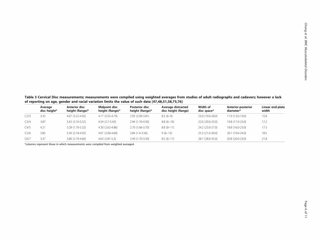
Table 3 Cervical Disc measurements; measurements were compiled using weighted averages from studies of adult radiographs and cadavers; however a lackof reporting on age, gender and racial variation limits the value of such data [47,48,51,58,75,76]
Averagedisc height*
Anterior discheight (Range)*
Midpoint discheight (Range)*
Posterior discheight (Range)*
Average distracteddisc height (Range)
Width ofdisc space*
Anterior-posteriordiameter*
Linear end-platewidth
C2/3 3.42 4.07 (3.22-4.92) 4.17 (3.55-4.79) 2.95 (2.09-3.81) 8.5 (6–9) 23.0 (19.0-28.0) 17.9 (13.0-19.0) 15.8
C3/4 3.87 3.42 (2.10-5.52) 4.54 (2.7-5.43) 2.94 (1.70-4.50) 8.8 (6–10) 22.0 (20.0-25.0) 19.8 (17.0-23.0) 17.2
C4/5 4.21 3.28 (1.70-5.32) 4.30 (2.62-4.86) 2.70 (1.66-3.70) 8.8 (8–11) 24.2 (23.0-27.0) 18.8 (16.0-23.0) 17.5
C5/6 3.85 3.30 (2.18-4.92) 4.01 (2.00-4.60) 2.84 (1.4-3.56) 9 (6–13) 25.3 (21.0-30.0) 20.7 (19.0-24.0) 18.5
C6/7 3.37 3.80 (2.79-4.60) 4.63 (2.81-5.3) 2.49 (1.70-3.50) 8.5 (6–11) 28.7 (28.0-35.0) 20.8 (20.0-23.0) 21.8
*columns represent those in which measurements were compiled from weighted averaged.
Chong
etal.BM
CMusculoskeletalD
isordersPage
6of
11
dictated by cervical anatomy; too small an implantwould provide inadequate stability and too large an im-plant would result in damage to the surrounding struc-tures [51]. Although lateral disc space width can rangebetween 20-30 mm in the cervical spine, cage implantwidth is limited laterally by the uncovertebral joint, withideal placement contacting bilaterally with the unci-nated processes [22,58]. Smith-Robinson recommendedimplants of 14 mm in width and depth, acknowledgingthe need to modify based on individual requirements[6]. Modern cage designs reflect these dimensions, withlateral widths ranging between 12-20 mm and depthsranging between 12-16 mm [34].
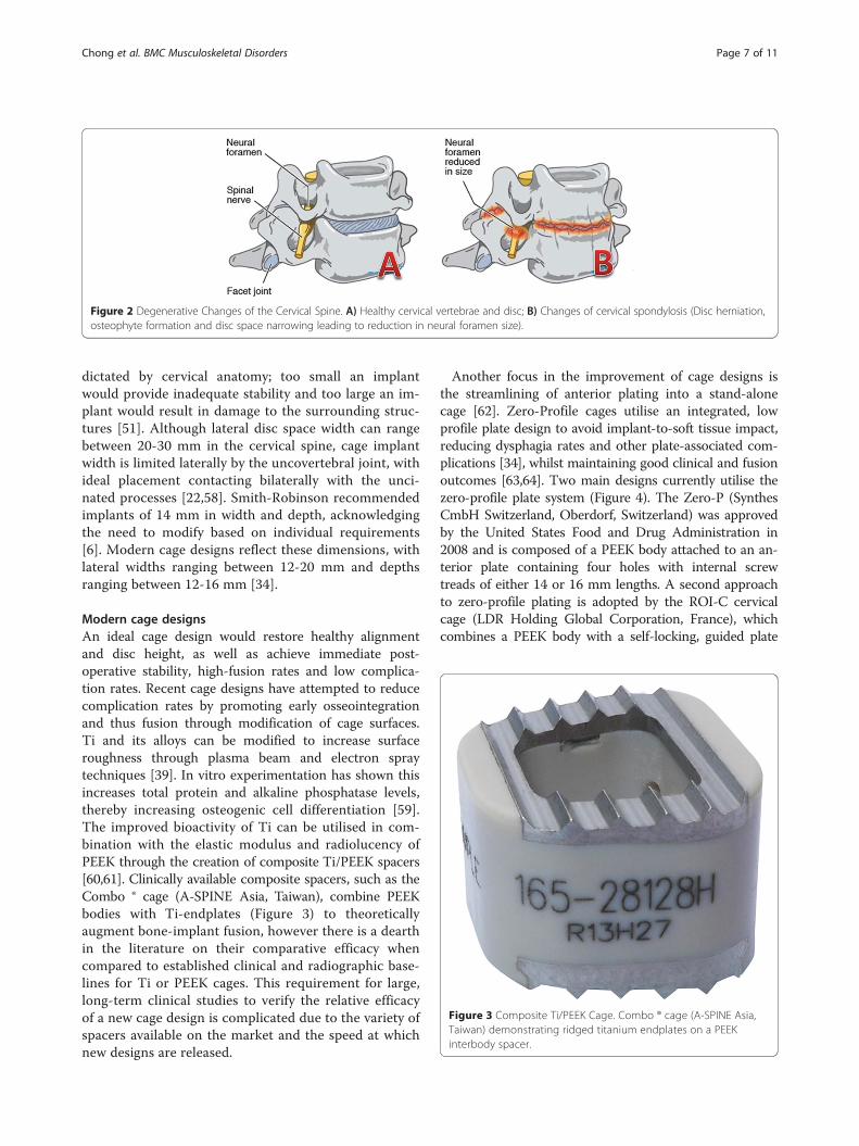
Modern cage designsAn ideal cage design would restore healthy alignmentand disc height, as well as achieve immediate post-operative stability, high-fusion rates and low complica-tion rates. Recent cage designs have attempted to reducecomplication rates by promoting early osseointegrationand thus fusion through modification of cage surfaces.Ti and its alloys can be modified to increase surfaceroughness through plasma beam and electron spraytechniques [39]. In vitro experimentation has shown thisincreases total protein and alkaline phosphatase levels,thereby increasing osteogenic cell differentiation [59].The improved bioactivity of Ti can be utilised in com-bination with the elastic modulus and radiolucency ofPEEK through the creation of composite Ti/PEEK spacers[60,61]. Clinically available composite spacers, such as theCombo ® cage (A-SPINE Asia, Taiwan), combine PEEKbodies with Ti-endplates (Figure 3) to theoreticallyaugment bone-implant fusion, however there is a dearthin the literature on their comparative efficacy whencompared to established clinical and radiographic base-lines for Ti or PEEK cages. This requirement for large,long-term clinical studies to verify the relative efficacyof a new cage design is complicated due to the variety ofspacers available on the market and the speed at whichnew designs are released.
Another focus in the improvement of cage designs isthe streamlining of anterior plating into a stand-alonecage [62]. Zero-Profile cages utilise an integrated, lowprofile plate design to avoid implant-to-soft tissue impact,reducing dysphagia rates and other plate-associated com-plications [34], whilst maintaining good clinical and fusionoutcomes [63,64]. Two main designs currently utilise thezero-profile plate system (Figure 4). The Zero-P (SynthesCmbH Switzerland, Oberdorf, Switzerland) was approvedby the United States Food and Drug Administration in2008 and is composed of a PEEK body attached to an an-terior plate containing four holes with internal screwtreads of either 14 or 16 mm lengths. A second approachto zero-profile plating is adopted by the ROI-C cervicalcage (LDR Holding Global Corporation, France), whichcombines a PEEK body with a self-locking, guided plate
Figure 3 Composite Ti/PEEK Cage. Combo ® cage (A-SPINE Asia,Taiwan) demonstrating ridged titanium endplates on a PEEKinterbody spacer.
Figure 2 Degenerative Changes of the Cervical Spine. A) Healthy cervical vertebrae and disc; B) Changes of cervical spondylosis (Disc herniation,osteophyte formation and disc space narrowing leading to reduction in neural foramen size).
Chong et al. BMC Musculoskeletal Disorders Page 7 of 11
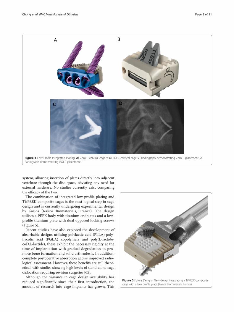
system, allowing insertion of plates directly into adjacentvertebrae through the disc space, obviating any need forexternal hardware. No studies currently exist comparingthe efficacy of the two.The combination of integrated low-profile plating and
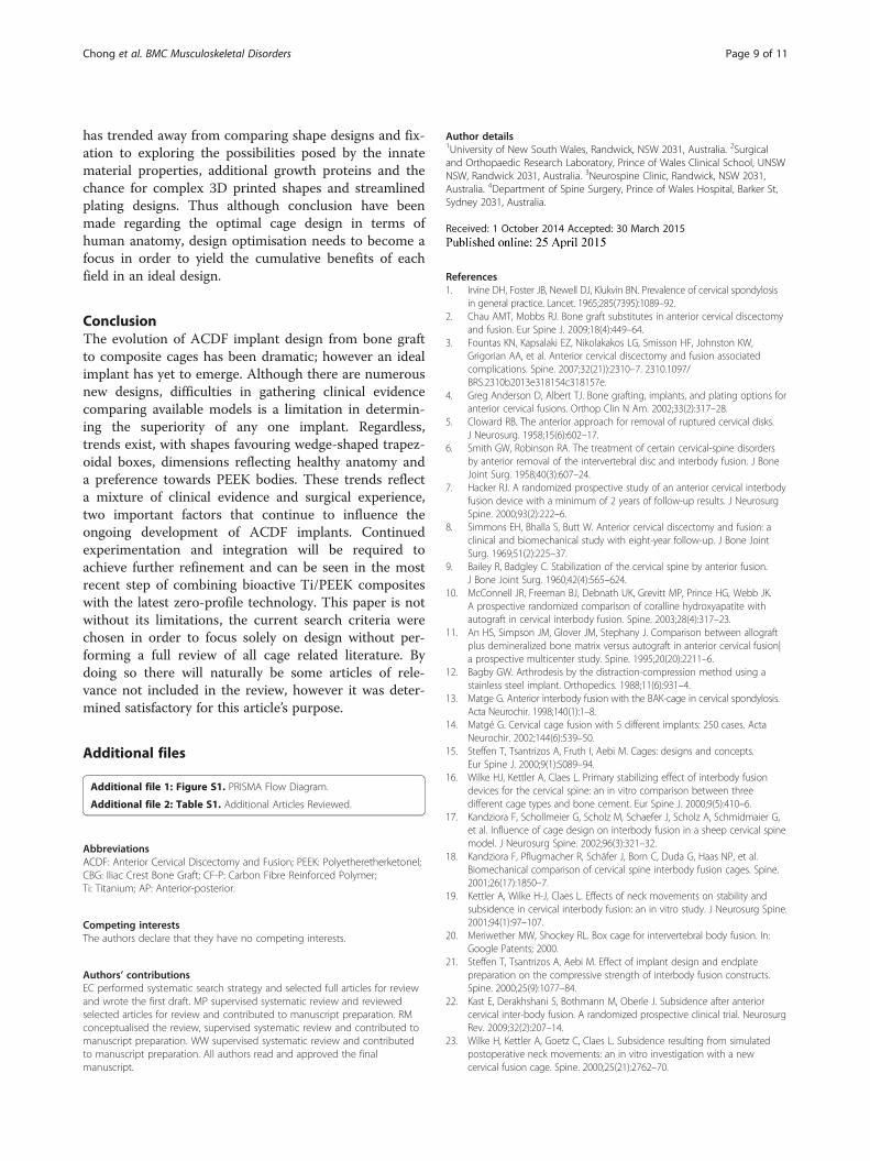
Ti/PEEK composite cages is the next logical step in cagedesign and is currently undergoing experimental designby Kasios (Kasios Biomaterials, France). The designutilises a PEEK body with titanium endplates and a low-profile titanium plate with dual opposed locking screws(Figure 5).Recent studies have also explored the development of
absorbable designs utilising polylactic acid (PLLA)-poly-flycolic acid (PGLA) copolymers and poly(L-lactide-coD,L-lactide), these exhibit the necessary rigidity at thetime of implantation with gradual degradation to pro-mote bone formation and solid arthrodesis. In addition,complete postoperative absorption allows improved radio-logical assessment. However, these benefits are still theor-etical, with studies showing high levels of stand-alone cagedislocation requiring revision surgeries [65].Although the variance in cage design availability has
reduced significantly since their first introduction, theamount of research into cage implants has grown. This
Figure 5 Future Designs. New design integrating a Ti/PEEK compositecage with a low profile plate (Kasios Biomaterials, France).
Figure 4 Low Profile Integrated Plating. A) Zero-P cervical cage V B) ROI-C cervical cage C) Radiograph demonstrating Zero-P placement D)Radiograph demonstrating ROI-C placement.
Chong et al. BMC Musculoskeletal Disorders Page 8 of 11
has trended away from comparing shape designs and fix-ation to exploring the possibilities posed by the innatematerial properties, additional growth proteins and thechance for complex 3D printed shapes and streamlinedplating designs. Thus although conclusion have beenmade regarding the optimal cage design in terms ofhuman anatomy, design optimisation needs to become afocus in order to yield the cumulative benefits of eachfield in an ideal design.
ConclusionThe evolution of ACDF implant design from bone graftto composite cages has been dramatic; however an idealimplant has yet to emerge. Although there are numerousnew designs, difficulties in gathering clinical evidencecomparing available models is a limitation in determin-ing the superiority of any one implant. Regardless,trends exist, with shapes favouring wedge-shaped trapez-oidal boxes, dimensions reflecting healthy anatomy anda preference towards PEEK bodies. These trends reflecta mixture of clinical evidence and surgical experience,two important factors that continue to influence theongoing development of ACDF implants. Continuedexperimentation and integration will be required toachieve further refinement and can be seen in the mostrecent step of combining bioactive Ti/PEEK compositeswith the latest zero-profile technology. This paper is notwithout its limitations, the current search criteria werechosen in order to focus solely on design without per-forming a full review of all cage related literature. Bydoing so there will naturally be some articles of rele-vance not included in the review, however it was deter-mined satisfactory for this article’s purpose.
Additional files
Additional file 1: Figure S1. PRISMA Flow Diagram.
Additional file 2: Table S1. Additional Articles Reviewed.
AbbreviationsACDF: Anterior Cervical Discectomy and Fusion; PEEK: PolyetheretherketoneI;CBG: Iliac Crest Bone Graft; CF-P: Carbon Fibre Reinforced Polymer;Ti: Titanium; AP: Anterior-posterior.
Competing interestsThe authors declare that they have no competing interests.
Authors’ contributionsEC performed systematic search strategy and selected full articles for reviewand wrote the first draft. MP supervised systematic review and reviewedselected articles for review and contributed to manuscript preparation. RMconceptualised the review, supervised systematic review and contributed tomanuscript preparation. WW supervised systematic review and contributedto manuscript preparation. All authors read and approved the finalmanuscript.
Author details1University of New South Wales, Randwick, NSW 2031, Australia. 2Surgicaland Orthopaedic Research Laboratory, Prince of Wales Clinical School, UNSWNSW, Randwick 2031, Australia. 3Neurospine Clinic, Randwick, NSW 2031,Australia. 4Department of Spine Surgery, Prince of Wales Hospital, Barker St,Sydney 2031, Australia.
Received: 1 October 2014 Accepted: 30 March 2015
References1. Irvine DH, Foster JB, Newell DJ, Klukvin BN. Prevalence of cervical spondylosis
in general practice. Lancet. 1965;285(7395):1089–92.2. Chau AMT, Mobbs RJ. Bone graft substitutes in anterior cervical discectomy
and fusion. Eur Spine J. 2009;18(4):449–64.3. Fountas KN, Kapsalaki EZ, Nikolakakos LG, Smisson HF, Johnston KW,
Grigorian AA, et al. Anterior cervical discectomy and fusion associatedcomplications. Spine. 2007;32(21)):2310–7. 2310.1097/BRS.2310b2013e318154c318157e.
4. Greg Anderson D, Albert TJ. Bone grafting, implants, and plating options foranterior cervical fusions. Orthop Clin N Am. 2002;33(2):317–28.
5. Cloward RB. The anterior approach for removal of ruptured cervical disks.J Neurosurg. 1958;15(6):602–17.
6. Smith GW, Robinson RA. The treatment of certain cervical-spine disordersby anterior removal of the intervertebral disc and interbody fusion. J BoneJoint Surg. 1958;40(3):607–24.
7. Hacker RJ. A randomized prospective study of an anterior cervical interbodyfusion device with a minimum of 2 years of follow-up results. J NeurosurgSpine. 2000;93(2):222–6.
8. Simmons EH, Bhalla S, Butt W. Anterior cervical discectomy and fusion: aclinical and biomechanical study with eight-year follow-up. J Bone JointSurg. 1969;51(2):225–37.
9. Bailey R, Badgley C. Stabilization of the cervical spine by anterior fusion.J Bone Joint Surg. 1960;42(4):565–624.
10. McConnell JR, Freeman BJ, Debnath UK, Grevitt MP, Prince HG, Webb JK.A prospective randomized comparison of coralline hydroxyapatite withautograft in cervical interbody fusion. Spine. 2003;28(4):317–23.
11. An HS, Simpson JM, Glover JM, Stephany J. Comparison between allograftplus demineralized bone matrix versus autograft in anterior cervical fusion|a prospective multicenter study. Spine. 1995;20(20):2211–6.
12. Bagby GW. Arthrodesis by the distraction-compression method using astainless steel implant. Orthopedics. 1988;11(6):931–4.
13. Matge G. Anterior interbody fusion with the BAK-cage in cervical spondylosis.Acta Neurochir. 1998;140(1):1–8.
14. Matgé G. Cervical cage fusion with 5 different implants: 250 cases. ActaNeurochir. 2002;144(6):539–50.
15. Steffen T, Tsantrizos A, Fruth I, Aebi M. Cages: designs and concepts.Eur Spine J. 2000;9(1):S089–94.
16. Wilke HJ, Kettler A, Claes L. Primary stabilizing effect of interbody fusiondevices for the cervical spine: an in vitro comparison between threedifferent cage types and bone cement. Eur Spine J. 2000;9(5):410–6.
17. Kandziora F, Schollmeier G, Scholz M, Schaefer J, Scholz A, Schmidmaier G,et al. Influence of cage design on interbody fusion in a sheep cervical spinemodel. J Neurosurg Spine. 2002;96(3):321–32.
18. Kandziora F, Pflugmacher R, Schäfer J, Born C, Duda G, Haas NP, et al.Biomechanical comparison of cervical spine interbody fusion cages. Spine.2001;26(17):1850–7.
19. Kettler A, Wilke H-J, Claes L. Effects of neck movements on stability andsubsidence in cervical interbody fusion: an in vitro study. J Neurosurg Spine.2001;94(1):97–107.
20. Meriwether MW, Shockey RL. Box cage for intervertebral body fusion. In:Google Patents; 2000.
21. Steffen T, Tsantrizos A, Aebi M. Effect of implant design and endplatepreparation on the compressive strength of interbody fusion constructs.Spine. 2000;25(9):1077–84.
22. Kast E, Derakhshani S, Bothmann M, Oberle J. Subsidence after anteriorcervical inter-body fusion. A randomized prospective clinical trial. NeurosurgRev. 2009;32(2):207–14.
23. Wilke H, Kettler A, Goetz C, Claes L. Subsidence resulting from simulatedpostoperative neck movements: an in vitro investigation with a newcervical fusion cage. Spine. 2000;25(21):2762–70.
Chong et al. BMC Musculoskeletal Disorders Page 9 of 11
24. Gödde S, Fritsch E, Dienst M, Kohn D. Influence of cage geometry onsagittal alignment in instrumented posterior lumbar interbody fusion. Spine.2003;28(15):1693–9.
25. Bartels RH, Donk R, van Dijk AR. Height of cervical foramina after anteriordiscectomy and implantation of a carbon fiber cage. J Neurosurg Spine.2001;95(1):40–2.
26. Schmieder K, Wolzik-Grossmann M, Pechlivanis I, Engelhardt M, Scholz M,Harders A. Subsidence of the wing titanium cage after anterior cervicalinterbody fusion: 2-year follow-up study. J Neurosurg Spine. 2006;4(6):447–53.
27. Emery SE, Fisher RJ, Bohlman HH. Three-level anterior cervical discectomyand fusion: radiographic and clinical results. Spine. 1997;22(22):2622–4.
28. Wang JC, McDonough PW, Endow KK, Delamarter RB. Increased fusion rateswith cervical plating for two-level anterior cervical discectomy and fusion.Spine. 2000;25(1):41.
29. Mobbs RJ, Rao P, Chandran NK. Anterior cervical discectomy and fusion:analysis of surgical outcome with and without plating. J Clin Neurosci.2007;14(7):639–42.
30. Schurman K, Busch G. Treatment of cervical luxation fractures with ventralfusion. CHIRURG. 1970;41(5):225–8.
31. Caspar W, Barbier DD, Klara PM. Anterior cervical fusion and Caspar platestabilization for cervical trauma. Neurosurgery. 1989;25(4):491–502.
32. Bohler J, Gaudernak T. Anterior plate stabilization for fracture-dislocations ofthe lower cervical spine. J Trauma Inj Infect Crit Care. 1980;20(3):203–5.
33. Song K-J, Taghavi CE, Lee K-B, Song J-H, Eun J-P. The efficacy of plateconstruct augmentation versus cage alone in anterior cervical fusion. Spine.2009;34(26):2886–92.
34. Scholz M, Schnake K, Pingel A, Hoffmann R, Kandziora F. A new zero-profileimplant for stand-alone anterior cervical interbody fusion. Clin Orthop RelatRes. 2011;469(3):666–73.
35. Bartels RH, Donk RD, Feuth T. Subsidence of stand-alone cervical carbonfiber cages. Neurosurgery. 2006;58(3):502–8. discussion 502–508.
36. Vavruch L, Hedlund R, Javid D, Leszniewski W, Shalabi A. A prospectiverandomized comparison between the cloward procedure and a carbonfiber cage in the cervical spine: a clinical and radiologic study. Spine.2002;27(16):1694–701.
37. Gercek E, Arlet V, Delisle J, Marchesi D. Subsidence of stand-alone cervicalcages in anterior interbody fusion: warning. Eur Spine J. 2003;12(5):513–6.
38. Mobbs RJ, Chau AM, Durmush D. Biphasic calcium phosphate containedwithin a polyetheretherketone cage with and without plating for anteriorcervical discectomy and fusion. Orthop Surg. 2012;4(3):156–65.
39. Rao PJ, Walsh WR, Pellitier MH, Mobbs RJ. Spine interbody implants: materialselection and modification, functionalization and bioactivation of surfaces toimprove osseointegration. Orthop Surg. 2014;6(2):81–9.
40. Williams DF. There is no such thing as a biocompatible material.Biomaterials. 2014;35(38):10009–14.
41. Kurtz SM, Devine JN. PEEK biomaterials in trauma, orthopedic, and spinalimplants. Biomaterials. 2007;28(32):4845–69.
42. Chou Y-C, Chen D-C, Hsieh WA, Chen W-F, Yen P-S, Harnod T, et al.Efficacy of anterior cervical fusion: Comparison of titanium cages,polyetheretherketone (PEEK) cages and autogenous bone grafts. J ClinNeurosci. 2008;15(11):1240–5.
43. Niu CC, Liao JC, Chen WJ, Chen LH. Outcomes of interbody fusion cages usedin 1 and 2-levels anterior cervical discectomy and fusion: titanium cages versuspolyetheretherketone (PEEK) cages. J Spinal Disord Tech. 2010;23(5):310–6.
44. Liao JC, Niu CC, Chen WJ, Chen LH. Polyetheretherketone (PEEK) cage filledwith cancellous allograft in anterior cervical discectomy and fusion. Int Orthop.2008;32(5):643–8.
45. Cabraja M, Oezdemir S, Koeppen D, Kroppenstedt S. Anterior cervicaldiscectomy and fusion: comparison of titanium and polyetheretherketonecages. BMC Musculoskelet Disord. 2012;13:172.
46. Chen Y, Wang X, Lu X, Yang L, Yang H, Yuan W, et al. Comparison oftitanium and polyetheretherketone (PEEK) cages in the surgical treatment ofmultilevel cervical spondylotic myelopathy: a prospective, randomized,control study with over 7-year follow-up. Eur Spine J. 2013;22(7):1539–46.
47. Frobin W, Leivseth G, Biggemann M, Brinckmann P. Vertebral height, discheight, posteroanterior displacement and dens–atlas gap in the cervicalspine: precision measurement protocol and normal data. Clin Biomech.2002;17(6):423–31.
48. Pait TG, Killefer JA, Arnautovic KI. Surgical anatomy of the anterior cervicalspine: the disc space, vertebral artery, and associated bony structures.Neurosurgery. 1996;39(4):769–76.
49. An HS, Evanich CJ, Nowicki BH, Haughton VM. Ideal thickness ofSmith-Robinson graft for anterior cervical fusion: a cadaveric study withcomputed tomographic correlation. Spine. 1993;18(14):2043–7.
50. Wilkinson M. The morbid anatomy of cervical spondylosis and myelopathy.Brain. 1960;83(4):589–617.
51. Lu J, Ebraheim N, Yang H, Rollins J, Yeasting R. Anatomic bases for anteriorspinal surgery: surgical anatomy of the cervical vertebral body and discspace. Surg Radiol Anat. 1999;21(4):235–9.
52. Lawrence J. Disc degeneration. Its frequency and relationship to symptoms.Ann Rheum Dis. 1969;28(2):121–38.
53. Friedenberg Z, Miller W. Degenerative disc disease of the cervical spine acomparative study of asymptomatic and symptomatic patients. J Bone JointSurg. 1963;45(6):1171–8.
54. Tanaka N, Fujimoto Y, An HS, Ikuta Y, Yasuda M. The anatomic relationamong the nerve roots, intervertebral foramina, and intervertebral discs ofthe cervical spine. Spine. 2000;25(3):286–91.
55. Kwon B, Kim DH, Marvin A, Jenis LG. Outcomes following anterior cervicaldiscectomy and fusion: the role of interbody disc height, angulation, andspinous process distance. J Spinal Disord Tech. 2005;18(4):304–8.
56. Bayley JC, Yoo JU, Kruger DM, Schlegel J. The role of distraction inimproving the space available for the cord in cervical spondylosis. Spine.1995;20(7):771–5.
57. Truumees E, Demetropoulos CK, Yang KH, Herkowitz HN. Effects of discheight and distractive forces on graft compression in an anterior cervicaldiscectomy model. Spine. 2002;27(22):2441–5.
58. Panjabi MM, Duranceau J, Goel V, Oxland T, Takata K. Cervical humanvertebrae quantitative three-dimensional anatomy of the middle and lowerregions. Spine. 1991;16(8):861–9.
59. Rosa AL, Beloti MM. Effect of cpTi surface roughness on human bonemarrow cell attachment, proliferation, and differentiation. Braz Dent J.2003;14(1):16–21.
60. Olivares-Navarrete R, Gittens RA, Schneider JM, Hyzy SL, Haithcock DA,Ullrich PF, et al. Osteoblasts exhibit a more differentiated phenotype andincreased bone morphogenetic protein production on titanium alloysubstrates than on poly-ether-ether-ketone. Spine J. 2012;12(3):265–72.
61. Han C-M, Lee E-J, Kim H-E, Koh Y-H, Kim KN, Ha Y, et al. The electron beamdeposition of titanium on polyetheretherketone (PEEK) and the resultingenhanced biological properties. Biomaterials. 2010;31(13):3465–70.
62. Scholz M, Reyes PM, Schleicher P, Sawa AG, Baek S, Kandziora F, et al. A newstand-alone cervical anterior interbody fusion device: biomechanical comparisonwith established anterior cervical fixation devices. Spine. 2009;34(2):156–60.
63. Qi M, Chen H, Liu Y, Zhang Y, Liang L, Yuan W. The use of a zero-profiledevice compared with an anterior plate and cage in the treatment ofpatients with symptomatic cervical spondylosis: a preliminary clinicalinvestigation. Bone Joint J. 2013;95(4):543–7.
64. Miao J, Shen Y, Kuang Y, Yang L, Wang X, Chen Y, et al. Early follow-upoutcomes of a new zero-profile implant used in anterior cervical discectomyand fusion. J Spinal Disord Tech. 2013;26(5):E193–7.
65. Brenke C, Kindling S, Scharf J, Schmieder K, Barth M. Short-term experience witha new absorbable composite cage (beta-tricalcium phosphate-polylactic acid)in patients after stand-alone anterior cervical discectomy and fusion. Spine(Phila Pa 1976). 2013;38(11):E635–40.
66. Cauthen JC, Theis RP, Allen AT. Anterior cervical fusion: a comparison ofcage, dowel and dowel-plate constructs. Spine J. 2003;3(2):106–17.
67. Bärlocher CB, Barth A, Krauss JK, Binggeli R, Seiler RW. Comparative evaluationof microdiscectomy only, autograft fusion, polymethylmethacrylateinterposition, and threaded titanium cage fusion for treatment of single-levelcervical disc disease: a prospective randomized study in 125 patients. NeurosurgFocus. 2002;12(1):1–7.
68. van Jonbergen HP, Spruit M, Anderson PG, Pavlov PW. Anterior cervicalinterbody fusion with a titanium box cage: early radiological assessment offusion and subsidence. Spine J. 2005;5(6):645–9. discussion 649.
69. Niu C-C, Chen L-H, Lai P-L, Fu T-S, Chen W-J. Trapezoidal titanium cage inanterior cervical interbody fusion: a clinical experience. Chang Gung Med J.2005;28(4):212–21.
70. Salame K, Ouaknine GE, Razon N, Rochkind S. The use of carbon fiber cagesin anterior cervical interbody fusion: report of 100 cases. Neurosurg Focus.2002;12(1):E1.
71. van der Haven I, Van Loon P, Bartels R, Van Susante J. Anterior cervicalinterbody fusion with radiolucent carbon fiber cages: clinical andradiological results. Acta Orthop Belg. 2005;71(5):604–9.
Chong et al. BMC Musculoskeletal Disorders Page 10 of 11
72. Moreland DB, Asch HL, Clabeaux DE, Castiglia GJ, Czajka GA, Lewis PJ, et al.Anterior cervical discectomy and fusion with implantable titanium cage:initial impressions, patient outcomes and comparison to fusion withallograft.[Erratum appears in Spine J. 2004 May-Jun;4(3):following table ofcontents]. Spine J. 2004;4(2):184–91. discussion 191.
73. Yang B, Li H, Zhang T, He X, Xu S. The incidence of adjacent segmentdegeneration after cervical disc arthroplasty (CDA): a meta analysis ofrandomized controlled trials. PLoS One. 2012;7(4):e35032.
74. Mastronardi L, Ducati A, Ferrante L. Anterior cervical fusion withpolyetheretherketone (PEEK) cages in the treatment of degenerative discdisease. Preliminary observations in 36 consecutive cases with a minimum12-month follow-up. Acta Neurochir. 2006;148(3):307–12. discussion 312.
75. Abuzayed B, Tutunculer B, Kucukyuruk B, Tuzgen S. Anatomic basis ofanterior and posterior instrumentation of the spine: morphometric study.Surg Radiol Anat. 2010;32(1):75–85.
76. Moroney SP, Schultz AB, Miller JA. Analysis and measurement of neck loads.J Orthop Res. 1988;6(5):713–20.
Submit your next manuscript to BioMed Centraland take full advantage of:
• Convenient online submission
• Thorough peer review
• No space constraints or color figure charges
• Immediate publication on acceptance
• Inclusion in PubMed, CAS, Scopus and Google Scholar
• Research which is freely available for redistribution
Submit your manuscript at www.biomedcentral.com/submit
Chong et al. BMC Musculoskeletal Disorders Page 11 of 11