Embed Size (px)
Citation preview
1
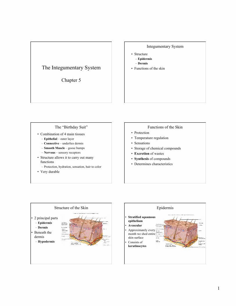
The Integumentary System
Chapter 5
Integumentary System
• Structure – Epidermis – Dermis
• Functions of the skin
The “Birthday Suit”
• Combination of 4 main tissues – Epithelial – outer layer – Connective – underlies dermis – Smooth Muscle – goose bumps – Nervous – sensory receptors
• Structure allows it to carry out many functions – Protection, hydration, sensation, hair to color
• Very durable
Functions of the Skin • Protection • Temperature regulation • Sensations • Storage of chemical compounds • Excretion of wastes • Synthesis of compounds • Determines characteristics
Human Anatomy, 3rd edition Prentice Hall, © 2001
Structure of the Skin
• 2 principal parts – Epidermis – Dermis
• Beneath the dermis – Hypodermis
Human Anatomy, 3rd edition Prentice Hall, © 2001
Epidermis
• Stratified squamous epithelium
• Avascular • Approximately every
month we shed entire skin surface
• Consists of keratinocytes
2
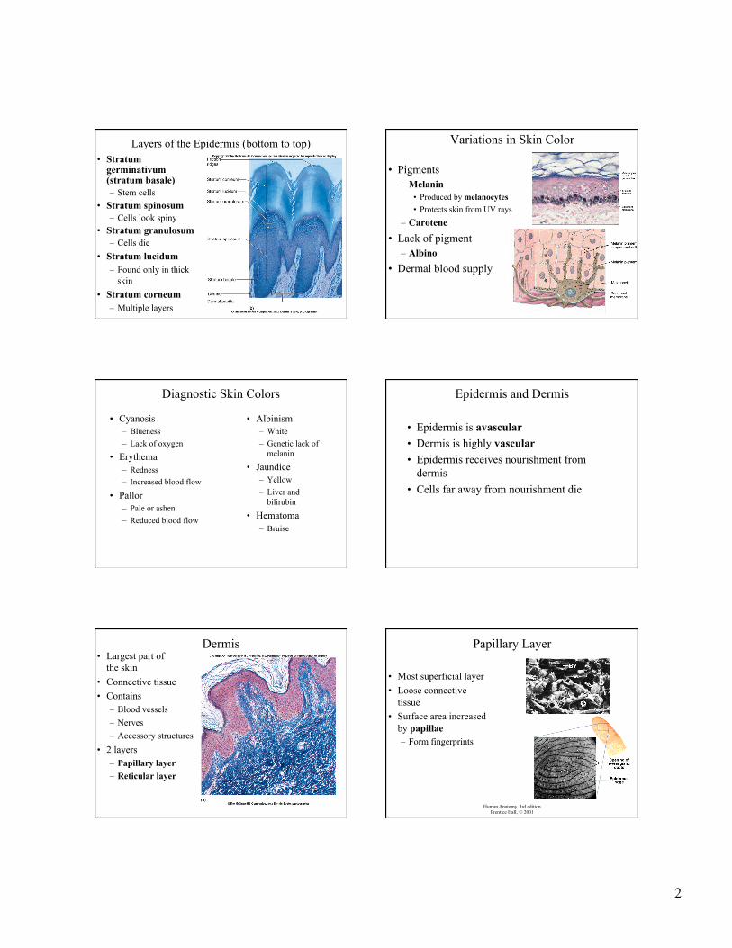
Layers of the Epidermis (bottom to top) • Stratum
germinativum (stratum basale) – Stem cells
• Stratum spinosum – Cells look spiny
• Stratum granulosum – Cells die
• Stratum lucidum – Found only in thick
skin • Stratum corneum
– Multiple layers
Variations in Skin Color
• Pigments – Melanin
• Produced by melanocytes • Protects skin from UV rays
– Carotene • Lack of pigment
– Albino • Dermal blood supply
Diagnostic Skin Colors
• Albinism – White – Genetic lack of
melanin
• Jaundice – Yellow – Liver and
bilirubin
• Hematoma – Bruise
Human Anatomy, 3rd edition Prentice Hall, © 2001
• Cyanosis – Blueness – Lack of oxygen
• Erythema – Redness – Increased blood flow
• Pallor – Pale or ashen – Reduced blood flow
Epidermis and Dermis
• Epidermis is avascular • Dermis is highly vascular • Epidermis receives nourishment from
dermis • Cells far away from nourishment die
Human Anatomy, 3rd edition Prentice Hall, © 2001
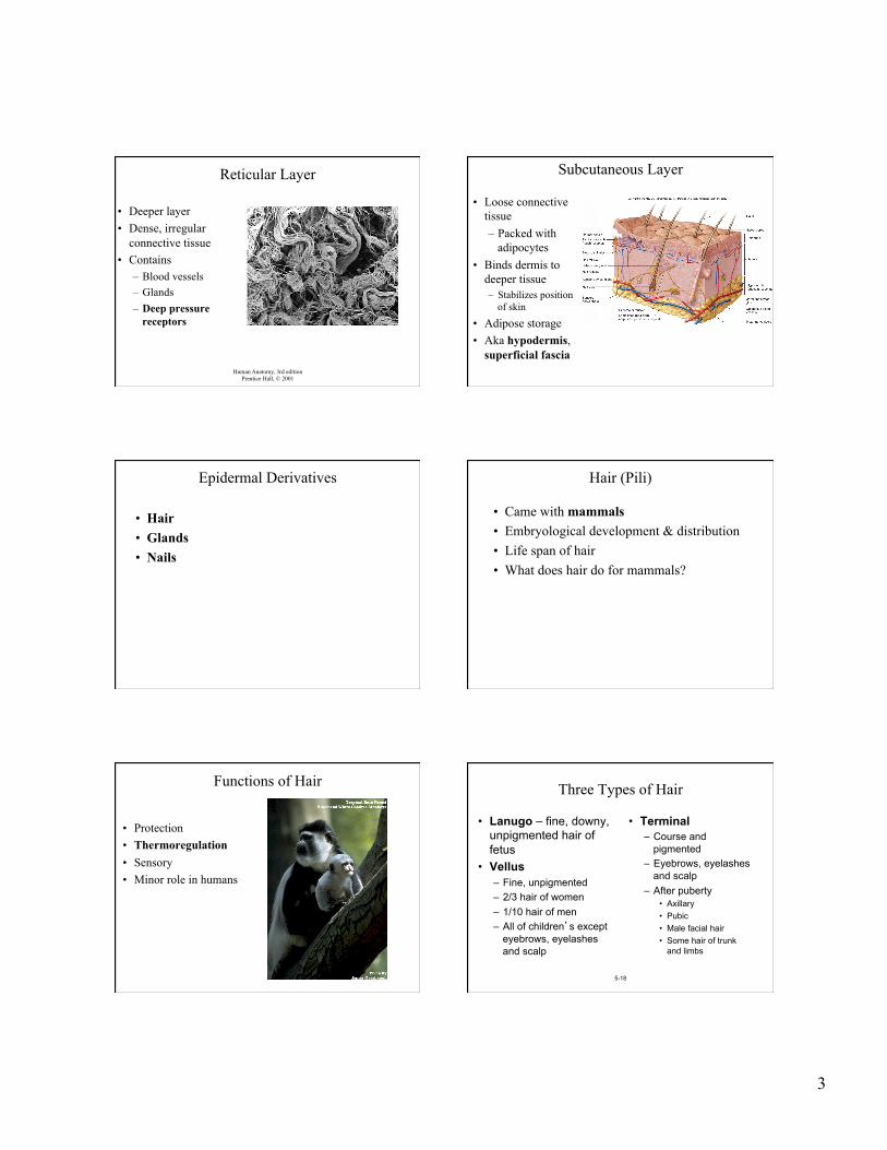
Dermis • Largest part of
the skin • Connective tissue • Contains
– Blood vessels – Nerves – Accessory structures
• 2 layers – Papillary layer – Reticular layer
Human Anatomy, 3rd edition Prentice Hall, © 2001
Papillary Layer
• Most superficial layer • Loose connective
tissue • Surface area increased
by papillae – Form fingerprints
3
Human Anatomy, 3rd edition Prentice Hall, © 2001
Reticular Layer
• Deeper layer • Dense, irregular
connective tissue • Contains
– Blood vessels – Glands – Deep pressure
receptors
Subcutaneous Layer
• Loose connective tissue – Packed with
adipocytes • Binds dermis to
deeper tissue – Stabilizes position
of skin
• Adipose storage • Aka hypodermis,
superficial fascia
Epidermal Derivatives
• Hair • Glands • Nails
Hair (Pili)
• Came with mammals • Embryological development & distribution • Life span of hair • What does hair do for mammals?
Functions of Hair
• Protection • Thermoregulation • Sensory • Minor role in humans
5-18
Three Types of Hair
• Lanugo – fine, downy, unpigmented hair of fetus
• Vellus – Fine, unpigmented – 2/3 hair of women – 1/10 hair of men – All of children’s except
eyebrows, eyelashes and scalp
• Terminal – Course and
pigmented – Eyebrows, eyelashes
and scalp – After puberty
• Axillary • Pubic • Male facial hair • Some hair of trunk
and limbs

4
Human Anatomy, 3rd edition Prentice Hall, © 2001
Hair Structure
• Shaft
– Superficial – Dead tissue
• Root – Remainder of hair
within follicle – Dead tissue
Human Anatomy, 3rd edition Prentice Hall, © 2001
Hair Structure
• Medulla – Soft core
• Cortex – Hard core
• Gives hair stiffness
• Cuticle – Outermost layer
Hair Develops in Follicles • Diagonal tube extending
deep in dermis • Two layers
– Epithelial root sheath – Connective tissue root
sheath • Bulb contains matrix • Hair papilla
– Contains blood vessels and nerves
• Associated structures – Hair receptors – Arrector pili
Arrector Pili
• Smooth muscle attaches to follicle • Raises hairs • Emotional response, cold • Function?
5-23
Hair Growth and Loss (Scalp)
Scalp – 4 to 8 years Eyelashes – 5 months Eyebrows – 2 months
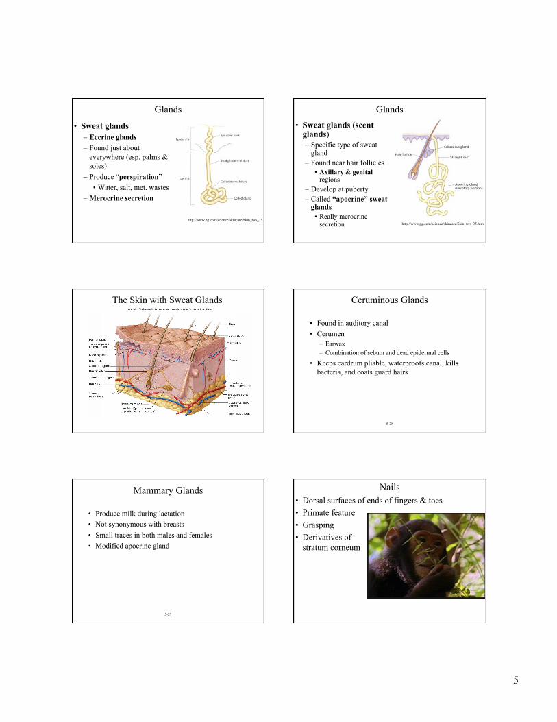
Glands • Sebaceous glands
– Usually connected to hair follicles – Secrete a waxy, oily substance (sebum) – Develop in utero at about 5 months – Secretion increases at puberty
5
Glands
• Sweat glands – Eccrine glands – Found just about
everywhere (esp. palms & soles)
– Produce “perspiration” • Water, salt, met. wastes
– Merocrine secretion
http://www.pg.com/science/skincare/Skin_tws_35.htm
Glands • Sweat glands (scent
glands) – Specific type of sweat
gland – Found near hair follicles
• Axillary & genital regions
– Develop at puberty – Called “apocrine” sweat
glands • Really merocrine
secretion http://www.pg.com/science/skincare/Skin_tws_35.htm
The Skin with Sweat Glands
5-28
Ceruminous Glands
• Found in auditory canal • Cerumen
– Earwax – Combination of sebum and dead epidermal cells
• Keeps eardrum pliable, waterproofs canal, kills bacteria, and coats guard hairs
5-29
Mammary Glands
• Produce milk during lactation • Not synonymous with breasts • Small traces in both males and females • Modified apocrine gland
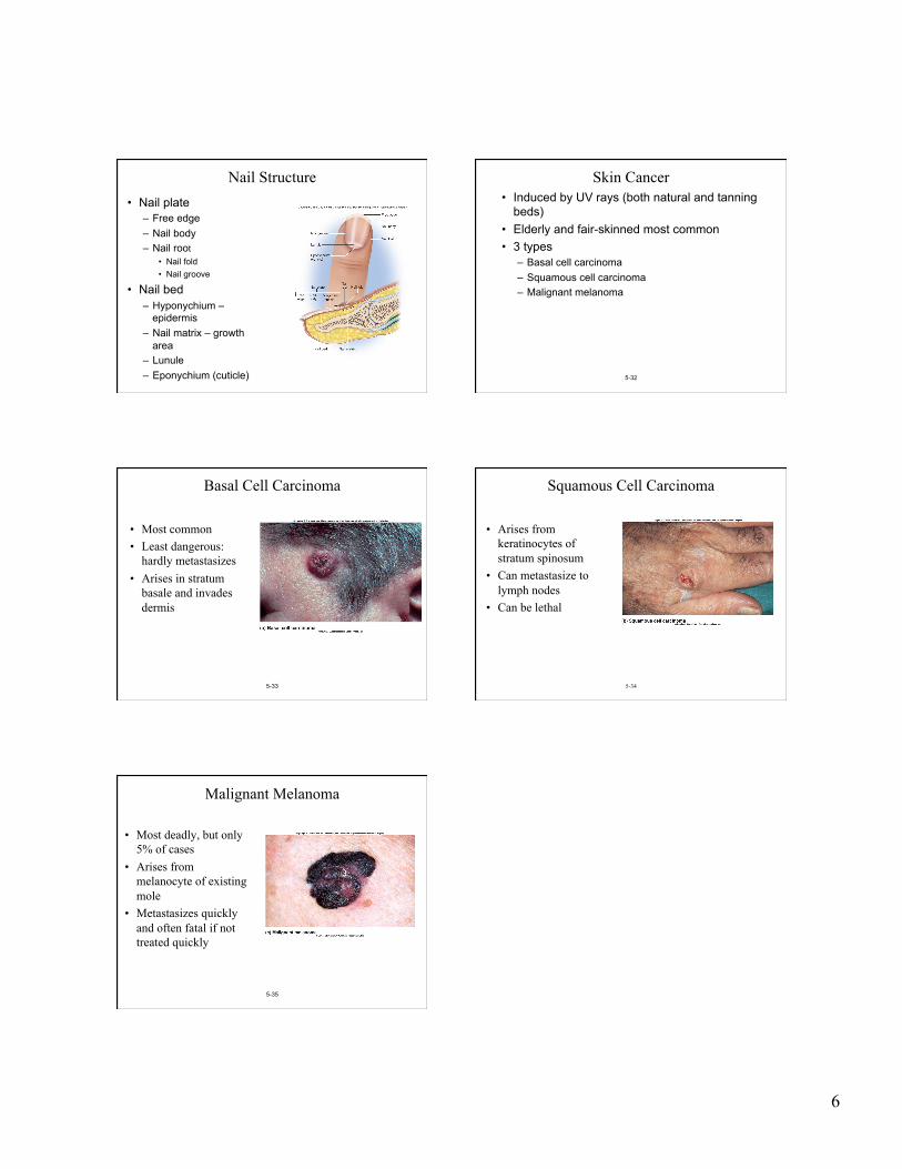
Nails • Dorsal surfaces of ends of fingers & toes • Primate feature • Grasping • Derivatives of
stratum corneum
6
Nail Structure • Nail plate
– Free edge – Nail body – Nail root
• Nail fold • Nail groove
• Nail bed – Hyponychium –
epidermis – Nail matrix – growth
area – Lunule – Eponychium (cuticle) 5-32
Skin Cancer • Induced by UV rays (both natural and tanning
beds) • Elderly and fair-skinned most common • 3 types
– Basal cell carcinoma – Squamous cell carcinoma – Malignant melanoma
5-33
Basal Cell Carcinoma
• Most common • Least dangerous:
hardly metastasizes • Arises in stratum
basale and invades dermis
5-34
Squamous Cell Carcinoma
• Arises from keratinocytes of stratum spinosum
• Can metastasize to lymph nodes
• Can be lethal
5-35
Malignant Melanoma
• Most deadly, but only 5% of cases
• Arises from melanocyte of existing mole
• Metastasizes quickly and often fatal if not treated quickly