Embed Size (px)
Citation preview
The Leg Muscles
BIO 238Instructor: Dr. Gourdine
1
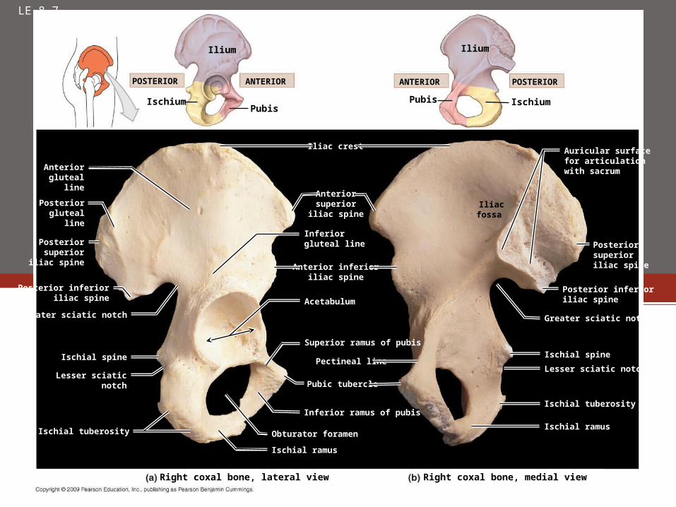
LE 8-7
Ilium
Right coxal bone, lateral view
Anteriorgluteal
line
Right coxal bone, medial view
IschiumPubis
POSTERIOR ANTERIOR
Pubis Ischium
ANTERIOR POSTERIOR
Ilium
Posteriorgluteal
line
Posteriorsuperior
iliac spine
Posterior inferioriliac spine
Greater sciatic notch
Ischial spine
Lesser sciaticnotch
Ischial tuberosity
Ischial ramus
Obturator foramen
Inferior ramus of pubis
Pubic tubercle
Pectineal line
Superior ramus of pubis
Acetabulum
Anterior inferioriliac spine
Inferiorgluteal line
Anteriorsuperior
iliac spine
Iliac crest
Iliac fossa
Auricular surfacefor articulationwith sacrum
Posteriorsuperioriliac spine
Posterior inferioriliac spine
Greater sciatic notch
Ischial spine
Lesser sciatic notch
Ischial tuberosity
Ischial ramus
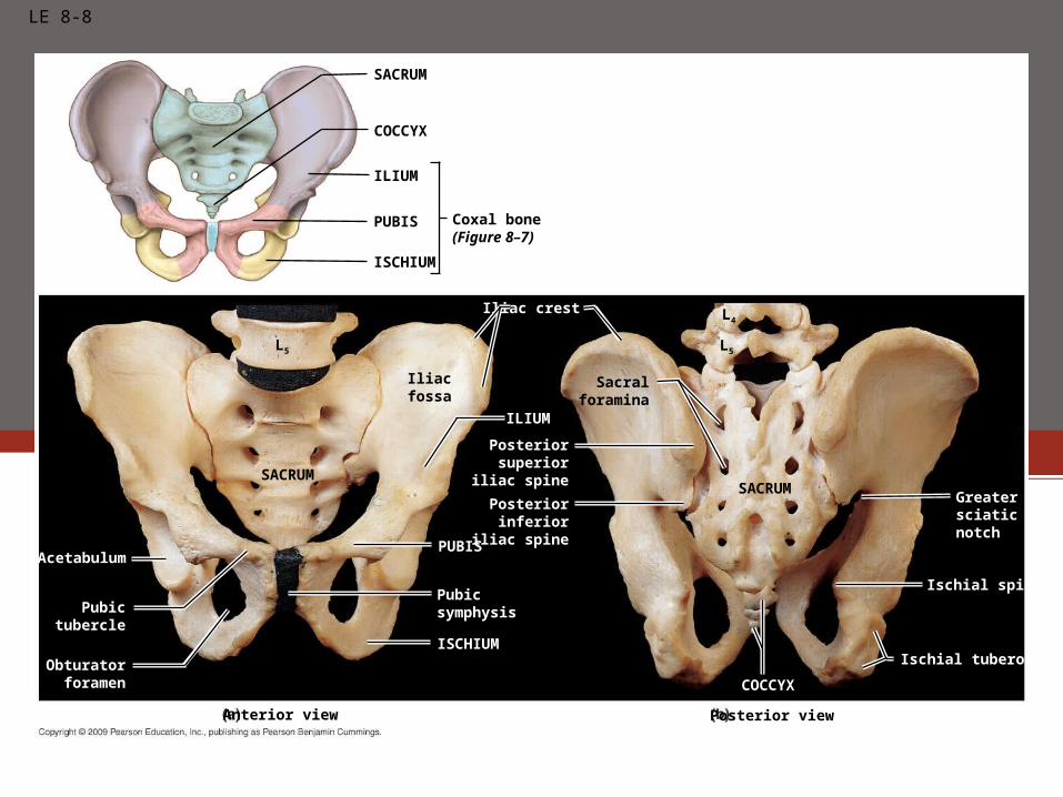
LE 8-8
SACRUM
COCCYX
ILIUM
PUBIS
ISCHIUM
Coxal bone(Figure 8–7)
Iliacfossa
SACRUM
L5
SACRUM
Sacralforamina
Acetabulum
Pubictubercle
Obturatorforamen
ISCHIUM
COCCYX
PUBIS
ILIUM
Pubicsymphysis
Posteriorinferior
iliac spine
Posteriorsuperior
iliac spine
Iliac crest
Anterior view Posterior view
L5
L4
Ischial tuberosity
Ischial spine
Greatersciaticnotch

LE 8-11
Greatertrochanter
Shaft
Neck
Lateral epicondyle
Patellar surface
Lateral condyle
Anterior surface Posterior surface
Lateral condyle
Lateral epicondyle
Popliteal surface
Neck
Greater trochanter
Femoral headIntertrochantericline
Lesser trochanter
Adductor tubercleMedial epicondyle
Medial condyle
Intertrochantericcrest
Gluteal tuberosity
Pectineal line
Linea aspera
Intercondylar fossa
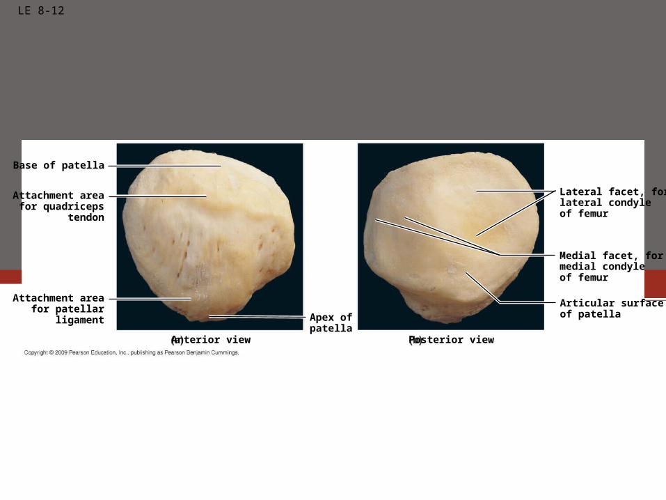
LE 8-12
Lateral facet, forlateral condyleof femur
Apex ofpatella
Attachment areafor quadriceps
tendon
Anterior view
Base of patella
Articular surfaceof patella
Posterior view
Attachment areafor patellar
ligament
Medial facet, formedial condyleof femur
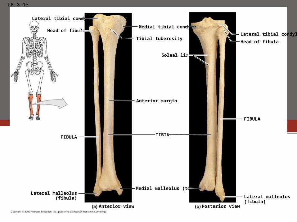
LE 8-13
Posterior viewAnterior view
Lateral tibial condyle
Head of fibula
Medial tibial condyle
Tibial tuberosity
Soleal line
Anterior margin
Lateral tibial condyle
Head of fibula
Lateral malleolus(fibula)
Medial malleolus (tibia)
Lateral malleolus(fibula)
FIBULA
FIBULA
TIBIA
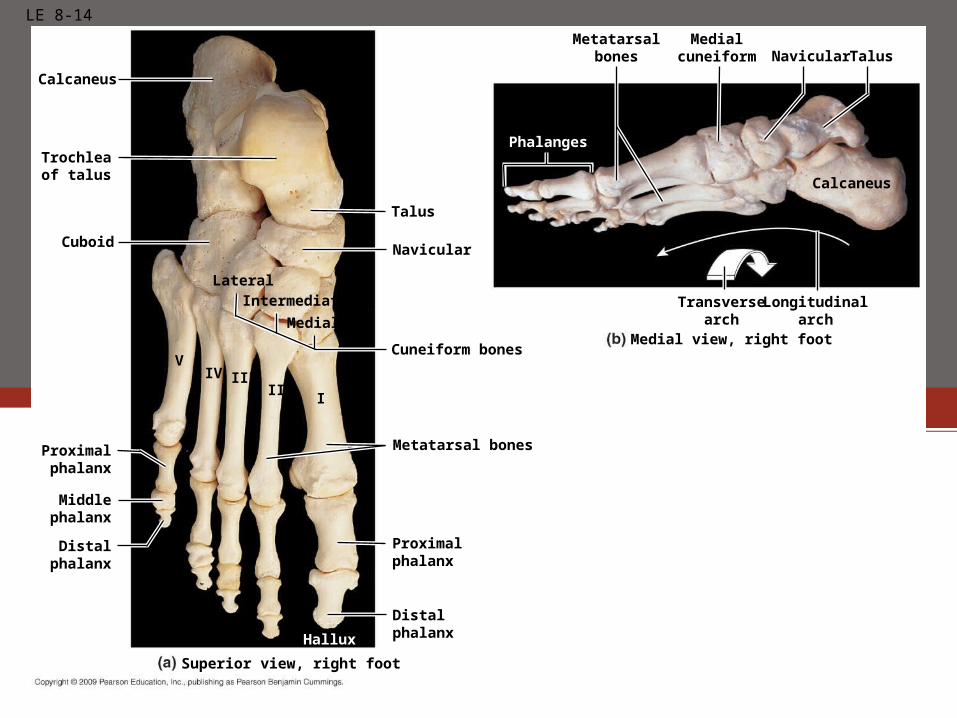
LE 8-14
Calcaneus
Distalphalanx
Cuboid
Superior view, right foot
Lateral
Medial view, right foot
Proximalphalanx
Middlephalanx
Proximalphalanx
Distalphalanx
Calcaneus
Trochleaof talus
Intermediate
Medial
Talus
Navicular
Cuneiform bones
Metatarsal bones
TalusNavicularMetatarsal
bonesMedial
cuneiform
Transversearch
Longitudinalarch
Phalanges
Hallux
VIV
III
III
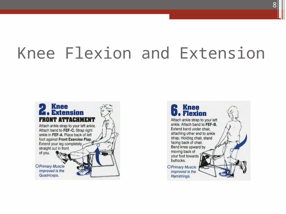
Knee Flexion and Extension
8
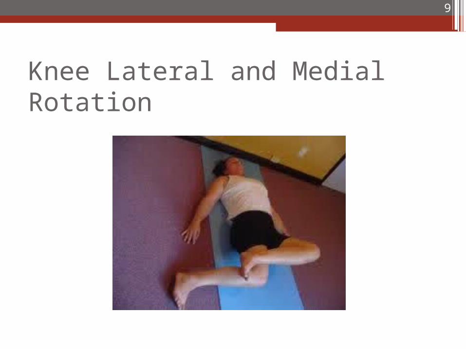
Knee Lateral and Medial Rotation
9
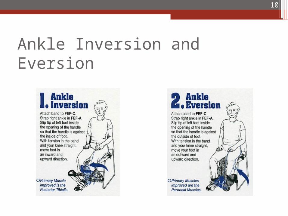
Ankle Inversion and Eversion
10
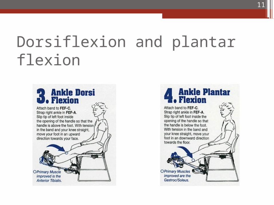
Dorsiflexion and plantar flexion
11
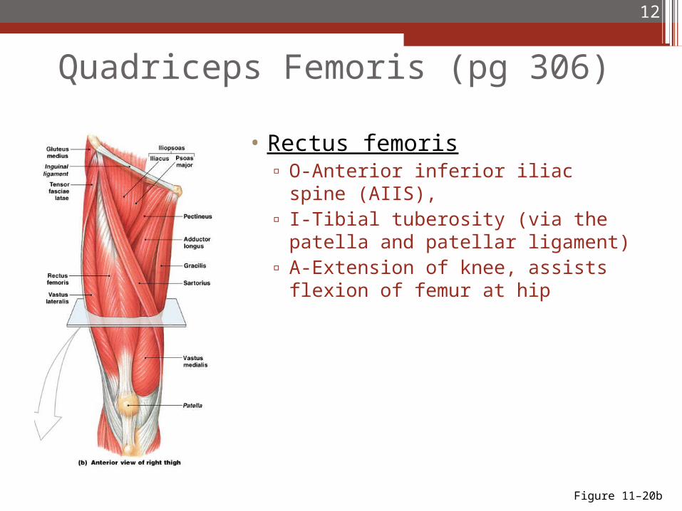
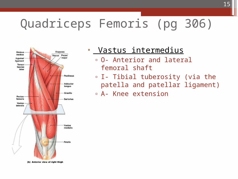
Quadriceps Femoris (pg 306)
• Rectus femoris▫ O-Anterior inferior iliac spine
(AIIS), ▫ I-Tibial tuberosity (via the patella
and patellar ligament)▫ A-Extension of knee, assists flexion
of femur at hip
12
Figure 11–20b
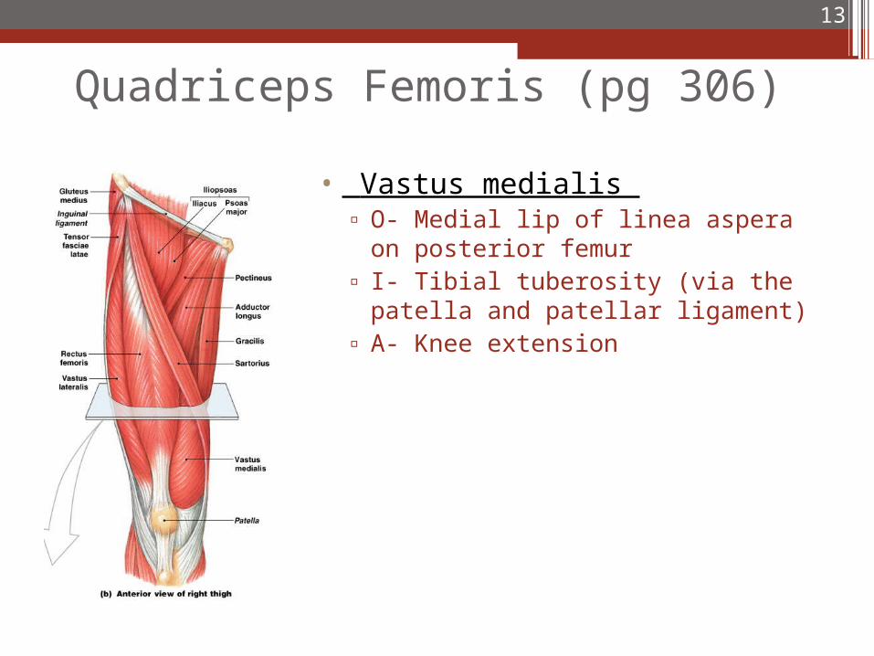
Quadriceps Femoris (pg 306)
• Vastus medialis ▫ O- Medial lip of linea aspera on
posterior femur▫ I- Tibial tuberosity (via the patella
and patellar ligament)▫ A- Knee extension
13
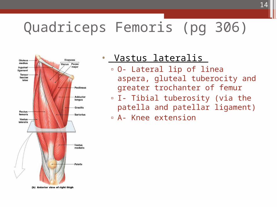
Quadriceps Femoris (pg 306)
• Vastus lateralis ▫ O- Lateral lip of linea aspera,
gluteal tuberocity and greater trochanter of femur
▫ I- Tibial tuberosity (via the patella and patellar ligament)
▫ A- Knee extension
14
Quadriceps Femoris (pg 306)
• Vastus intermedius▫ O- Anterior and lateral femoral
shaft▫ I- Tibial tuberosity (via the patella
and patellar ligament)▫ A- Knee extension
15
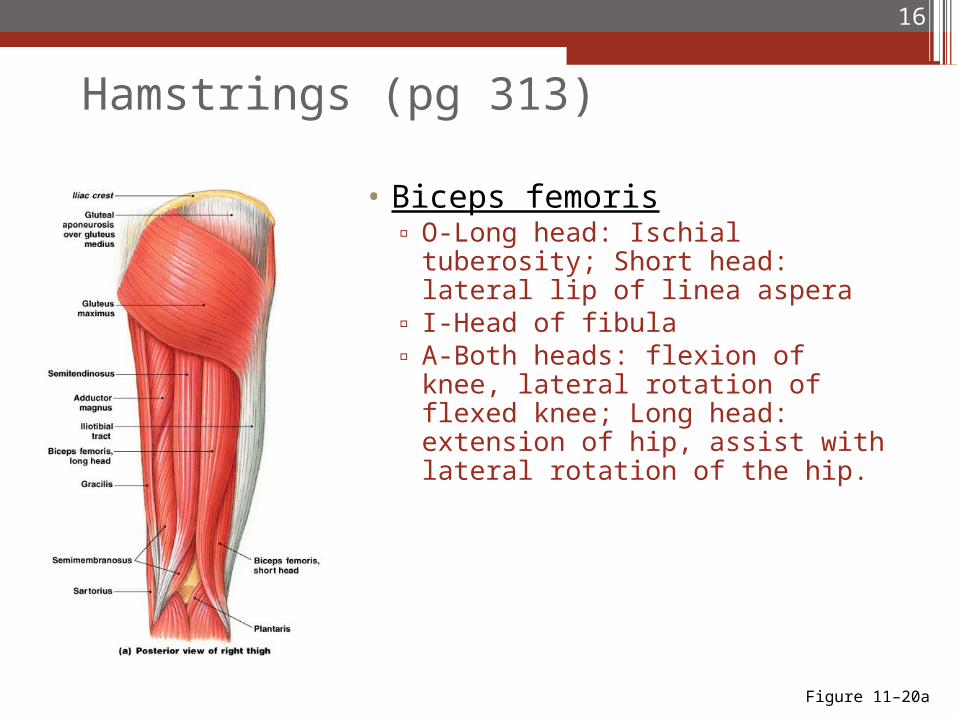
Hamstrings (pg 313)
• Biceps femoris▫ O-Long head: Ischial tuberosity;
Short head: lateral lip of linea aspera
▫ I-Head of fibula▫ A-Both heads: flexion of knee,
lateral rotation of flexed knee; Long head: extension of hip, assist with lateral rotation of the hip.
16
Figure 11–20a
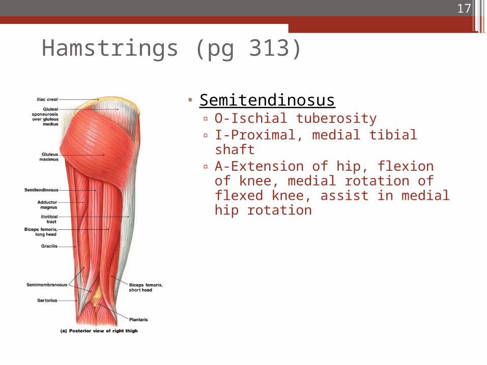
Hamstrings (pg 313)
• Semitendinosus▫ O-Ischial tuberosity▫ I-Proximal, medial tibial shaft▫ A-Extension of hip, flexion of knee,
medial rotation of flexed knee, assist in medial hip rotation
17
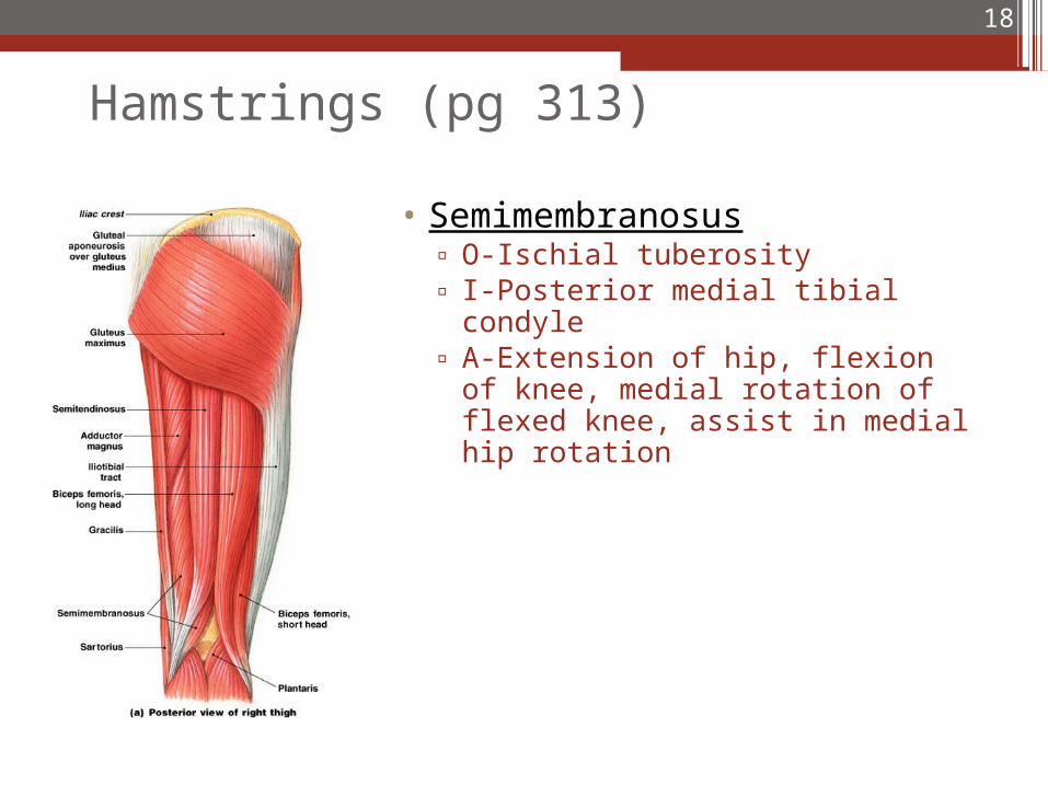
Hamstrings (pg 313)
• Semimembranosus▫ O-Ischial tuberosity▫ I-Posterior medial tibial condyle▫ A-Extension of hip, flexion of knee,
medial rotation of flexed knee, assist in medial hip rotation
18
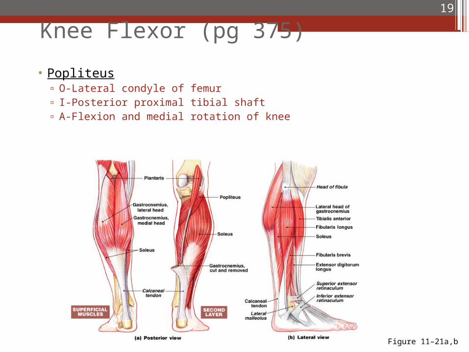
Knee Flexor (pg 375)
• Popliteus▫ O-Lateral condyle of femur▫ I-Posterior proximal tibial shaft▫ A-Flexion and medial rotation of knee
19
Figure 11–21a,b
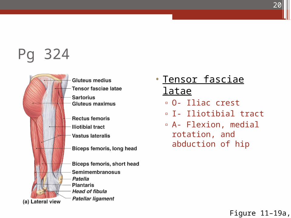
Pg 324
• Tensor fasciae latae▫ O- Iliac crest ▫ I- Iliotibial tract ▫ A- Flexion, medial
rotation, and abduction of hip
20
Figure 11–19a,b
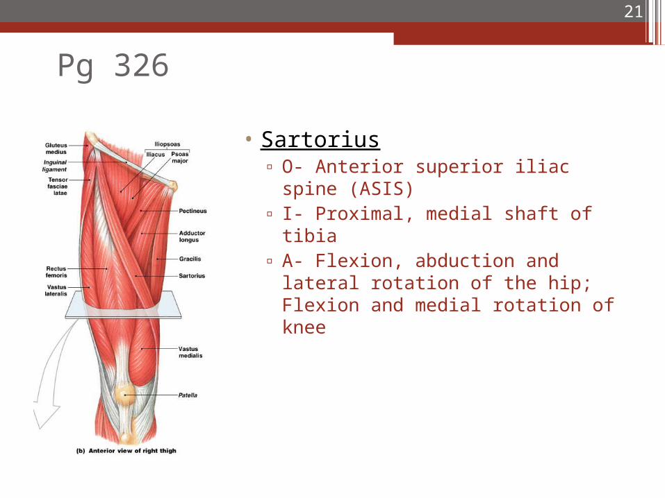
Pg 326
• Sartorius▫ O- Anterior superior iliac spine
(ASIS) ▫ I- Proximal, medial shaft of tibia▫ A- Flexion, abduction and lateral
rotation of the hip; Flexion and medial rotation of knee
21
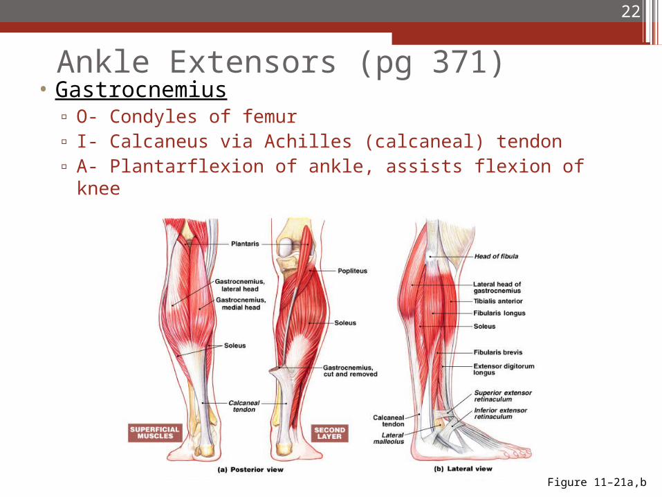
Ankle Extensors (pg 371)• Gastrocnemius
▫ O- Condyles of femur▫ I- Calcaneus via Achilles (calcaneal) tendon▫ A- Plantarflexion of ankle, assists flexion of knee
22
Figure 11–21a,b
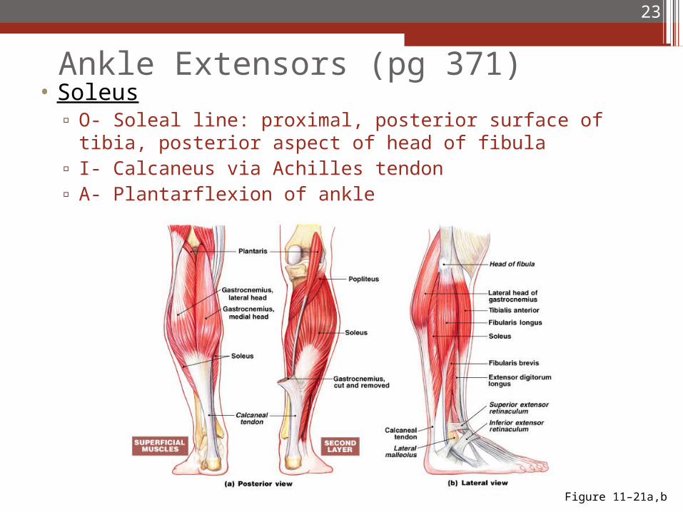
Ankle Extensors (pg 371)• Soleus
▫ O- Soleal line: proximal, posterior surface of tibia, posterior aspect of head of fibula
▫ I- Calcaneus via Achilles tendon▫ A- Plantarflexion of ankle
23
Figure 11–21a,b
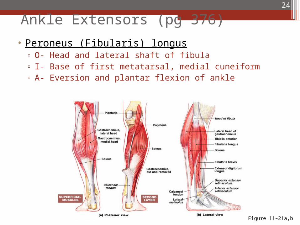
Ankle Extensors (pg 376)
• Peroneus (Fibularis) longus▫ O- Head and lateral shaft of fibula▫ I- Base of first metatarsal, medial cuneiform▫ A- Eversion and plantar flexion of ankle
24
Figure 11–21a,b
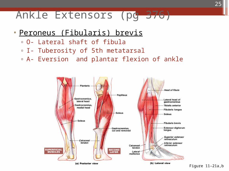
Ankle Extensors (pg 376)
• Peroneus (Fibularis) brevis▫ O- Lateral shaft of fibula▫ I- Tuberosity of 5th metatarsal▫ A- Eversion and plantar flexion of ankle
25
Figure 11–21a,b
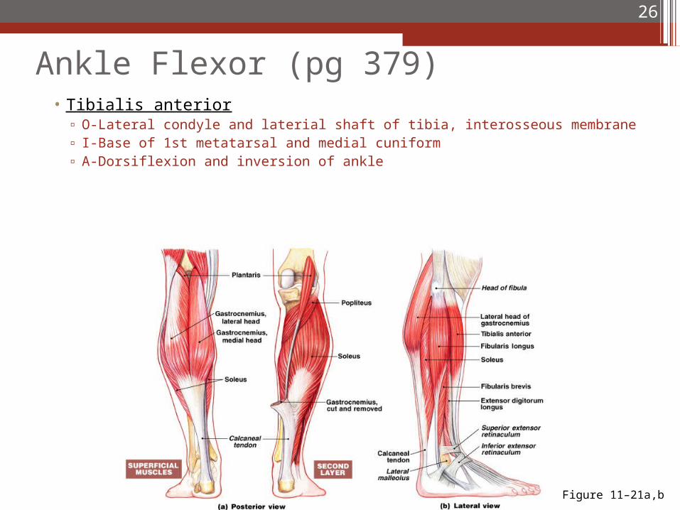
Ankle Flexor (pg 379)• Tibialis anterior
▫ O-Lateral condyle and laterial shaft of tibia, interosseous membrane▫ I-Base of 1st metatarsal and medial cuniform▫ A-Dorsiflexion and inversion of ankle
26
Figure 11–21a,b
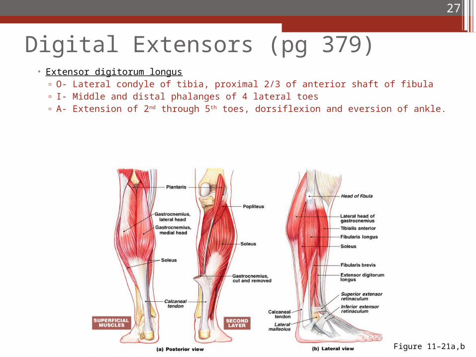
Digital Extensors (pg 379)• Extensor digitorum longus
▫ O- Lateral condyle of tibia, proximal 2/3 of anterior shaft of fibula▫ I- Middle and distal phalanges of 4 lateral toes▫ A- Extension of 2nd through 5th toes, dorsiflexion and eversion of ankle.
27
Figure 11–21a,b