Embed Size (px)
Citation preview
IMF YJMBI-64687; No. of pages: 9; 4C: 3, 4, 5, 6
FeaturedAr�cle
James Ho1, †, H
0022-2836/© 2015 Elsevi
Please cite this article aJ Mol Biol (2015), http:/
The Molecular Motor F-ATP Synthase IsTargeted by the Tumoricidal Protein HAMLET
endrik Sielaff 2, †, Aftab Na
deem1,Catharina Svanborg1 and Gerhard Grüber21 - Department of Microbiology, Immunology and Glycobiology, Institute of Laboratory Medicine, Lund University, Sölvegatan 23,S-223 62 Lund, Sweden2 - School of Biological Sciences, Nanyang Technological University, 60 Nanyang Drive, Singapore 637551, Republic of Singapore
Correspondence to Catharina Svanborg andGerhard Grüber: [email protected];[email protected]://dx.doi.org/10.1016/j.jmb.2015.01.024Edited by B. Poolman
Legend: HAMLET (beige), approaching the tumor cell and identifyingthe F-ATP synthase with which it associates to inhibit ATP synthesis.Thestructureof theα3:β3:γcomplexof theF-ATPsynthase is shown ingreen, orange and yellow, respectively.
James Ho, Hendrik Sielaff, Catharina Svanborg andGerhard Grüber
Abstract
HAMLET (human alpha-lactalbumin made lethal totumor cells) interacts with multiple tumor cellcompartments, affecting cell morphology,metabolism,proteasome function, chromatin structure and viability.This study investigated if these diverse effects ofHAMLETmight be caused, in part, by a direct effect onthe ATP synthase and a resulting reduction in cellularATP levels. A dose-dependent reduction in cellularATP levelswas detected in A549 lung carcinoma cells,and by confocal microscopy, co-localization ofHAMLET with the nucleotide-binding subunits α(non-catalytic) and β (catalytic) of the energy convert-ing F1F0 ATP synthase was detected. As shown byfluorescence correlation spectroscopy, HAMLETbinds to the F1 domain of the F1F0 ATP synthasewith a dissociation constant (KD) of 20.5 μM.Increasing concentrations of the tumoricidal proteinHAMLET added to the enzymatically active α3β3γcomplex of the F-ATP synthase lowered its ATPaseactivity, demonstrating that HAMLET binding to theF-ATP synthase effects the catalysis of this molecularmotor. Single-molecule analysis was applied to studyHAMLET–α3β3γ complex interaction. Whereas theα3β3γ complex of the F-ATP synthase rotated in acounterclockwise direction with a mean rotational rateof 3.8 ± 0.7 s−1, no rotation could be observed in thepresence of bound HAMLET. Our findings suggestthat direct effects of HAMLET on the F-ATP synthasemay inhibit ATP-dependent cellular processes.
© 2015 Elsevier Ltd. All rights reserved.
er Ltd. All rights reserved. J Mol Biol (2015) xx, xxx–xxx
s: Ho James, et al, The Molecular Motor F-ATP Synthase Is Targeted by the Tumoricidal Protein HAMLET,/dx.doi.org/10.1016/j.jmb.2015.01.024

2 Interaction of HAMLET and the F-ATP
Introduction
unfolded α-lactalbumin and several oleate residues[1]. HAMLET in solution shows a two-domain confor-
HAMLET (human alpha-lactalbumin made lethal totumor cells) is a proteolipid complex of partially
mation with a large globular domain and an extendedC-terminal part [2]. Its efficacy as a selective killer oftumor cells has been documented in vitro and in vivo inseveral animal models, including human brain tumorxenografts in nude rats, murine bladder cancer andcolon cancer in theAPCMin+/−mice, resembling humandisease [3]. In clinical studies, HAMLET has showntherapeutic efficacy against skin papillomas anddramatic effects in bladder cancer patients [3,4].HAMLET initiates cell death by perturbation of theplasma membrane and activation of ion fluxes and thisresponse distinguishes tumor cells from healthy cells.The interactions of HAMLET with different cellulartargets including histonesH2,H3andH4; hexokinase I;α-actinin 1/4; and proteasomal subunits have beenstudied [5–8]. The sensitivity of tumor cells of differentorigins suggests that HAMLET may act on moleculartargets that are shared among tumor cells, therebysucceeding to kill those cells, rather than healthydifferentiated cells with a more inert cell membrane.HAMLET is also internalized by tumor cells, changingmorphology, gene expression and phosphorylation. Inparallel, a reduction in the content of ATPaccompaniescell death [9,10].
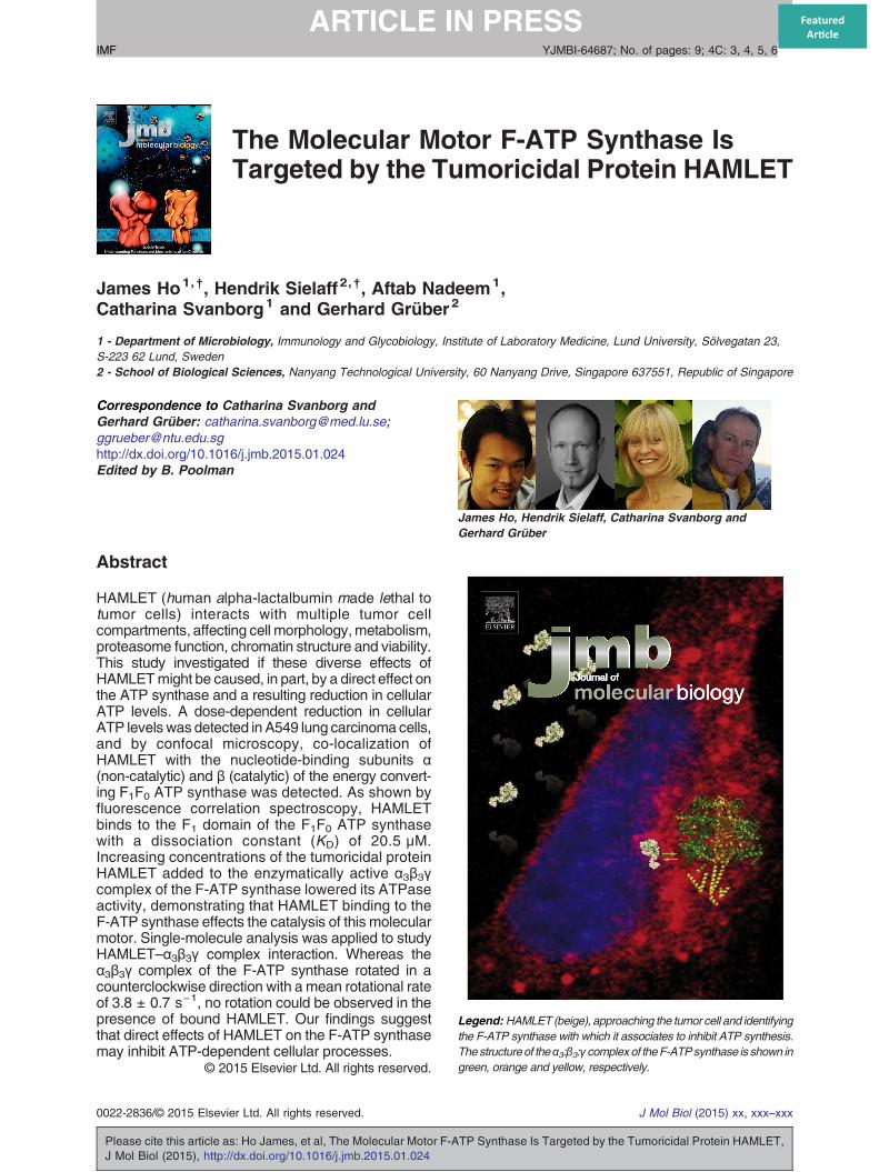
Fig. 1. (A) The effect of HAMLET on intracellular ATP levels o35 μMHAMLET,whichwas producedas described byHakanssonin intracellular ATP levels. Error bars represent ±SEM (standardprocured from American Type Culture Collection and weremaintapyruvate (Fisher Scientific), non-essential amino acids (1:100) (Fis5% fetal calf serum (FCS). Cells were cultured at 37 °C, 90%humi(for PrestoBlue™ and ATP assays), in 6-well plates (for WesAfterwards, cells were detached fromculture flaskswith 10–15 ml800 ml phosphate-buffered solution (PBS) and 200 ml H2O) andCellswere seeded in a 96-well plate at a concentration of 1 × 10⁴ ccell 150; Heraeus). Adherent cells werewashedwith PBS twice antime durations. Cell culture medium was removed and cellulaPerkinElmer) according to themanufacturer's instructions. (B andCsynthase subunits α and β in lung carcinoma cells. (B) Suspensionsubunits α (top) and β (bottom) from the periphery to the cytoplasmwas replaced by the formation of larger subunit α aggregates afterwas less pronounced. HAMLET showed stronger co-localization wcaused an increase in the staining for both α and β subunits in the ethe catalytic β subunit was observed at 21 μM HAMLET. At 35 μsuspension cell experiments, A549 lung carcinoma cells were adoses of HAMLET (7, 21 and 35 μM; 10% Alexa-HAMLET) for 18-well glass chamber slides (Lab-Tek, Chamber Slide, Thermoovernight at 37 °C.Cellswerewashed twicewithPBSand treatedCells were fixed with 2% paraformaldehyde, non-permeabilizedsynthase subunitαorβmonoclonal antibodies (1:40 in 10%FCS/Pwith secondary Alexa-488 conjugated antibodies (1:100 in 10%(Abcam) and examined using a LSM 510 META laser scanning cfluorescence quantification were performed using LSM510 image
Please cite this article as: Ho James, et al, The Molecular Motor F-AJ Mol Biol (2015), http://dx.doi.org/10.1016/j.jmb.2015.01.024
The enzyme catalyzing ATP synthesis is the F1F0ATP synthase (F-ATP synthase), a membrane-boundmulti-subunit complex consisting of two rotarymotors inthe F0 and F1 sector, respectively. The membrane-bound proton translocating ATP synthase catalyzesATP synthesis and ATP hydrolysis in the F1 part, whichis coupled to proton translocation across the F0 sector.The F1 domain consists of subunits α3β3γδε [11] andthe membrane-integrated F0 of most bacteria is madeup of subunits a and b (a:b2), and the c-ring rotorsubunits, with a stoichiometry of 9–15 c subunits [12].The subcomplex α3β3 forms a hexamer with a centralcavity that allows for the penetration of subunit γ.Subunits γ and ε form the soluble part of the rotor shaft,called the central stalk [13]. Rotation of the central stalksubunit γ within the α3β3 cavity causes conformationalchanges in the three catalytic sites located at the α–βinterfaces leading to ATP hydrolysis [14]. The two partsof the ATP synthase are connected by two stalks, thatis, one central rotating shaft formed by the subunits γand ε and a thin stalk at the periphery, composed of thesubunitsbandδholding together theF1andF0portions[15].Here, we demonstrate that the interaction of
HAMLET with the F1 sector of the F-ATP synthaseresults in a reduction of catalytic activity, providingone mechanism for the reduction in cellular ATP.Monoclonal antibodies against the nucleotide-bind-ing subunits α and β of the F-ATP synthase identifythe co-localization of HAMLET with the molecular
f tumor cells. A549 lung carcinoma cells treated with 7, 21 oret al. [1], showeda rapid time- and dose-dependent reductionerror of the mean). The lung carcinoma cells (A549) were
ined in RPMI 1640 medium supplemented with 1 mM sodiumher Scientific), 50 μg/ml gentamicin (Gibco, Paisley, UK) anddity and 5%CO2. Cells were grown in 96-well plates overnighttern blots) and in 75-mm flasks (for immunoprecipitation).versene (200 mgethylenediaminetetraacetic acid dissolved inresuspended in RPMI 1640 complete medium with 5% FCS.ells perwell, overnight at 37 °C in ahumidified incubator (Herad treatedwith different concentrations of HAMLET for differentr ATP was quantified using the ATPlite Kit (Infinite F200;) The effects of HAMLET on the cellular localization of F-ATPcells: HAMLET triggered the translocation of F-ATP synthaseand the nuclei. A punctate staining pattern in untreated cellsHAMLET exposure (1 h). The β subunit aggregate formationith subunit α than with subunit β. (C) Adherent cells: HAMLETntire cell (see Fig. 2) and stronger co-localization (yellow) withM, co-localization with the α and β subunits was similar. Forllowed to partially adhere to glass slip and treated with threeh. For adherent cell experiments, A549 cells were grown onFisher Scientific) at a concentration 2.5 × 10⁴ cells per wellwithHAMLET (7, 21 and35 μM;10%Alexa-HAMLET) for 1 h., blocked (10% FCS, 1 h), incubated with primary F-ATPBS; Life Technologies) for 2 hat room temperature, incubatedFCS/PBS; Molecular Probes), counterstained with DRAQ-5onfocal microscope (Carl Zeiss). Co-localization analysis andbrowser software and Photoshop CS5, respectively.
TP Synthase Is Targeted by the Tumoricidal Protein HAMLET,
3Interaction of HAMLET and the F-ATP
motor F-ATP synthase in vivo. With the use of theenzymatic active α3β3γ complex of the thermophilicBacillus PS3 F-ATP synthase, the quantitative and
Fig. 1 (legend on
Please cite this article as: Ho James, et al, The Molecular Motor F-AJ Mol Biol (2015), http://dx.doi.org/10.1016/j.jmb.2015.01.024
qualitative interaction of both proteins was investi-gated, and a mechanistic model of HAMLET–F-ATPsynthase assembly is proposed.
previous page).
TP Synthase Is Targeted by the Tumoricidal Protein HAMLET,
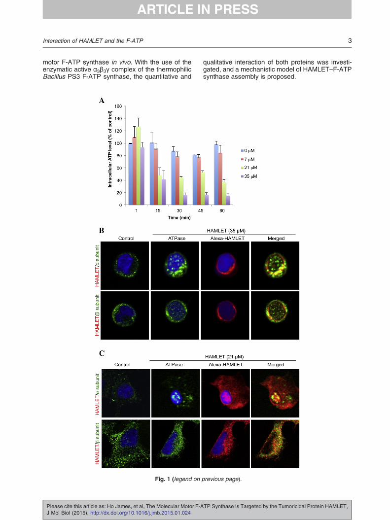
Fig. 2. The effects of HAMLET on the cellular localization of F-ATP synthase subunits α and β in adherent lung carcinomacells. (A) HAMLET triggered a concentration-dependent change in F-ATP synthase subunit α and β staining from a punctatecytoplasmic staining pattern in untreated cells to the formation of larger aggregates for α subunit and occasional smalleraggregates for β subunit in cell nucleus after HAMLET exposure (7, 21 or 35 μM, 1 h). (B) Stronger co-localization (yellow) withthe catalytic β subunit was observed at 21 μM HAMLET. At 35 μM, co-localization with the α and β subunits was similar.
4 Interaction of HAMLET and the F-ATP
Results and discussion
Reduction in intracellular ATP levels
The effect of HAMLET on intracellular ATP levels wasquantified in A549 lung carcinoma cells. A dose-dependent reduction was observed after 15 min. At35 μM HAMLET, the ATP level was reduced to about40% of untreated cells after 15 min, and ATP levelsremained low from 30 min onwards (15% of control;Fig. 1A). At 21 μMHAMLET, theATP levelwas reducedto about 45% after 15 min and was further reduced to
Please cite this article as: Ho James, et al, The Molecular Motor F-AJ Mol Biol (2015), http://dx.doi.org/10.1016/j.jmb.2015.01.024
35% after 60 min, as compared to control. Lowerconcentrations of HAMLET (7 μM) had a modest effect(about 20% control).
HAMLET co-localizes with the F-ATP synthase
The enzyme responsible for ATP synthesis ineukaryotic cells is the F1F0 ATP synthase, with thealternating nucleotide-binding subunits α and βforming a hexameric headpiece of the F1 part. Theinterface of each α–β pair forms the nucleotide-binding sites. Besides subunit c of the F0 part, which
TP Synthase Is Targeted by the Tumoricidal Protein HAMLET,

5Interaction of HAMLET and the F-ATP
is membrane embedded, both subunits α and βshow the highest levels of homology among F-ATPsynthases. To examine if HAMLET affects thecellular distribution of F1F0 ATP synthase, westained Alexa-HAMLET-treated A549 cells withmonoclonal antibodies directed against subunits αand β from human mitochondrial F-ATP synthase. Arapid increase in staining was observed by confocalmicroscopy of cells in suspension (Fig. 1B).HAMLET triggered a change in F-ATP synthasesubunit α staining (Fig. 1B, top) from a punctate,peripheral α subunit staining pattern in untreatedcells to the formation of larger aggregates afterHAMLET exposure (15 min). At a HAMLET con-centration of 35 μM, nuclear staining was observed.
Please cite this article as: Ho James, et al, The Molecular Motor F-AJ Mol Biol (2015), http://dx.doi.org/10.1016/j.jmb.2015.01.024
To further address if HAMLET is localized in thesame cellular compartments as the F-ATP synthasesubunits, co-localization of the α and β subunits withAlexa-HAMLET was investigated by confocal mi-croscopy and weak co-localization was observed(Fig. 1B, right panel). The β subunit showed a similartranslocation from the cell periphery but aggregateformation was less pronounced. Cytoplasmicand nuclear aggregates were formed at 35 μMHAMLET. Co-localizations of Alexa-HAMLET withthe two subunits were observed at the perinuclearand the cytoplasmic region where HAMLET accu-mulation was the strongest.The experiment was extended to include adherent
cells (Figs. 1C and 2). HAMLET triggered aconcentration-dependent change in F-ATP syn-thase subunit α and β staining from a punctatecytoplasmic staining pattern in untreated cells to theformation of larger aggregates for subunit α and
Fig. 3. TF1–HAMLET binding studied by fluorescencecorrelation spectroscopy. (A) SDS-PAGE of purified α3β3γcomplex of the F-ATP synthase from thermophilic BacillusPS3 (lane 2) and a molecular weight marker (lane 1).(B)Normalized autocorrelation functions of theα3β3γ complexand HAMLET-Atto647N (HL) obtained by increasing thequantity of the α3β3γ complex (from left to right: 0 μM, 0.5 μM,5 μM, 10 μM and, 50 μM). (C) Concentration-dependentbinding of α3β3γ to HAMLET. The percentage of complexformation for each concentration was calculated using atwo-component fitting model. The binding constant, KD, wasderived by fitting the data with the Hill equation. For thefluorescence correlation spectroscopy experiments, thecysteine in subunit γ of α3β3γ was labeled with Atto647N.The free dye was removed by washing the sample in buffer A[20 mM Mops (pH 7.0), 50 mM KCl and 5 mM MgCl2],followed by centrifugation in a centrifugal filter column(exclusion size of 100 kDa; Centricon, Millipore) for at leastthree times. In case of HAMLET, lyophilized HAMLET wasresuspended in 250 μl of buffer B [50 mM Tris (pH 7.5) and250 mM NaCl] to a final concentration of 50–100 μM.Atto647N-maleimide (ATTO-TEC)wasadded in aprotein:dyeratio of 1:0.9 and incubated for 5 min on ice in the dark, beforeinactivation of the maleimide moiety by adding 10 mM DTT.The reaction time and labeling ratio were kept low to avoiddouble labeling. The labeled protein was separated from freedye by gel filtration via a S75 column (GE Healthcare) withbuffer C. Measurements were performed on a LSM510Meta/ConfoCor 3 microscope (Carl Zeiss, Germany) with a waterimmersion objective (40×/1.2W Corr UV-VIS-IR; Zeiss) andthe 633 nm line of a 5 mWHeNe633 laser. Samples (in bufferA) of 15 μl were placed in Nunc 8-well chambers treated with3% gelatin to prevent unspecific binding of proteins [20]. Cy5in water was used as references for the calibration of theconfocal microscope. The fluorescence intensities of fluores-cent particles in the confocal volume (HAMLET-Atto647Nandα3β3γ-Atto647N) were measured at 25 °C for up to 10 minwith 10-s repetitions. From the fit of the autocorrelationfunction, the number of particles in the confocal volume, thediffusion times of fluorescent particles, the intrinsic triplet stateof the dye and the percentage of complex formation werederived.
TP Synthase Is Targeted by the Tumoricidal Protein HAMLET,
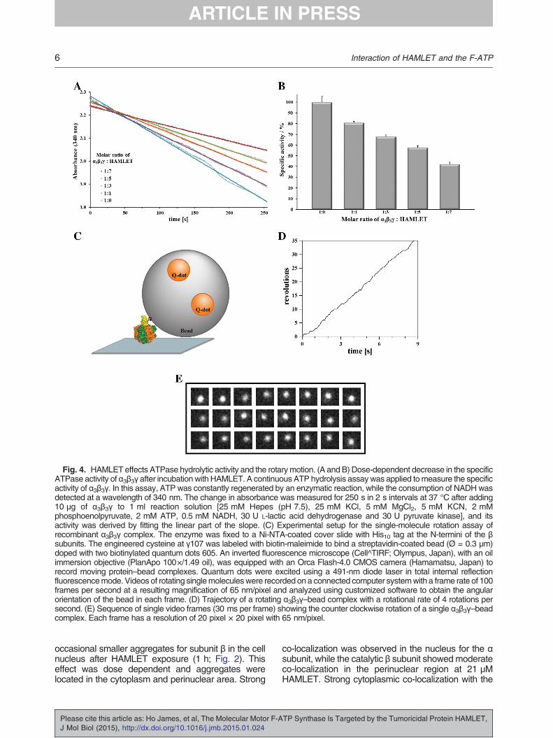
Fig. 4. HAMLET effects ATPase hydrolytic activity and the rotarymotion. (A and B) Dose-dependent decrease in the specificATPase activity of α3β3γ after incubation with HAMLET. A continuous ATP hydrolysis assaywas applied tomeasure the specificactivity of α3β3γ. In this assay, ATP was constantly regenerated by an enzymatic reaction, while the consumption of NADH wasdetected at a wavelength of 340 nm. The change in absorbance was measured for 250 s in 2 s intervals at 37 °C after adding10 μg of α3β3γ to 1 ml reaction solution [25 mM Hepes (pH 7.5), 25 mM KCl, 5 mM MgCl2, 5 mM KCN, 2 mMphosphoenolpyruvate, 2 mM ATP, 0.5 mM NADH, 30 U L-lactic acid dehydrogenase and 30 U pyruvate kinase], and itsactivity was derived by fitting the linear part of the slope. (C) Experimental setup for the single-molecule rotation assay ofrecombinant α3β3γ complex. The enzyme was fixed to a Ni-NTA-coated cover slide with His10 tag at the N-termini of the βsubunits. The engineered cysteine at γ107 was labeled with biotin-maleimide to bind a streptavidin-coated bead (Ø = 0.3 μm)doped with two biotinylated quantum dots 605. An inverted fluorescence microscope (Cell^TIRF; Olympus, Japan), with an oilimmersion objective (PlanApo 100×/1.49 oil), was equipped with an Orca Flash-4.0 CMOS camera (Hamamatsu, Japan) torecord moving protein–bead complexes. Quantum dots were excited using a 491-nm diode laser in total internal reflectionfluorescencemode. Videos of rotating singlemoleculeswere recorded on a connected computer systemwith a frame rate of 100frames per second at a resulting magnification of 65 nm/pixel and analyzed using customized software to obtain the angularorientation of the bead in each frame. (D) Trajectory of a rotating α3β3γ–bead complex with a rotational rate of 4 rotations persecond. (E) Sequence of single video frames (30 ms per frame) showing the counter clockwise rotation of a single α3β3γ–beadcomplex. Each frame has a resolution of 20 pixel × 20 pixel with 65 nm/pixel.
6 Interaction of HAMLET and the F-ATP
occasional smaller aggregates for subunit β in the cellnucleus after HAMLET exposure (1 h; Fig. 2). Thiseffect was dose dependent and aggregates werelocated in the cytoplasm and perinuclear area. Strong
Please cite this article as: Ho James, et al, The Molecular Motor F-AJ Mol Biol (2015), http://dx.doi.org/10.1016/j.jmb.2015.01.024
co-localization was observed in the nucleus for the αsubunit, while the catalytic β subunit showedmoderateco-localization in the perinuclear region at 21 μMHAMLET. Strong cytoplasmic co-localization with the
TP Synthase Is Targeted by the Tumoricidal Protein HAMLET,
7Interaction of HAMLET and the F-ATP
catalytic β subunit was observed at 21 μM HAMLET.Our observations are consistent with earlier studiesin which F-ATP synthase subunits were alsolocalized at cellular compartments distinct from theinnermembraneofmitochondria [16,17]. Furthermore,ectopic cell surface F-ATP synthase has been shownto be a receptor for angiostatin [18]. In addition to theexisting knowledge, our present findings on the drasticchange in localization of F-ATP synthase subunits,from a punctate cell surface staining pattern to acytoplasmic pattern with nuclear aggregates, mightsuggest a scenario whereby the motor is uncoupledfrom the proton gradient, leading to an inhibition of theATP synthesis process. The outcome of this is evidentas a rapid reduction in intracellular ATP level wasobserved after HAMLET treatment.
Quantitative and qualitative binding of HAMLETto the F1 domain of F-ATP synthase
To examine the hypothesis that HAMLET inhibitioncauses a malfunctioning F-ATP synthase, we appliedfluorescence correlation spectroscopy to confirm andquantify the interaction of HAMLET with the F-ATPsynthase. We used the mechanistically best under-stood F-ATP synthase from thermophilic BacillusPS3 as a prototype. The enzymatically active α3β3γcomplex of the F1F0 ATP synthase (TF1) waspurified as previously described (Fig. 3A and seeRef. [19]). Both HAMLET (14.1 kDa) and the α3β3γcomplex (352 kDa) were labeled with Atto647N-maleimide and their individual diffusion times weredetermined from a single-component fit of theresulting autocorrelation function to be 209 μs and440 μs, respectively, which correlate well with theirmolecular size. Next, we measured the diffusiontime of HAMLET-Atto647N after addition of increasingconcentrations of unlabeled α3β3γ complex. In theseexperiments, the autocorrelation function was fittedwith a two-component fit, where the diffusion time ofthe smaller component was fixed to 209 μs. Weobserved that, with increasing concentrations ofα3β3γ, an increasing fraction of HAMLET-Atto647Nshowed a diffusion time of about 700 μs, which wasattributable to the binding of HAMLET to the α3β3γdomain (Fig. 3B). These data proved that HAMLET-Atto647N indeed binds to the F1 domain of the F-ATPsynthase. Figure 3C shows the ratio of the formedcomplex depending on the α3β3γ complex concentra-tion in the range from 0.3 to 50 μM α3β3γ. From the fitwith the Hill equation, a dissociation constant (KD) of20.5 μM was determined.
HAMLET-F-ATP synthase interaction decreasesATPase activity
The data presented here raise the questionwhether the HAMLET-F-ATP synthase interactioncauses enzymatic alterations in the F-ATP synthase.
Please cite this article as: Ho James, et al, The Molecular Motor F-AJ Mol Biol (2015), http://dx.doi.org/10.1016/j.jmb.2015.01.024
In order to address this question, we have studiedthe effect of HAMLET to F-ATP synthase binding byperforming an NADH-coupled ATP hydrolysis assaywith the α3β3γ complex in the presence and absenceof HAMLET (Fig. 4A). The α3β3γ complex wasincubated with different molar ratios of HAMLET at37 °C. As a positive control, we used the α3β3γdomain, while HAMLET alone served as a negativecontrol. Depending on the incubation time, the α3β3γcomplex alone showed a specific activity of around4.0 U/mg, which we set to 100%, and we calculatedthe specific activity of HAMLET that inhibited α3β3γaccordingly. As shown in Fig. 4A, HAMLET alone didnot show any hydrolytic activity. In comparison, adose-dependent inhibition of the hydrolytic activity ofα3β3γ reaching more than 55% inhibition at 7-foldmolar excess of HAMLET was observed (Fig. 4B).
Mechanistic implications of HAMLET–F-ATPsynthase interaction
The F-ATP synthase is made up of two motors: themembrane-embedded F0 motor, responsible for iontranslocation, and the F1 motor, whose movementsare coupled by the central stalk subunit γ [15]. Tofurther confirm the enzymatic effect of HAMLET on theF-ATP synthase and to gain insight into a possiblemechanistic event, we tested the effects of HAMLET ina single-molecule rotation assay. Single molecules ofthe enzymatically active α3β3γ complex were attachedvia the N-terminal His tags in the three β subunits to aNi-NTA-covered cover slip as described previously(Fig. 4C and see Ref. [21]). On the opposite end, anengineered cysteine in the γ subunit wasbiotinylated inorder to attach a streptavidin coated bead to theprotein complex. The bead was further doped withbiotinylated quantum dots to visualize its movement inan inverted fluorescencemicroscope.Upon addition ofa saturating ATP concentration (4 mM), some beadsstarted to rotate counterclockwise (when viewed fromthe membrane side), indicating that α3β3γ is hydrolyz-ing ATP. We actively scanned the cover slide forrotating enzyme–bead complexes and found onaverage one rotating bead in 7 min with a meanrotational rate of 3.8 ± 0.7 rotations per second. Insome cases, when a 0.6-μm bead duplex wasattached to the protein, the mean rotational ratedropped to 1.8 ± 0.4 rotations per second by 50%due to a higher hydrodynamic friction of the beadduplex. These results are inline with the rotational rateSakaki et al. [21] found previously for a rotating α3β3γcomplex at saturating ATP concentration (about 3rotations per second for a 0.49 μm bead duplex).Occasionally, the complexes stopped rotating for a fewseconds. Instead, they were fluctuating around acertain position with a mean angular distribution of41° ± 10°, as if theywere inhibited byMg-ADP [15,22].The trajectory of a rotating enzyme–bead complex
TP Synthase Is Targeted by the Tumoricidal Protein HAMLET,
8 Interaction of HAMLET and the F-ATP
is given in Fig. 4D, while Fig. 4E shows a sequenceof 24 frames of the rotating complex (the wholevideo sequence is provided as SupplementaryMovie S1).In another set of experiments, α3β3γ was incubat-
ed with HAMLET in a ratio of 1:10 before it wasused in the rotation assay. Under this experimentalcondition, the time course did not show any clearunidirectional rotation in a counterclockwise direc-tion as observed for the α3β3γ complex alone (seeabove). Despite intensively scanning for rotatingbeads, no rotating complex was found within 45 minof total searching time. These results reveal that therotation in the α3β3γ part is blocked due to HAMLETbinding and that HAMLET is influencing the catalyticprocess of ATP hydrolysis of the F-ATP synthasemotor protein.
Conclusions
Thepresent study reports qualitative and quantitativestudies demonstrating the direct binding betweenHAMLET and the F1 domain of the F-ATP synthaseand functional consequences of this interaction. TheHAMLET–F-ATP synthase association reduces enzy-matic activity and rotary motion of the motor proteinF-ATP synthase. Being the key enzyme in the processof oxidative phosphorylation, a reduction in the catalyticactivity of the F-ATP synthase inhibits ATP formationand reduces cellular ATP levels. As glycolysis, whichtumor cells are heavily dependent on, is driven by ATPin the first rate-limiting step, a reduced F-ATP synthasefunction caused by HAMLET is likely to impairglycolysis and thereby drives the energy-deprivedtumor cell to their death.Supplementary data to this article can be found
online at http://dx.doi.org/10.1016/j.jmb.2015.01.024.
Acknowledgements
This research was supported by Ministry of Educa-tion MoE Tier 2, Singapore (MOE2011-T2-2-156; ARC18/12), the Sharon D. Lund Foundation grant and theAmerican Cancer Society, as well as the NationalCancer Institute, National Institutes of Health GrantU54 CA 112970, the Swedish Cancer Society, theMedical Faculty (Lund University), the SöderbergFoundation, the Segerfalk Foundation, the Anna-Lisaand Sven-Erik Lundgren Foundation for MedicalResearch, the Knut and Alice Wallenberg Foundation,the Lund City Jubileumsfond, the John and AugustaPersson Foundation for Medical Research, theMaggieStephens Foundation, the Gunnar Nilsson CancerFoundation, the Inga-Britt and Arne LundbergFoundation, the HJ Forssman Foundation for Medical
Please cite this article as: Ho James, et al, The Molecular Motor F-AJ Mol Biol (2015), http://dx.doi.org/10.1016/j.jmb.2015.01.024
Research and the Royal Physiographic Society.Support was also obtained from the Danish Councilfor Independent Research (Medical Sciences). Wethank Lavanya Sundararaman for technical assistancewith the ATP hydrolysis and rotation assay.
Received 19 November 2014;Received in revised form 17 January 2015;
Accepted 20 January 2015Available online xxxx
Keywords:HAMLET;
tumoricidal protein;ATP synthase;bioenergetics;
molecular motor
†J.H.C.S. and H.S. contributed equally to this work.
References
[1] Hakansson A, Zhivotovsky B, Orrenius S, Sabharwal H,Svanborg C. Apoptosis induced by a human milk protein.Proc Natl Acad Sci USA 1995;92:8064–8.
[2] Ho CS, Rydstrom A, Manimekalai MS, Svanborg C, Grüber G.Low resolution solution structure ofHAMLETand the importanceof its alpha-domains in tumoricidal activity. PLoS One 2012;7:e53051.
[3] HoCSJ, RydströmA, TrulssonM,Balfors J, StormP,PuthiaM,et al. HAMLET: functional properties and therapeutic potential.Future Oncol 2012;8:1301–13.
[4] Puthia M, Storm P, Nadeem A, Hsiung S, Svanborg C.Prevention and treatment of colon cancer by peroral administra-tion of HAMLET (human α-lactalbumin made lethal to tumourcells). Gut 2014;63:131–42.
[5] Düringer C, Hamiches A, Gustafsson L, Kimura H,Svanborgh C. HAMLET interacts with histones and chroma-tin in tumor cell nuclei. J Biol Chem 2002;278:42131–5.
[6] Storm P, Aits S, Puthia MK, Urbano A, Northen T, Powers S,et al. Conserved features of cancer cells define their sensitivityby HAMLET-induced cell death; c-Myc and glycolysis. Onco-gene 2011;30:4765–79.
[7] Trulsson M, Yu H, Gisselsson L, Chao Y, Urbano A, Aits S,et al. HAMLET binding to alpha-actinin facilitates tumor celldetachment. PLoS One 2011;6:e17179.
[8] Gustafsson L, Aits S, Onnerfjord P, Trulsson M, Storm P,Svanborg C. Changes in proteasome structure and functioncaused by HAMLET in tumor cells. PLoS One 2009;4:e5229.
[9] Storm P, Klausen TK, Trulsson M, Dosnon M, Westergren T,ChaoY, et al. Aunifyingmechanism for cancer cell death throughion channel activation by HAMLET. PLoS One 2013;8:e58578.
[10] HoCSJ,StormP,RydstromA,BowenB,AlsinF,SullivanL, et al.Lipids as tumoricidal components of human alpha-lactalbuminmade lethal to tumor cells (HAMLET): unique and shared effectson signaling and death. J Biol Chem 2013;288:17460–71.
[11] Grüber G. Structural and functional features of the Escher-ichia coli F1-ATPase. J Bioenerg Biomembr 2000;32:341–6.
[12] Junge W, Lill H, Engelbrecht S. ATP synthase: anelectrochemical transducer with rotatory mechanics. TIBS1997;22:420–3.
TP Synthase Is Targeted by the Tumoricidal Protein HAMLET,
9Interaction of HAMLET and the F-ATP
[13] Noji H, Yasuda R, Yoshida M, Kinosita K. Direct observationof the rotation of F1-ATPase. Nature 1997;386:299–302.
[14] Martin JL, Ishmukhametov R, Hornung T, Ahmad Z, FraschWD. Anatomy of F1-ATPase powered rotation. Proc NatlAcad Sci USA 2014;111:3715–20.
[15] Junge W, Sielaff H, Engelbrecht S. Torque generation andelastic power transmission in the rotary FOF1-ATPase.Nature 2009;459:364–70.
[16] Moser TL, Kenan DJ, Ashley TA, Roy JA, Goodman MD, MisraUK, et al. Endothelial cell surfaceF1F0ATPsynthase is active inATP synthesis and is inhibited by angiostatin. ProcNatl AcadSciUSA 2001;98:6656–61.
[17] RaiAK,SpolaoreB,HarrisDA,Dabbeni-SalaF, LippeG.EctopicFOF1 ATP synthase contains both nuclear and mitochondrially-encoded subunits. J Bioenerg Biomembr 2013;45:569–79.
[18] Chang H-Y, Huang H-C, Huang TC, Yang P-C, Wang Y-C,Juan H-F. Ectopic ATP synthase blockade suppresses lung
Please cite this article as: Ho James, et al, The Molecular Motor F-AJ Mol Biol (2015), http://dx.doi.org/10.1016/j.jmb.2015.01.024
adenocarcinoma growth by activating the unfolded proteinresponse. Cancer Res 2012;72:4696–706.
[19] Montemagno C, Bachand G, Stelick S, Bachand M.Constructing biological motor powered nanomechanicaldevices. Nanotechnology 1999;10:225–31.
[20] Hunke C, Chen W-Y, Schäfer H-J, Grüber G. Cloning,purification, and nucleotide-binding traits of the catalyticsubunit A of the catalytic V1VO ATPase from Aedesalbopictus. Protein Expression Purif 2007;53:378–83.
[21] Sakaki N, Shimo-Kon R, Adachi K, Itoh H, Furuike S,Muneyuki E, et al. One rotary mechanism for F1-ATPase overATP concentrations from millimolar down to nanomolar.Biophys J 2005;88:2047–56.
[22] Hirono-Hara Y, Noji H, Nishiura M, Muneyuki E, HaraKY, Yasuda R, et al. Pause and rotation of F1-ATPaseduring catalysis. Proc Natl Acad Sci USA 2001;98:13649–54.
TP Synthase Is Targeted by the Tumoricidal Protein HAMLET,


![ATP Synthase Subunit a Supports Permeability Transition in ...Mitochondrial ATP synthase, an enzyme that provides cellular energy in the form of ATP, is composed of 17 subunits [1]](https://img.pdfslide.net/doc/110x75/5f101bf57e708231d4477d9e/atp-synthase-subunit-a-supports-permeability-transition-in-mitochondrial-atp.jpg)