Embed Size (px)
Citation preview
The Morphology of the Gut of the Brown Trout(Salmo trutta)
By G. BURNSTOCK
(From the Department of Zoology, King's College, London; present address,Pharmacology Department, University of Oxford)
With two plates (figs. 3 and 4)
SUMMARY
1. In the trout gut a short oesophagus containing only striated circular musclesopens into a large cardiac stomach possessing inner circular and outer longitudinalsmooth muscle-coats, as well as a muscularis mucosae. About 45 pyloric caeca comeoff the intestine, which, while containing muscle-coats, does not possess a muscularismucosae. In the rectum, the longitudinal muscle is as thick as the circular muscle-coat,but in other regions the circular muscle is dominant, especially in the pyloric stomachwhere it is over 10 times as thick as the longitudinal layer.
2. The mucosa is distinguished by the presence of a prominent layer of dense col-lagen, the stratum compactum, which is perforated only by nerves and blood-vessels.This layer forms a firm and relatively inextensible (approximately 10% extensibility)basis to the gut-wall. It limits the extensibility of the smooth muscle to 75 % radiallyin the stomach and 25% radially and longitudinally in the intestine. In contrast, thestomachs of the pike and perch, which do not possess a stratum compactum, extendup to 200%.
3. A detailed description of the regional junctions and sphincters gives a basis forthe interpretation of events occurring in the living system. Valves at the junction of thepneumatic duct with the oesophagus, and between the duodenum and pyloric stomach,serve to prevent the regurgitation of gas and semi-digested food respectively. A com-plex sphincter mechanism exists at the pylorus, and to a lesser extent at the antrum.A series of about five circular muscle-constrictors represents the anus.
4. It is suggested that the cells forming the stratum granulosum, a layer closelyassociated with the stratum compactum, are composed of active fibroblast cellsproducing collagen.
5. The rectum contains a muscular annulo-spiral septum of unknown functionwhich protrudes into the lumen.
INTRODUCTION
THE main purpose of this paper is to display the structure of the troutgut with special reference to its mechanical potentialities. It forms the
morphological basis of studies of the distribution of the nervous elements inthe gut-wall (Burnstock, 1959), of the physiology (Burnstock, 1957, 1958a),and of the pharmacology (Burnstock, 19586) of the fish gut. The arrangementof the smooth and striated muscle, the relative thicknesses of the circular andlongitudinal coats in the different regions of the gut, and also the sphincterand valve systems found at the various regional junctions, give a structuralbasis for movements occurring in the living system. An account of the nature
[Quarterly Journal of Microscopical Science, Vol. 100, part 2, pp. 183-198, June 1959.]421.2 O
184 Burnstock—Morphology of Gut of Trout
and mechanical function of the stratum compactum is included in relation tostudies of the extensibility of the gut-wall in the different regions.
Previous work on the morphology of the alimentary tract of the brown trout(Salmo trutta or S. fario) was concerned mainly with the structure of themucosa. Cajetan (1883) found large glands at the upper end of the stomach,small glands more posteriorly and slime glands only at the pylorus. Oppel(1896) included a short description of the structure of the stomach wall ofthe trout in a systematic account of the anatomy of the vertebrate stomach.His diagram showed longitudinal and circular muscle-coats, but the prominentmuscularis mucosae and the extremely narrow glandular portion do not agreewith the sections of the stomach wall described in this paper. He was the firstperson to give the name 'stratum compactum' to the dense layer of tissue socharacteristic of the mucosa of salmonids. More recently Weinreb and Bilstad(1955) described the histology of the digestive tract of the rainbow trout (S.gairdneri irideus). Particular attention was given to the cytochemistry of thegranule cells.
The morphology of the gut of the salmon, which is closely related to thetrout, has also been described. Early papers by Agassiz and Vogt (1843),Valatour (1861), Barton (1900), and Gulland (1898) included descriptions ofthe digestive mucosa, while Greene (1912) gave a more comprehensive mor-phological account of the whole gut. Detailed descriptions of the layers presentin the walls of the oesophagus, stomach, intestine, and pyloric caeca wereincluded. The stomach was said to be characterized by the extensive differ-entiation of the mucosa, and the intestine to possess no sub-mucosa or mus-cularis mucosae.
MATERIAL AND METHODS
For microscopic studies the gut was removed immediately after the fishhad been killed and washed through with formaldehyde-saline (formalin(40%) 10 ml., saline (o-8%) 90 ml). In the preparation of transverse sections thegut was cut up into approximately i f c m lengths, pumped up with fixativeto preserve a natural cylindrical shape, and identified by a series of cottonlengths before being placed in a dish of the same fixative. For longitudinalsections, lengths of about 2 cm. were flattened between slides during fixation.After about 18 h in formaldehyde-saline, the preparations were dehydrated,cleared, embedded, and sectioned at about 6/u. The sections were stained withEhrlich's haematoxylin and eosin, the stratum compactum becoming a dis-tinctive pink colour. Bouin's fixative followed by iron haematoxylin orMallory's triple stain was used for checking only.
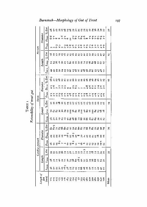
In a general survey of the gut musculature, a comparative study of thevarious thicknesses of the circular and longitudinal coats of a trout 180 mmlong was made. For this purpose transverse sections of different regions wereprepared by pumping up segments with fixative to a size which was approxi-mately midway between the fully extended and fully contracted (empty)dimensions (see table 1, p. 197). By means of preparations showing sections
Burnstock—Morphology of Gut of Trout 185
cut in three planes at right angles to one another, a detailed examination ofthe mechanical arrangement of the sphincters and of the regional junctionswas possible.
For examination of the form and arrangement of individual muscle-fibres,the tissue was either examined fresh, after maceration in nitric acid, or instained sections of material fixed in various ways. In the study of the limits ofextensibility of the gut, 20 trout which had been starved for about 3 weekswere killed by severing the brain from the spinal cord. The gut was removedand the 'empty', fully contracted lengths and diameters of the cardiac stomach,ileum, and rectum were recorded, together with the total length of the fishmeasured from the tip of the snout to the extreme tip of the tail fin. Thedimensions of the fully extended gut regions were examined by tying the gutbehind the pyloric sphincter and at the 'ileo-duodenal' junction, and pump-ing up the two regions to bursting point with a strong pipette containingo-8% saline solution. The relaxed and extended diameters were measured withcallipers.
For gross comparison of the extensibility of the gut of teleostean carnivoreswhich do not possess a stratum compactum, the pike and perch were treatedin a similar manner. Measurements were also taken of the extensibility of thestomach of the char, a salmonid fish possessing a stratum compactum, butfeeding on plankton.
Stained stratum compactum has the appearance of dense collagen and sincehigh-angle diffraction patterns are a useful and direct way of identifyingcollagen (Randall, 1953), this method was employed to establish the fact.The stratum compactum was isolated from the gut by carefully dissecting offthe other layers in o-8% saline solution. The sample of stratum compactumto be X-rayed was dried and held in a frame so that it was slightly stretched.The final diffraction pattern was registered as a photographic film.
A method was devised for measuring the percentage stretch of the isolatedstratum compactum. Wet strips, 5 mm wide but of variable length, weresuspended by a spring paper clip and stretched by means of weights untilthey ruptured. The difference between the initial and final lengths gave ameasure of the extensibility of the tissue, providing an interesting com-parison with the extensibility figures of the intact gut-wall itself.
RESULTS
General structure of the gut
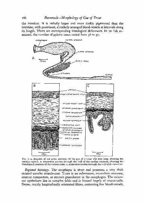
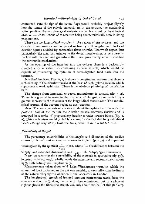
Gross anatomy. Fig. 1, A shows the gross anatomy of the trout gut. Ashort muscular oesophagus opens into the large anterior cardiac portion ofthe stomach with its prominent longitudinal folds. A shorter, more compact,cylindrical pyloric limb opens through a powerful pyloric sphincter into theintestine. The intestine has been divided for the sake of convenience into'duodenal' and 'ileal' portions, the 'duodenal-ileal' junction being defined asthe point beyond which no pyloric caeca appear. There is no histological dis-tinction between these regions. The rectum, however, is clearly defined from
186 Burnstock—Morphology of Gut of Trout
the intestine. It is radially larger and more darkly pigmented than theintestine, with prominent, circularly arranged blood-vessels at intervals alongits length. There are corresponding histological differences. In 20 fish ex-amined, the number of pyloric caeca varied from 36 to 57.
oesophagus cardiac stomach
pyloric stomach
SOJJ,
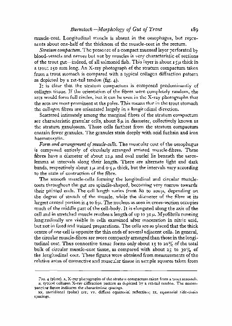
FIG. 1. A, diagram of the gross anatomy of the gut of a trout 180 mm long, showing thevarious regions. B, transverse section through the wall of the cardiac stomach, showing thehistological relations of the various coats, c, transverse section through the wall of the intestine.
Regional histology. The oesophagus is short and possesses a very thickstriated circular muscle-coat. There is no submucosa, muscularis mucosae,stratum compactum, or stratum granulosum in the oesophagus. The colum-nar epithelium lies in complex folds and is formed largely of mucus-cells.Dense, mainly longitudinally orientated fibres, containing few blood-vessels,
Burnstock—Morphology of Gut of Trout 187
form the sub-epithelial connective tissue-layer. A prominent subserous regioncontaining numerous blood-vessels is characteristic of this region.
The stomach wall (fig. 1, B) possesses all four coats characteristically foundin a vertebrate gut, but is distinguished by the extensive differentiation of themucosa. The serous coat consists of a single layer of plate-like epithelialcells. The circular muscle-coat is more than twice as thick as the longitudinalcoat and contains anteriorly some striated fibre-bundles extending from theoesophagus. An irregular connective tissue region, containing blood-vesselsand muscle-fibres, represents the submucosa and lies next to the outer layerof the mucosa. This consists of a muscularis mucosae, which contains bothcircular and longitudinal fibres as well as free fibre-bundles distributed irregu-larly in the folds of the submucosa. A prominent specialized layer of densecollagen known as the stratum compactum characterizes the mucosa. It isperforated by blood-vessels and nerves, but not by muscles. The stratumgranulosum consists of specialized granular cells, which are interposed betweenthe more diffuse marginal fibres of the stratum compactum and are moredensely packed on the side closest to the muscularis mucosae. The tunicapropria is a thick layer of compact areolar connective tissue, swollen withmany gastric glands and containing blood-vessels and nerves. Columnarepithelial cells line the lumen and lie in prominent longitudinal and circularfolds.
The intestine differs from the stomach principally by the absence of sub-mucosa and muscularis mucosae (fig. 1, c). A layer of serosal cells lies outsidethe muscle-coats, the circular layer being about twice as thick as the longi-tudinal layer. The stratum compactum and granulosum are very prominent.The tunica propria contains no glands and is bound by a much-folded mucosalepithelium containing many mucus-cells. The 'duodenal' portion of the intes-tine is characterized by the presence of diverticula, the pyloric caeca, whichappear to be histologically identical with the intestine.
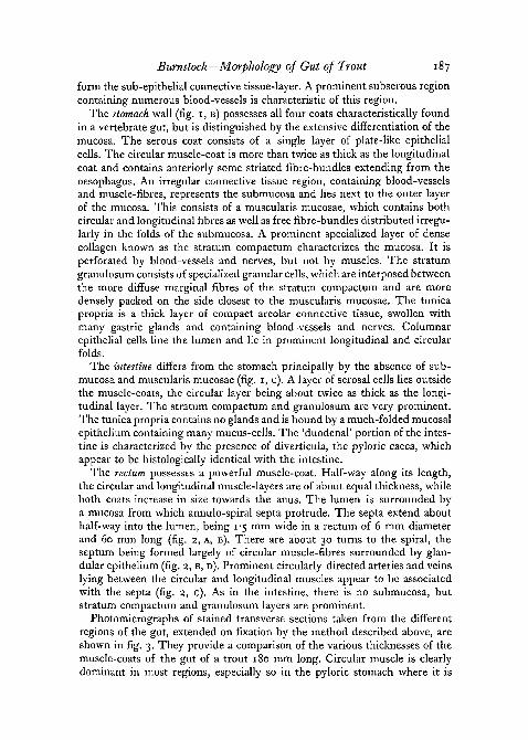
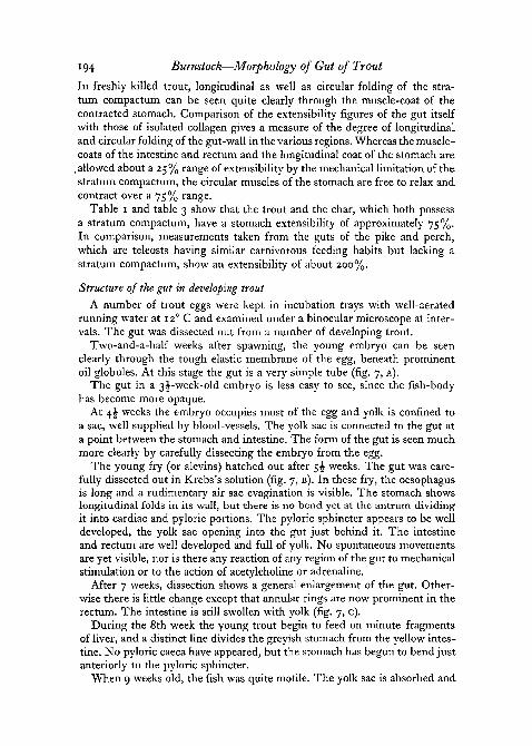
The rectum possesses a powerful muscle-coat. Half-way along its length,the circular and longitudinal muscle-layers are of about equal thickness, whileboth coats increase in size towards the anus. The lumen is surrounded bya mucosa from which annulo-spiral septa protrude. The septa extend abouthalf-way into the lumen, being 1-5 mm wide in a rectum of 6 mm diameterand 60 mm long (fig. 2, A, B). There are about 30 turns to the spiral, theseptum being formed largely of circular muscle-fibres surrounded by glan-dular epithelium (fig. 2, B, D). Prominent circularly directed arteries and veinslying between the circular and longitudinal muscles appear to be associatedwith the septa (fig. 2, c). As in the intestine, there is no submucosa, butstratum compactum and granulosum layers are prominent.
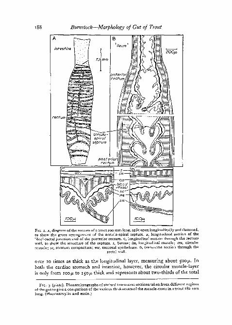
Photomicrographs of stained transverse sections taken from the differentregions of the gut, extended on fixation by the method described above, areshown in fig. 3. They provide a comparison of the various thicknesses of themuscle-coats of the gut of a trout 180 mm long. Circular muscle is clearlydominant in most regions, especially so in the pyloric stomach where it is
188 Burnstock—Morphology of Gut of Trout
infesHna
rectum
200jj.
FIG. 2. A, diagram of the rectum of a trout 200 mm long, split open longitudinally and flattened,to show the gross arrangement of the annulo-spiral septum. B, longitudinal section of the'ileo'-rectal junction and of the posterior rectum. C, longitudinal section through the rectumwall, to show the structure of the septum, s, Serosa; Im, longitudinal muscle; cm, circularmuscle; sc, stratum compactum; me, mucosal epithelium. D, transverse section through the
rectal wall.
over 10 times as thick as the longitudinal layer, measuring about 500 ju. Inboth the cardiac stomach and intestine, however, the circular muscle-layeris only from 100/x. to 150/x thick and represents about two-thirds of the total
FIG. 3 (plate). Photomicrographs of stained transverse sections taken from different regionsof the gut to give a comparison of the various thicknesses of the muscle-coats in a trout 180 mmlong. (Haematoxylin and eosin.)
py/ori'c stomach
oesophagus
T£ — cardiac stoma<
IOOJJ.
FIG. 3
G. BURNSTOCK
If it0-9
-215-
3- 65
Fie. 4
G. BURNSTOCK
characteristic kpacings
Burnstock—Morphology of Gut of Trout 189
muscle-coat. Longitudinal muscle is absent in the oesophagus, but repre-sents about one-half of the thickness of the muscle-coat in the rectum.
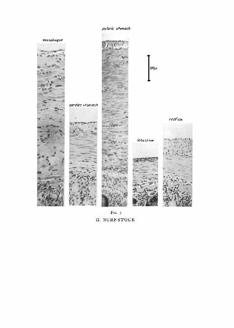
Stratum compactum. The presence of a compact mucosal layer perforated byblood-vessels and nerves but not by muscles is very characteristic of sectionsof the trout gut—indeed, of all salmonid fish. This layer is about 15 /u. thick ina trout 150 mm long. An X-ray photograph of the stratum compactum takenfrom a trout stomach is compared with a typical collagen diffraction patternas depicted by a rat-tail tendon (fig. 4).
It is clear that the stratum compactum is composed predominantly ofcollagen tissue. If the orientation of the fibres were completely random, thearcs would form full circles, but it can be seen in the X-ray photographs thatthe arcs are most prominent at the poles. This means that in the trout stomachthe collagen fibres are orientated largely in a longitudinal direction.
Scattered intimately among the marginal fibres of the stratum compactumare characteristic granular cells, about 8 ju, in diameter, collectively known asthe stratum ganulosum. Those cells farthest from the stratum compactumcontain fewer granules. The granules stain deeply with acid fuchsin and ironhaematoxylin.
Form and arrangement of muscle-cells. The muscular coat of the oesophagusis composed entirely of circularly arranged striated muscle-fibres. Thesefibres have a diameter of about 12/u. and oval nuclei lie beneath the sarco-lemma at intervals along their length. There are alternate light and darkbands, respectively about 1 ju. and 0-5 /J, thick, but the intervals vary accordingto the state of contraction of the fibre.
The smooth muscle-cells forming the longitudinal and circular muscle-coats throughout the gut are spindle-shaped, becoming very narrow towardstheir pointed ends. The cell length varies from 80 to 200 fi, depending onthe degree of stretch of the muscle, while the diameter of the fibre at itslargest central portion is 4 to 6/x. The nucleus as seen in cross-section occupiesmuch of the middle part of the cell-body. It is elongated along the axis of thecell and in stretched muscle reaches a length of up to 30 JU.. Myofibrils runninglongitudinally are visible in cells examined after maceration in nitric acid,but not in fixed and stained preparations. The cells are so placed that the thickcentre of one cell is opposite the thin ends of several adjacent cells. In general,the circular muscle-fibres are more compactly arranged than those in the longi-tudinal coat. Thus connective tissue forms only about 15 to 20% of the totalbulk of circular muscle-coat tissue, as compared with about 25 to 30% ofthe longitudinal coat. These figures were obtained from measurements of therelative areas of connective and muscular tissue in sample squares taken from
FIG. 4 (plate). A, X-ray photographs of the stratum compactum taken from a trout stomach.B, typical collagen X-ray diffraction pattern as depicted by a rat-tail tendon. The accom-
panying figure indicates the characteristic spacings.xx, meridional (polar) arc; YY, diffuse equatorial reflection; zz, equatorial side-chain
spacings.
190 Burnstock—Morphology of Gut of Trout
different regions. Throughout the gut the muscle-fibres are bound together insmall bundles by collagen. Although the fibres within each bundle lie alongthe same plane, the bundles themselves are often slightly, and sometimesgrossly, out of line with each other. Sometimes several small bundles areenclosed in a larger, thicker collagen sheath, particularly in the region ofsphincters.
Regional junctions and sphincters.
The following parts of the gut show structural characteristics which differfrom the regional histology described above.
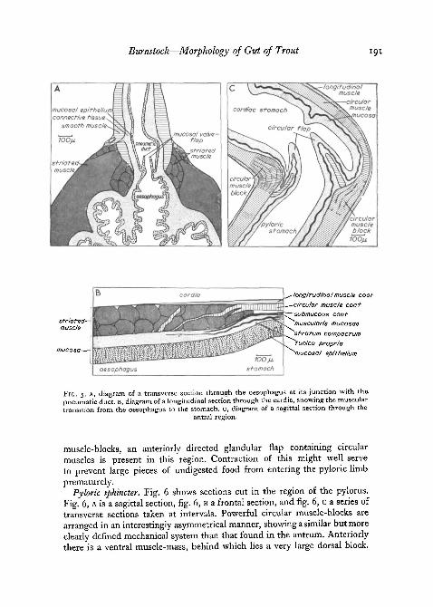
Pneumatic duct. Salmonid fish are physostomatous. Fig. 5, A shows thejunction of the pneumatic duct with the oesophagus. The diagram shows asection cut transversely through the oesophagus, so that the pneumatic ductappears in longitudinal section. The duct itself is surrounded by smoothcircular muscle, while at its origin in the oesophagus, blocks of striated musclehave become reorientated to form a sphincter-like base to the duct. A non-muscular, valve-like, epithelial flap protrudes up into the lumen of the ductin this region and, together with the contraction of the basal striated muscle,presumably serves to prevent gas escaping from the swim bladder.
Cardia. The arrangement of the muscles at the junction of the oesophagusand stomach can be seen in fig. 5, B, which is a longitudinal section cutthrough the wall of this region. There is clearly no sphincter, but the regionshows a marked morphological transition. The prominent striated circularmuscle-blocks of the oesophagus give way to a narrower layer of smoothcircular muscle in the stomach. The longitudinal muscle-coat makes its firstappearance at the cardia together with the stratum compactum, stratumgranulosum, and muscularis mucosae. The tunica propria is composed of verydensely packed connective tissue. In the living system, peristaltic waves passdown the stomach from this region; no movements pass beyond the cardia inthe orally directed vomiting reflex.
Antrum. Fig. 5, c shows a sagittal section of the antral region. A divisioninto cardiac and pyloric regions of the stomach, though well marked ana-tomically, especially in the extended stomach, is well defined histologically.Large blocks of circular muscle lie at the apex of the inner bend of the stomachand just posterior to the curvature in the outer bend. The orientation of themuscle-fibres shows that their action is not directly antagonistic but ratherthat they act across each other, so that the inner block tends to pull in an-teriorly to the outer one. This means that whereas the contraction of the innerblock acts in a plane along the radius of curvature of the bend, the outermuscle pulls through a line at right angles to the longitudinal plane of thepyloric stomach. These morphological findings are in fact consistent withthe frequent occurrence of constrictions in exactly these positions, seen in theliving stomach. In fish showing the vomiting reaction, waves running orallyappear to originate in this region of the stomach. In addition to the circular
Burnstock—Morphology of Gid of Trout
longitudinal muscle coot•circular muscle cootsubmucous coot"muscu/aris mucosae'stratum compactum'tunica propria'mucosal epithelium
FIG. 5. A, diagram of a transverse section through the oesophagus at its junction with the.pneumatic duct. B, diagram of a longitudinal section through the cardia, showing the musculartransition from the oesophagus to the stomach, c, diagram of a sagittal section through the
antral region.
muscle-blocks, an anteriorly directed glandular flap containing circularmuscles is present in this region. Contraction of this might well serveto prevent large pieces of undigested food from entering the pyloric limbprematurely.
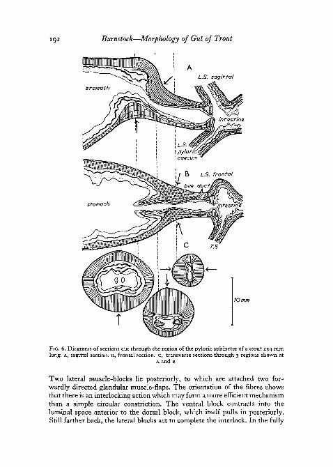
Pyloric sphincter. Fig. 6 shows sections cut in the region of the pylorus.Fig. 6, A is a sagittal section, fig. 6, B a frontal section, and fig. 6, c a series oftransverse sections taken at intervals. Powerful circular muscle-blocks arearranged in an interestingly asymmetrical manner, showing a similar but moreclearly defined mechanical system than that found in the antrum. Anteriorlythere is a ventral muscle-mass, behind which lies a very large dorsal block.
192 Burnstock—Morphology of Gut of Trout
10 mm
FIG. 6. Diagrams of sections cut through the region of the pyloric sphincter of a trout 254 mmlong. A, sagittal section. B, frontal section. C, transverse sections through 3 regions shown at
A and B.
Two lateral muscle-blocks lie posteriorly, to which are attached two for-wardly directed glandular muscle-flaps. The orientation of the fibres showsthat there is an interlocking action which may form a more efficient mechanismthan a simple circular constriction. The ventral block contracts into theluminal space anterior to the dorsal block, which itself pulls in posteriorly.Still farther back, the lateral blocks act to complete the interlock. In the fully
Burnstock—Morphology of Gut of Trout 193
contracted state the tips of the lateral flaps would probably project slightlyinto the lumen of the pyloric stomach. As in the antrum, the mechanicalaction predicted by morphological analysis is in fact borne out by physiologicalobservation, constrictions of this nature being characteristically seen in livingpreparations.
There are no longitudinal muscles in the region of the pylorus, and thecircular muscle-masses are composed of from 4 to 8 longitudinal blocks ofcircular figures divided by connective-tissue sheaths. The whole region, butparticularly the area just anterior to the dorsal muscle-mass, is very heavilypacked with collagen and granular cells. These presumably serve to stabilizethe contractile mechanism.
At the opening of the intestine into the pylorus there is a backwardlydirected circular valve flap containing circular muscle, which would becapable of preventing regurgitation of semi-digested food back into thestomach.
Intestinal junctions. Figs. 6, A, B shows in longitudinal section that there isa thickening of the circular muscle at the base of each pyloric caecum whichrepresents a weak sphincter. There is no obvious physiological correlationhere.
The change from intestinal to rectal musculature is gradual (fig. 3, B).There is a general increase in the diameter of the gut, accompanied by agradual increase in the thickness of the longitudinal muscle-coat. The annulo-spiral septum of the rectum begins at this junction.
Anus. The anus consists of a series of about five sphincters. Towards theposterior end of the rectum the circular muscle becomes thicker and isarranged in a series of progressively heavier circular muscle-blocks (fig. 3,B). This mechanism would probably account for the fact that long cylindricalfaeces emerge very slowly from the anus, rather than in a sudden rush.
Extensibility of the gut
The percentage extensibilities of the lengths and diameters of the cardiacstomach, 'ileum', and rectum are shown in table 1 (p. 197) and represent
values given by the quotient X ioo, where / = the difference between the
'empty' and extended dimensions and Lmin = the 'empty 'gut dimensions.It can be seen that the extensibility of the stomach is approximately 35%
longitudinally and 79% radially, while the intestine and rectum stretch about25% both radially and longitudinally.
Measurements taken from wild Lake Windermere trout, in which theamount of food contained in the gut was variable, always fell within the limitsof the extensibility figures obtained in the laboratory in London.
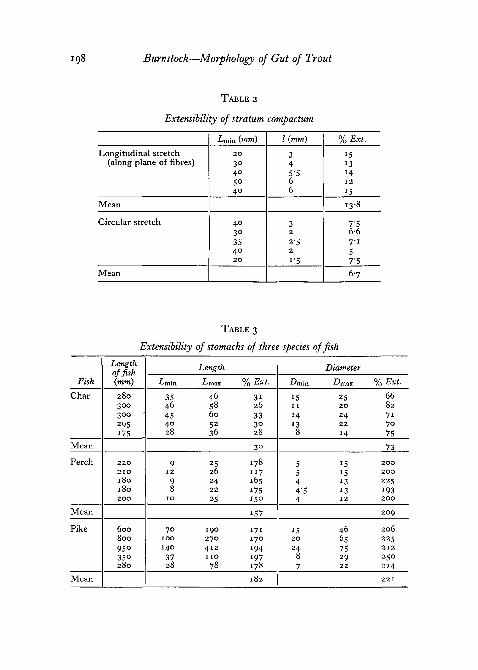
The longitudinal stretch of isolated stratum compactum taken from thestomach is about 14% along the plane of fibre orientation, but in a plane atright angles to the fibres the stretch was only about one-half of this (table 2).
194 Burnstock—Morphology of Gut of Trout
In freshly killed trout, longitudinal as well as circular folding of the stra-tum compactum can be seen quite clearly through the muscle-coat of thecontracted stomach. Comparison of the extensibility figures of the gut itselfwith those of isolated collagen gives a measure of the degree of longitudinaLand circular folding of the gut-wall in the various regions. Whereas the muscle-coats of the intestine and rectum and the longitudinal coat of the stomach are
, allowed about a 25% range of extensibility by the mechanical limitation of thestratum compactum, the circular muscles of the stomach are free to relax andcontract over a 75% range.
Table 1 and table 3 show that the trout and the char, which both possessa stratum compactum, have a stomach extensibility of approximately 75%.In comparison, measurements taken from the guts of the pike and perch,which are teleosts having similar carnivorous feeding habits but lacking astratum compactum, show an extensibility of about 200%.
Structure of the gut in developing trout
A number of trout eggs were kept in incubation trays with well-aeratedrunning water at 120 C and examined under a binocular microscope at inter-vals. The gut was dissected out from a number of developing trout.
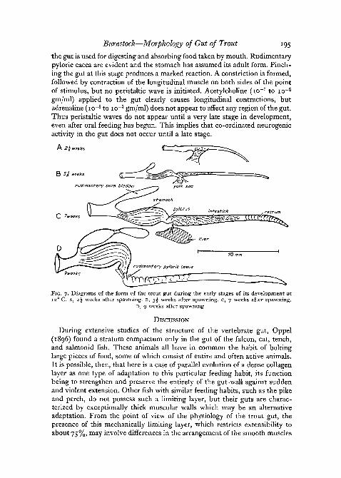
Two-and-a-half weeks after spawning, the young embryo can be seenclearly through the tough elastic membrane of the egg, beneath prominentoil globules. At this stage the gut is a very simple tube (fig. 7, A).
The gut in a 3 ̂ -week-old embryo is less easy to see, since the fish-bodyhas become more opaque.
At 4J weeks the embryo occupies most of the egg and yolk is confined to-a sac, well supplied by blood-vessels. The yolk sac is connected to the gut ata point between the stomach and intestine. The form of the gut is seen muchmore clearly by carefully dissecting the embryo from the egg.
The young fry (or alevins) hatched out after 5^ weeks. The gut was care-fully dissected out in Krebs's solution (fig. 7, B). In these fry, the oesophagusis long and a rudimentary air sac evagination is visible. The stomach showslongitudinal folds in its wall, but there is no bend yet at the antrum dividingit into cardiac and pyloric portions. The pyloric sphincter appears to be welldeveloped, the yolk sac opening into the gut just behind it. The intestineand rectum are well developed and full of yolk. No spontaneous movementsare yet visible, nor is there any reaction of any region of the gut to mechanicalstimulation or to the action of acetylcholine or adrenaline.
After 7 weeks, dissection shows a general enlargement of the gut. Other-wise there is little change except that annular rings are now prominent in therectum. The intestine is still swollen with yolk (fig. 7, c).
During the 8th week the young trout begin to feed on minute fragmentsof liver, and a distinct line divides the greyish stomach from the yellow intes-tine. No pyloric caeca have appeared, but the stomach has begun to bend justanteriorly to the pyloric sphincter.
When 9 weeks old, the fish was quite motile. The yolk sac is absorbed and
Burnstock—Morphology of Gut of Trout 195
the gut is used for digesting and absorbing food taken by mouth. Rudimentarypyloric caeca are evident and the stomach has assumed its adult form. Pinch-ing the gut at this stage produces a marked reaction. A constriction is formed,followed by contraction of the longitudinal muscle on both sides of the pointof stimulus, but no peristaltic wave is initiated. Acetylcholine (io~7 to io~5
gm/ml) applied to the gut clearly causes longitudinal contractions, butadrenaline (io~8 to io~5 gm/ml) does not appear to affect any region of the gut.Thus peristaltic waves do not appear until a very late stage in development,even after oral feeding has begun. This implies that co-ordinated neurogenicactivity in the gut does not occur until a late stage.
FIG. 7. Diagrams of the form of the trout gut during the early stages of its development at12° C. A, 2^ weeks after spawning, B, $\ weeks after spawning, c, 7 weeks after spawning.
D, 9 weeks after spawning.
DISCUSSION
During extensive studies of the structure of the vertebrate gut, Oppel{1896) found a stratum compactum only in the gut of the falcon, cat, tench,and salmonid fish. These animals all have in common the habit of boltinglarge pieces of food, some of which consist of entire and often active animals.It is possible, then, that here is a case of parallel evolution of a dense collagenlayer as one type of adaptation to this particular feeding habit, its functionbeing to strengthen and preserve the entirety of the gut-wall against suddenand violent extension. Other fish with similar feeding habits, such as the pikeand perch, do not possess such a limiting layer, but their guts are charac-terized by exceptionally thick muscular walls which may be an alternativeadaptation. From the point of view of the physiology of the trout gut, thepresence of this mechanically limiting layer, which restricts extensibility toabout 75%, may involve differences in the arrangement of the smooth muscles
196 Burnstock—Morphology of Gut of Trout
and possibly of their innervation, as compared to that of the pike and perchwhere the muscle is free to extend up to 200%. Studies of the orientation ofthe fibres in relation to the collagen lattices upon which they pull is desirable.
There has been a considerable controversy as to the function of the cellsof the stratum granulosum. Greene suggested that 'their function is one ofinternal secretion', while Gulland (1898) described these cells as 'largeeosinophile leucocytes'. A more likely explanation is that the stratum granu-losum is composed of active fibroblast cells. Fibroblast cells are almost cer-tainly responsible for the formation of collagen fibrils and while engaged inthis activity, fine granules appear in the cytoplasm (Randall, 1953). Further,their position in the gut-wall scattered among the marginal fibres of the col-lagenous stratum compactum supports this hypothesis.
The pyloric sphincter is very powerful and appears to function by meansof interacting circular muscle-blocks, rather than by a single circular closure.Anteriorly-directed lateral processes containing some circular muscles are alsopresent. A reduced but similar arrangement of muscle-blocks and processesexists in the antrum. No separate extrinsic innervation of these sphinctermuscles has been discovered.
A muscular annulo-spiral septum protruding into the lumen of the rectumhas been described. Further study will be necessary to discover whether thisstructure has a defaecatory, osmo-regulatory, or even respiratory function.
This work consists of part of a Ph.D. thesis carried out under the supervisionof Dr. M. E. Brown and Professor J. Z. Young. I would like to thank Mr.North (Biophysics Unit) for taking X-ray photographs of the stratum com-pactum, and the Department of Scientific and Industrial Research for thegrant which allowed this work to be carried out. Fig. 1, A, B has been repro-duced from the Journal of Physiology by kind permission of the EditorialBoard.
REFERENCESAGASSIZ, L. R., and VOCT, C. C, 1845. Anatomie des Salmones. NeuchStel (Wolfrath).BARTON, J. K., 1900. J. Anat. Lond., 34, 295.BURNSTOCK, G., 1957. Nature, 180, 1491.
1958a. J. Physiol., 141, 35.19586. Brit. J. Pharmacol., 13, 216.'959' Quart. J. micr. Sci., 100, 199.
CAJETAN, J., 1883. Inaug. Diss. Bonn.GREENE, C. W., 1912. Bull. U.S. Bur. Fish, 32, 73.GULLAND, L., 1898. Anat. Anz., 14, 16.OPPEL, A., 1896. Lehrbuch. vergl. mikr. Anat. Wirbelt., Bd. i , 70; Bd. 2, 7.RANDALL, J. T., 1953. Nature and structure of collagen. London (Academic Press).VALATOUR, M., iSSr. Ana. S:i. Nat. ZODL, I 6 , 219.WEINREB, E. L., and BILSTAD, N. M., 1955. Copeia, 194.
TA
BL
E I
Exte
nsi
bil
ity o
f tr
out-
gut
Leng
th o
ffis
h(m
m)
20
4
21
4
275
175
14
01
06
'83
32
5
15
42
74 97 373
167
28
0
27
03
20
26
02
30
24
0
12
0
Mea
n
Lm
; n
36 40 54 3° 25 16
538 55 2
9 45 14 66 26 36 40 55 51
42
41 23
Car
diac
st
omac
h
Leng
th
imax
%
48 54 74 40 34 22 5° 75 38 60 18
59
0 35 5° 54 76 68 57 55 3i
Ext
.
33 35 37 33 36 33 32 36 3i 33 32 37 34-5
39 35 38 33 35 34 35 35
Ani
n
16 13 14 8-5
7 5T
O 13 8-5
11 5 16 8
15 13 13 12
10
10
5
55
Dia
met
e
Ana
x
29
24
24 14
13 9 18
525 1
6s
20 8 3° 25
22
24
21 18
20
10
%E
xt.
81 84
-57
i 65 86 80 85 92 94 82 60 87 75 66 70 85 75 80
90 69 79
56 52
70
25
24 19 82
28 66 19 88 32 66 59 83 69 54 55 23
Leng
th
imax
%
70 65 85 32
3O 24
10
0 36 81
24
no 40
80 76
10
0 84 65 68 28
Ileu
m
Ext
.
25 25 21 28
25 25 22
27
23
25 25 25 23
27 22
22
20
23
21 24
An
ir
3"5
4"2 5-5
2- 9 2-8
2-7
4-8
2- 9
5- i
2- 4
4-
i
2'5 3-8
3-7
4-9
4*9
4-3
4"4
2-6
Dia
met
er
Ana
x
4'5
5-6
7 3-7
3-5
3"5
6 3-5
6-5
3 5 3 4- 8 4-6
6-2
6- i 5'4
5-6
3-2
% E
xt.
28.5
33
27
28
25
29
25
20
27
25
22
20
26
24
26
24
26
27
23
26
imin
42 45 59 32
24
23 66 23 58 16 59 28 58 59 66 55 47 48 22
Len
gth
T 0/
•^m
ax
/o
54 55 76 40
30
29 80
29
72
20 75 34 72 76 82 68 57 61
27
Rec
tum
Ext
.
28 22 27 25 25 26 23 26 24
25 27
21 24 27 24
23 21
27 23 25
An
in
5 5 6-3
4-5
3 4"3
4-8
7-8
4-6
6-3
4- 2 6-8
4-2
6-3
5-4
7-6
5-2
4-7
49
2- 9
Dia
met
er
Ana
x
6-8
7- 1 8 6 3-8
5-4
61
0 5-9
82
5'4
8-4
5-6
8 7-4
9"4
7-1
6-3
6-4
37
% E
xt.
28
40
27
33
27
26
25 28 35 3° 29
23 33 27 37 24 36 34 3°
27 3°
Burnstock—Morphology of Gut of Trout
TABLE 2
Extensibility of stratum compactum
Longitudinal stretch(along plane of fibres)
Mean
Circular stretch
Mean
Lmin (mm)
2 0
3O405O40
40
3O35402 0
/ (mm)
345-566
32
2 52
i-5
% .Ex*.
i s13H1 2
IS
1 3 8
7"56-67 1
57'5
6 7
TABLE 3
Extensibility of stomachs of three species of fish
Fish
Char
Mean
Perch
Mean
Pike
Mean
Lengthoffish(mm)
2 8 03 0 0300 '29S175
2 2 0
2 1 0
1 8 01 8 02 0 0
6 0 08 0 0
95°35°2 8 0
•t-min
3546
454028
91 2
98
1 0
701 0 0
1 4 0
3728
Length
-Crnax
4658605236
2526242 2
2 5
I 9 O27O4 1 2n o
78
% Ext.
3i2633
3°28
3°
1781 1 71651751 5 0
157
1711 7 0194197178
1 8 2
•Dmin
15I I
1 413
8
5544'54
152 0
2487
Diameter
-Dmax
252 0
242 2
1 4
151513131 2
466575292 2
% £*«.
6682717075
73
2 0 0
2 0 0
2 2 5
1932 0 0
2 0 9
2 0 62 2 52 1 2
2 5 02 1 4
2 2 1