Embed Size (px)
Citation preview
26
The Plant–Type Ferredoxin-NADP+ Reductases
Matías A. Musumeci, Eduardo A. Ceccarelli and Daniela L. Catalano-Dupuy
Instituto de Biología Molecular y Celular de Rosario (IBR), CONICET, Facultad de Ciencias Bioquímicas y Farmacéuticas, Universidad Nacional de Rosario
Argentina
1. Introduction
Ferredoxin-NADP+ reductases (FNRs, EC 1.18.1.2) constitute a family of hydrophilic, monomeric enzymes that contain non-covalently bound FAD as prosthetic group. These flavoenzymes deliver NADPH or low potential one-electron donors (ferredoxin, flavodoxin, adrenodoxin) to redox-based metabolisms in plastids, mitochondria and bacteria. The main physiological role of the chloroplast FNR is to catalyze the final step of photosynthetic electron transport, namely, the electron transfer from the ferredoxin (Fd), reduced by photosystem I, to NADP+ (Eqn. 1) (Shin & Arnon, 1965). This reaction provides the NADPH necessary for CO2 assimilation in plants and cyanobacteria. FNRs also participate in others electron transfer metabolic processes as nitrogen fixation, isoprenoid biosynthesis, steroid metabolism, xenobiotic detoxification, oxidative-stress response and iron-sulfur cluster biogenesis (Carrillo & Ceccarelli, 2003, Ceccarelli et al., 2004, Medina & Gomez-Moreno, 2004, Rohrich et al., 2005, Seeber et al., 2005). Eqn. 1 represents the electron flow through FNR as it occurs in the photosynthetic electron chain. However, the physiological direction of the reaction catalyzed by FNRs involved in the other pathways is opposite, i.e. toward the production of reduced Fd. On this basis, FNRs are sometimes classified as autotrophic (photosynthetic FNRs) and heterotrophic (all other FNRs) (Aliverti et al., 2008, Arakaki et al., 1997).
2 Fd(Fe2+) + NADP+ + H+ ↔ 2 Fd (Fe3+) + NADPH (1)
Some bacteria and algae posses the FMN-containing flavodoxin (Fld), that is able to efficiently replace Fd as the electron partner of FNR in different metabolic routes, including photosynthesis (Razquin et al., 1996). In cyanobacteria, Fld expression is induced under condition of iron deficit, when the [2Fe-2S] cluster of Fd cannot be assembled (Razquin et al., 1996). In other prokaryotes, flavodoxins are constitutively expressed, or induced by oxidants (Zheng et al., 1999). Both Fld and FNR participate in the detoxification of reactive oxygen species in aerobic and facultative bacteria (Krapp et al., 2002, Zheng et al., 1999). The FNR displays strong preference for NADP(H) and is a very poor NAD(H) oxidoreductase. In contrast, various redox compounds, including complexed metals and aromatic molecules, can replace Fd or Fld as electron acceptors in vitro, in the so-called diaphorase activity (Avron & Jagendorf, 1956). All FNR-mediated reactions can thus be
www.intechopen.com
Advances in Photosynthesis – Fundamental Aspects
540
interpreted consisting of two-steps: hydride exchange with pyridine nucleotide (Eqn. 2) and electron transfer to and from the other partner (Eqn. 3).
NADPH + H+ + FNRox ↔ NADP+ + FNRH2 (2)
FNRH2 + nAox → FNRox + nAred (3)
A, electron acceptor n = 1, 2
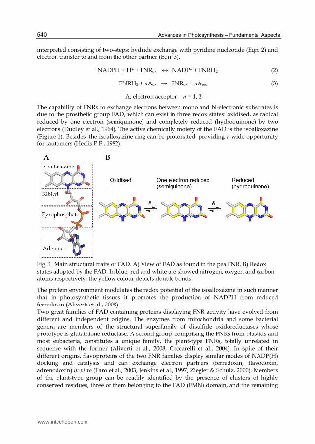
The capability of FNRs to exchange electrons between mono and bi-electronic substrates is due to the prosthetic group FAD, which can exist in three redox states: oxidised, as radical reduced by one electron (semiquinone) and completely reduced (hydroquinone) by two electrons (Dudley et al., 1964). The active chemically moiety of the FAD is the isoalloxazine (Figure 1). Besides, the isoalloxazine ring can be protonated, providing a wide opportunity for tautomers (Heelis P.F., 1982).
Fig. 1. Main structural traits of FAD. A) View of FAD as found in the pea FNR. B) Redox states adopted by the FAD. In blue, red and white are showed nitrogen, oxygen and carbon atoms respectively; the yellow colour depicts double bonds.
The protein environment modulates the redox potential of the isoalloxazine in such manner that in photosynthetic tissues it promotes the production of NADPH from reduced ferredoxin (Aliverti et al., 2008). Two great families of FAD containing proteins displaying FNR activity have evolved from different and independent origins. The enzymes from mitochondria and some bacterial genera are members of the structural superfamily of disulfide oxidoreductases whose prototype is glutathione reductase. A second group, comprising the FNRs from plastids and most eubacteria, constitutes a unique family, the plant-type FNRs, totally unrelated in sequence with the former (Aliverti et al., 2008, Ceccarelli et al., 2004). In spite of their different origins, flavoproteins of the two FNR families display similar modes of NADP(H) docking and catalysis and can exchange electron partners (ferredoxin, flavodoxin, adrenodoxin) in vitro (Faro et al., 2003, Jenkins et al., 1997, Ziegler & Schulz, 2000). Members of the plant-type group can be readily identified by the presence of clusters of highly conserved residues, three of them belonging to the FAD (FMN) domain, and the remaining
www.intechopen.com
The Plant–Type Ferredoxin-NADP
+ Reductases
541
to the NADP(H) region (Arakaki et al., 1997, Bruns & Karplus, 1995, Ceccarelli et al., 2004, Karplus et al., 1991). This group can be further classified into a plastidic and a bacterial class, which differ not only in their sequences, but also in the environment of the active site, FAD conformations and catalytic efficiencies (Carrillo & Ceccarelli, 2003). Plastidic FNRs display high catalytic efficiencies (turnover numbers in the range 100-600 s-1), whereas bacterial reductases are much less active (Ceccarelli et al., 2004). In plants and cyanobacteria, optimization for FNR catalytic efficiency might be related to the demands of the photosynthetic process that requires a very fast electron flow to sustain CO2 fixation rates. In organisms growing on heterotrophic metabolisms or anoxygenic photosynthesis, FNR is involved in pathways that proceed at a much lower pace, acting as a shuttle between the abundant NAD(P)H pool and the low potential electron carriers. This chapter will focus on structural and functional aspects of the ferredoxin-NADP(H) reductase in association with the metabolic process of photosynthesis. Special attention will be pay to techniques and approaches that could help to appreciate the importance of the enzyme and to understand, conceive and/or execute research on FNR.
2. FNR in the photosynthetic electron transport
The generation of reducing power is crucial for all biosynthetic processes within chloroplasts. The main source of reduction equivalents is the light-driven photosynthesis. The FNR is a key enzyme of photosynthetic electron transport. FNR transfers electrons between the one-electron carrier ferredoxin and the two-electron carrier NADP(H) at the end of the photosynthetic electron transport chain. FNR also participates in others relevant processes as the electron cyclic flow around the photosystem I and in the control of the NADPH/NADP+ homeostasis of stressed chloroplasts (Palatnik et al., 1997). Photosynthetic electron flow is driven by two photochemical reactions catalyzed by photosystem II (PSII) and photosystem I (PSI), which are linked by the electron transport chain (Figure 2). Linear electron transport starts with the photo-induced water oxidation catalyzed by PSII. Electrons are transferred from PSII through the plastoquinone pool to cytochrome b6f. The electron transport is coupled to proton translocation into the thylakoid lumen, and the resulting pH gradient drives the ATP synthase to produce ATP. Next, electrons move from cytochrome b6f to the soluble electron carrier plastocyanin and then to PSI, which acts as light-driven plastocyanin ferredoxin oxidoreductase. Ultimately these electrons can be used by ferredoxin-NADP+ reductase to produce NADPH that together with the ATP generated by the ATP synthase will drive the Calvin cycle for CO2 assimilation (Rochaix, 2011). The primary function of PSI is to reduce NADP+ to NADPH, which is then used in the assimilation of CO2 (Setif, 2006, Vishniac & Ochoa, 1951). In plants, it occurs via reduction of the soluble [2Fe-2S] ferredoxin (Fd) by PSI. Subsequent reduction of NADP+ by Fdrd is catalyzed by FNR (Arakaki et al., 1997). In most cyanobacteria, and algae under low iron conditions, Fld, in particular Fldsq/Fldhq, substitutes for the Fdox/Fdrd pair in this reaction (Eqn. 4) (Bottin & Lagoutte, 1992, Medina & Gomez-Moreno, 2004). Two Fldsq molecules transfer two electrons from two PSI molecules to one FNR. FNR becomes fully reduced through formation of the intermediate, FNRsq, and later transfers both electrons simultaneously to NADP+ (Eqn. 5) (Medina, 2009). It was demonstrated that FNR is one of the rate-limiting steps in photosynthesis, and controls the balance between the demand for redox equivalents and photosynthetic activity
www.intechopen.com
Advances in Photosynthesis – Fundamental Aspects
542
under a wide range of environmental conditions (Hajirezaei et al., 2002). It gives to this enzyme relevance as a possible target for crop improvement and treatment of weeds.
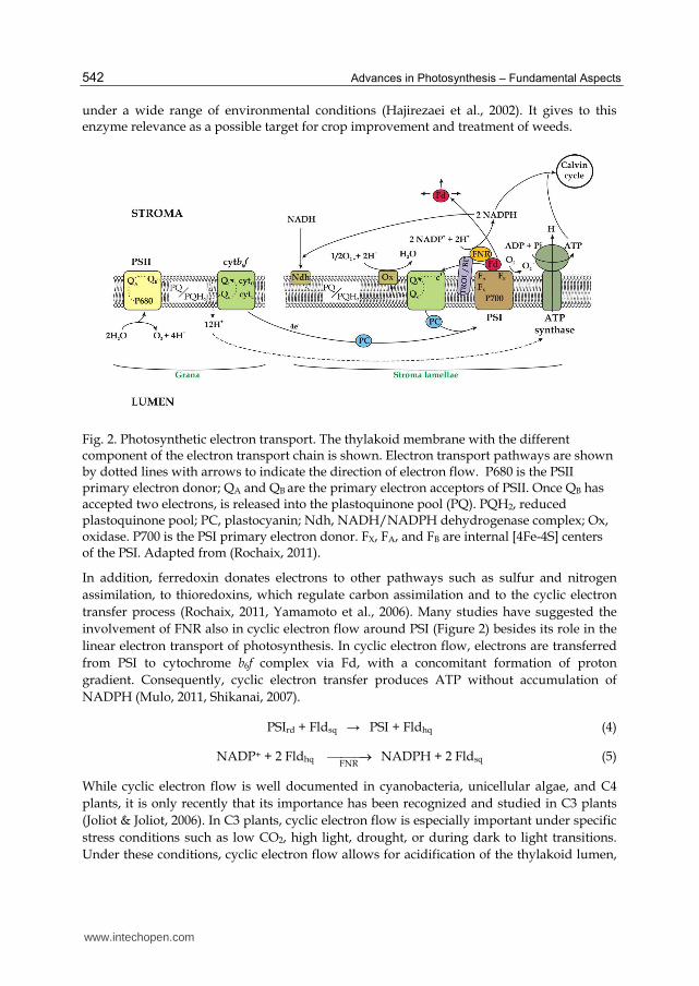
Fig. 2. Photosynthetic electron transport. The thylakoid membrane with the different component of the electron transport chain is shown. Electron transport pathways are shown by dotted lines with arrows to indicate the direction of electron flow. P680 is the PSII primary electron donor; QA and QB are the primary electron acceptors of PSII. Once QB has accepted two electrons, is released into the plastoquinone pool (PQ). PQH2, reduced plastoquinone pool; PC, plastocyanin; Ndh, NADH/NADPH dehydrogenase complex; Ox, oxidase. P700 is the PSI primary electron donor. FX, FA, and FB are internal [4Fe-4S] centers of the PSI. Adapted from (Rochaix, 2011).
In addition, ferredoxin donates electrons to other pathways such as sulfur and nitrogen assimilation, to thioredoxins, which regulate carbon assimilation and to the cyclic electron transfer process (Rochaix, 2011, Yamamoto et al., 2006). Many studies have suggested the involvement of FNR also in cyclic electron flow around PSI (Figure 2) besides its role in the linear electron transport of photosynthesis. In cyclic electron flow, electrons are transferred from PSI to cytochrome b6f complex via Fd, with a concomitant formation of proton gradient. Consequently, cyclic electron transfer produces ATP without accumulation of NADPH (Mulo, 2011, Shikanai, 2007).
PSIrd + Fldsq → PSI + Fldhq (4)
NADP+ + 2 Fldhq FNR
NADPH + 2 Fldsq (5)
While cyclic electron flow is well documented in cyanobacteria, unicellular algae, and C4 plants, it is only recently that its importance has been recognized and studied in C3 plants (Joliot & Joliot, 2006). In C3 plants, cyclic electron flow is especially important under specific stress conditions such as low CO2, high light, drought, or during dark to light transitions. Under these conditions, cyclic electron flow allows for acidification of the thylakoid lumen,
www.intechopen.com
The Plant–Type Ferredoxin-NADP
+ Reductases
543
which is required for inducing ATP synthesis and for triggering non-photochemical quenching, which in turn down-regulates PSII. Cyclic electron flow is also important when photorespiration is active because more ATP is required for CO2 fixation under these conditions (Osmond, 1981). Several routes for cyclic electron flow have been proposed. In the first, ferredoxin transfers its electrons through FNR to NADP+ and the NAD(P)H dehydrogenase (Ndh) complex and ultimately to the plastoquinone pool (Figure 2). However, in plants, the level of this complex is probably too low for mediating sufficient cyclic electron flow required for ATP production under steady state conditions (Burrows et al., 1998). FNR, which is found both in the stroma and associated with thylakoid membranes (through TROL or the FNR binding protein, see below), has been proposed to modulate partitioning between the cyclic and linear electron pathways (Rochaix, 2011). In the second pathway, ferredoxin is thought to transfer electrons to the plastoquinone pool through a ferredoxin–plastoquinone oxidoreductase (Cleland & Bendall, 1992), an enzyme that has however not yet been identified. This function has been recently assigned to the FNR (Szymanska et al., 2011). In the third, ferredoxin may interact directly with cytochrome b6f and transfer its electrons to cytochrome c′, a new component identified in the crystal structure of the cytochrome b6f complex, using a Q-cycle derived mechanism (reviewed in Joliot & Joliot, 2006, Shikanai, 2007). It has been proposed that the transhydrogenase activity of the FNR may function in vivo as an intra-chloroplastic source of NADH (Chopowick & Israelstam, 1971, Krawetz & Israelstam, 1978). However, the presence of NADH in chloroplasts is most likely to be related to the malate/oxalacetate shuttle (Carrillo & Vallejos, 1987, Krause & Heber, 1976).
3. Purification and characterization of FNR
3.1 Purification procedures
FNRs can be obtained using transgenic expression in Escherichia coli cells or from biological samples such as plant leaves, plant roots or cyanobacteria. Nevertheless, the purification procedures further applied to obtain FNR are similar in any case. In this section we will discuss how to obtain the soluble protein extracts from diverse sources, and how to apply different procedures to obtain pure FNR.
3.1.1 Preparation of soluble protein extracts from FNR transgenic expression in Escherichia coli
High throughput preparations of FNRs can be obtained from recombinant expression in E. coli cells by using vectors of the pET (Novagen, USA) or pQE (QIAGEN, USA) series. For the culture of FNR-expressing E. coli cells, Luria-Bertani or 2YT (16 g/l tryptone, 10 g/l yeast extract, 5.0 g/l NaCl) media are recommended. The expression conditions may vary depending on the features of the FNR being studied. Usually, for the wild-type FNR from plants cloned in vectors under the control of the Lac promoter 0.2–1 mM IPTG is used for the induction of protein expression during 2 to 5 h at 25-37ºC (Aliverti et al., 1990, Catalano-Dupuy et al., 2006, Onda et al., 2000). For the Anabaena variabilis FNR no inductor was applied and the culture maintained overnight at 30ºC (Tejero et al., 2003). For unstable FNR mutants, longer induction periods (10-14 h) at low temperature (18–20ºC) are recommended. In these cases, lower inductor concentration (0.1 mM IPTG) gives better yields (Musumeci et al., 2008, Musumeci et al., 2011). Nevertheless, extremely long induction periods (20 h or
www.intechopen.com
Advances in Photosynthesis – Fundamental Aspects
544
more) are detrimental for the quality of the obtained FNR due to the incorporation of a modified flavin or an improper protein folding (Figure 3A). After the induction, E. coli cells that over-expressed FNR are harvested by centrifugation and resuspended in an appropriate buffer solution (25-50 mM Tris-HCl, pH 7.5–8.0, 100–150 mM NaCl). Addition of 5 mM benzamidine hydrochloride or 1 mM phenylmethylsulfonyl fluoride (PMSF) can be used to inhibit unwanted FNR proteolytic degradation. Cells are disrupted by sonication or French Press at 60 MPa. After 20 min incubation with DNase and RNase, centrifugation at 40,000 X g (60 min, 4 °C) is applied in order to remove cell debris and to obtain the soluble protein extract.
3.1.2 Preparation of soluble protein extracts from photosynthetic tissues
The biochemical purification of FNR from biological tissues was neglected with the advent of the recombinant DNA technology, which ensures the production of high amounts of enzyme in lesser times. However, the biochemical purification from photosynthetic tissues can be applied for some specific purposes. Several methodologies have been described in the literature for the purification of the FNR from paprika leaves (Dorowski et al., 2000), spinach leaves (Grzyb et al., 2004), soluble extracts of wheat chloroplasts (Grzyb et al., 2008) and from different plant types roots (Green et al., 1991, Morigasaki et al., 1990, Onda et al., 2000). FNR can be also purified from cyanobacteria cell cultures grown in BG 11 medium as described (Sancho et al., 1988).
3.1.3 Purification of FNR from soluble protein extract
All purification protocol steps should be carried out at 4ºC to ensure good quality preparations of FNR. Soluble protein extract can be subjected to different purification procedures including precipitation with ammonium sulphate, affinity-, anion exchange- and hydrophobic-chromatography. A typical purification protocol for FNR is as follows: The soluble extract is load onto a dye (reactive red or cibacron blue) affinity chromatography matrix. The FNR remains bound to the resin by interaction with the adenosine-like structure of the dye. After washing off the unabsorbed material, bound FNR is eluted with a linear gradient of NaCl or NADP+
(Aliverti et al., 1990, Carrillo & Vallejos, 1983, Dorowski et al., 2000). The FNR containing fractions are dialyzed and applied to an ionic exchange chromatography, such as DEAE Sepharose column. After extensive washing, a gradient of NaCl in the same buffer is applied. The FNR containing fractions are pooled and dialyzed. If a further purification is needed, the sample can be applied to a phenyl-Sepharose column. The FNR is eluted with a linear decreasing gradient of (NH4)2SO4 (Dorowski et al., 2000). Affinity chromatography also can be applied by using immobilized ferredoxin. This methodology has been used for purification purposes (Sakihama et al., 1992) and also for FNR analysis (Onda & Hase, 2004). Purification procedures using specific tags as glutathion S-transferase (Serra et al., 1993) or polihystidines (Catalano-Dupuy et al., 2006) have been reported. In these cases, a recognition site for specific proteases (as factor Xa, thrombin, TEV protease) is inserted between the FNR sequence and the tag to allow removal of the carrier protein if necessary. Amino terminal extensions are preferred to the carboxy-terminal due to the involvement of this latter region in substrate binding and catalysis. For Ni-NTA affinity chromatography the protein extract containing the His-tagged FNR is loaded onto a column containing the resin. After intensive washing with Tris-HCl buffer the
www.intechopen.com
The Plant–Type Ferredoxin-NADP
+ Reductases
545
fusion protein is eluted with 100-200 mM imidazole in the same buffer. Then, the histidine tag can be removed by digestion with the appropriate protease and an additional Ni-NTA chromatography (Catalano-Dupuy et al., 2006). Further purification steps have been applied to improve enzyme quality after the Ni-NTA affinity chromatography (Tejero et al., 2003).
3.1.4 Long term storage conditions
FNR frozen at -20ºC retain the activity for several months (Zanetti et al., 1984). FNR can be stored for longer periods (years) at -70ºC in 50 mM Tris-HCl, pH 8.0, 150 mM NaCl. Addition of 1 mM 2-mercaptoethanol or DTT avoid covalent aggregation of some FNRs (Nakajima et al., 2002).
3.2 Properties of the purified FNR 3.2.1 FNR spectroscopic properties
The transition energies of the isoalloxazine portion of FAD are susceptible to variations by the near environment. These perturbations are detectable by different spectroscopic techniques, which provide value information about the structure and the functional performance of the prosthetic group and the flavoenzyme.
3.2.2 Analysis of FNR by UV-visible spectroscopy
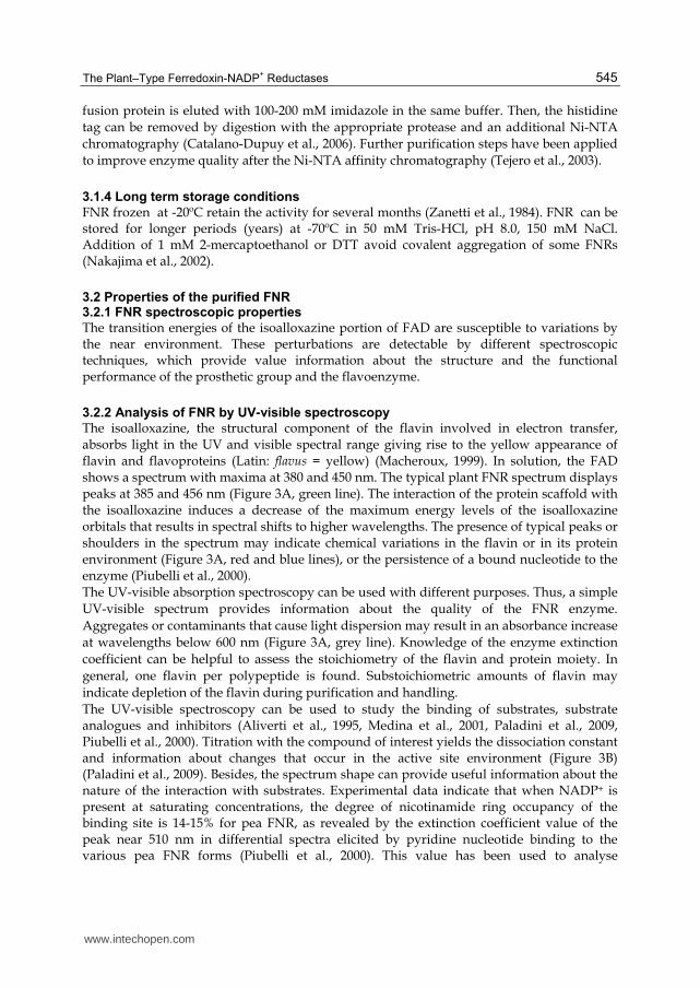
The isoalloxazine, the structural component of the flavin involved in electron transfer, absorbs light in the UV and visible spectral range giving rise to the yellow appearance of flavin and flavoproteins (Latin: flavus = yellow) (Macheroux, 1999). In solution, the FAD shows a spectrum with maxima at 380 and 450 nm. The typical plant FNR spectrum displays peaks at 385 and 456 nm (Figure 3A, green line). The interaction of the protein scaffold with the isoalloxazine induces a decrease of the maximum energy levels of the isoalloxazine orbitals that results in spectral shifts to higher wavelengths. The presence of typical peaks or shoulders in the spectrum may indicate chemical variations in the flavin or in its protein environment (Figure 3A, red and blue lines), or the persistence of a bound nucleotide to the enzyme (Piubelli et al., 2000). The UV-visible absorption spectroscopy can be used with different purposes. Thus, a simple UV-visible spectrum provides information about the quality of the FNR enzyme. Aggregates or contaminants that cause light dispersion may result in an absorbance increase at wavelengths below 600 nm (Figure 3A, grey line). Knowledge of the enzyme extinction coefficient can be helpful to assess the stoichiometry of the flavin and protein moiety. In general, one flavin per polypeptide is found. Substoichiometric amounts of flavin may indicate depletion of the flavin during purification and handling. The UV-visible spectroscopy can be used to study the binding of substrates, substrate analogues and inhibitors (Aliverti et al., 1995, Medina et al., 2001, Paladini et al., 2009, Piubelli et al., 2000). Titration with the compound of interest yields the dissociation constant and information about changes that occur in the active site environment (Figure 3B) (Paladini et al., 2009). Besides, the spectrum shape can provide useful information about the nature of the interaction with substrates. Experimental data indicate that when NADP+ is present at saturating concentrations, the degree of nicotinamide ring occupancy of the binding site is 14-15% for pea FNR, as revealed by the extinction coefficient value of the peak near 510 nm in differential spectra elicited by pyridine nucleotide binding to the various pea FNR forms (Piubelli et al., 2000). This value has been used to analyse
www.intechopen.com
Advances in Photosynthesis – Fundamental Aspects
546
nicotinamide occupancy when NADP+ is bound to different mutant FNR enzymes (Figure 3C). On the other hand, the FNR spectral UV-visible properties also depend on the redox state of the flavin. These spectral data are very useful to analyse the flavoprotein function (Macheroux, 1999, Nogues et al., 2004).
Fig. 3. Analysis of FNR by UV-visible spectroscopy. A) Absorption spectra of pea FNR obtained after 6 (green, good quality), 19 (blue) and 25 (red) hours of induction in E. coli. FNR preparation containing insoluble aggregates (grey line). B) Differential spectra obtained upon titration of wild-type FNR with NADP+. Inset: absorbance change at 515 nm plotted as function of NADP+ concentration. C) Differential spectra elicited by NADP+ binding to different FNR mutants (grey, yellow, cyan, red and blue corresponds to the spectra of wild-type, C266A, C266AL266A and C266M FNR forms respectively (Musumeci et al., 2008)).
3.2.3 Application of fluorescence spectroscopy
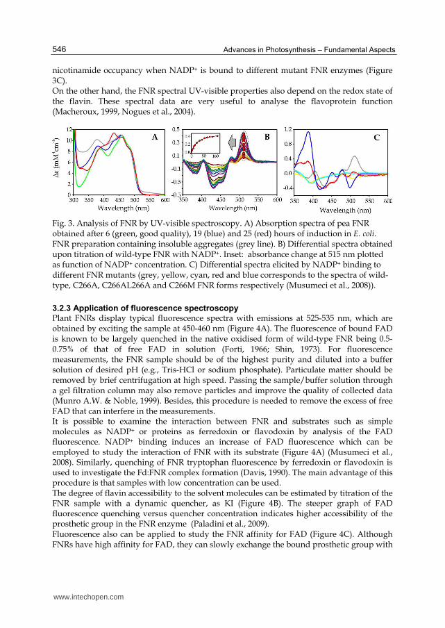
Plant FNRs display typical fluorescence spectra with emissions at 525-535 nm, which are obtained by exciting the sample at 450-460 nm (Figure 4A). The fluorescence of bound FAD is known to be largely quenched in the native oxidised form of wild-type FNR being 0.5-0.75% of that of free FAD in solution (Forti, 1966; Shin, 1973). For fluorescence measurements, the FNR sample should be of the highest purity and diluted into a buffer solution of desired pH (e.g., Tris-HCl or sodium phosphate). Particulate matter should be removed by brief centrifugation at high speed. Passing the sample/buffer solution through a gel filtration column may also remove particles and improve the quality of collected data (Munro A.W. & Noble, 1999). Besides, this procedure is needed to remove the excess of free FAD that can interfere in the measurements. It is possible to examine the interaction between FNR and substrates such as simple molecules as NADP+ or proteins as ferredoxin or flavodoxin by analysis of the FAD fluorescence. NADP+ binding induces an increase of FAD fluorescence which can be employed to study the interaction of FNR with its substrate (Figure 4A) (Musumeci et al., 2008). Similarly, quenching of FNR tryptophan fluorescence by ferredoxin or flavodoxin is used to investigate the Fd:FNR complex formation (Davis, 1990). The main advantage of this procedure is that samples with low concentration can be used. The degree of flavin accessibility to the solvent molecules can be estimated by titration of the FNR sample with a dynamic quencher, as KI (Figure 4B). The steeper graph of FAD fluorescence quenching versus quencher concentration indicates higher accessibility of the prosthetic group in the FNR enzyme (Paladini et al., 2009). Fluorescence also can be applied to study the FNR affinity for FAD (Figure 4C). Although FNRs have high affinity for FAD, they can slowly exchange the bound prosthetic group with
www.intechopen.com
The Plant–Type Ferredoxin-NADP
+ Reductases
547
the FAD in solution. Thus, it is possible follow the process of FAD dissociation by measuring the increase in FAD fluorescence of a FNR sample at different times. The obtained data can be used to calculate the FNR:FAD complex dissociation constant or the FAD dissociation rate constant (koff) (Musumeci et al., 2011, Paladini et al., 2009).
Fig. 4. Application of FAD fluorescence to study structural features of FNR. A) Fluorescence spectra obtained upon titration with NADP+. Inset: The increase of fluorescence at 525 nm was plotted against NADP+ concentration, and then Kd value can be obtained by adjusting the plot to the sigmoidal function. B) Study of FAD accessibility by titration with a dynamic quencher (KI). Pea Y308S mutant in which the tyrosine facing the isoalloxazine was replaced by a serine shows an increase in the flavin exposure (red) with respect to wild-type FNR (green) (Paladini et al., 2009). C) Fluorescence in solution as a function of time of wild-type FNR (green) and C266M mutant (blue). The koff for FAD of the mutant is higher than the one obtained for wild-type enzyme.
3.2.4 Use of CD spectroscopy
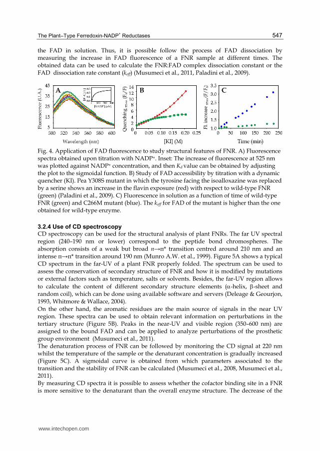
CD spectroscopy can be used for the structural analysis of plant FNRs. The far UV spectral region (240–190 nm or lower) correspond to the peptide bond chromospheres. The absorption consists of a weak but broad n→π* transition centred around 210 nm and an intense π→π* transition around 190 nm (Munro A.W. et al., 1999). Figure 5A shows a typical CD spectrum in the far-UV of a plant FNR properly folded. The spectrum can be used to assess the conservation of secondary structure of FNR and how it is modified by mutations or external factors such as temperature, salts or solvents. Besides, the far-UV region allows to calculate the content of different secondary structure elements (-helix, -sheet and random coil), which can be done using available software and servers (Deleage & Geourjon, 1993, Whitmore & Wallace, 2004). On the other hand, the aromatic residues are the main source of signals in the near UV region. These spectra can be used to obtain relevant information on perturbations in the tertiary structure (Figure 5B). Peaks in the near-UV and visible region (350–600 nm) are assigned to the bound FAD and can be applied to analyze perturbations of the prosthetic group environment (Musumeci et al., 2011). The denaturation process of FNR can be followed by monitoring the CD signal at 220 nm whilst the temperature of the sample or the denaturant concentration is gradually increased (Figure 5C). A sigmoidal curve is obtained from which parameters associated to the transition and the stability of FNR can be calculated (Musumeci et al., 2008, Musumeci et al., 2011). By measuring CD spectra it is possible to assess whether the cofactor binding site in a FNR is more sensitive to the denaturant than the overall enzyme structure. The decrease of the
www.intechopen.com
Advances in Photosynthesis – Fundamental Aspects
548
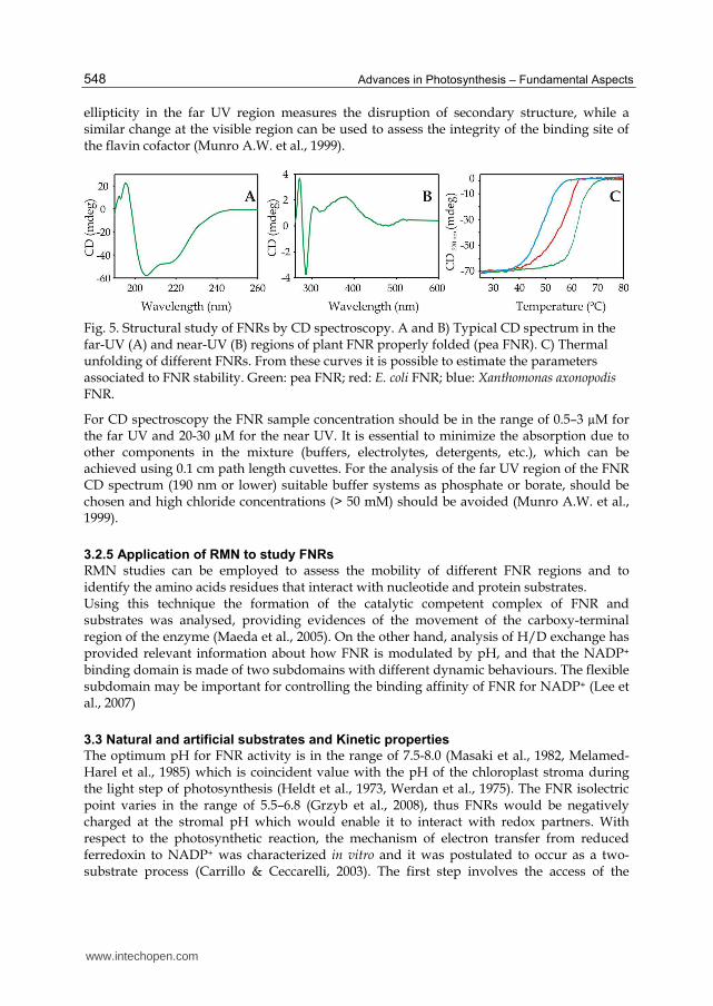
ellipticity in the far UV region measures the disruption of secondary structure, while a similar change at the visible region can be used to assess the integrity of the binding site of the flavin cofactor (Munro A.W. et al., 1999).
Fig. 5. Structural study of FNRs by CD spectroscopy. A and B) Typical CD spectrum in the far-UV (A) and near-UV (B) regions of plant FNR properly folded (pea FNR). C) Thermal unfolding of different FNRs. From these curves it is possible to estimate the parameters associated to FNR stability. Green: pea FNR; red: E. coli FNR; blue: Xanthomonas axonopodis FNR.
For CD spectroscopy the FNR sample concentration should be in the range of 0.5–3 µM for the far UV and 20-30 µM for the near UV. It is essential to minimize the absorption due to other components in the mixture (buffers, electrolytes, detergents, etc.), which can be achieved using 0.1 cm path length cuvettes. For the analysis of the far UV region of the FNR CD spectrum (190 nm or lower) suitable buffer systems as phosphate or borate, should be chosen and high chloride concentrations (> 50 mM) should be avoided (Munro A.W. et al., 1999).
3.2.5 Application of RMN to study FNRs
RMN studies can be employed to assess the mobility of different FNR regions and to identify the amino acids residues that interact with nucleotide and protein substrates. Using this technique the formation of the catalytic competent complex of FNR and substrates was analysed, providing evidences of the movement of the carboxy-terminal region of the enzyme (Maeda et al., 2005). On the other hand, analysis of H/D exchange has provided relevant information about how FNR is modulated by pH, and that the NADP+
binding domain is made of two subdomains with different dynamic behaviours. The flexible subdomain may be important for controlling the binding affinity of FNR for NADP+ (Lee et al., 2007)
3.3 Natural and artificial substrates and Kinetic properties
The optimum pH for FNR activity is in the range of 7.5-8.0 (Masaki et al., 1982, Melamed-Harel et al., 1985) which is coincident value with the pH of the chloroplast stroma during the light step of photosynthesis (Heldt et al., 1973, Werdan et al., 1975). The FNR isolectric point varies in the range of 5.5–6.8 (Grzyb et al., 2008), thus FNRs would be negatively charged at the stromal pH which would enable it to interact with redox partners. With respect to the photosynthetic reaction, the mechanism of electron transfer from reduced ferredoxin to NADP+ was characterized in vitro and it was postulated to occur as a two-substrate process (Carrillo & Ceccarelli, 2003). The first step involves the access of the
www.intechopen.com
The Plant–Type Ferredoxin-NADP
+ Reductases
549
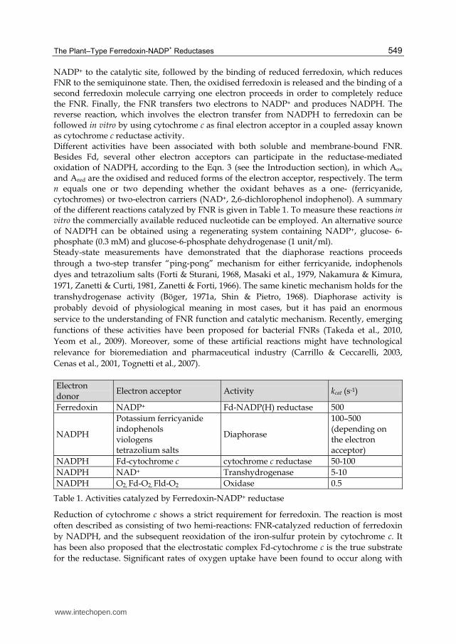
NADP+ to the catalytic site, followed by the binding of reduced ferredoxin, which reduces FNR to the semiquinone state. Then, the oxidised ferredoxin is released and the binding of a second ferredoxin molecule carrying one electron proceeds in order to completely reduce the FNR. Finally, the FNR transfers two electrons to NADP+ and produces NADPH. The reverse reaction, which involves the electron transfer from NADPH to ferredoxin can be followed in vitro by using cytochrome c as final electron acceptor in a coupled assay known as cytochrome c reductase activity. Different activities have been associated with both soluble and membrane-bound FNR. Besides Fd, several other electron acceptors can participate in the reductase-mediated oxidation of NADPH, according to the Eqn. 3 (see the Introduction section), in which Aox and Ared are the oxidised and reduced forms of the electron acceptor, respectively. The term n equals one or two depending whether the oxidant behaves as a one- (ferricyanide, cytochromes) or two-electron carriers (NAD+, 2,6-dichlorophenol indophenol). A summary of the different reactions catalyzed by FNR is given in Table 1. To measure these reactions in vitro the commercially available reduced nucleotide can be employed. An alternative source of NADPH can be obtained using a regenerating system containing NADP+, glucose- 6-phosphate (0.3 mM) and glucose-6-phosphate dehydrogenase (1 unit/ml). Steady-state measurements have demonstrated that the diaphorase reactions proceeds through a two-step transfer “ping-pong” mechanism for either ferricyanide, indophenols dyes and tetrazolium salts (Forti & Sturani, 1968, Masaki et al., 1979, Nakamura & Kimura, 1971, Zanetti & Curti, 1981, Zanetti & Forti, 1966). The same kinetic mechanism holds for the transhydrogenase activity (Böger, 1971a, Shin & Pietro, 1968). Diaphorase activity is probably devoid of physiological meaning in most cases, but it has paid an enormous service to the understanding of FNR function and catalytic mechanism. Recently, emerging functions of these activities have been proposed for bacterial FNRs (Takeda et al., 2010, Yeom et al., 2009). Moreover, some of these artificial reactions might have technological relevance for bioremediation and pharmaceutical industry (Carrillo & Ceccarelli, 2003, Cenas et al., 2001, Tognetti et al., 2007). Electron donor
Electron acceptor Activity kcat (s-1)
Ferredoxin NADP+ Fd-NADP(H) reductase 500
NADPH
Potassium ferricyanide indophenols viologens tetrazolium salts
Diaphorase
100–500 (depending on the electron acceptor)
NADPH Fd-cytochrome c cytochrome c reductase 50-100 NADPH NAD+ Transhydrogenase 5-10 NADPH O2, Fd-O2, Fld-O2 Oxidase 0.5
Table 1. Activities catalyzed by Ferredoxin-NADP+ reductase
Reduction of cytochrome c shows a strict requirement for ferredoxin. The reaction is most often described as consisting of two hemi-reactions: FNR-catalyzed reduction of ferredoxin by NADPH, and the subsequent reoxidation of the iron-sulfur protein by cytochrome c. It has been also proposed that the electrostatic complex Fd-cytochrome c is the true substrate for the reductase. Significant rates of oxygen uptake have been found to occur along with
www.intechopen.com
Advances in Photosynthesis – Fundamental Aspects
550
citochrome c reductase activity under aerobic conditions (Carrillo & Vallejos, 1987). The oxidase activity of FNR is very low (Carrillo & Vallejos, 1987, Gomez-Moreno et al., 1994). This reaction is enhanced several-fold by different electronic acceptors, including one-electron reduced ferredoxin or flavodoxin, viologens, nitroderivates and quinones, that can readily engage in oxygen-dependent redox cycling leading to superoxide formation (Gomez-Moreno et al., 1994, Shah & Spain, 1996). In addition, FNR catalyses the transhydrogenation between NADPH and NAD+ (Böger, 1971b). The stopped-flow technique allows to study fast enzyme kinetic events in the range of milliseconds. This approach has been employed to characterize pre-steady state processes which involve the interaction and electron transfer between oxidised FNR and NADPH, reduced FNR and NADP+ and reduced ferredoxin or flavodoxin and oxidised FNR (Anusevicius et al., 2005, Hurley et al., 1995, Martinez-Julvez et al., 1998, Medina et al., 2001, Paladini et al., 2009, Tejero et al., 2007). The first reduction of pea FNR by Fd produces a semiquinone form too fast to be measured by rapid mixing techniques (Carrillo & Ceccarelli, 2003) and references therein. Fortunately, this step was characterized for Anabaena FNR (Hurley et al., 1995, Martinez-Julvez et al., 1998). The electron transfer processes that involve the dissociation of oxidised ferredoxin, the binding of reduced ferredoxin and flavin reduction occurs more slowly and have been characterized in wild-type and mutant enzymes (Medina, 2009, Nogues et al., 2004).
4. Crystal structure of FNR and its complex with natural substrates
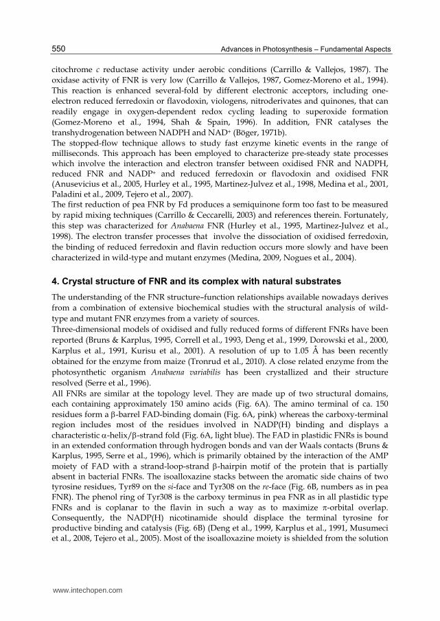
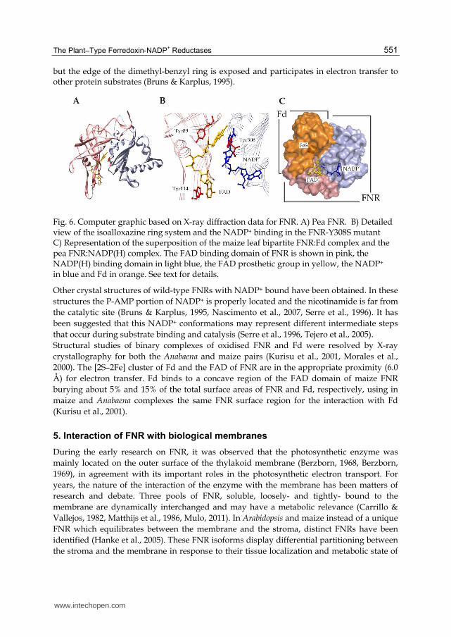
The understanding of the FNR structure–function relationships available nowadays derives from a combination of extensive biochemical studies with the structural analysis of wild-type and mutant FNR enzymes from a variety of sources. Three-dimensional models of oxidised and fully reduced forms of different FNRs have been reported (Bruns & Karplus, 1995, Correll et al., 1993, Deng et al., 1999, Dorowski et al., 2000, Karplus et al., 1991, Kurisu et al., 2001). A resolution of up to 1.05 Å has been recently obtained for the enzyme from maize (Tronrud et al., 2010). A close related enzyme from the photosynthetic organism Anabaena variabilis has been crystallized and their structure resolved (Serre et al., 1996). All FNRs are similar at the topology level. They are made up of two structural domains, each containing approximately 150 amino acids (Fig. 6A). The amino terminal of ca. 150 residues form a -barrel FAD-binding domain (Fig. 6A, pink) whereas the carboxy-terminal region includes most of the residues involved in NADP(H) binding and displays a characteristic -helix/-strand fold (Fig. 6A, light blue). The FAD in plastidic FNRs is bound in an extended conformation through hydrogen bonds and van der Waals contacts (Bruns & Karplus, 1995, Serre et al., 1996), which is primarily obtained by the interaction of the AMP moiety of FAD with a strand-loop-strand -hairpin motif of the protein that is partially absent in bacterial FNRs. The isoalloxazine stacks between the aromatic side chains of two tyrosine residues, Tyr89 on the si-face and Tyr308 on the re-face (Fig. 6B, numbers as in pea FNR). The phenol ring of Tyr308 is the carboxy terminus in pea FNR as in all plastidic type FNRs and is coplanar to the flavin in such a way as to maximize -orbital overlap. Consequently, the NADP(H) nicotinamide should displace the terminal tyrosine for productive binding and catalysis (Fig. 6B) (Deng et al., 1999, Karplus et al., 1991, Musumeci et al., 2008, Tejero et al., 2005). Most of the isoalloxazine moiety is shielded from the solution
www.intechopen.com
The Plant–Type Ferredoxin-NADP
+ Reductases
551
but the edge of the dimethyl-benzyl ring is exposed and participates in electron transfer to other protein substrates (Bruns & Karplus, 1995).
Fig. 6. Computer graphic based on X-ray diffraction data for FNR. A) Pea FNR. B) Detailed view of the isoalloxazine ring system and the NADP+ binding in the FNR-Y308S mutant C) Representation of the superposition of the maize leaf bipartite FNR:Fd complex and the pea FNR:NADP(H) complex. The FAD binding domain of FNR is shown in pink, the NADP(H) binding domain in light blue, the FAD prosthetic group in yellow, the NADP+ in blue and Fd in orange. See text for details.
Other crystal structures of wild-type FNRs with NADP+ bound have been obtained. In these structures the P-AMP portion of NADP+ is properly located and the nicotinamide is far from the catalytic site (Bruns & Karplus, 1995, Nascimento et al., 2007, Serre et al., 1996). It has been suggested that this NADP+ conformations may represent different intermediate steps that occur during substrate binding and catalysis (Serre et al., 1996, Tejero et al., 2005). Structural studies of binary complexes of oxidised FNR and Fd were resolved by X-ray crystallography for both the Anabaena and maize pairs (Kurisu et al., 2001, Morales et al., 2000). The [2S–2Fe] cluster of Fd and the FAD of FNR are in the appropriate proximity (6.0 Å) for electron transfer. Fd binds to a concave region of the FAD domain of maize FNR burying about 5% and 15% of the total surface areas of FNR and Fd, respectively, using in maize and Anabaena complexes the same FNR surface region for the interaction with Fd (Kurisu et al., 2001).
5. Interaction of FNR with biological membranes
During the early research on FNR, it was observed that the photosynthetic enzyme was mainly located on the outer surface of the thylakoid membrane (Berzborn, 1968, Berzborn, 1969), in agreement with its important roles in the photosynthetic electron transport. For years, the nature of the interaction of the enzyme with the membrane has been matters of research and debate. Three pools of FNR, soluble, loosely- and tightly- bound to the membrane are dynamically interchanged and may have a metabolic relevance (Carrillo & Vallejos, 1982, Matthijs et al., 1986, Mulo, 2011). In Arabidopsis and maize instead of a unique FNR which equilibrates between the membrane and the stroma, distinct FNRs have been identified (Hanke et al., 2005). These FNR isoforms display differential partitioning between the stroma and the membrane in response to their tissue localization and metabolic state of
www.intechopen.com
Advances in Photosynthesis – Fundamental Aspects
552
the chloroplast (Grzyb et al., 2008, Hanke et al., 2005, Lintala et al., 2007, Okutani et al., 2005, Zanetti & Arosio, 1980). FNR solubilisation from the membrane plays a role in maintaining the NADPH/NADP+ homeostasis of the stressed plastid. The enzyme changes its function from a membrane-bound NADPH producer to a soluble NADPH consumer to overcome the accumulation of NADPH. The excess of reducing power might otherwise increase the risk of oxidative damage through the production of hydroxyl radicals (Palatnik et al., 1997). The persistence of a fraction that remains tightly bound to the membrane has drive to several research groups to search for the existence of an internal membrane protein that acts as binding site for the reductase. The soluble and membrane-bound forms of FNR show different allotropic properties which may reflect conformational changes of the enzyme upon binding to the membrane (Carrillo et al., 1981, Schneeman & Krogmann, 1975). In this way, the membrane component may serve to transmit internal thylakoid conditions as pH changes and membrane fluidity to the bound reductase, which then changes its kinetics behaviour. Several years ago a polypeptide of 17.5 kDa was identified as the binding protein for the reductase (Ceccarelli et al., 1985, Chan et al., 1987, Vallejos et al., 1984). The 17.5-FNR complex was detected in different higher plants, including C3, C4, and Crassulacean acid metabolism species (Soncini & Vallejos, 1989). During solubilisation of FNR under stress condition the reductase-binding protein was released together with FNR, suggesting that it might be the target of some regulation of the membrane bound state (Palatnik et al., 1997). The reductase-binding protein was identified to be the same than the PsbQ like protein (Soncini & Vallejos, 1989). This localization challenges its participation as a FNR binding protein. It has been proposed that the PsbQ may also contribute to the integrity of grana thylakoids stacks (Anderson et al., 2008). Moreover, PsbQ-like homologs have been identified as essential members of the chloroplast NAD(P)H dehydrogenase complex in Arabidopsis (Yabuta et al., 2010). The reductase-binding protein (PsbQ like protein) may probably has a ubiquitous function on the thylakoid membrane structure. That makes this topic an interesting issue to pursue. More recently, a thylakoidial transmembrane protein with a rhodanase like structure was identified as binding site for FNR (Juric et al., 2009). This integral membrane protein contains a conserved carboxy-terminal domain that interacts with high affinity with the FNR enzyme. Three imperfect of such domain were found in one of the proteins of the translocon at the inner envelope membrane of chloroplasts Tic62. Tic62 was proposed as a redox sensor of the translocon and may possibly act as a regulator during the translocation process and serves as a docking site for FNR. Moreover, Tic62 was found bound to thylakoids, soluble in the chloroplast stroma and attached to the inner membrane of the organelle envelope (Benz et al., 2009, Peltier et al., 2004). An unquestionable evidence of the role of TROL protein in FNR binding is that its absence in knockout plants disables FNR from being tethered to the membrane. In these plants, a decrease of the electron transfer rates under high light intensity was detected (Juric et al., 2009). Nevertheless, the authors suggest that TROL is an important, but not exclusive site for FNR since the enzyme was found in a number of other protein complexes. It was recently observed by crystallographic studies that the FNR-binding motif is a polyproline type II helix, which is located in the carboxy terminus of Tic62 (Alte et al., 2010). The polyproline type II helix mediates self-assembly of the FNR dimer without influence on the active sites of the enzyme. Interestingly, this type of secondary structure was detected on
www.intechopen.com
The Plant–Type Ferredoxin-NADP
+ Reductases
553
the crystal structure of the PsbQ (the 17,5 kDa identified as the reductase binding protein) on residues Pro9-Pro10-Pro11-Pro12, which forms a typical left-handed helix (or a polyproline type II structure) (Balsera et al., 2005). These structural elements have been implicated in protein–protein interactions in various cytosolic signal transduction pathways and eucariotic proteins. In addition, the carboxy terminus of TROL may have a similar structure as observed by sequence comparison (Juric et al., 2009). Thus, all proteins that have been identified up to date to firmly interact with FNR contain this structure. FNR interacts with different integral components of the thylakoid membrane. Several year ago Wagner et al by using flash kinetic spectroscopy demonstrated that the enzyme undergoes a very rapid rotational diffusion on the thylakoid membrane surface (Wagner et al., 1982). This movement was significantly reduced upon addition of Fd due to the formation of a ternary complex between the reductase and the photosystem I protein complex. Direct interaction with subunit of PSI was also detected (Andersen et al., 1992), but the relevance or significance of this interaction is unknown. As in the case of PSI, FNR has also been detected in citochrome b6f preparations (Clark et al., 1984, Zhang et al., 2001). FNR associated with the cytochrome b₆f complex can participate in the cyclic electron transport as PSI-plastoquinone or NADPH-plastoquinone oxidoreductases, which can explain the presence of FNR in citochrome b6f preparations (Szymanska et al., 2011). Some authors have identified FNR in the Ndh complex. The NAD(P)H dehydrogenase (Ndh) complex in chloroplast thylakoid membranes functions in cyclic electron transfer and in chlororespiration. Ndh is composed of at least 15 subunits, including both chloroplast- and nuclear-encoded proteins (Suorsa et al., 2009). As already mentioned, among the detected subunits of the Ndh complex is the PsbQ which was found associated with FNR. However, the amount of Ndh complex is a minor component of the chloroplast and it is present at ~1.5% of the level of PSII on a molar basis (Burrows et al., 1998) and probably makes a small contribution to the total cyclic electron transport measured in vivo (Joliot et al., 2004). FNR is an abundant protein in chloroplast. Moreover, protein concentration in the chloroplast stroma was determined to be within the range of 520 to 730 g/dm3 for spinach and pea (Lilley, 1983). FNR structure is suitable for interaction with membrane and proteins. It is therefore likely to detect many interactions of the FNR with other structures inside the chloroplast, many of which may have metabolic relevance.
6. Concluding remarks
For over six decades much effort has been devoted to the elucidation of the structure, catalytic mechanism and regulation of the FNR. Similarly, enormous progress has been made in understanding the insertion and importance of this enzyme on the various metabolic processes in plants and other organisms. The ability to produce FNR by transgenic expression and the modern techniques available nowadays have provided the means to analyze more deeply this flavoprotein. However, multiple aspects of this enzyme are still unknown as the structural bases for the increase of its catalytic competence and the ways to change the nucleotide substrate specificity. Furthermore, the enzyme may be an attractive target for the development of new herbicides and for the design of bactericides. Likewise, the ability of FNR to metabolize xenobiotics and environmental pollutants remain to be exploited. The widespread distribution of FNR will allow in the near future to increase the knowledge of the enzyme by comparative analysis of FNRs from different backgrounds
www.intechopen.com
Advances in Photosynthesis – Fundamental Aspects
554
and to advance in the study and manipulation of this key enzyme in the photosynthetic process.
7. Acknowledgment
This work was supported by grants from CONICET, Agencia de Promoción Científica y Tecnológica (ANPCyT) and Fundación Bunge y Born, Argentina.
8. References
Aliverti, A., Bruns, C. M., Pandini, V. E., Karplus, P. A., Vanoni, M. A., Curti, B., & Zanetti, G. (1995). Involvement of serine 96 in the catalytic mechanism of ferredoxin-NADP+ reductase: structure--function relationship as studied by site-directed mutagenesis and X-ray crystallography. Biochemistry 34, 8371-8379.
Aliverti, A., Jansen, T., Zanetti, G., Ronchi, S., Herrmann, R. G., & Curti, B. (1990). Expression in Escherichia coli of ferredoxin:NADP+ reductase from spinach. Bacterial synthesis of the holoflavoprotein and of an active enzyme form lacking the first 28 amino acid residues of the sequence. Eur J Biochem 191, 551-555.
Aliverti, A., Pandini, V., Pennati, A., de Rosa, M., & Zanetti, G. (2008). Structural and functional diversity of ferredoxin-NADP(+) reductases. Arch. Biochem. Biophys. 474, 283-291.
Alte, F., Stengel, A., Benz, J. P., Petersen, E., Soll, J., Groll, M., & Bolter, B. (2010). Ferredoxin:NADPH oxidoreductase is recruited to thylakoids by binding to a polyproline type II helix in a pH-dependent manner. Proc. Natl. Acad. Sci. U. S. A 107, 19260-19265.
Andersen, B., Scheller, H. V., & Moller, B. L. (1992). The PSI-E subunit of photosystem I binds ferredoxin:NADP+ oxidoreductase. FEBS Lett 311, 169-173.
Anderson, J. M., Chow, W. S., & De Las, R. J. (2008). Dynamic flexibility in the structure and function of photosystem II in higher plant thylakoid membranes: the grana enigma. Photosynth. Res. 98, 575-587.
Anusevicius, Z., Miseviciene, L., Medina, M., Martinez-Julvez, M., Gomez-Moreno, C., & Cenas, N. (2005). FAD semiquinone stability regulates single- and two-electron reduction of quinones by Anabaena PCC7119 ferredoxin:NADP+ reductase and its Glu301Ala mutant. Arch. Biochem. Biophys. 437, 144-150.
Arakaki, A. K., Ceccarelli, E. A., & Carrillo, N. (1997). Plant-type ferredoxin-NADP+ reductases: a basal structural framework and a multiplicity of functions. FASEB J. 11, 133-140.
Avron, M. & Jagendorf, A. T. (1956). A TPNH diaphorase from chloroplast. Arch. Biochem. Biophys. 65, 475-490.
Balsera, M., Arellano, J. B., Revuelta, J. L., De Las, R. J., & Hermoso, J. A. (2005). The 1.49 A resolution crystal structure of PsbQ from photosystem II of Spinacia oleracea reveals a PPII structure in the N-terminal region. J. Mol. Biol. 350, 1051-1060.
Benz, J. P., Stengel, A., Lintala, M., Lee, Y. H., Weber, A., Philippar, K., Gugel, I. L., Kaieda, S., Ikegami, T., Mulo, P., Soll, J., & Bolter, B. (2009). Arabidopsis Tic62 and ferredoxin-NADP(H) oxidoreductase form light-regulated complexes that are integrated into the chloroplast redox poise. Plant Cell 21, 3965-3983.
www.intechopen.com
The Plant–Type Ferredoxin-NADP
+ Reductases
555
Berzborn, R. (1968). On soluble and insoluble chloroplast antigens. Demonstration of ferredoxin-NADP-reductase in the surface of the chloroplast lamellar system with the aid of specific antibodies. Z Naturforsch B 23, 1096-1104.
Berzborn, R. J. (1969). Studies on the surface structure of the thylakoid system of chloroplasts using antibodies against ferredoxin-NADP-reductase. Z Naturforsch B 24, 436-446.
Böger, P. (1971a). Relationship of transhydrogenase and diaphorase activity of ferredoxin- NADP+ reductase with photosynthetic NADP+ reduction. Z Naturforsch B 26, 807-815.
Böger, P. (1971b). Einfluß von Ferredoxin auf Ferredoxin-NADP-Reduktase. Planta 99, 319-338.
Bottin, H. & Lagoutte, B. (1992). Ferredoxin and flavodoxin from the cyanobacterium Synechocystis sp PCC 6803. Biochim. Biophys. Acta 1101, 48-56.
Bruns, C. M. & Karplus, P. A. (1995). Refined crystal structure of spinach ferredoxin reductase at 1.7 A resolution: oxidised, reduced and 2'-phospho-5'-AMP bound states. J. Mol. Biol. 247, 125-145.
Burrows, P. A., Sazanov, L. A., Svab, Z., Maliga, P., & Nixon, P. J. (1998). Identification of a functional respiratory complex in chloroplasts through analysis of tobacco mutants containing disrupted plastid ndh genes. EMBO J. 17, 868-876.
Carrillo, N. & Vallejos, R. H. (1987). Ferredoxin-NADP+ oxidoreductase. In: The Light Reactions (Topics in photosynthesis), edited by J. Barber, pp. 527-560. Amsterdam-New York-Oxford: Elsevier.
Carrillo, N. & Ceccarelli, E. A. (2003). Open questions in ferredoxin-NADP+ reductase catalytic mechanism. Eur. J. Biochem. 270, 1900-1915.
Carrillo, N., Lucero, H. A., & Vallejos, R. H. (1981). Light modulation of chloroplast membrane-bound ferredoxin-NADP+ oxidoreductase. J. Biol. Chem. 256, 1058-1059.
Carrillo, N. & Vallejos, R. H. (1982). Interaction of Ferredoxin-NADP+ Oxidoreductase with the Thylakoid Membrane. Plant Physiol 69, 210-213.
Carrillo, N. & Vallejos, R. H. (1983). Interaction of ferredoxin-NADP+ oxidoreductase with triazine dyes. A rapid purification method by affinity chromatography. Biochim. Biophys. Acta 742, 285-294.
Catalano-Dupuy, D. L., Orecchia, M., Rial, D. V., & Ceccarelli, E. A. (2006). Reduction of the pea ferredoxin-NADP(H) reductase catalytic efficiency by the structuring of a carboxyl-terminal artificial metal binding site. Biochemistry 45, 13899-13909.
Ceccarelli, E. A., Arakaki, A. K., Cortez, N., & Carrillo, N. (2004). Functional plasticity and catalytic efficiency in plant and bacterial ferredoxin-NADP(H) reductases. Biochim. Biophys. Acta 1698, 155-165.
Ceccarelli, E. A., Chan, R. L., & Vallejos, R. H. (1985). Trimeric structure and other properties of the chloroplast reductase binding protein. FEBS Letters 190, 165-168.
Cenas, N., Nemeikaite-Ceniene, A., Sergediene, E., Nivinskas, H., Anusevicius, Z., & Sarlauskas, J. (2001). Quantitative structure-activity relationships in enzymatic single-electron reduction of nitroaromatic explosives: implications for their cytotoxicity. Biochim. Biophys. Acta 1528, 31-38.
Chan, R. L., Ceccarelli, E. A., & Vallejos, R. H. (1987). Immunological studies of the binding protein for chloroplast ferredoxin-NADP+ reductase. Arch. Biochem. Biophys. 253, 56-61.
www.intechopen.com
Advances in Photosynthesis – Fundamental Aspects
556
Chopowick, R. & Israelstam, G. F. (1971). Pyridine nucleotide transhydrogenase from Chlorella. Planta 101, 171-173.
Clark, R. D., Hawkesford, M. J., Coughlan, S. J., Bennett, J., & Hind, G. (1984). Association of ferredoxin-NADP+ oxidoreductase with the chloroplast cytochrome b-f complex. FEBS Letters 174, 137-142.
Cleland, R. E. & Bendall, D. S. (1992). Photosystem I cyclic electron transport: Measurement of ferredoxin-plastoquinone reductase activity. Photosynthesis Research 34, 409-418.
Correll, C. C., Ludwig, M., Bruns, C. M., & Karplus, P. A. (1993). Structural prototypes for an extended family of flavoprotein reductases: comparison of phthalate dioxygenase reductase with ferredoxin reductase and ferredoxin. Protein Sci 2, 2112-2133.
Davis, D. J. (1990). Tryptophan fluorescence studies of ferredoxin:NADP+ reductase indicate the presence of tryptophan in or near the ferredoxin binding site. Arch. Biochem. Biophys. 276, 1-5.
Deleage, G. & Geourjon, C. (1993). An interactive graphic program for calculating the secondary structure content of proteins from circular dichroism spectrum. Comput. Appl. Biosci. 9, 197-199.
Deng, Z., Aliverti, A., Zanetti, G., Arakaki, A. K., Ottado, J., Orellano, E. G., Calcaterra, N. B., Ceccarelli, E. A., Carrillo, N., & Karplus, P. A. (1999). A productive NADP+ binding mode of ferredoxin-NADP+ reductase revealed by protein engineering and crystallographic studies. Nat. Struct. Biol. 6, 847-853.
Dorowski, A., Hofmann, A., Steegborn, C., Boicu, M., & Huber, R. (2000). Crystal structure of paprika ferredoxin-NADP+ reductase - implications for the electron transfer pathway. J. Biol. Chem. 276, 9253-9263.
Dudley, K. H., Ehrenberg, A., Hemmerich, P., & Müller, F. (1964). Spektren und Strukturen der am Flavin-Redoxsystem beteiligten Partikeln. Studien in der Flavinreihe IX. Helv. Chim. Acta 47, 1354-1383.
Faro, M., Schiffler, B., Heinz, A., Nogues, I., Medina, M., Bernhardt, R., & Gomez-Moreno, C. (2003). Insights into the design of a hybrid system between Anabaena ferredoxin-NADP+ reductase and bovine adrenodoxin. Eur. J. Biochem. 270, 726-735.
Forti, G. & Sturani, E. (1968). On the structure and function of reduced nicotinamide adenine dinucleotide phosphate-cytochrome f reductase of spinach chloroplasts. Eur. J. Biochem. 3, 461-472.
Gomez-Moreno, C., Medina, M., Hurley, J. K., Cusanovich, M., Markley, J., Cheng, H., Xia, B., Chae, Y. K., & Tollin, G. (1994). Protein engineering for the elucidation of the mechanism of electron transfer in redox proteins. Biochem Soc. Trans. 22, 796-800.
Green, L. S., Yee, B. C., Buchanan, B. B., Kamide, K., Sanada, Y., & Wada, K. (1991). Ferredoxin and Ferredoxin-NADP+ Reductase from Photosynthetic and Nonphotosynthetic Tissues of Tomato. Plant Physiology 96, 1207-1213.
Grzyb, J., Malec, P., Rumak, I., Garstka, M., & Strzalka, K. (2008). Two isoforms of ferredoxin:NADP(+) oxidoreductase from wheat leaves: purification and initial biochemical characterization. Photosynth. Res. 96, 99-112.
Grzyb, J., Waloszek, A., Latowski, D., & Wieckowski, S. (2004). Effect of cadmium on ferredoxin:NADP+ oxidoreductase activity. J. Inorg. Biochem. 98, 1338-1346.
Hajirezaei, M. R., Peisker, M., Tschiersch, H., Palatnik, J. F., Valle, E. M., Carrillo, N., & Sonnewald, U. (2002). Small changes in the activity of chloroplastic NADP(+)-
www.intechopen.com
The Plant–Type Ferredoxin-NADP
+ Reductases
557
dependent ferredoxin oxidoreductase lead to impaired plant growth and restrict photosynthetic activity of transgenic tobacco plants. Plant J. 29, 281-293.
Hanke, G. T., Ookutani, S. , Satomi, Y., Takao, T., Suzuki, A., & Hase, T. (2005). Multiple iso-proteins of FNR in Arabidopsis: evidence for different contributions to chloroplast function and nitrogen assimilation. Plant, Cell & Environment 28, 1146-1157.
Heelis P.F. (1982). The photophysical and photochemical properties of flavins (isoalloxazines). Chem. Soc. Rev. 11, 15-39.
Heldt, W. H., Werdan, K., Milovancev, M., & Geller, G. (1973). Alkalization of the chloroplast stroma caused by light-dependent proton flux into the thylakoid space. Biochim. Biophys. Acta 314, 224-241.
Hurley, J. K., Fillat, M., Gomez-Moreno, C., & Tollin, G. (1995). Structure-function relationships in the ferredoxin/ferredoxin: NADP+ reductase system from Anabaena. Biochimie 77, 539-548.
Jenkins, C. M., Genzor, C. G., Fillat, M. F., Waterman, M. R., & Gomez-Moreno, C. (1997). Negatively charged Anabaena flavodoxin residues (Asp144 and Glu145) are important for reconstitution of cytochrome P450 17alpha-hydroxylase activity. J. Biol. Chem. 272, 22509-22513.
Joliot, P., Beal, D., & Joliot, A. (2004). Cyclic electron flow under saturating excitation of dark-adapted Arabidopsis leaves. Biochim. Biophys. Acta 1656, 166-176.
Joliot, P. & Joliot, A. (2006). Cyclic electron flow in C3 plants. Biochim. Biophys. Acta 1757, 362-368.
Juric, S., Hazler-Pilepic, K., Tomasic, A., Lepedus, H., Jelicic, B., Puthiyaveetil, S., Bionda, T., Vojta, L., Allen, J. F., Schleiff, E., & Fulgosi, H. (2009). Tethering of ferredoxin:NADP+ oxidoreductase to thylakoid membranes is mediated by novel chloroplast protein TROL. Plant J. 60, 783-794.
Karplus, P. A., Daniels, M. J., & Herriott, J. R. (1991). Atomic structure of ferredoxin-NADP+ reductase: prototype for a structurally novel flavoenzyme family. Science 251, 60-66.
Krapp, A. R., Rodriguez, R. E., Poli, H. O., Paladini, D. H., Palatnik, J. F., & Carrillo, N. (2002). The flavoenzyme ferredoxin (flavodoxin)-NADP(H) reductase modulates NADP(H) homeostasis during the soxRS response of Escherichia coli. J. Bacteriol. 184, 1474-1480.
Krause, G. H. & Heber, U. (1976).The intact chloroplast , edited by H. Barber, pp. 171-214. Elsevier, Amsterdam.
Krawetz, S. A. & Israelstam, G. F. (1978). Kinetics of pyridine nucleotide transhydrogenase from Chlorella. Plant Science Letters 12, 323-326.
Kurisu, G., Kusunoki, M., Katoh, E., Yamazaki, T., Teshima, K., Onda, Y., Kimata-Ariga, Y., & Hase, T. (2001). Structure of the electron transfer complex between ferredoxin and ferredoxin-NADP+ reductase. Nat. Struct. Biol. 8, 117-121.
Lee, Y. H., Tamura, K., Maeda, M., Hoshino, M., Sakurai, K., Takahashi, S., Ikegami, T., Hase, T., & Goto, Y. (2007). Cores and pH-dependent dynamics of ferredoxin-NADP+ reductase revealed by hydrogen/deuterium exchange. J. Biol. Chem. 282, 5959-5967.
Lilley, R. (1983). Chloroplast metabolism: the pathways of primary carbon metabolism in C3 plants. Plant, Cell & Environment 6, 329-343.
Lintala, M., Allahverdiyeva, Y., Kidron, H., Piippo, M., Battchikova, N., Suorsa, M., Rintamaki, E., Salminen, T. A., Aro, E. M., & Mulo, P. (2007). Structural and
www.intechopen.com
Advances in Photosynthesis – Fundamental Aspects
558
functional characterization of ferredoxin-NADP+-oxidoreductase using knock-out mutants of Arabidopsis. Plant J. 49, 1041-1052.
Macheroux, P. (1999). Flavoprotein Protocols, edited by Stephen K.Chapman & Graeme A.Reid, Springer Science.
Maeda, M., Lee, Y. H., Ikegami, T., Tamura, K., Hoshino, M., Yamazaki, T., Nakayama, M., Hase, T., & Goto, Y. (2005). Identification of the N- and C-terminal substrate binding segments of ferredoxin-NADP+ reductase by NMR. Biochemistry 44, 10644-10653.
Martinez-Julvez, M., Hermoso, J., Hurley, J. K., Mayoral, T., Sanz-Aparicio, J., Tollin, G., Gomez-Moreno, C., & Medina, M. (1998). Role of Arg100 and Arg264 from Anabaena PCC 7119 ferredoxin-NADP+ reductase for optimal NADP+ binding and electron transfer. Biochemistry 37, 17680-17691.
Masaki, R., Wada, K., & Matsubara, H. (1979). Isolation and characterization of two ferredoxin-NADP+ reductases from Spirulina platensis. J Biochem (Tokyo) 86, 951-962.
Masaki, R., Yoshikawa, S., & Matsubara, H. (1982). Steady-state kinetics of oxidation of reduced ferredoxin with ferredoxin-NADP+ reductase. Biochim. Biophys. Acta 700, 101-109.
Matthijs, H. C., Coughlan, S. J., & Hind, G. (1986). Removal of ferredoxin:NADP+ oxidoreductase from thylakoid membranes, rebinding to depleted membranes, and identification of the binding site. J. Biol. Chem. 261, 12154-12158.
Medina, M. (2009). Structural and mechanistic aspects of flavoproteins: photosynthetic electron transfer from photosystem I to NADP+. FEBS J. 276, 3942-3958.
Medina, M. & Gomez-Moreno, C. (2004). Interaction of Ferredoxin-NADP+ Reductase with Its Substrates: Optimal Interaction for Efficient Electron Transfer. Photosynth. Res. 79, 113-131.
Medina, M., Luquita, A., Tejero, J., Hermoso, J., Mayoral, T., Sanz-Aparicio, J., Grever, K., & Gomez-Moreno, C. (2001a). Probing the determinants of coenzyme specificity in ferredoxin-NADP+ reductase by site-directed mutagenesis. J. Biol. Chem. 276, 11902-11912.
Melamed-Harel, H., Tel-Or, E., & Pietro, A. S. (1985). Effect of Ferredoxin on the Diaphorase Activity of Cyanobacterial Ferredoxin-NADP+ Reductase. Plant Physiology 77, 229-231.
Morales, R., Kachalova, G., Vellieux, F., Charon, M. H., & Frey, M. (2000). Crystallographic studies of the interaction between the ferredoxin-NADP+ reductase and ferredoxin from the cyanobacterium Anabaena: looking for the elusive ferredoxin molecule. Acta Crystallogr D Biol Crystallogr 56, 1408-1412.
Morigasaki, S., Takata, K., Suzuki, T., & Wada, K. (1990). Purification and Characterization of a Ferredoxin-NADP+ Oxidoreductase-Like Enzyme from Radish Root Tissues. Plant Physiol 93, 896-901.
Mulo, P. (2011). Chloroplast-targeted ferredoxin-NADP(+) oxidoreductase (FNR): Structure, function and location. Biochim. Biophys. Acta 1807, 927-934.
Munro A.W., Kelly S., & Price N. (1999). Fluorescence analysis of Flavoproteins. In: Flavoprotein protocols , edited by G. A. R. Stephen K.Chapman, pp. 111-129. Humana press. NJ, USA.
www.intechopen.com
The Plant–Type Ferredoxin-NADP
+ Reductases
559
Munro A.W. & Noble, M. A. (1999) Circular Dichroism studies of flavoproteins. In: Flavoprotein protocols edited by G. A. R. Stephen K.Chapman, pp. 25-48. Humana Press. NJ, USA.
Musumeci, M. A., Arakaki, A. K., Rial, D. V., Catalano-Dupuy, D. L., & Ceccarelli, E. A. (2008). Modulation of the enzymatic efficiency of ferredoxin-NADP(H) reductase by the amino acid volume around the catalytic site. FEBS J. 275, 1350-1366.
Musumeci, M. A., Botti, H., Buschiazzo, A., & Ceccarelli, E. A. (2011). Swapping FAD Binding Motifs between Plastidic and Bacterial Ferredoxin-NADP(H) Reductases. Biochemistry 50, 2111-2122.
Nakajima, M., Sakamoto, T., & Wada, K. (2002). The complete purification and characterization of three forms of ferredoxin-NADP(+) oxidoreductase from a thermophilic cyanobacterium Synechococcus elongatus. Plant Cell Physiol 43, 484-493.
Nakamura, S. & Kimura, T. (1971). Studies on spinach ferredoxin-nicotinamide adenine dinucleotide phosphate reductase. Kinetic studies on the interactions of the reductase and ferredoxin and a possible regulation of enzyme activities by ionic strength. J. Biol. Chem. 246, 6235-6241.
Nascimento, A. S., Catalano-Dupuy, D. L., Bernardes, A., de Oliveira, N. M., Santos, M. A., Ceccarelli, E. A., & Polikarpov, I. (2007). Crystal structures of Leptospira interrogans FAD-containing ferredoxin-NADP+ reductase and its complex with NADP+. BMC. Struct. Biol. 7, 69.
Nogues, I., Tejero, J., Hurley, J. K., Paladini, D., Frago, S., Tollin, G., Mayhew, S. G., Gomez-Moreno, C., Ceccarelli, E. A., Carrillo, N., & Medina, M. (2004). Role of the C-terminal tyrosine of ferredoxin-nicotinamide adenine dinucleotide phosphate reductase in the electron transfer processes with its protein partners ferredoxin and flavodoxin. Biochemistry 43, 6127-6137.
Okutani, S., Hanke, G. T., Satomi, Y., Takao, T., Kurisu, G., Suzuki, A., & Hase, T. (2005). Three maize leaf ferredoxin:NADPH oxidoreductases vary in subchloroplast location, expression, and interaction with ferredoxin. Plant Physiol 139, 1451-1459.
Onda, Y. & Hase, T. (2004). FAD assembly and thylakoid membrane binding of ferredoxin:NADP+ oxidoreductase in chloroplasts. FEBS Lett. 564, 116-120.
Onda, Y., Matsumura, T., Kimata-Ariga, Y., Sakakibara, H., Sugiyama, T., & Hase, T. (2000). Differential interaction of maize root Ferredoxin:NADP(+) oxidoreductase with photosynthetic and non-photosynthetic ferredoxin isoproteins. Plant Physiol 123, 1037-1046.
Osmond, C. B. (1981). Photorespiration and photoinhibition : Some implications for the energetics of photosynthesis. Biochim. Biophys. Acta 639, 77-98.
Paladini, D. H., Musumeci, M. A., Carrillo, N., & Ceccarelli, E. A. (2009). Induced fit and equilibrium dynamics for high catalytic efficiency in ferredoxin-NADP(H) reductases. Biochemistry 48, 5760-5768.
Palatnik, J. F., Valle, E. M., & Carrillo, N. (1997). Oxidative stress causes ferredoxin-NADP+ reductase solubilization from the thylakoid membranes in methyl viologen-treated plants. Plant Physiol 115, 1721-1727.
Peltier, J. B., Ytterberg, A. J., Sun, Q., & van Wijk, K. J. (2004). New functions of the thylakoid membrane proteome of Arabidopsis thaliana revealed by a simple, fast, and versatile fractionation strategy. J. Biol. Chem. 279, 49367-49383.
www.intechopen.com
Advances in Photosynthesis – Fundamental Aspects
560
Piubelli, L., Aliverti, A., Arakaki, A. K., Carrillo, N., Ceccarelli, E. A., Karplus, P. A., & Zanetti, G. (2000). Competition between C-terminal tyrosine and nicotinamide modulates pyridine nucleotide affinity and specificity in plant ferredoxin- NADP(+) reductase. J. Biol. Chem. 275, 10472-10476.
Razquin, P., Fillat, M. F., Schmitz, S., Stricker, O., Bohme, H., Gomez-Moreno, C., & Peleato, M. L. (1996). Expression of ferredoxin-NADP+ reductase in heterocysts from Anabaena sp. Biochem. J. 316 ( Pt 1), 157-160.
Rochaix, J. D. (2011). Regulation of photosynthetic electron transport. Biochim. Biophys. Acta 1807, 375-383.
Rohrich, R. C., Englert, N., Troschke, K., Reichenberg, A., Hintz, M., Seeber, F., Balconi, E., Aliverti, A., Zanetti, G., Kohler, U., Pfeiffer, M., Beck, E., Jomaa, H., & Wiesner, J. (2005). Reconstitution of an apicoplast-localised electron transfer pathway involved in the isoprenoid biosynthesis of Plasmodium falciparum. FEBS Lett. 579, 6433-6438.
Sakihama, N., Nagai, K., Ohmori, H., Tomizawa, H., Tsujita, M., & Shin, M. (1992). Immobilized ferredoxins for affinity chromatography of ferredoxin-dependent enzymes. J. Chromatogr. 597, 147-153.
Sancho, J., Peleato, M. L., Gomez-Moreno, C., & Edmondson, D. E. (1988). Purification and properties of ferredoxin-NADP+ oxidoreductase from the nitrogen-fixing cyanobacteria Anabaena variabilis. Arch. Biochem. Biophys. 260, 200-207.
Schneeman, R. & Krogmann, D. W. (1975). Polycation interactions with spinach ferredoxin-nicotinamide adenine dinucleotide phosphate reductase. J. Biol. Chem. 250, 4965-4971.
Seeber, F., Aliverti, A., & Zanetti, G. (2005). The plant-type ferredoxin-NADP+ reductase/ferredoxin redox system as a possible drug target against apicomplexan human parasites. Curr. Pharm. Des 11, 3159-3172.
Serra, E. C., Carrillo, N., Krapp, A. R., & Ceccarelli, E. A. (1993). One-step purification of plant ferredoxin-NADP+ oxidoreductase expressed in Escherichia coli as fusion with glutathione S-transferase. Protein. Expr. Purif. 4, 539-546.
Serre, L., Vellieux, F. M., Medina, M., Gomez-Moreno, C., Fontecilla-Camps, J. C., & Frey, M. (1996). X-ray structure of the ferredoxin:NADP+ reductase from the cyanobacterium Anabaena PCC 7119 at 1.8 A resolution, and crystallographic studies of NADP+ binding at 2.25 A resolution. J. Mol. Biol. 263, 20-39.
Setif, P. (2006). Electron Transfer from the Bound Iron–Sulfur Clusters to Ferredoxin/Flavodoxin: Kinetic and Structural Properties of Ferredoxin/Flavodoxin Reduction by Photosystem I. In. Photosystem I: The LightDriven Plastocyanin:Ferredoxin Oxidoreductase, edited by J. H. Golbeck, pp. 439-454. Springer, Dordrecht. Berlin
Shah, M. M. & Spain, J. C. (1996). Elimination of nitrite from the explosive 2,4,6- trinitrophenylmethylnitramine (tetryl) catalyzed by ferredoxin NADP+ oxidoreductase from spinach. Biochem. Biophys. Res. Commun. 220, 563-568.
Shikanai, T. (2007). Cyclic electron transport around photosystem I: genetic approaches. Annu. Rev. Plant Biol. 58, 199-217.
Shin, M. & Arnon, D. I. (1965). Enzymatic mechanisms of pyridine nucleotide reduction in chloroplast. J. Biol. Chem. 240, 1405-1411.
Shin, M. & Pietro, A. S. (1968). Complex formation of ferredoxin-NADP+ reductase with ferredoxin and with NADP+. Biochem. Biophys. Res. Commun. 33, 38-42.
www.intechopen.com
The Plant–Type Ferredoxin-NADP
+ Reductases
561
Soncini, F. C. & Vallejos, R. H. (1989). The chloroplast reductase-binding protein is identical to the 16.5-kDa polypeptide described as a component of the oxygen-evolving complex. J. Biol. Chem. 264, 21112-21115.
Suorsa, M., Sirpio, S., & Aro, E. M. (2009). Towards characterization of the chloroplast NAD(P)H dehydrogenase complex. Mol. Plant 2, 1127-1140.
Szymanska, R., Dluzewska, J., S'lesak, I., & Kruk, J. (2011). Ferredoxin:NADP+ oxidoreductase bound to cytochrome b6f complex is active in plastoquinone reduction: Implications for cyclic electron transport. Physiologia Plantarum 141, 289-298.
Takeda, K., Sato, J., Goto, K., Fujita, T., Watanabe, T., Abo, M., Yoshimura, E., Nakagawa, J., Abe, A., Kawasaki, S., & Niimura, Y. (2010). Escherichia coli ferredoxin-NADP+ reductase and oxygen-insensitive nitroreductase are capable of functioning as ferric reductase and of driving the Fenton reaction. Biometals 23, 727-737.
Tejero, J., Martinez-Julvez, M., Mayoral, T., Luquita, A., Sanz-Aparicio, J., Hermoso, J. A., Hurley, J. K., Tollin, G., Gomez-Moreno, C., & Medina, M. (2003). Involvement of the pyrophosphate and the 2'-phosphate binding regions of ferredoxin-NADP+ reductase in coenzyme specificity. J. Biol. Chem. 278, 49203-49214.
Tejero, J., Peregrina, J. R., Martinez-Julvez, M., Gutierrez, A., Gomez-Moreno, C., Scrutton, N. S., & Medina, M. (2007). Catalytic mechanism of hydride transfer between NADP+/H and ferredoxin-NADP+ reductase from Anabaena PCC 7119. Arch. Biochem. Biophys. 459, 79-90.
Tejero, J., Perez-Dorado, I., Maya, C., Martinez-Julvez, M., Sanz-Aparicio, J., Gomez-Moreno, C., Hermoso, J. A., & Medina, M. (2005). C-terminal tyrosine of ferredoxin-NADP+ reductase in hydride transfer processes with NAD(P)+/H. Biochemistry 44, 13477-13490.
Tognetti, V. B., Monti, M. R., Valle, E. M., Carrillo, N., & Smania, A. M. (2007). Detoxification of 2,4-dinitrotoluene by Transgenic Tobacco Plants Expressing a Bacterial Flavodoxin. Environmental Science & Technology 41, 4071-4076.
Tronrud, D. E., Berkholz, D. S., & Karplus, P. A. (2010). Using a conformation-dependent stereochemical library improves crystallographic refinement of proteins. Acta Crystallogr. D. Biol. Crystallogr. 66, 834-842.
Vallejos, R. H., Ceccarelli, E., & Chan, R. (1984). Evidence for the existence of a thylakoid intrinsic protein that binds ferredoxin-NADP+ oxidoreductase. J. Biol. Chem. 259, 8048-8051.
Vishniac, W. & Ochoa, S. (1951). Photochemical reduction of pyridine nucleotides by spinach grana and coupled carbon dioxide fixation. Nature 167, 768-769.
Wagner, R., Carrillo, N., Junge, W., & Vallejos, R. H. (1982). On the conformation of reconstituted ferredoxin:NADP+ oxidoreductase in the thylakoid membrane. Studies via triplet lifetime and rotational diffusion with eosin isothiocyanate as label. Biochimica et Biophysica Acta (BBA) - Bioenergetics 680, 317-330.
Werdan, K., Heldt, H. W., & Milovancev, M. (1975). The role of pH in the regulation of carbon fixation in the chloroplast stroma. Studies on CO2 fixation in the light and dark. Biochim. Biophys. Acta 396, 276-292.
Whitmore, L. & Wallace, B. A. (2004). DICHROWEB, an online server for protein secondary structure analyses from circular dichroism spectroscopic data. Nucleic Acids Res. 32, W668-W673.
www.intechopen.com
Advances in Photosynthesis – Fundamental Aspects
562
Yabuta, S., Ifuku, K., Takabayashi, A., Ishihara, S., Ido, K., Ishikawa, N., Endo, T., & Sato, F. (2010). Three PsbQ-like proteins are required for the function of the chloroplast NAD(P)H dehydrogenase complex in Arabidopsis. Plant Cell Physiol 51, 866-876.
Yamamoto, H., Kato, H., Shinzaki, Y., Horiguchi, S., Shikanai, T., Hase, T., Endo, T., Nishioka, M., Makino, A., Tomizawa, K., & Miyake, C. (2006). Ferredoxin limits cyclic electron flow around PSI (CEF-PSI) in higher plants-stimulation of CEF-PSI enhances non-photochemical quenching of Chl fluorescence in transplastomic tobacco. Plant Cell Physiol 47, 1355-1371.
Yeom, J., Jeon, C. O., Madsen, E. L., & Park, W. (2009). Ferredoxin-NADP+ reductase from Pseudomonas putida functions as a ferric reductase. J. Bacteriol. 191, 1472-1479.
Zanetti, G., Aliverti, A., & Curti, B. (1984). A cross-linked complex between ferredoxin and ferredoxin-NADP+ reductase. J. Biol. Chem. 259, 6153-6157.
Zanetti, G. & Arosio, P. (1980). Solubilization from spinach thylakoids of a higher molecular weight form of ferredoxin-NADP+ reductase. FEBS Lett 111, 373-376.
Zanetti, G. & Curti, B. (1981). Interactions between ferredoxin-NADP+ reductase and ferredoxin at different reduction levels of the two proteins. FEBS Lett 129, 201-204.
Zanetti, G. & Forti, G. (1966). Studies on the triphosphopyridine nucleotide-cytochrome f reductase of chloroplasts. J. Biol. Chem. 241, 279-285.
Zhang, H., Whitelegge, J. P., & Cramer, W. A. (2001). Ferredoxin:NADP+ oxidoreductase is a subunit of the chloroplast cytochrome b6f complex. J. Biol. Chem. 276, 38159-38165.
Zheng, M., Doan, B., Schneider, T. D., & Storz, G. (1999). OxyR and SoxRS regulation of fur. J. Bacteriol. 181, 4639-4643.
Ziegler, G. A. & Schulz, G. E. (2000). Crystal structures of adrenodoxin reductase in complex with NADP+ and NADPH suggesting a mechanism for the electron transfer of an enzyme family. Biochemistry 39, 10986-10995.
www.intechopen.com
Advances in Photosynthesis - Fundamental AspectsEdited by Dr Mohammad Najafpour
ISBN 978-953-307-928-8Hard cover, 588 pagesPublisher InTechPublished online 15, February, 2012Published in print edition February, 2012
InTech EuropeUniversity Campus STeP Ri Slavka Krautzeka 83/A 51000 Rijeka, Croatia Phone: +385 (51) 770 447 Fax: +385 (51) 686 166www.intechopen.com
InTech ChinaUnit 405, Office Block, Hotel Equatorial Shanghai No.65, Yan An Road (West), Shanghai, 200040, China
Phone: +86-21-62489820 Fax: +86-21-62489821
Photosynthesis is one of the most important reactions on Earth. It is a scientific field that is the topic of manyresearch groups. This book is aimed at providing the fundamental aspects of photosynthesis, and the resultscollected from different research groups. There are three sections in this book: light and photosynthesis, thepath of carbon in photosynthesis, and special topics in photosynthesis. In each section important topics in thesubject are discussed and (or) reviewed by experts in each book chapter.
How to referenceIn order to correctly reference this scholarly work, feel free to copy and paste the following:
Matías A. Musumeci, Eduardo A. Ceccarelli and Daniela L. Catalano-Dupuy (2012). The Plant–TypeFerredoxin-NADP+ Reductases, Advances in Photosynthesis - Fundamental Aspects, Dr MohammadNajafpour (Ed.), ISBN: 978-953-307-928-8, InTech, Available from:http://www.intechopen.com/books/advances-in-photosynthesis-fundamental-aspects/the-plant-type-ferredoxin-nadp-reductases

© 2012 The Author(s). Licensee IntechOpen. This is an open access articledistributed under the terms of the Creative Commons Attribution 3.0License, which permits unrestricted use, distribution, and reproduction inany medium, provided the original work is properly cited.