Embed Size (px)
Citation preview
Plant Physiol. (1 997) 1 15: 1699-1 705
The PsaD Subunit of Photosystem I'
Mutations in the Basic Domain Reduce the Leve1 of PsaD in the Membranes
Vaishali P. Chitnis, A n Ke', and Parag R. Chitnis*
Depar tment of Biochemistry and Biophysics, lowa State University, Ames, lowa 5001 1
Fd. PsaD and PsaE facilitate Fd docking, and PsaE may be The PsaD subunit of photosystem I (PSI) i s a peripheral protein involved in cyclic electron flow "nd PSI (Chimis, 1996).
that provides a docking site for ferredoxin and interacts with the PsaD is a crucial component on the reducing side of PSI. PsaB, PsaC, and PsaL subunits of PSI. We used site-directed mu- The insertional inactivation of the psaD gene in the PSI tagenesis to determine the function of a basic region in PsaD of the complexes of the cyanobacterium Syneckocystis sp. PCC cyanobacterium Synechocystis sp. PCC 6803. We generated five 6803 indicated that PsaD is essential for efficient function of mutant strains in which one or more charged residues were altered. the cyanobacterial p s ~ (Chimis et al., 1989). PsaD has sev- Western blotting showed that replacement of IYSine (LYS)-74 with era1 roles in the function and organization of PSI. First, it glutamine or glutamic acid led to a substantial decrease in the leve1 interacts with at least three proteins of PSI and stabilizes of PsaD in the membranes. The mutant PSI complexes showed their organization within the complex. cross-1hking exper- reduced NADP+ photoreduction activity mediated by ferredoxin; iments have demonstrated a close association of PsaD with
PsaC and PsaL (Xu et al., 1994a; Armbrust et al., 1996; the decrease in activity correlated with the reduced leve1 of PsaD. Using protein synthesis inhibitors we showed that the degradation
Jansson et al., 1996). Limited proteolysis experiments rates of the mutant and wild-type PsaD were similar, indicating a defect in the assembly of the mutant protein. Treatment of the showed that lo0Ps Of
mutant PSI complexes with a different concentration of Na1 showed (sun et 1997b). Secondj influentes assembly Of
that the mutations decreased affinity between PsaD and the trans- PsaC into paramagnetic membrane components of PSI. With glutaraldehyde, the mutant and reSOnance ProPerties of the terminal electron donors FA wild-type PsaD proteins could be cross-linked with PsaC, but the and FB (Chitnis et al., 1996; Hanley et a]., 1996). T h i d PsaD-PsaL cross-linked product was reduced drastically when PsaD is an essential component of the docking site for Fd. arginine-72, Lys-74, and Lys-76 were mutated simultaneously. It can be cross-linked to Fd using a hydrophilic, zero- These studies demonstrate that the basic residues in the central length cross-linker, N-ethyl-3(3-dimethylaminopropyl)- region of PsaD, especially Lys-74, are crucial in the assembly of carbodiimide (Zanetti and Merati, 1987; Zilber and Malkin, PsaD into the PSI complex. 1988); the cross-linked product is redox-active (Lelong et
al., 1996). The Lys-106 residue of PsaD in Synechocystis sp. PCC 6803 can be cross-linked with the Glu-93 residue of Fd (Lelong et al., 1994). The PsaD-less mutants of Syneckocystis sp. PCC 6803 were used to demonstrate the functional significance of PsaD in electron transfer to Fd (Xu et al., 1994c; Hanley et al., 1996). The Lys-106 residue is a dis- pensable component of the docking site, and an ionic in- teraction between Lys-106 of PsaD and Glu-93 of Fd is not essential for electron transfer to Fd (Chitnis et al., 1996; Hanley et al., 1996). Analysis of site-directed mutations in
PsaC on the reaction center and in facilitating Fd-mediated NADp+ photoreduction on the reducing side of psI are hdependent of each other (Chitnis et al,, 1996).
The three-dimensional structure of PsaD is not known. The x-ray crystallographic analysis of PSI at 4A resolution indicates that PsaD may contain one cw-helix ( K ~ ~ ~ ~ ~ et al., 1996). Biochemical and molecular genetic studies were used to identify the domains in PsaD that are exposed on the surface of PSI. Modification of surface residues and limited proteolysis with endoproteinase G1u-C and ther- molysin have been used to understand the topography of PsaD (XU et al., 199413). These studies indicated that the surface-exposed proteolytic sites and lysyl residues are
shields
(Li et al.f 1991) and
PSI is a multiheteromeric pigment-protein complex in the thylakoid membranes of cyanobacteria and chloro- plasts (Golbeck, 1994; Chitnis, 1996). It catalyzes electron transfer from reduced plastocyanin (or Cyt c6) to oxidized Fd (or flavodoxin). The PsaA and PsaB subunits form a heterodimeric core that harbors approximately 100 antenna chlorophyll u molecules, the primary electron donor, P700,
PsaD, and PsaE subunits of PSI constitute its reducing side, on which Fd can dock and accept electrons. PsaC contains the (4Fe-4s) clusters F, and F,, which donate electrons to
This work was supported in part by grants from the Nationa1 Science Foundation (nos. MCB9696170 and MCB9723001). The Kansas Agricultura1 Experiment Station supported initial stages of this work. This is journal paper no. J-17,457 of the Iowa Agricul- ture and Home Economics Experiment Station, Ames, project no. 3416, and was supported by Hatch Act and State of Iowa funds,
* Present address: Division of Biology, Kansas State University, Manhattan, KS 66506.
* Corresponding author; e-mail [email protected]; fax 1-515- 294- 045.
and a chain Of acceptorst and FX' The PsaC, PsaD indicated that the roles of PsaD in stabilization of
1699 www.plantphysiol.orgon February 7, 2018 - Published by Downloaded from Copyright © 1997 American Society of Plant Biologists. All rights reserved.
1700 Chitnis et al. Plant Physiol. Vol. 11 5, 1997
located in the C-terminal domain of PsaD. Examination of the primary sequence of PsaD reveals a basic domain be- tween Arg-72 and Lys-86 in the central region of the pro- tein (Manna and Chitnis, 1997). Although the length and exact sequence of the basic domain vary in different spe- cies, it contains four conserved basic residues. One or two additional lysyl or arginyl residues are found at various positions in the basic region. To investigate the role of the basic domain in assembly and function of PsaD, we gen- erated site-directed mutations in two conserved and one variable basic residue in this region and introduced the mutant genes in a PsaD-less strain of Syneckocystis sp. PCC 6803. Here we present biochemical characterization of the mutant strains.
MATERIALS A N D METHODS
PsaD Mutagenesis
AI1 PsaD mutants used in this experiment were gener- ated by a PCR-mediated mutation technique (Higuchi, 1989). The detailed strategy for PsaD mutagenesis was described previously (Chitnis et al., 1996). A degenerate oligonucleotide was used for replacing the residues in the basic domain. The sequence of the mutagenic primer was T(TGC) GGG TT(TGC) AAA TT(TGC) TGT (CG)(CT)(CG) TAG CTG G. The psaD gene was amplified by PCR using
' the mutagenic primer and cloned into an appropriate vec- tor (Chitnis et al., 1996). The amplified regions in 20 DNA clones were sequenced completely to identify sequence alterations. Although the primer was designed to make one or more replacements in Arg-72, Lys-74, Lys-76, and Lys- 78, none of the sequenced DNAs contained mutations that altered Lys-78. In one case, an additional residue, Glu-62, was mutated, probably because of an error by Taq DNA polymerase. Five DNAs with representative mutations were selected for further studies (Table I). The natural transformation was used to introduce the mutated DNAs into the PsaD-less ADC4 strain of Syneckocystis sp. PCC 6803 (Xu et al., 1994~). After severa1 generations of segre- gation on BG-11 plates with 30 pg/mL kanamycin, stable mutant strains were generated. We extracted the genomic DNA from the mutant strains, amplified the mutant psaD genes by PCR, and confirmed the appropriate nucleotide changes in the genome by DNA sequencing. The KD1 strain contains the unaltered psaD gene introduced back into the ADC4 strain and was used in these studies as the wild type.
Characterization of Mutants
The strains were grown aerobically in BG-11 medium under 21 pmol m-'s-' light for isolation of membranes and PSI complexes. Previously published methods were used for isolation of thylakoid membranes (Chitnis and Chitnis, 1993), purification of PSI complexes using Triton X-100 (Chitnis et al., 1993), isolation of monomeric PSI complexes with n-dodecyl P-maltoside (Chitnis and Chit- nis, 1993), treatment of membranes with Na1 (Xu et al., 1994e), cross-linking of PSI proteins with glutaraldehyde
(Xu et al., 1994a), and analytical PAGE and immunodetec- tion (Xu et al., 1994~). The reductase activity of the mutants was determined using an Fd-mediated NADP+ photore- duction assay (Jung et al., 1995). The reaction mixture contained thylakoid membranes (5 pg chlorophyll/mL), 5 p~ Fd, 50 I ~ M Tricine, pH 8.0, 10 mM MgC1,15 FM Cyt c6, 5 mM sodium ascorbate, 0.05% n-dodecyl P-maltoside, 0.5 mM NADE'+, and 0.8 PM Fd:NADP+ oxidoreductase. The changes in the absorption of NADPH at 340 nm were monitored using a 160A spectrophotometer (Shimadzu, Tokyo, Japan) fitted with red-light-emitting diodes (LS1, Hansatech, Norfolk, UK) for sample illumination. To ex- amine protein turnover, we grew cells for 6,11, and 24 h at 30°C in B G l l medium that was supplemented with two protein-synthesis inhibitors, streptomycin and tetracycline, each at 80 pg/mL. Membranes were prepared and relative levels of PSI proteins was determined by western blotting. The anti-PsaD and anti-PsaC antibodies were kindly pro- vided by Dr. John H. Golbeck (Pennsylvania State Univer- sity, State College, PA). Other antibodies were raised against respective proteins from Syneckocystis sp. PCC 6803 (Sun et al., 1997a). Cyt c6 was purified from Microcystis aeruginosa according to published procedures (Gomez Lojero and Krogmann, 1996). Fd was overproduced in Esckerichia coli from a gene of Synechocystis sp. PCC 6803 and was purified by previously published methods (Xu et al., 1995b).
RESULTS
Function of PSI in the Mutant Strains
To study the physiological impact of mutations, we es- timated the doubling time of the mutant strains under photoautotrophic growth conditions. The growth of cul- tures in BGll medium was monitored from the changes at A,,,. A11 strains contained similar amounts of chlorophyll (approximately 4 p g ) per unit at A730. Consistent with a previous report (Xu et al., 1994c), the doubling time for the PsaD-less mutant was longer than that for the KD1 strain (Table I). The strains with site-directed mutations grew at approximately the same rate as the wild type. PsaD func- tions as an essential docking site for Fd. Consequently, Fd-mediated electron transfer is reduced drastically in the PsaD-less PSI complexes (Chitnis et al., 1996). When we determined Fd-mediated NADP+ photoreduction as a measure of the reductase activity of PSI, the rate in the wild-type membranes was 285 pmol mg-I chlorophyll h-l, whereas the activity in the PsaD-less membranes was barely detected (Table I). The mutations reduced the reduc- tase activity by 26 to 81% when compared with the wild- type membranes. The thylakoids of strain 7 showed 19% of the wild-type activity, whereas the membranes of strain 3 had 74% of the normal activity. Other mutations had inter- mediate activities. Despite the decreased Fd-mediated NADE'+ reduction activity, the growth rates were not sig- nificantly affected by the mutations. This could be due to the presence of flavodoxin in cells during growth condi- tions. Traces of flavodoxin can be recovered from the cells grown under normal conditions and the amount of fla-
www.plantphysiol.orgon February 7, 2018 - Published by Downloaded from Copyright © 1997 American Society of Plant Biologists. All rights reserved.
Mutations in the PsaD Subunit of PSI 1701
Table I. Effects of mutations on properties and function of PsaD
Strain
KD1357
1319ADC4
Mutation(s)
Wild typeK74QE62V, K74E, K76ER72W, K74ER72WR72W, K74Q, K76EPsaD-less strain
Predicted ProteinFeatures
MassD
15,62615,74415,71615,77515,77415,775
P'
9.389.067.177.179.065.82
Doubling MADP+ Photoreduction RateTime
h
16151818161528
jumo/ mg ' chlorophyll h '
2852106055
215195
15
vodoxin dramatically increases under iron-deficientgrowth conditions (Bottin and Lagoutte, 1992). Alterna-tively, the normal growth rates in the mutants indicated thatPSI activity is not a limiting factor for autotrophic growth.
Accumulation of PSI Proteins in the Mutant Strains
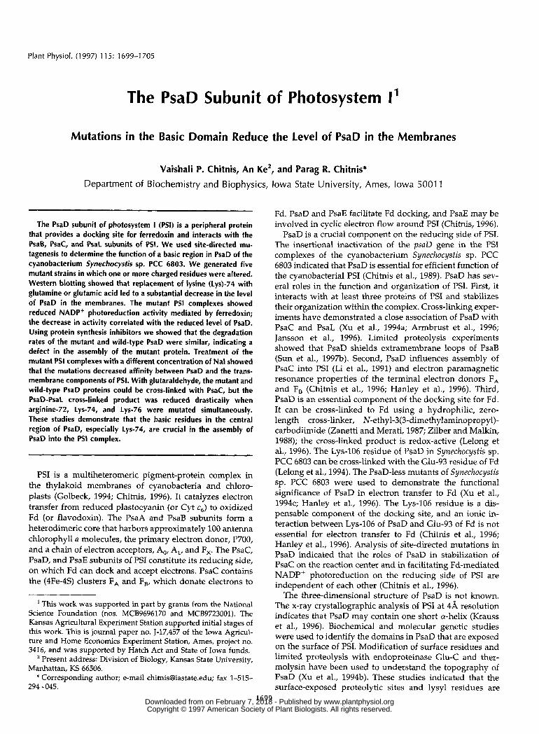
The effects of mutations on PSI function could resultfrom a defect in Fd docking or from decreased accumula-tion of mutant proteins in the PSI complexes. To examinethese possibilities, relative levels of PsaD and other PSIproteins in thylakoids and in purified PSI complexes wereestimated by western analyses (Fig. 1). The epitopes for theanti-PsaD polyclonal antibody are located in the regionbetween Glu-93 and the carboxyl terminus (Xu et al.,1994b). Therefore, mutations in the basic domain betweenArg-72 and Lys-86 are not expected to alter the affinity ofthis antibody for PsaD. In addition, when the protein sub-units of the purified PSI complexes were resolved by elec-trophoresis and the gel was stained with Coomassie blue,the band intensity of PsaD correlated with the correspond-
ing signal in western blotting (data not shown). Thus,immunodetection was expected to provide reliable esti-mates of the relative levels for PsaD.
Western analysis of thylakoid proteins was performedon an equal chlorophyll basis. The level of PsaD in mutantstrains 5 and 7 was considerably lower than in the wild-type strain (Fig. 1). From densitometry, we estimated thatthese strains contained approximately 25% of the wild-typelevel of PsaD. In contrast, thylakoids of the mutant strain19 had approximately 65% of the wild-type level of PsaD.Other mutants had intermediate levels of PsaD in theirthylakoid membranes. Mutations in the surface-exposedresidues, such as Lys-106, do not affect the steady-statelevel of PsaD in membranes (Chitnis et al., 1996). Mutantstrain 19 contained a reduced level of PsaK. Since thePsaD-PsaK interaction has not been demonstrated or antic-ipated, we do not know the significance of this observation.Mutations in PsaD did not have a major influence on theaccumulation of other PSI subunits in thylakoid mem-branes (Fig. 1). When the monomeric PSI complexes thathad been purified in the presence of Triton X-100 were
Thylakoid Membranes
I<
t.0
Strain KD1
PsaB
PsaC
13 19Photosystem I Complexes
KD1 3 5 7 13 19
PsaD
PsaE
PsaF
PsaK
PsaL
Figure 1. Western analyses of PSI subunits inthe thylakoid membranes and purified PSI com-plexes. The thylakoids and PSI complexes werepurified from the PsaD-less (ADC4), the wild-type (KD1), and the PsaD mutant strains. Themembranes and PSI complexes (containing 10jug of chlorophyll) were solubilized and re-solved by Tricine/urea/SDS-PAGE. The sepa-rated proteins were transferred to anImmobilon-P membrane (Millipore) and probedwith several antibodies. The immunoreactionwas visualized by enhanced chemilumines-cence.
www.plantphysiol.orgon February 7, 2018 - Published by Downloaded from Copyright © 1997 American Society of Plant Biologists. All rights reserved.
1702 Chitnis et al. Plant Physiol. Vol. 115, 1997
Strain KD1 #7Time (h) 0 11 24 0 11
Antibodyagainst
PsaD
PsaL
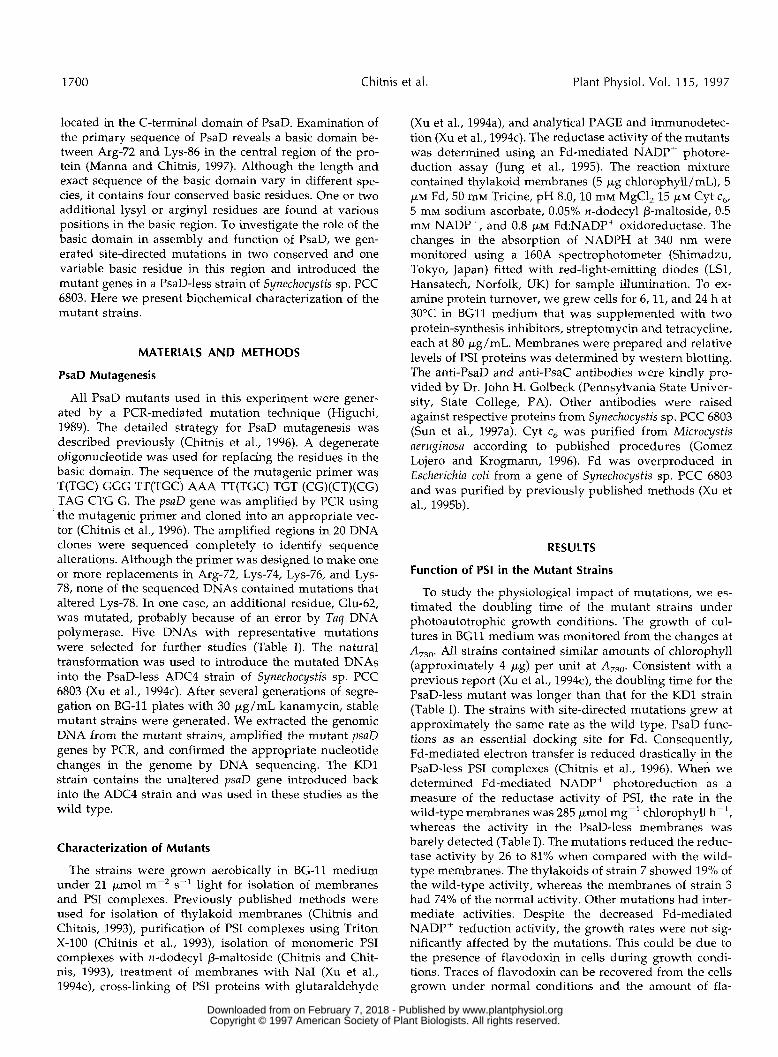
Figure 2. Levels of PsaD and PsaL after treatment with protein syn-thesis inhibitors. The cells of the wild type and mutant 7 strains (#7)in the wild-type and DE strains were grown at 38°C in the presenceof the protein-synthesis inhibitors streptomycin and tetracycline at afinal concentration of 80 /j.g/ml each. The cells were harvested at 6,11, and 24 h after initiation of the experiment. Subsequently, thephotosynthetic membranes were isolated. The proteins in the prep-arations containing 10 /j,g of chlorophyll were separated on Tricine-SDS-urea-PAGE, transferred to Immobilon-P membranes, and probedwith anti-PsaD and anti-PsaL antibodies. The immunoreaction wasvisualized by enhanced chemiluminescence.
used for western blotting, we observed similar trends inaccumulation of PsaD (Fig. 2). The PSI complexes of mu-tant strains 5 and 7 contained the least amount of PsaDamong the PSI complexes used in this experiment. In ad-dition, the level of PsaL in these complexes was reduced; asimilar decrease in accumulation of PsaL has been reportedfor the PsaD-less strain of Synechocystis sp. PCC 6803 (Xu etal., 1994a). It is interesting that PsaD of mutant strains 3, 5,and 7 migrated slightly faster than the wild-type PsaDprotein (Fig. 1). The changes in charges on the proteinmight affect SDS binding and thus alter migration duringelectrophoresis. Thus, the site-directed mutations in thebasic region reduced the abundance of PsaD in the PSIcomplexes and in the thylakoid membranes. The decreasedlevels of PsaD might have caused a decrease in theFd-mediated electron transfer activity of the mutant PSIcomplexes.
Stability of PsaD in Mutant Strain 7
The reduced level of PsaD in the mutant strains could bedue to the decreased expression of the mutant genes, im-paired assembly of mutant proteins into membranes, orincreased turnover of mutant proteins. Although we can-not rule out effects of mutations on transcription, transla-tion, or RNA stability, these possibilities are less likely thanthe effects on assembly or degradation. The mutationscaused relatively minor changes in the nucleotide sequenceand are more than 200 nucleotides downstream from theregions involved in transcription of the psaD gene or trans-lation of its mRNA. To test the effects of mutations on PsaDturnover, we grew the KD1 and strain-7 cells in the pres-ence of antibiotics that inhibited protein synthesis. Wemonitored levels of PsaD and PsaL in thylakoid mem-branes by western blotting (Fig. 2). The wild-type PSIcomplexes and its protein constituents are turned over veryslowly (Chitnis and Nelson, 1992b). Cellular chlorophyllcontent, PSI activity, or level of the PSI proteins is notaltered for up to 24 h of antibiotic treatment (data notshown). In contrast, the level of the Dl protein of PSIIrapidly decreased under these conditions. The relative
amounts of PsaD and PsaL did not decrease significantlyafter up to 24 h of treatment with antibiotics in the mutantand KD1 strains. Variation in the immunodetection inten-sity was within the error of these analyses. Therefore, themutation in strain 7 does not cause increased degradationof PsaD that has been assembled in the complexes. Whenwe used cytoplasmic fractions in western analysis, wefailed to detect soluble PsaD protein. It is likely that theunassembled PsaD is rapidly degraded by the cytoplasmicproteases. Overall, our results imply that the mutation instrain 7 affects assembly of PsaD into PSI.
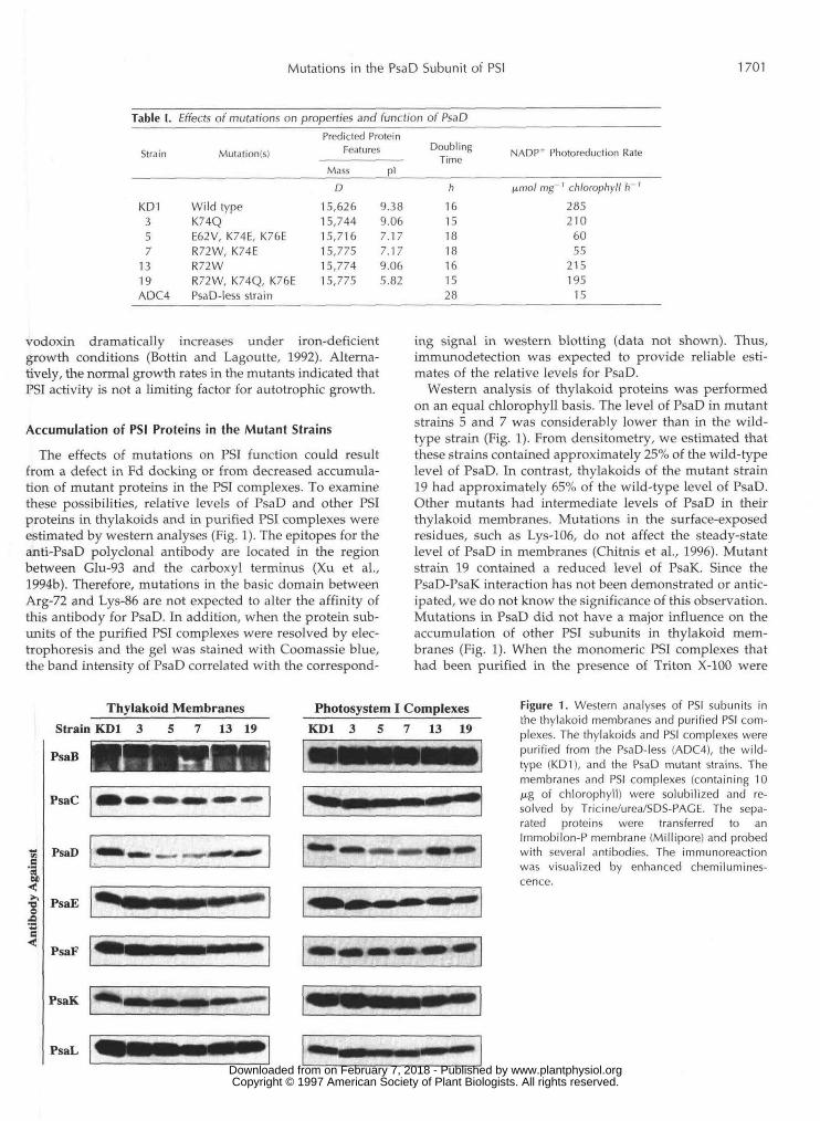
Treatment of Membranes with Nal
To study the defect in assembly of the mutant PsaDproteins, we examined interaction of PsaD with other PSIproteins. Resistance of PSI subunits to chaotropic treat-ments has been used to assess the relative strength of theirinteractions within the PSI complexes (Xu et al., 1994d,1994e; Chitnis et al., 1996). When thylakoid membranes ofthe wild-type and PsaD mutants were incubated with 0,1,2, 3, and 4 M Nal, their subunits differed in their suscepti-bility to removal by Nal (Fig. 3), PsaC and PsaE in thePsaD-less ADC4 strain were more susceptible to removalby Nal treatment than in the wild type (KD1) or in mutantstrain 7. One mole of Nal could remove PsaC and PsaEsubunits from the thylakoid membranes from ADC4. Toremove PsaC and PsaE from the thylakoids of the KD1strain, 3 M Nal or more was needed. Treatment with 2 MNal could remove PsaD from the thylakoid membranes ofmutant strain 7, but 3 M Nal was needed to remove PsaC,showing different effects of the PsaD mutations on thesesubunits. The results of Nal treatment demonstrated that
•oo
Strain____Nal(M) 0 1
PsaC
ADC4 KD1 #72 3 4 0 1 2 3 4 0 1 2 3 4
PsaD
PsaE
PsaL
Figure 3. Removal of peripheral proteins of PSI by Nal treatment.Relative contents of the PsaC, PsaD, PsaE, and PsaL subunits in thethylakoid membranes purified from the PsaD-less (ADC4), the wild-type (KD1), and a PsaD mutant (#7) strain after treatment with Nal.The membranes were incubated with 0, 1, 2, 3, or 4 M Nal for 15 minon ice. Samples containing 10 ju.g of chlorophyll were solubilizedand resolved by Tricine/urea/SDS-PAGE. Separated proteins weretransferred to an Immobilon-P membrane and probed with differentantibodies. The immunoreaction was visualized by enhanced chemi-luminescence. For immunodetection of PsaD in membranes of mu-tant strain 7, samples with 25 jug of chlorophyll were used in eachlane and the blots were exposed longer to the x-ray film. www.plantphysiol.orgon February 7, 2018 - Published by Downloaded from
Copyright © 1997 American Society of Plant Biologists. All rights reserved.
Mutations in the PsaD Subunit of PSI 1703
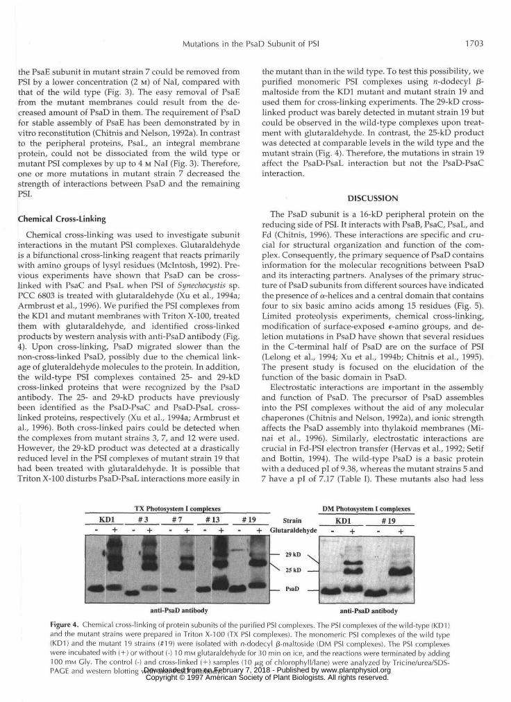
the PsaE subunit in mutant strain 7 could be removed fromPSI by a lower concentration (2 M) of Nal, compared withthat of the wild type (Fig. 3). The easy removal of PsaEfrom the mutant membranes could result from the de-creased amount of PsaD in them. The requirement of PsaDfor stable assembly of PsaE has been demonstrated by invitro reconstitution (Chitnis and Nelson, 1992a). In contrastto the peripheral proteins, PsaL, an integral membraneprotein, could not be dissociated from the wild type ormutant PSI complexes by up to 4 M Nal (Fig. 3). Therefore,one or more mutations in mutant strain 7 decreased thestrength of interactions between PsaD and the remainingPSI.
Chemical Cross-Linking
Chemical cross-linking was used to investigate subunitinteractions in the mutant PSI complexes. Glutaraldehydeis a bifunctional cross-linking reagent that reacts primarilywith amino groups of lysyl residues (Mclntosh, 1992). Pre-vious experiments have shown that PsaD can be cross-linked with PsaC and PsaL when PSI of Synechocystis sp.PCC 6803 is treated with glutaraldehyde (Xu et al., 1994a;Armbrust et al., 1996). We purified the PSI complexes fromthe KD1 and mutant membranes with Triton X-100, treatedthem with glutaraldehyde, and identified cross-linkedproducts by western analysis with anti-PsaD antibody (Fig.4). Upon cross-linking, PsaD migrated slower than thenon-cross-linked PsaD, possibly due to the chemical link-age of gluteraldehyde molecules to the protein. In addition,the wild-type PSI complexes contained 25- and 29-kDcross-linked proteins that were recognized by the PsaDantibody. The 25- and 29-kD products have previouslybeen identified as the PsaD-PsaC and PsaD-PsaL cross-linked proteins, respectively (Xu et al., 1994a; Armbrust etal., 1996). Both cross-linked pairs could be detected whenthe complexes from mutant strains 3, 7, and 12 were used.However, the 29-kD product was detected at a drasticallyreduced level in the PSI complexes of mutant strain 19 thathad been treated with glutaraldehyde. It is possible thatTriton X-100 disturbs PsaD-PsaL interactions more easily in
the mutant than in the wild type. To test this possibility, wepurified monomeric PSI complexes using n-dodecyl j3-maltoside from the KD1 mutant and mutant strain 19 andused them for cross-linking experiments. The 29-kD cross-linked product was barely detected in mutant strain 19 butcould be observed in the wild-type complexes upon treat-ment with glutaraldehyde. In contrast, the 25-kD productwas detected at comparable levels in the wild type and themutant strain (Fig. 4). Therefore, the mutations in strain 19affect the PsaD-PsaL interaction but not the PsaD-PsaCinteraction.
DISCUSSION
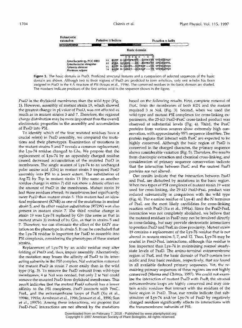
The PsaD subunit is a 16-kD peripheral protein on thereducing side of PSI. It interacts with PsaB, PsaC, PsaL, andFd (Chitnis, 1996). These interactions are specific and cru-cial for structural organization and function of the com-plex. Consequently, the primary sequence of PsaD containsinformation for the molecular recognitions between PsaDand its interacting partners. Analyses of the primary struc-ture of PsaD subunits from different sources have indicatedthe presence of a-helices and a central domain that containsfour to six basic amino acids among 15 residues (Fig. 5).Limited proteolysis experiments, chemical cross-linking,modification of surface-exposed e-amino groups, and de-letion mutations in PsaD have shown that several residuesin the C-terminal half of PsaD are on the surface of PSI(Lelong et al., 1994; Xu et al., 1994b; Chitnis et al., 1995).The present study is focused on the elucidation of thefunction of the basic domain in PsaD.
Electrostatic interactions are important in the assemblyand function of PsaD. The precursor of PsaD assemblesinto the PSI complexes without the aid of any molecularchaperones (Chitnis and Nelson, 1992a), and ionic strengthaffects the PsaD assembly into thylakoid membranes (Mi-nai et al., 1996). Similarly, electrostatic interactions arecrucial in Fd-PSI electron transfer (Hervas et al., 1992; Setifand Bottin, 1994). The wild-type PsaD is a basic proteinwith a deduced pi of 9.38, whereas the mutant strains 5 and7 have a pi of 7.17 (Table I). These mutants also had less
TX Photosystem I complexesKD1 #3 #7 #13 #19
DM Photosystem I complexes
Strain KD1 #19Glutaraldehyde - + - +
anti-PsaD antibody anti-PsaD antibody
Figure 4. Chemical cross-linking of protein subunits of the purified PSI complexes. The PSI complexes of the wild-type (KD1)and the mutant strains were prepared in Triton X-100 (TX PSI complexes). The monomeric PSI complexes of the wild type(KD1) and the mutant 19 strains (#19) were isolated with n-dodecyl /3-maltoside (DM PSI complexes). The PSI complexeswere incubated with ( + ) or without (-) 10 HIM glutaraldehyde for 30 min on ice, and the reactions were terminated by adding100 mM Gly. The control (-) and cross-linked ( + ) samples (10 /ng of chlorophyll/lane) were analyzed by Tricine/urea/SDS-PACE and western blotting with anti-PsaD antibody. www.plantphysiol.orgon February 7, 2018 - Published by Downloaded from
Copyright © 1997 American Society of Plant Biologists. All rights reserved.
1704
PsaD in
Chitnis et al.
Eukaryotic extension Putative a helices
Plant Physiol. Vol. 115, 1997
Putative a helix
Basic domain
Y C + , + - + ca Synechocysfis sp. PCC 6803 6 6 Q- Synechococcus elongatus 66 QQ
91 R- R- Spinacia oleracea
Hordeum vulgare i a
Figure 5. The basic domain in PsaD. Predicted structural features and a comparison of selected sequences of the basic domain are shown. Although two to three regions of PsaD are predicted to form a-helices, only one a-helix has been assigned to PsaD in the 4 A structure of PSI (Krauss et al., 1996). The conserved residues in the basic domain are shaded. The numbers indicate positions of the first amino acid in the sequence shown in the figure.
the thylakoid membranes than the wild type (Fig. 1). However, assembly of mutant strain 19, which showed the greatest change in pI value of PsaD, was not affected as much as in mutant strains 5 and 7. Therefore, the regional charge distribution may be more important than the overall electrostatic properties in the assembly and accumulation of PsaD into PSI.
To identify which of the four mutated residues have a crucial role(s) in PsaD assembly, we compared the muta- tions and their phenotypes. Examination of mutations in the mutant strains 5 and 7 reveals a common replacement; the Lys-74 residue changed to Glu. We propose that the replacement of Lys-74 by an oppositely charged residue caused decreased accumulation of the mutated PsaD in membranes. The single mutant of Lys-74 to an uncharged polar amino acid (Gln) in mutant strain 3 impaired PsaD assembly into PSI to a lesser extent. The substitution of Arg-72 by Trp in mutant strain 13 (the same as another residue change in strain 7) did not show a drastic effect on the amount of PsaD in the membranes. Mutant strain 19 had three residues altered; its membranes had significantly more PsaD than mutant strain 5. This mutant had an iden- tical replacement (K76E) as one of the mutations in mutant strain 5, and its other residue substitution (R72W) was also present in mutant strain 7. The third residue change in strain 19 was Lys-74 replaced by Gln (the same as that in mutant strain 3) instead of by Glu, as that in strains 5 and 7. Therefore, we can eliminate the effect of the E62V mu- tation on the phenotype in strain 5. It can be concluded that the Lys-74 residue is important for PsaD to assemble into PSI complexes, considering the phenotypes of these mutant str ains .
Replacement of Lys-74 by an acidic residue may alter folding of PsaD and, thus, impair assembly. Alternatively, the mutation may lessen the affinity of PsaD to its inter- acting subunits in the PSI complex. Na1 extraction removed the mutant PsaD in strain 7 more easily than in the wild type (Fig. 3). To remove the PsaD subunit from wild-type membranes, 4 M Na1 was needed, but only 2 M Na1 could remove the mutated PsaD from thylakoid membranes. This result indicates that the mutant PsaD subunit has a lower affinity to the PSI complexes. PsaD interacts with PsaC, PsaL, and the extramembrane loops of PsaB (Xu et al., 1994a, 1995a; Armbrust et al., 1996; Jansson et al., 1996; Sun et al., 199%). Among these interactions, we propose that PsaD-PsaC interactions are not altered by the mutations
based on the following results. First, complete remova1 of PsaC from the membranes of both KD1 and the mutant required 3 M NaI, (Fig. 3). Second, when we used the wild-type and mutant PSI complexes for cross-linking ex- periments, the 25-kD PsaD-PsaC cross-linked product was observed at substantial levels (Fig. 4). Third, the PsaC proteins from various sources show extremely high con- servation, with approximately 95% sequence identities. The protein regions that interact with PsaC are expected to be highly conserved. Although the basic region of PsaD is conserved in the charged character, the primary sequence shows considerable variation (Fig. 5). Therefore, the results from chaotropic extraction and chemical cross-linking, and consideration of primary sequence conservation indicate that the interaction between PsaC and the mutant PsaD proteins are not altered.
Our results indicate that the interaction between PsaD and PsaL are disturbed by mutations in the basic region. When two types of PSI complexes of mutant strain 19 were used for cross-linking, the 29-kD PsaD-PsaL product was reduced substantially in comparison with the wild type (Fig. 4). The eamino residue of Lys-41 and the N terminus of PsaL are the most likely candidates for cross-linking reaction with PsaD (Xu et al., 1994a). Since the PsaD-PsaL interaction was not completely abolished, we believe that the mutated residues in PsaD may not be involved directly in cross-linking, but may provide electrostatic interactions to position PsaD and PsaL in close proximity. Mutant strain 19 contains a replacement of the Lys-76 residue that is not altered in mutant strains 3, 7, and 12. Thus, Lys-76 may be crucial in PsaD-PsaL interactions, although this residue is less important than Lys-74 in maintaining normal steady- state levels of PsaD. The amino-terminal extramembrane region of PsaL and the basic domain of PsaD contain two acidic and four basic residues, respectively, that are found in a11 available deduced primary sequences. Yet, the re- maining primary sequences of these regions are not highly conserved (Manna and Chitnis, 1997). We could not exam- ine the interaction of mutant PsaD with PsaB; the stromal extramembrane loops are highly conserved and may con- tain acidic residues that interact with the residues of the basic region in PsaD. Thus, our results indicate that sub- stitution of Lys-74 and/or Lys-76 of PsaD by negatively charged residues significantly affects its interactions with the transmembrane subunits of PSI.
www.plantphysiol.orgon February 7, 2018 - Published by Downloaded from Copyright © 1997 American Society of Plant Biologists. All rights reserved.
Mutations in the PsaD Subunit of PSI 1705
In summary, the present study shows that the basic domain of PsaD, especially its Lys-74 residue, is crucial for in vivo accumulation of PsaD. This region may interact with the acidic residues i n the amino-terminal region of PsaL or the stromal extramembrane loops of PsaB.
ACKNOWLEDCMENTS
We thank Dr. David Krogmann (Purdue University, West Lafayette, IN) for the gift of Cyt c6 and for critically reading the manuscript. We also acknowledge Dr. John Golbeck for anti-PsaD and anti-PsaC antibodies.
Received June 23, 1997; accepted September 4, 1997. Copyright Clearance Center: 0032-0889/97/115/1699/07.
LITERATURE ClTED
Armbrust TS, Chitnis PR, Guikema JA (1996) Organization of photosystem I polypeptides examined by chemical cross- linking. Plant Physiol 111: 1307-1312
Bottin H, Lagoutte B (1992) Ferredoxin and flavodoxin from the cyanobacterium Synechocystis sp PCC 6803. Biochim Biophys Acta 1101: 48-56
Chitnis PR (1996) Photosystem I. Plant Physiol 111: 661-669 Chitnis PR, Chitnis VP, Xu Q, Jung Y-S, Yu L, Golbeck JH (1995)
Mutational analysis of photosystem I polypeptides. In P Mathis, ed, Photosynthesis: from Light to Biosphere. Kluwer Academic Publishers, Dordrecht, The Netherlands, pp 17-22
Chitnis PR, Nelson N (1992a) Assembly of two subunits of the cyanobacterial photosystem I on the n-side of thylakoid mem- branes. Plant Physiol99: 239-246
Chitnis PR, Nelson N (1992b) Biogenesis of photosystem I: the subunit PsaE is important for the stability of PS I complex. ln J Argyroudi-Akoyunoglou, ed, Chloroplast Biogenesis. Plenum Press, New York, pp 285-290
Chitnis PR, Reilly PA, Nelson N (1989) Insertional inactivation of the gene encoding subunit I1 of photosystem I from the cyanobac- terium Synechocystis sp. PCC 6803. J Biol Chem 264 18381-18385
Chitnis VP, Chitnis PR (1993) PsaL subunit is required for the formation of photosystem I trimers in the cyanobacterium Syn- eckocystis sp. PCC 6803. FEBS Lett 336: 330-334
Chitnis VP, Jung Y-S, Albee L, Golbeck JH, Chitnis PR (1996) Mutational analysis of photosystem I polypeptides: role of PsaD and the lysyl 106 residue in the reductase activity of photosys- tem 1. J Biol Chem 271: 11772-11780
Chitnis VP, Xu Q, Yu L, Golbeck JH, Nakamoto H, Xie DL, Chitnis PR (1993) Targeted inactivation of the gene psaL encod- ing a subunit of photosystem I of the cyanobacterium Synecko- cystis sp. PCC 6803. J Biol Chem 268: 11678-11684
Golbeck JH (1994) Photosystem I in cyanobacteria. ln DA Bryant, ed, The Molecular Biology of Cyanobacteria. Kluwer Academic Publishers, Dordrecht, The Netherlands, pp 179-220
Gomez Lojero C, Krogmann DW (1996) Large scale preparations of photosynthetic catalysts from cyanobacteria. Photosynth Res
Hanley J, Setif P, Bottin H, Lagoutte B (1996) Mutagenesis of photosystem I in the region of the ferredoxin cross-linking site: modifications of positively charged amino acids. Biochemistry
Hervas M, Navarro J, Tollin G (1992) A laser-flash spectroscopy study of the kinetics of electron transfer from spinach photosys- tem I to spinach and alga1 ferredoxins. Photochem Photobiol 56:
Higuchi R (1989) Using PCR to engineer DNA. In HA Erlich, ed, PCR Technology. Stockton Press, New York, pp 61-70
Jansson S, Anderson B, Scheller H V (1996) Nearest-neighbor analysis of higher plant photosystem I holocomplex. Plant Physiol 112: 409-420
47 293-299
35: 8563-8571
319-324
Jung Y-S, Yu L, Golbeck JH (1995) Reconstitution of iron-sulfur center F, results in complete restoration of NADP+ photoreduc- tion in Hg-treated photosystem I complexes from Syneckococcus sp. PCC 6301. Photosynth Res 46: 249-255
Krauss N, Schubert W-D, Klukas O, Fromme P, Witt HT, Saenger W (1996) Photosystem I at 4 A resolution represents the first structural model of a joint photosynthetic reaction centre and core antenna system. Nature Struct Biol 3: 965-973
Lelong C, Boekema E, Kruip J, Bottin H, Rogner M, Setif P (1996) Characterization of a redox-active cross-linked complex be- tween cyanobacterial photosystem I and soluble ferredoxin.
Lelong C, Setif P, Lagoutte B, Bottin H (1994) Identification of the amino acids involved in the functional interaction between pho- tosystem I and ferredoxin from Syneckocystis sp. PCC 6803 by chemical cross-linking. J Biol Chem 269 10034-10039
Li N, Zhao J, Warren PV, Warden JT, Bryant DA, Golbeck JH (1991) PsaD is required for the stable binding of PsaC to the photosystem I core protein of Synechococcus sp. PCC 6301. Bio- chemistry 30: 7863-7872
Manna P, Chitnis PR (1997) Function and molecular genetics of photosystem I. In GS Singhal, G Renger, SK Sapory, eds, Con- cepts in Photobiology: Photosynthesis and Photomorphogen- esis. Kluwer Academic Publishers, Dordrecht, The Netherlands (in press)
Mchtosh DB (1992) Glutaraldehyde cross-links Lys-492 and Arg- 678 at the active site of sarcoplasmic reticulum Ca2+-ATPase. J Biol Chem 31: 22328-22335
Minai L, Cohen Y, Chitnis PR, Nechushtai R (1996) The precursor of PsaD assembles into the photosystem I complex in two steps. Proc Natl Acad Sci USA 93: 6338-6342
Setif PQ, Bottin H (1994) Laser flash absorption spectroscopy study of ferredoxin reduction by photosystem I in Syneckocystis sp. PCC 6803: evidence for submicrosecond and microsecond kinetics. Biochemistry 33: 8495-8504
Sun J, Ke A, Jin P, Chitnis VP, Chitnis PR (1997a) Isolation and functional study of photosystem I subunits in the cyanobacte- rium Synechocystis sp. PCC 6803. Methods Enzymol (in press)
Sun J, Xu Q, Chitnis VP, Jin P, Chitnis PR (1997b) Topography of the photosystem I core proteins of the cyanobacterium Synecho- cystis sp. PCC 6803. J Biol Chem 272: 21793-21802
Xu Q, Armbrust TS, Guikema JA, Chitnis PR (1994a) Organiza- tion of photosystem I polypeptides. A structural interaction between PsaD and PsaL subunits. Plant Physiol 106 1057-1063
Xu Q, Chitnis VP, Ke A, Chitnis PR (1995a) Structural Organiza- tion of Photosystem I. In P Mathis, ed, Photosynthesis: From Light to Biosphere. Kluwer Academic Publishers, Dordrecht, The Netherlands, pp 87-90
Xu Q, Guikema JA, Chitnis PR (1994b) Identification of surface- exposed domains on the reducing side of photosystem I. Plant Physiol 106 617-624
Xu Q, Hoppe D, Chitnis VP, Odom WR, Guikema JA, Chitnis PR (199510) Mutational analysis of photosystem I polypeptides in the cyanobacterium Syneckocystis sp. PCC 6803. Targeted inac- tivation of psaI reveals the function of PsaI in the structural organization of PsaL. J Biol Chem 270 16243-16250
Xu Q, Jung YS, Chitnis VP, Guikema JA, Golbeck JH, Chitnis PR (1994~) Mutational analysis of photosystem I polypeptides in Syneckocystis sp. PCC 6803. Subunit requirements for reduction of NADPt mediated by ferredoxin and flavodoxin. J Biol Chem
Xu Q, Odom WR, Guikema JA, Chitnis VP, Chitnis PR (1994d) Targeted deletion of psaJ from the cyanobacterium Syneckocystis sp. PCC 6803 indicates structural interactions between the PsaJ and PsaF subunits of photosystem I. Plant Mo1 Biol2624 291-302
Xu Q, Yu L, Chitnis VP, Chitnis PR (1994e) Function and orga- nization of photosystem I in a cyanobacterial mutant strain that lacks PsaF and PsaJ subunits. J Biol Chem 269: 3205-3211
Zanetti G, Merati G (1987) Interaction between photosystem I and ferredoxin: identification by chemical cross-linking of the polypeptide which binds ferredoxin. Eur J Biochem 169: 143-146
Zilber A, Malkin R (1988) Ferredoxin cross-links to a 22-kD sub- unit of photosystem I. Plant Physiol 88: 810-814
EMBO J 15: 2160-2168
2693: 21512-21518
www.plantphysiol.orgon February 7, 2018 - Published by Downloaded from Copyright © 1997 American Society of Plant Biologists. All rights reserved.