Embed Size (px)
Citation preview
The Radiological Diagnostics of the
Respiratory System
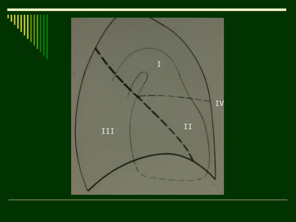
I
IIIII
IV
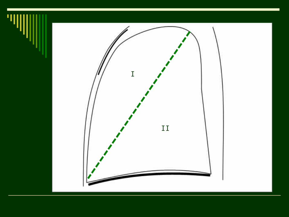
I
II
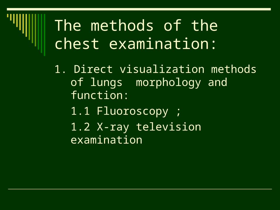
The methods of the chest examination:
1. Direct visualization methods of lungs morphology and function:
1.1 Fluoroscopy ;
1.2 X-ray television examination
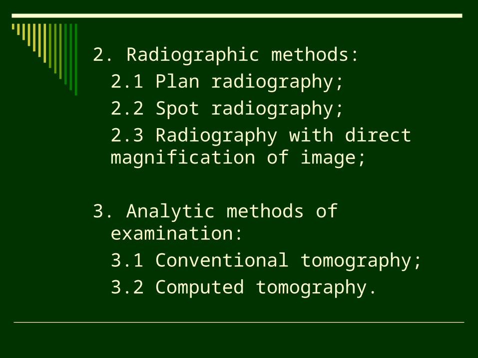
2. Radiographic methods:
2.1 Plan radiography;
2.2 Spot radiography;
2.3 Radiography with direct magnification of image;
3. Analytic methods of examination:
3.1 Conventional tomography;
3.2 Computed tomography.
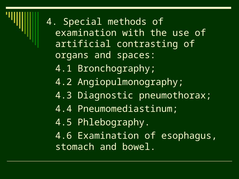
4. Special methods of examination with the use of artificial contrasting of organs and spaces:
4.1 Bronchography;
4.2 Angiopulmonography;
4.3 Diagnostic pneumothorax;
4.4 Pneumomediastinum;
4.5 Phlebography.
4.6 Examination of esophagus, stomach and bowel.
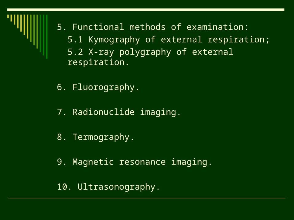
5. Functional methods of examination:
5.1 Kymography of external respiration;
5.2 X-ray polygraphy of external respiration.
6. Fluorography.
7. Radionuclide imaging.
8. Termography.
9. Magnetic resonance imaging.
10. Ultrasonography.
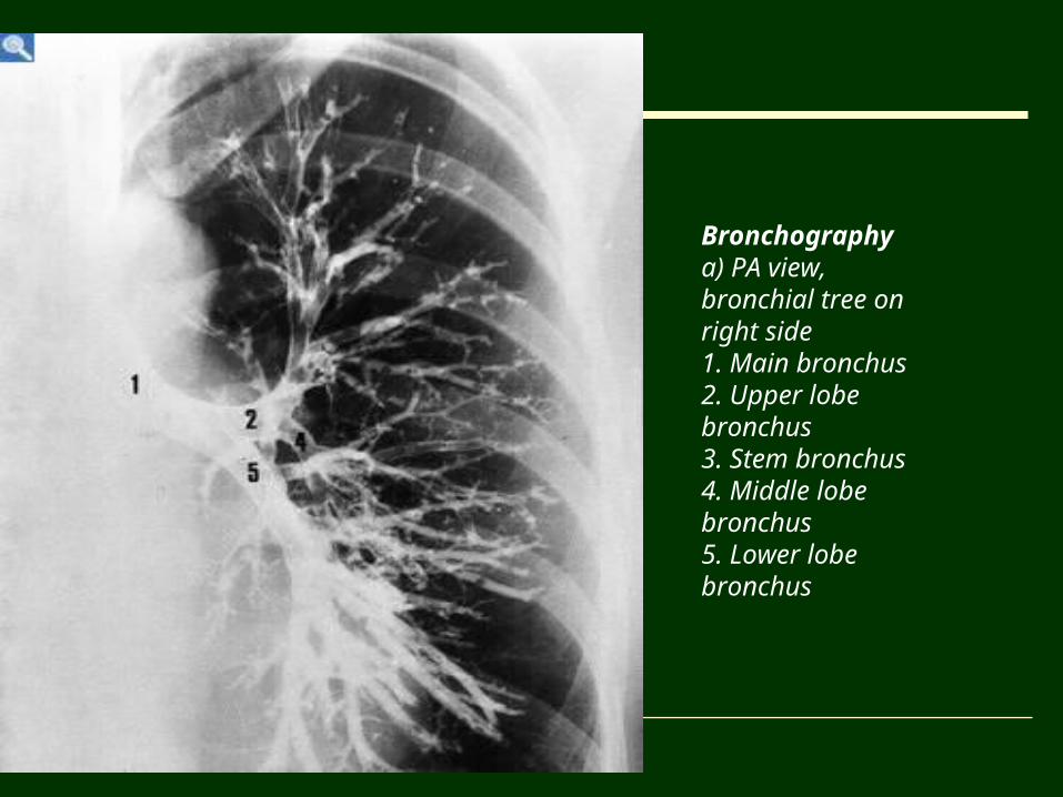
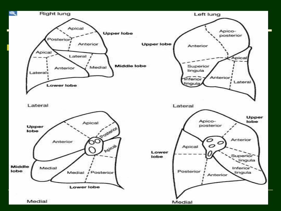
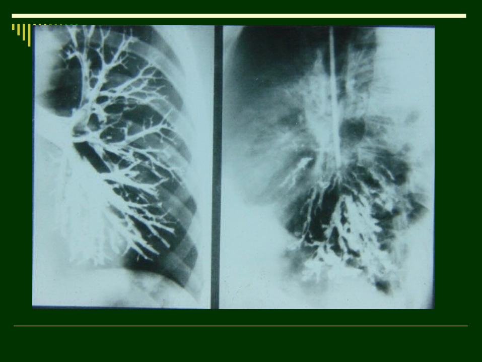
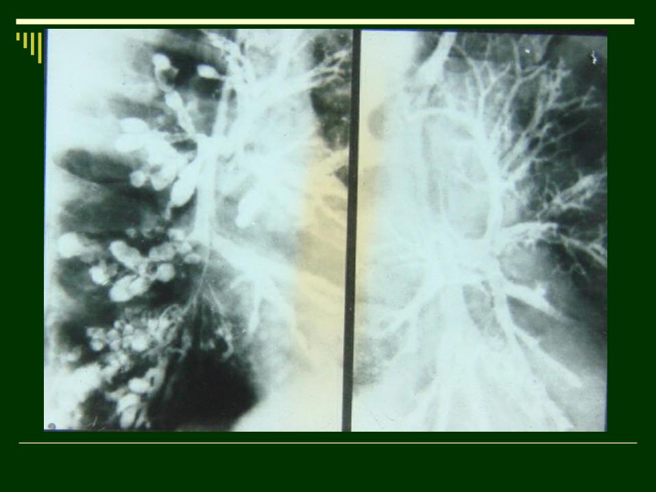
Bronchographya) PA view, bronchial tree on right side 1. Main bronchus2. Upper lobe bronchus3. Stem bronchus4. Middle lobe bronchus5. Lower lobe bronchus
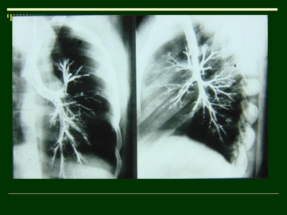
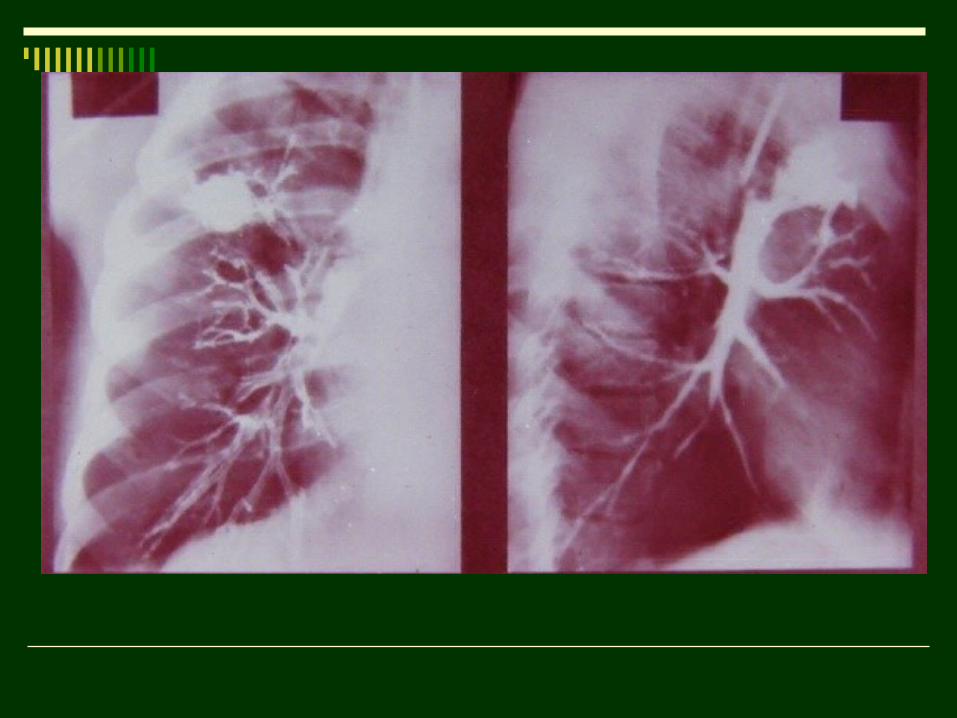
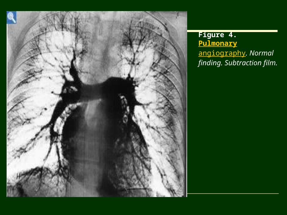
Figure 4. Pulmonary angiography. Normal finding. Subtraction film.
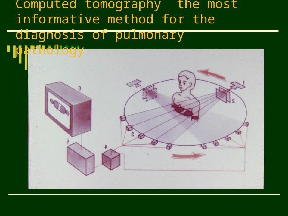
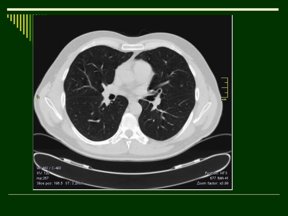
Сomputed tomography the most informative method for the diagnosis of pulmonary pathology
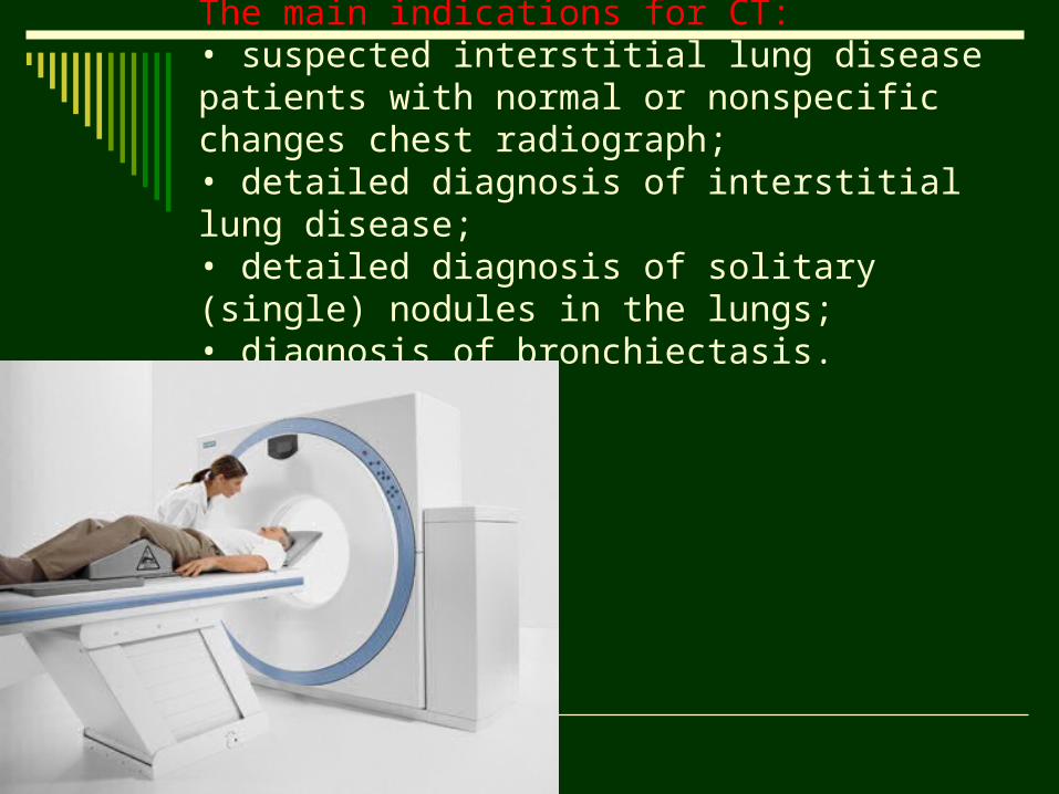
The main indications for CT: • suspected interstitial lung disease patients with normal or nonspecific changes chest radiograph; • detailed diagnosis of interstitial lung disease; • detailed diagnosis of solitary (single) nodules in the lungs; • diagnosis of bronchiectasis.
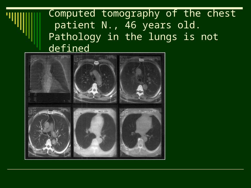
Computed tomography of the chest patient N., 46 years old. Pathology in the lungs is not defined
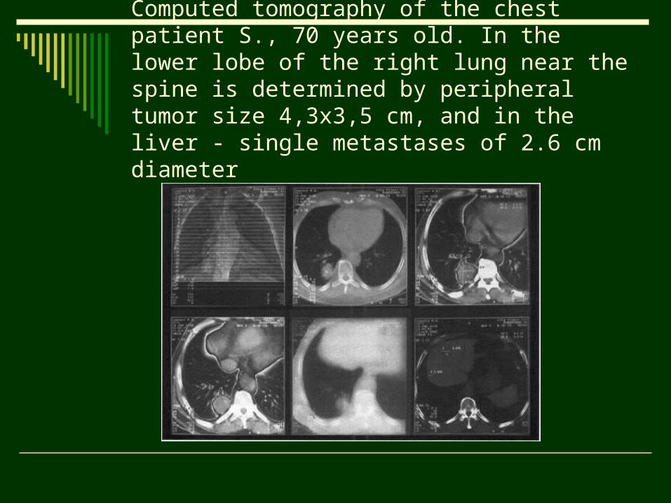
Computed tomography of the chest patient S., 70 years old. In the lower lobe of the right lung near the spine is determined by peripheral tumor size 4,3x3,5 cm, and in the liver - single metastases of 2.6 cm diameter
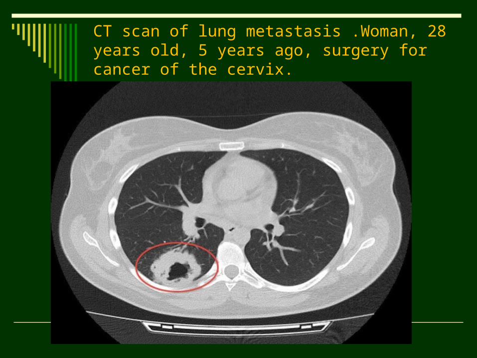
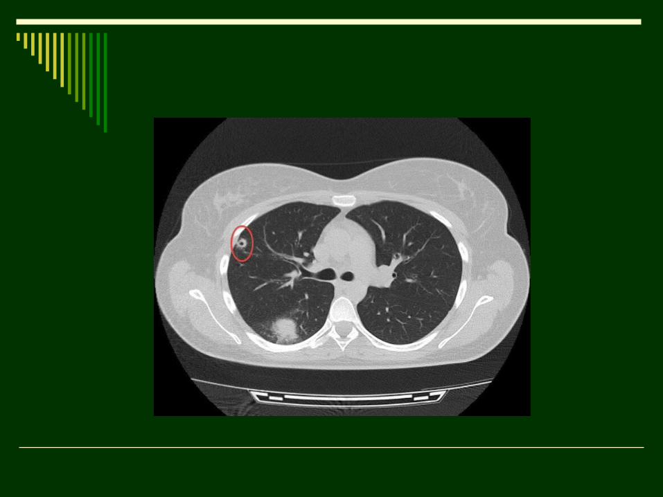
CT scan of lung metastasis .Woman, 28 years old, 5 years ago, surgery for cancer of the cervix.
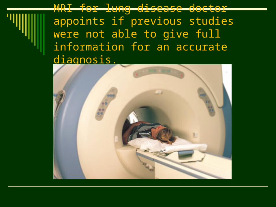
MRI for lung disease doctor appoints if previous studies were not able to give full information for an accurate diagnosis.
MRI Thanks to a high-quality image anatomy of blood vessels and
the formation of a multi-dimensional MRI images is the best way to diagnose congenital cardiovascular anomalies, and aortic aneurysm ruptures.
Most authors isolated following clinical situations in which MRI chest preferred over computer tomography:
• assessment of the neurovascular structures in the Pancoast tumor - evaluation of operability;
• differential diagnosis of recurrent lymphoma and fibrosis appeared after treatment of lymphoma;
• diagnosis of lung tumor invasion into the chest wall, the roots of the lungs, mediastinum;
• congenital cardiovascular anomalies; • diagnosis of pericarditis
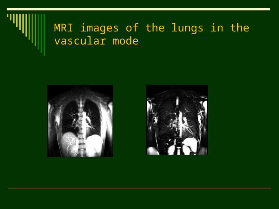
MRI images of the lungs in the vascular mode
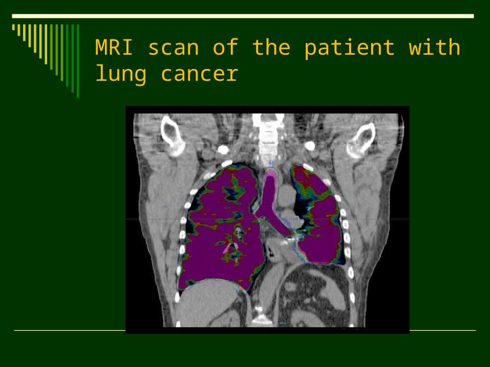
MRI scan of the patient with lung cancer
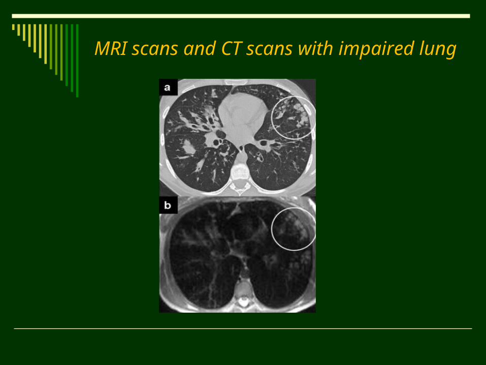
MRI scans and CT scans with impaired lung
Absolute contraindications to MRI are implanted pacemaker or any metal structures in the human body, which can unexpectedly shift work or in a magnetic field, causing tissue damage. Relative contraindications include claustrophobia and pregnancy, although there is no conclusive evidence about the harmful effects of MRI on the fetus.
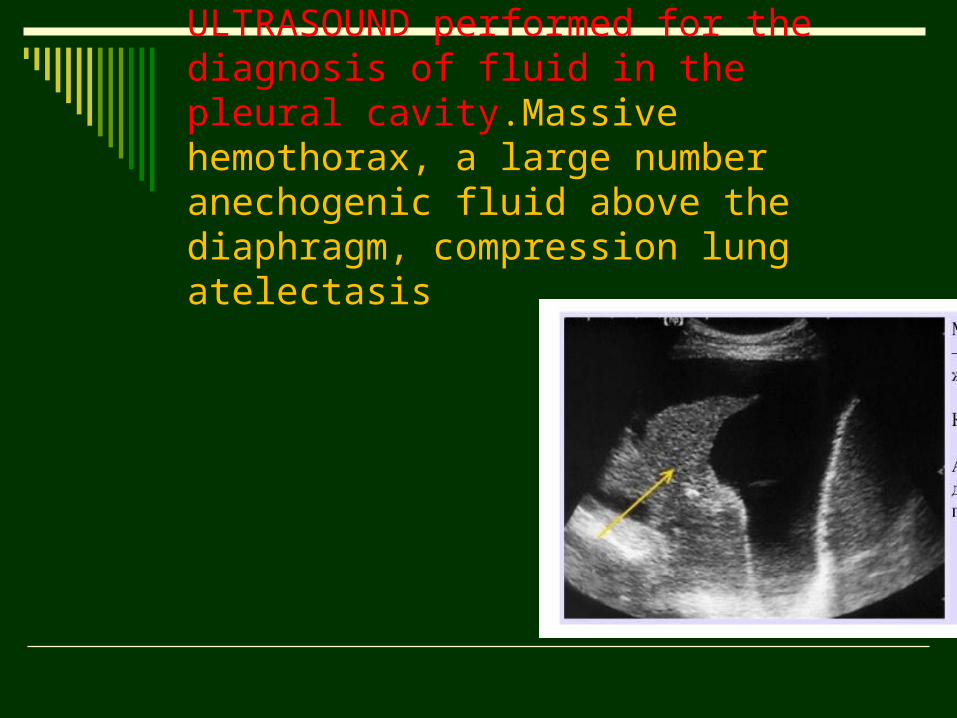
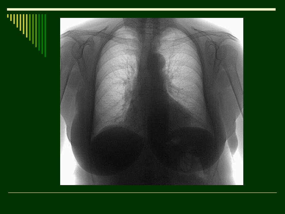
ULTRASOUND performed for the diagnosis of fluid in the pleural cavity.Massive hemothorax, a large number anechogenic fluid above the diaphragm, compression lung atelectasis
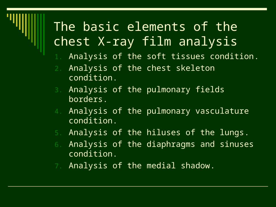
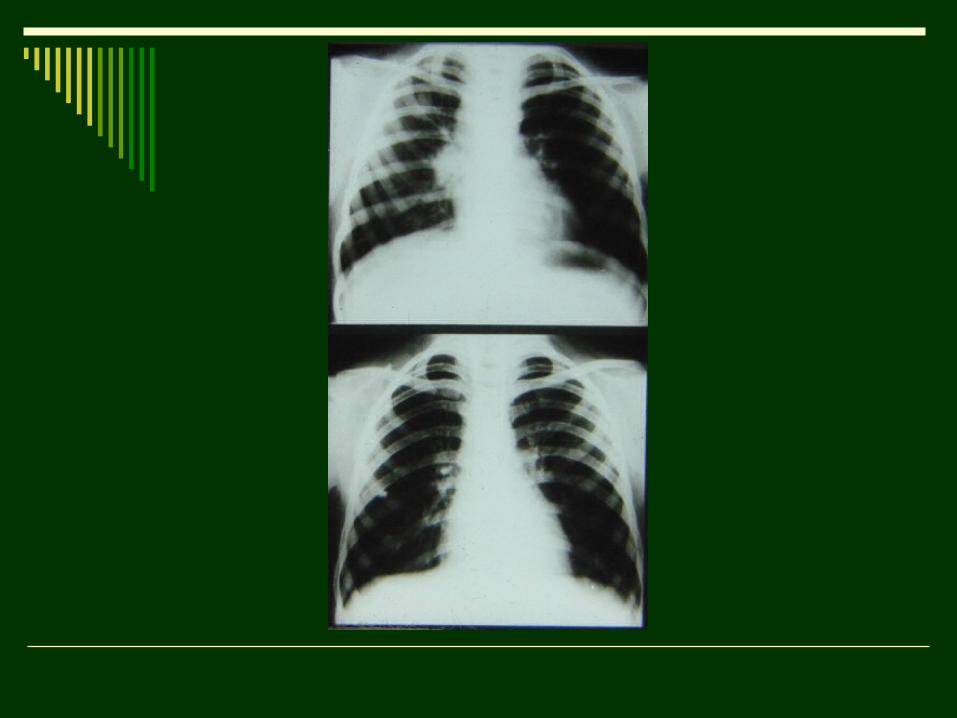
The basic elements of the chest X-ray film analysis1. Analysis of the soft tissues condition.
2. Analysis of the chest skeleton condition.
3. Analysis of the pulmonary fields borders.
4. Analysis of the pulmonary vasculature condition.
5. Analysis of the hiluses of the lungs.
6. Analysis of the diaphragms and sinuses condition.
7. Analysis of the medial shadow.
The main task of radiololgist is to differentiate normal and pathological condition. There are three symptoms of pulmonary diseases:
а. Air-free – opacity.
b. Clarification.
c. Changes of pulmonary vasculature.
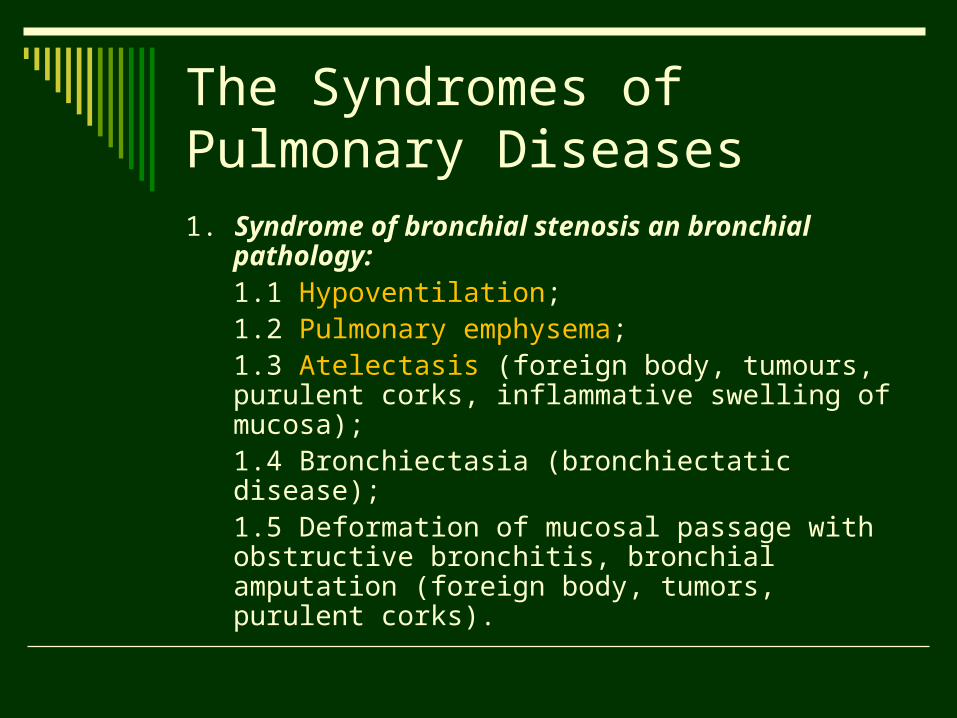
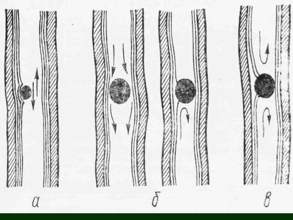
The Syndromes of Pulmonary Diseases1. Syndrome of bronchial stenosis an
bronchial pathology:1.1 Hypoventilation;1.2 Pulmonary emphysema;1.3 Atelectasis (foreign body, tumours, purulent corks, inflammative swelling of mucosa);1.4 Bronchiectasia (bronchiectatic disease);1.5 Deformation of mucosal passage with obstructive bronchitis, bronchial amputation (foreign body, tumors, purulent corks).
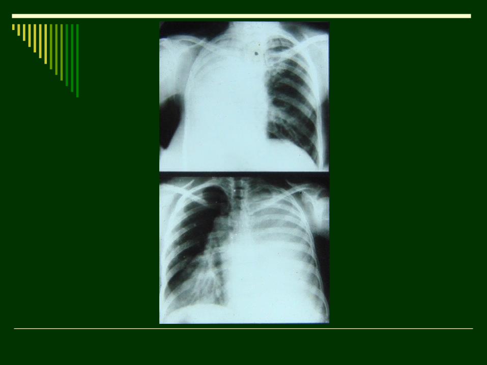
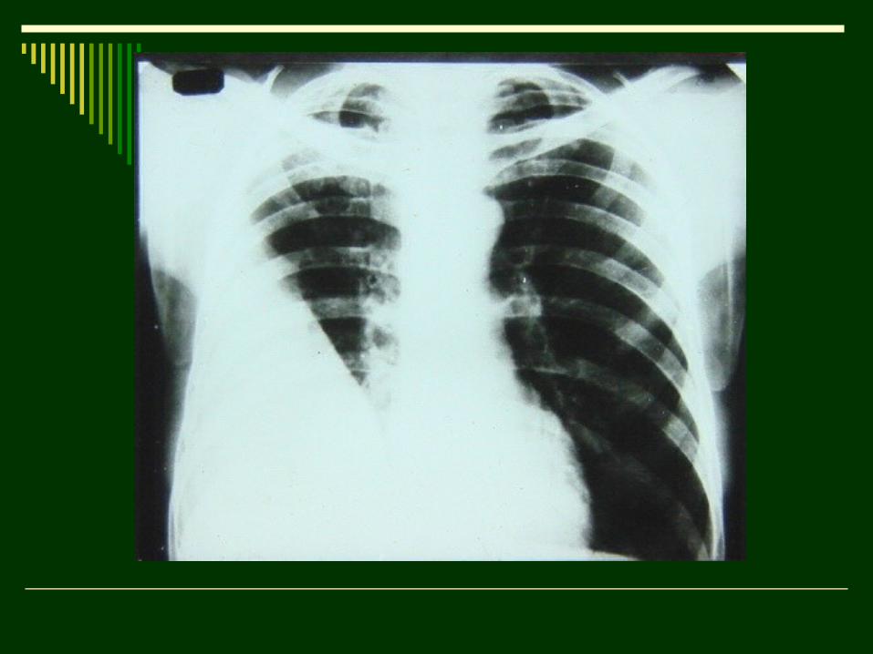
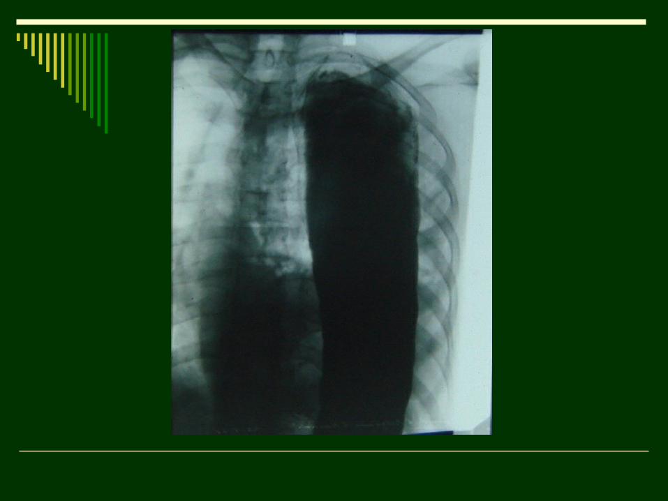
2. Syndrome of total and subtotal opacity:
Depends on pathology:
2.1 Of Lungs (atelectasis; inflammation – tuberculosis, pneumonia, cirrhosis);
2.2 Of Pleura (fluid; massive pleural adhesions; fibrothorax);
2.3 Of Diaphragm (hernia).
3. Syndrome of limited opacity:3.1 Atelectasis (lobar, segmental – cancer, foreign
body of lobar segmental bronchus);3.2 Inflammation of the part of lung (pneumonia,
infiltrative tuberculosis);3.3 Cirrhosis of the part of lung (bronchiectatic
disease, tuberculosis);3.4 Tumor (cancer in main bronchus without
atelectasis, peripheral tumor);3.5 Plural effusion (hydrothorax, hemothorax,
exudative pleuritis);3.6 Pleural adhesions (after operations, injury,
pleuritis);3.7 Abdominal organs in thoracic cavity.
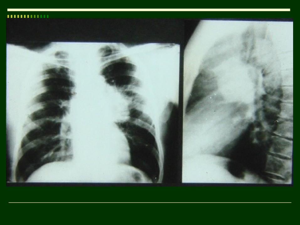
4. Syndrome of Round Shadow:4.1 Inflammation (acute pneumonia,
eosynophilic infiltration, tuberculous infiltration, tubelculoma);
4.2 Cysts (retention, echinococcus, mediastinal);
4.3 Tumor (primary carcinoma, metastases, mediastinal tumor);
4.4 Encapsulated fluid accumulation in pleural cavity (pleuritis – costal, mediastinal, diaphragmatic, interlobar).
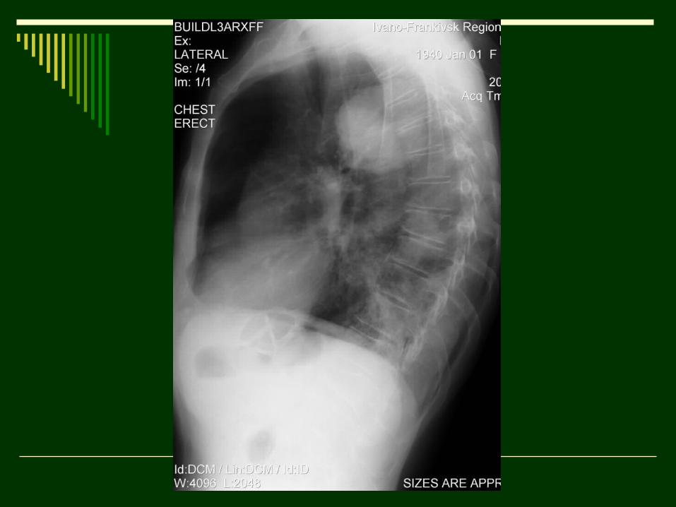
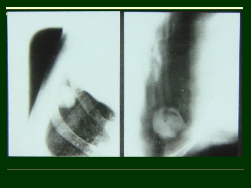
5. Syndrome of Cavity:
5.1 Inflammation (abscess, cavernous tuberculosis);
5.2 Tumor (carcinoma, which is destroying);
5.3 Abnormalities (air cysts of lungs, cystic bronchiectases - polycystosis).
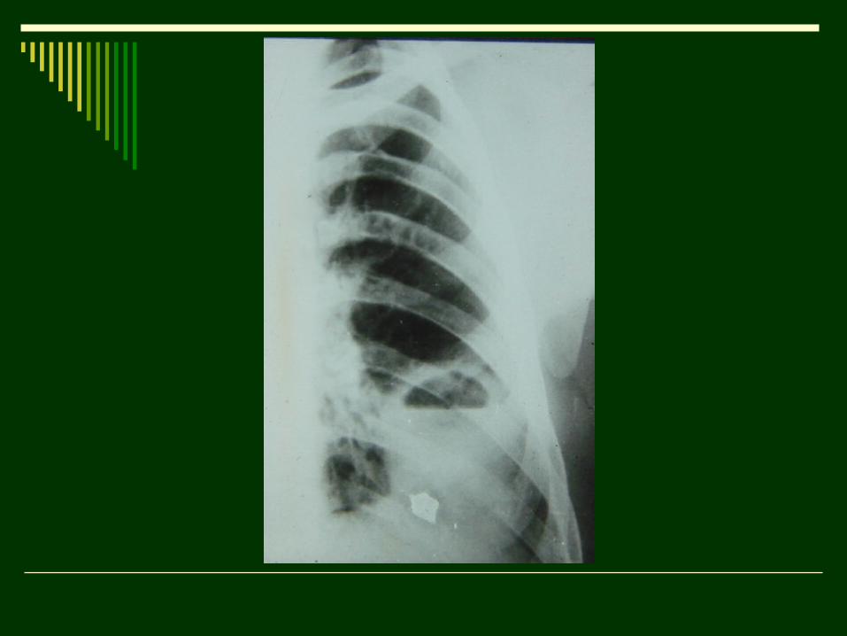
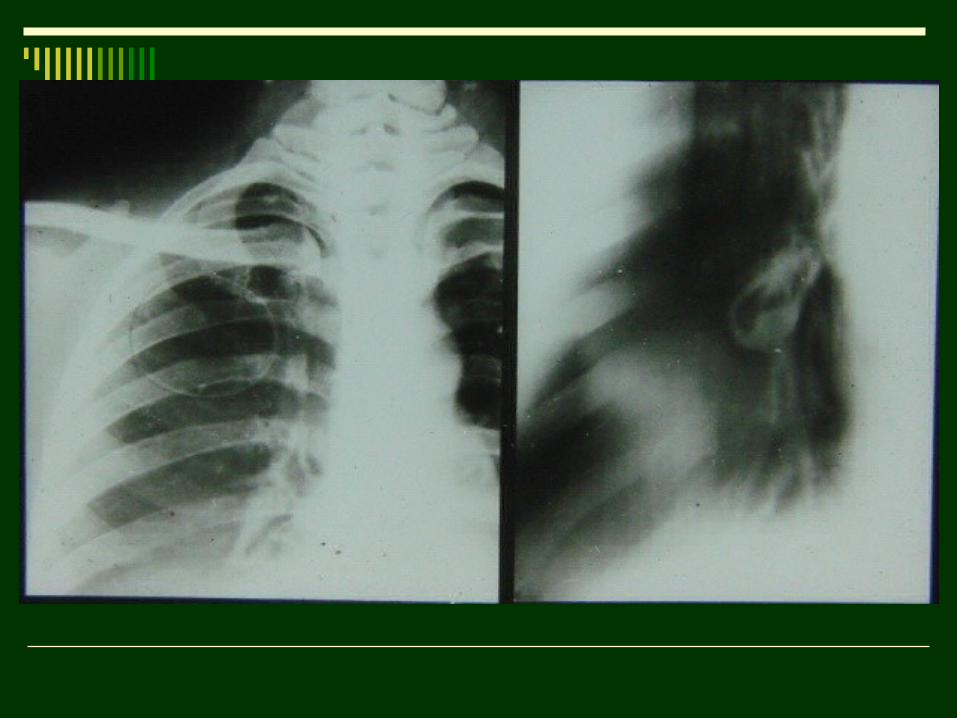
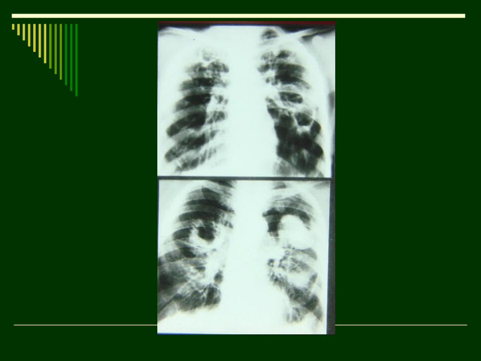
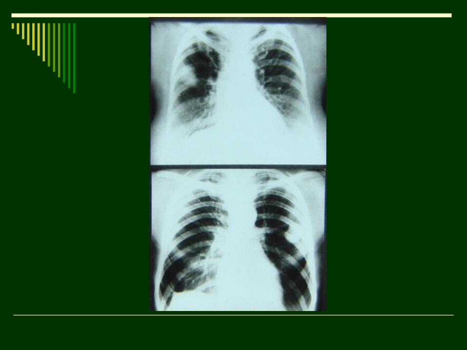
6. Syndrome of Lesion and Limited Dissemination (two intercostal spaces).
Intensity:- low (intensity of vessels);- medium (ribs crossing);- high (mediastinum).
Inflammative changes of pulmonary parenchyma (tuberculosis, pneumonia);
Peripheral carcinoma, metastases, pneumoconiosis.
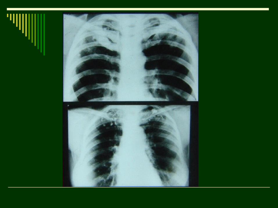
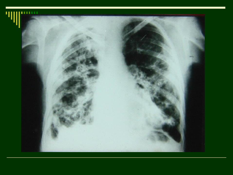
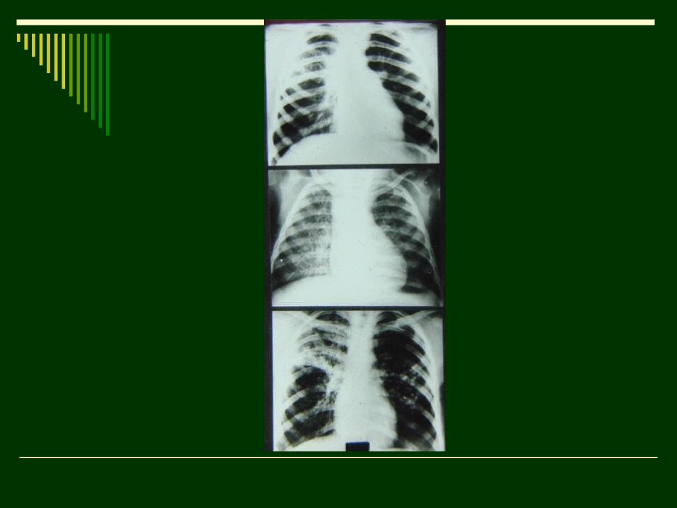
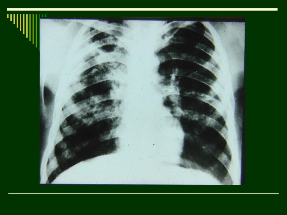
7. Syndrome of Diffuse Dissemination:
Multiple lesions.
Types of lesions:
А. Miliary – 1-2мм;
Б. Small – 3-4мм;
В. Medium – 5-8мм;
Г. Large – up to 10мм.
7.1 Miliary (influenza pneumonia, miliary tuberculosis, lymphohematogenic tuberculosis, pneumoconioses);
7.2 Small focal (acute and chronic hematogenic disseminative tuberculosis, small focal pneumonia, pneumoconioses, collagenoses);
7.3 Medium focal (pneumonia,metastases, acute hematogenic disseminative tuberculosis);
7.4 Large focal (pneumonia, lung swelling, metastases).
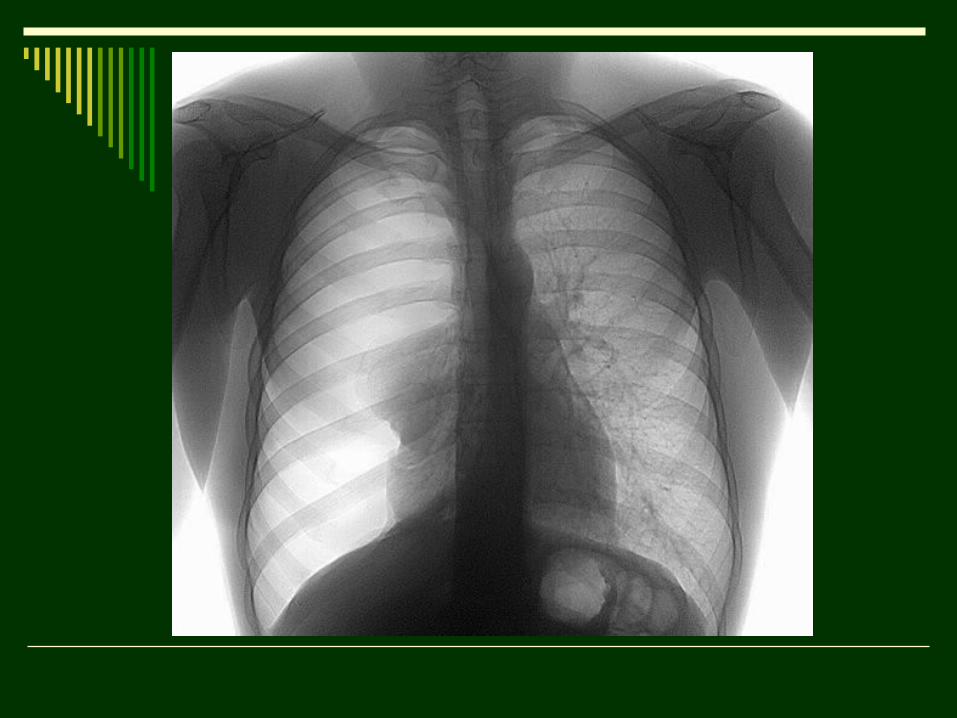
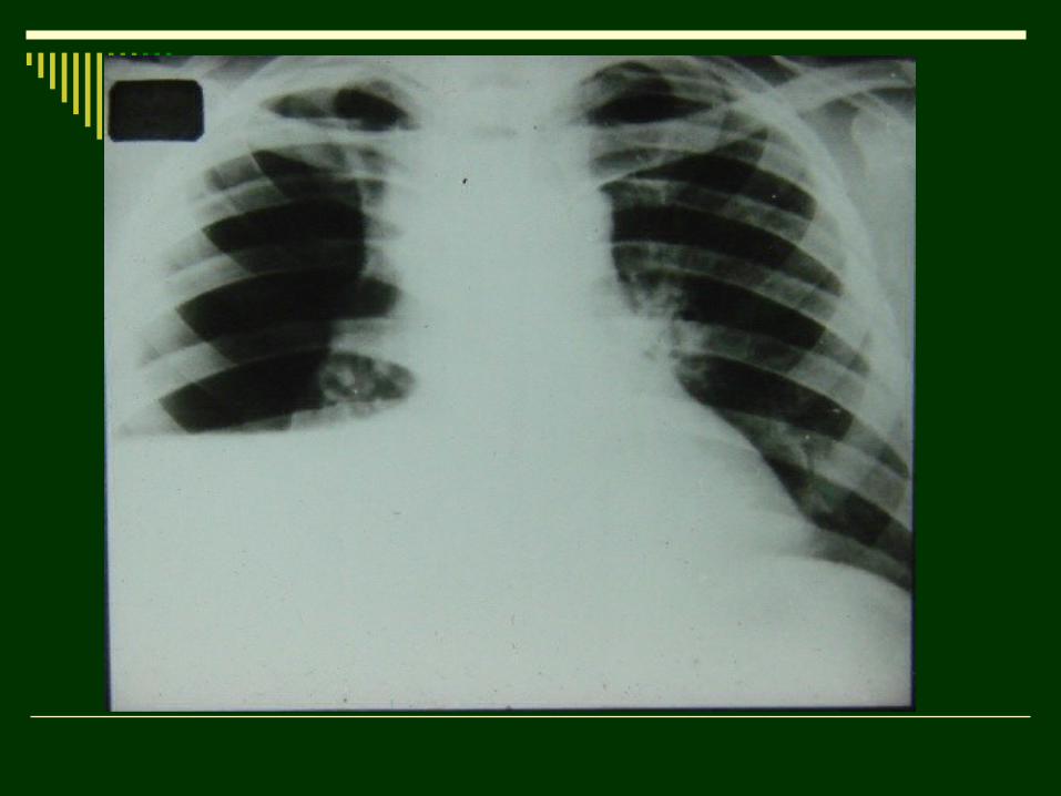
8. Syndrome of Total and Subtotal clarification:
8.1 Emphysema (bronchial asthma, chronic bronchitis);
8.2 Valvular bronchial occlusion;
8.3 Giant air cyst;
8.4 Pulmonary abnormality;
8.5 Pneumothorax.
9. Syndrome of the Pulmonary Vasculature Changes:
9.1 Gain of vasculature – increasing of the number and caliber of vasculature elements in the unit of area of pulmonary field (blood circulation disorders, acute interstitial inflammation, collagenoses);
9.2 Depletion of vasculature (arterial depletion of lungs; pulmonary tissue swelling – obturative emphysema);
9.3 Reduction (weakening) of vasculature (diffuse focal dissemination);
9.4 Gain and deformation of vasculature – change of normal structure of elements (chronic bronchitis, pneumonia, limpho-, hematogenic tuberculosis, pneumoscleroses, cancerous lymphangoitis).
10. Syndrome of the lungs’ hiluses changes:
10.1 Hiluses’ congestion – cardiac abnormalities (both sides), gain of vasculature;
10.2 Infiltration of hiluses (inflamation of fat – enlargement, loss of structure);
10.3 Scar deformation of hiluses (gain and deformation of the shadow );
10.4 Enlargement of the hilar lymph nodes.10.5 Calcification of the hilar lymph nodes.