Embed Size (px)
Citation preview
Review ArticleThe Role of Proteinase-Activated Receptors 1 and 2 in theRegulation of Periodontal Tissue Metabolism and Disease
E. S. Rovai and M. Holzhausen
Division of Periodontics, Department of Stomatology, School of Dentistry, University of São Paulo, São Paulo, SP, Brazil
Correspondence should be addressed to M. Holzhausen; [email protected]
Received 16 November 2016; Revised 13 January 2017; Accepted 5 March 2017; Published 19 April 2017
Academic Editor: Kristen M. Kahle
Copyright © 2017 E. S. Rovai and M. Holzhausen. This is an open access article distributed under the Creative CommonsAttribution License, which permits unrestricted use, distribution, and reproduction in any medium, provided the original workis properly cited.
Proteinase-activated receptors 1 (PAR1) and 2 (PAR2) are the most highly expressed members of the PAR family in theperiodontium. These receptors regulate periodontal inflammatory and repair processes through their activation by endogenousand bacterial enzymes. PAR1 is expressed by the periodontal cells such as human gingival fibroblasts, gingival epithelial cells,periodontal ligament cells, osteoblasts, and monocytic cells and can be activated by thrombin, matrix metalloproteinase 1(MMP-1), MMP-13, fibrin, and gingipains from Porphyromonas gingivalis. PAR2 is expressed by neutrophils, osteoblasts, oralepithelial cells, and human gingival fibroblasts, and its possible activators in the periodontium are gingipains, neutrophilproteinase 3, and mast cell tryptase. The mechanisms through which PARs can respond to periodontal enzymes and result inappropriate immune responses have until recently been poorly understood. This review discusses recent findings that arebeginning to identify a cardinal role for PAR1 and PAR2 on periodontal tissue metabolism.
1. Introduction
Periodontium is characterized by the tissues that involve andsupport the teeth such as the gingiva, alveolar mucosa,cementum, periodontal ligament, and alveolar bone. Peri-odontitis, an oral disease which leads to alveolar bone loss,can be mediated by some of the endogenous host enzymes,as well as bacterial proteinases present in the periodontalpocket (e.g., neutrophil serine proteinase 3),mast cell tryptase,and gingipain fromPorphyromonas gingivalis. Interestingly, itwas recently shown that the biological activities of theseproteinases can be mediated through specific proteinase-activated receptor (PAR) activation. PARs are members ofthe G-protein-coupled family, seven-transmembrane domainreceptors, and their activation occurs through proteolyticcleavage of the N-terminal domain by proteinases, leadingto the generation of a new N-terminal “tethered ligand,”which binds to the receptor itself resulting in its autoacti-vation [1, 2]. Until now, four members of the PAR familywere discovered: PAR1, PAR3, and PAR4 which are activatedby thrombin and PAR2 that can be activated by trypsin,neutrophil proteinase 3, tissue factor/factor VIIa/factor Xa,
mast cell tryptase, membrane-tethered serine proteinase-1,or gingipains [3, 4]. PARs represent a component of theinnate inflammatory response, being involved in neutrophilrecruitment, increased perfusion, pain, and swelling. Thus,since PARs are present in periodontal epithelial cells andare capable of recognizing and responding to bacterial infec-tions, it is believed that they can act as a first “alarm system”for bacterial invasions [5]. In addition, studies have suggestedan important role for PARs in regulating the inflammatoryresponse intensity to bacterial infection, as well as inperiodontitis [5, 6].
Although the structures and mechanisms related to theactivation of these receptors are similar, they can be expressedby different cells; hence, in each cell, their activation maylead to distinct roles in pathophysiological processes, suchas growth, development, inflammation, tissue repair, andpain [2, 7–10]. PAR1 is expressed by platelets, osteoblast endo-thelial cells, epithelial cells, fibroblasts, myocytes, neurons,and astrocytes, and it seems to play an important role ininjured tissues. In the periodontium, the literature have alsoimplicated PAR1 in bone repair and homeostasis [11–13], aswell as proliferation of gingival fibroblasts mediated by the
HindawiJournal of Immunology ResearchVolume 2017, Article ID 5193572, 13 pageshttps://doi.org/10.1155/2017/5193572
protein synthesis of endothelin-1 (ET-1) and subsequentactivation of ET receptor type A [14] and transactivation oflatent transforming growth factor beta 1 (TGF-β1) [15]. Inaddition, Rohani et al. [16] showed that in gingival epithelialcells, the PAR1 activation by thrombin can result in theinduction of chemokines leading to granulocyte attraction.Interestingly, gingipain-R (RgpB and HRgpA), a proteinasefrom Porphyromonas gingivalis, can activate PAR1 in mono-cytic cells triggering an overproduction of proinflammatorycytokines [17] and in the surface of platelets leading toplatelet aggregation [18]. This mechanism may constitutethe biological plausibility of the association between peri-odontitis and cardiovascular disease and deserves furtherclarification by future studies.
PAR3 and PAR4 are expressed by platelets, endothelialcells, myocytes, and astrocytes, and their activation has beenassociated with the formation of pathologic thrombus [1].The functional role of PAR3 is controversial, whereas someinvestigators have described it as a nonsignaling receptor thatacts along with PAR1 and PAR4, others have claimed thatPAR3 can signal independently of PAR1 activation. Thus,since PAR3 and PAR4 are less abundant than PAR1 inperiodontal cells, and since their possible function on theperiodontium still needs to be investigated [16], they arenot going to be part of the present review.
PAR2 has been demonstrated to be expressed by epithe-lial cells, endothelial cells, fibroblasts, osteoblasts, myocytes,neurons, astrocytes, lymphocytes, neutrophils, and mastcells [1, 17, 19–21], where it has been associated to severalroles in the inflammation process such as increased vascularpermeability, blood vessel relaxation, hypotension, granulocyteinfiltration, release of cytokines, and pain [3, 4, 17, 22–26].Moreover, inflammatory events in the joints, skin, colon,kidney, and airways have also been associated to PAR2activation [1, 2, 24, 27–29]. More recently, studies haveshown that PAR2 activation may play an important rolein the inflammatory process and tissue breakdown in peri-odontitis [6, 30–33].
In this review, we will discuss the possible roles of themost highly expressed members of the PAR family in theperiodontium, PAR1 and PAR2, as important molecules thatmediate mammalian and bacterial enzyme’s effects on cells inthe regulation of periodontal inflammation and repair.
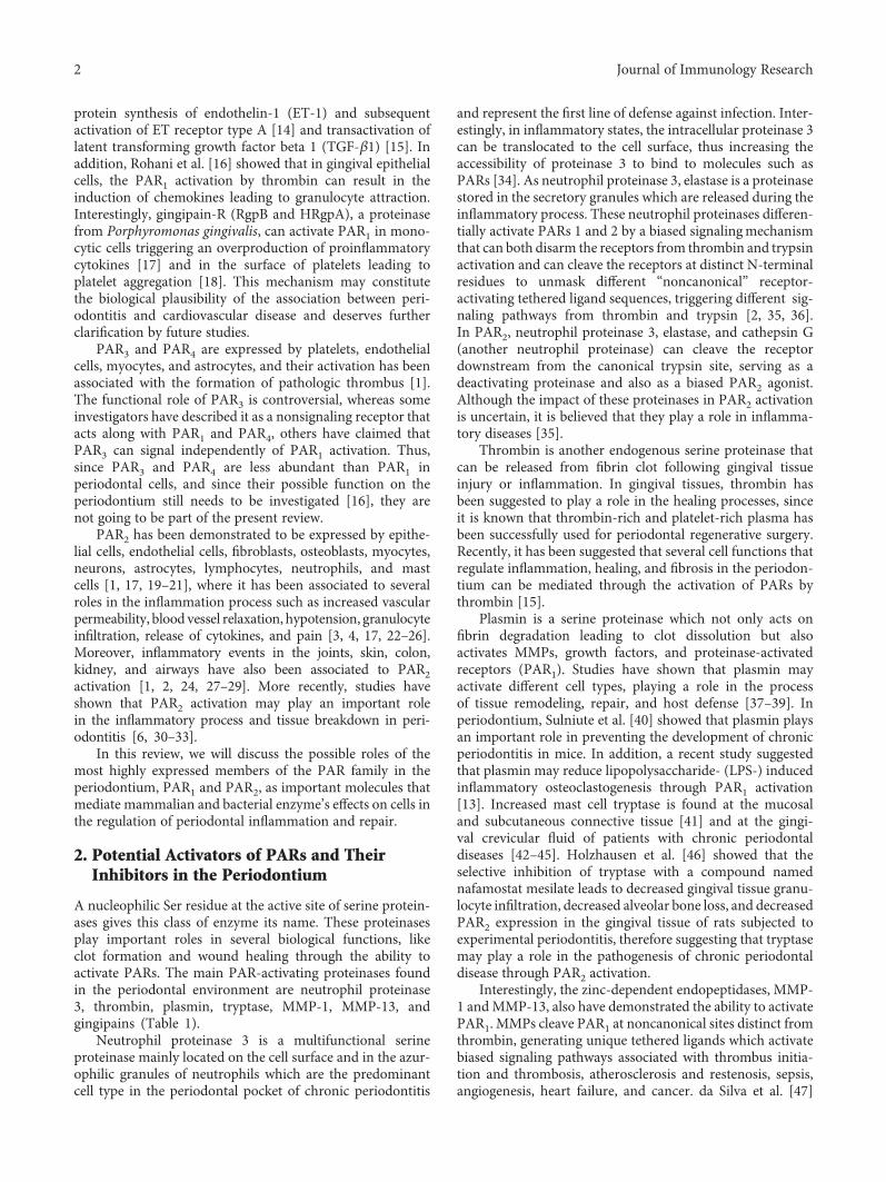
2. Potential Activators of PARs and TheirInhibitors in the Periodontium
A nucleophilic Ser residue at the active site of serine protein-ases gives this class of enzyme its name. These proteinasesplay important roles in several biological functions, likeclot formation and wound healing through the ability toactivate PARs. The main PAR-activating proteinases foundin the periodontal environment are neutrophil proteinase3, thrombin, plasmin, tryptase, MMP-1, MMP-13, andgingipains (Table 1).
Neutrophil proteinase 3 is a multifunctional serineproteinase mainly located on the cell surface and in the azur-ophilic granules of neutrophils which are the predominantcell type in the periodontal pocket of chronic periodontitis
and represent the first line of defense against infection. Inter-estingly, in inflammatory states, the intracellular proteinase 3can be translocated to the cell surface, thus increasing theaccessibility of proteinase 3 to bind to molecules such asPARs [34]. As neutrophil proteinase 3, elastase is a proteinasestored in the secretory granules which are released during theinflammatory process. These neutrophil proteinases differen-tially activate PARs 1 and 2 by a biased signalingmechanismthat can both disarm the receptors from thrombin and trypsinactivation and can cleave the receptors at distinct N-terminalresidues to unmask different “noncanonical” receptor-activating tethered ligand sequences, triggering different sig-naling pathways from thrombin and trypsin [2, 35, 36].In PAR2, neutrophil proteinase 3, elastase, and cathepsin G(another neutrophil proteinase) can cleave the receptordownstream from the canonical trypsin site, serving as adeactivating proteinase and also as a biased PAR2 agonist.Although the impact of these proteinases in PAR2 activationis uncertain, it is believed that they play a role in inflamma-tory diseases [35].
Thrombin is another endogenous serine proteinase thatcan be released from fibrin clot following gingival tissueinjury or inflammation. In gingival tissues, thrombin hasbeen suggested to play a role in the healing processes, sinceit is known that thrombin-rich and platelet-rich plasma hasbeen successfully used for periodontal regenerative surgery.Recently, it has been suggested that several cell functions thatregulate inflammation, healing, and fibrosis in the periodon-tium can be mediated through the activation of PARs bythrombin [15].
Plasmin is a serine proteinase which not only acts onfibrin degradation leading to clot dissolution but alsoactivates MMPs, growth factors, and proteinase-activatedreceptors (PAR1). Studies have shown that plasmin mayactivate different cell types, playing a role in the processof tissue remodeling, repair, and host defense [37–39]. Inperiodontium, Sulniute et al. [40] showed that plasmin playsan important role in preventing the development of chronicperiodontitis in mice. In addition, a recent study suggestedthat plasmin may reduce lipopolysaccharide- (LPS-) inducedinflammatory osteoclastogenesis through PAR1 activation[13]. Increased mast cell tryptase is found at the mucosaland subcutaneous connective tissue [41] and at the gingi-val crevicular fluid of patients with chronic periodontaldiseases [42–45]. Holzhausen et al. [46] showed that theselective inhibition of tryptase with a compound namednafamostat mesilate leads to decreased gingival tissue granu-locyte infiltration, decreased alveolar bone loss, and decreasedPAR2 expression in the gingival tissue of rats subjected toexperimental periodontitis, therefore suggesting that tryptasemay play a role in the pathogenesis of chronic periodontaldisease through PAR2 activation.
Interestingly, the zinc-dependent endopeptidases, MMP-1 andMMP-13, also have demonstrated the ability to activatePAR1. MMPs cleave PAR1 at noncanonical sites distinct fromthrombin, generating unique tethered ligands which activatebiased signaling pathways associated with thrombus initia-tion and thrombosis, atherosclerosis and restenosis, sepsis,angiogenesis, heart failure, and cancer. da Silva et al. [47]
2 Journal of Immunology Research
Table1:Potentialactivators
ofPARsin
theperiod
ontium
.
Protease
Protease-3
Throm
bin
Plasm
inTryptase
MMP-1
&MMP-13
Gingipains
Dentilisin
Aaprotease
Origin
Neutrop
hils
Fibrin
clot
Fibrin
clot
Mastcells
Mon
ocytes/
macroph
ages
Porphyromonas
gingivalis
Treponemadenticola
Aggregatibacter
actinomycetem
comitan
s
Type
Serine
protease
Serine
protease
Serine
protease
Chymotrypsin-like
protease
Collagenases
Cysteineproteases
(i)Arginine
(HRgpAandRgpB)
(ii)Lysine
(Kgp)
Chymotrypsin-likeenzyme
Arginineand
lysine
proteases
PAR
interaction
PAR2
activation
PAR1activation
PAR3activation
PAR4activation
PAR1activation
PAR2activation
PAR1activation
PAR1activation
PAR2activation
PAR2disarm
ing
PAR2activation
PAR:p
roteaseactivatedreceptor;M
MP:m
atrixmetalloproteinase;A
a:Aggregatibacter
actinomycetem
comitan
s.
3Journal of Immunology Research
demonstrated that increased MMP-13 levels were associatedwith an increased PAR1 expression at the gingival crevicularfluid of patients with chronic periodontitis after nonsurgicalperiodontal treatment.
In addition to host origin proteinases, exogenous ser-ine proteinases originated by periodontopathic bacteriacan also play a role in the innate response mediated byPARs. Porphyromonas gingivalis, for instance, produces andreleases the cysteine proteinases, arginine-gingipain (HRgpAand RgpB) and lysine-gingipain (Kgp) which are stronglyassociated with periodontal breakdown and disruption ofhost defense. Some of the mechanisms played by gingipainsare mediated by PARs 1 and 2, due to their potential tointeract with host cell surface receptors modulating theinnate response.
Other nonmammalian proteinases produced by peri-odontal pathogens have already been suggested to play a roleon PAR2 function. Dentilisin, a chymotrypsin-like enzymeproduced by Treponema denticola, is suggested to causePAR2 disarming or inhibition to further activation [6]. Inter-estingly, a study by Euzebio Alves et al. [32] has demonstratedan inverse relationship between PAR2 expression and theexpression of dentilisin in the periodontal sites of moderatechronic periodontitis patients. Another bacterial proteinase,an arginine- and lysine-specific proteinase produced byAggregatibacter actinomycetemcomitans, was shown to induceinterleukin (IL)-8 and intercellular adhesion molecule-1(ICAM-1) expression in gingival epithelial cells throughPAR2activation [48]. It canbesuggested thatbacterialprotein-ases produced by other periodontal pathogens could alsoplay a role on the activation or suppression of PAR2 functionor expression.
Interestingly, the plasma contains serine proteinaseinhibitors (serpins) that can regulate proteolytic events intissues [2, 49]. The easy accessibility of the reactive site loopsof serpins guarantees the rapid inhibition of specific hostproteinases, but it also makes them easy targets for bacterialproteinases, which can specifically inactivate them. In fact,the ability to resist inhibition by serpins is also important inhost defense evasion by bacterial pathogens. Accordingly,Euzebio Alves et al. [32] have demonstrated that elevatedlevels of gingipain and proteinase 3 and decreased levelsof secretory leucocyte proteinase inhibitor (SLPI) were asso-ciated to PAR2 overexpression. SLPI is expressed by epithelialand immune cells where they play a role as an alarm pro-teinase inhibitor mediating anti-inflammatory and antimi-crobial effects. In this study, decreased levels of SLPI werefound in chronic periodontitis patients, whereas periodontaltreatment led to its upregulation. The authors suggestedthat these results might be explained by the ability of thearginine-specific gingipains (Rgps) to degrade SLPI. Similarly,reduced SLPI levels and higher serine proteinase acti-vities correlating with PAR2 overexpression were foundin the gastric mucosa of Helicobacter pylori-infected indi-viduals. This fact may be associated to the loss of hostprotective capacity and increase susceptibility to breakdownfrom chronic infection. These data reinforce the roleplayed by Porphyromonas gingivalis on PAR2-mediatedperiodontal inflammation.
3. Biological Effects of PAR1 Activation inPeriodontal Cells and Tissues
PAR1 involvement in periodontal tissue metabolism has beensuggested by several in vitro studies which have shownexpression of its receptor by the periodontal cells such ashuman gingival fibroblasts, gingival epithelial cells, peri-odontal ligament cells, osteoblasts, and monocytic cells andpresence of its possible activators, thrombin, plasmin,MMPs, and gingipains, in the periodontal environment(Table 2).
The biological effects of PAR1 activation on the peri-odontium are still not well clarified. Some studies have shownthat PAR1 activation has a tissue destructive profile, leadingto the induction of proinflammatory mediators that regulateperiodontal breakdown, while others highlighted its possibleinvolvement with the repair of periodontal tissues [11, 12].
Uehara et al. [50] have showed that production ofhepatocyte growth factor (HGF) by human gingival fibro-blasts upon stimulation with gingipains occurred throughPARs, specifically PAR1 and PAR2. HGF plays a role inwound healing, through its mitogenic activity to gingivalepithelial cells, and enhances matrix metalloproteinasesproduction, therefore playing a fundamental role in tissueremodeling. Moreover, HGF also stimulates blood vesselformation and promotes vascularization, a later process inwound healing.
In gingival fibroblasts, thrombin and a specific PAR1-activating peptide-induced proliferation through endothelin-1 (ET-1) are involved indrug-inducedproliferationof gingivalfibroblasts [14]. In this context, an in vitro study [15] demon-strated that thrombin and PAR1 agonist induced connectivetissue growth factor (CTGF) synthesis and TGF-β1 activa-tion in gingival fibroblasts. TGF-β1and CTGF are proteinsthat regulate many biological effects, such as cell adhesion,migration, differentiation, proliferation, extracellular matrixproduction, angiogenesis, and wound healing. As well asET-1, it is suggested that its overexpression may be involvedin gingival overgrowth.
Moreover, Rohani et al. [16] showed that activation ofPAR1by thrombin ingingival epithelial cells leads to inductionof chemokines which are important chemo-attractants forneutrophils and have a role in wound healing.
Thrombin exerts multiple effects upon osteoblastsincluding stimulating proliferation and inhibiting osteoblastdifferentiation and apoptosis. Some of these effects such assynthesis and secretion of growth factors and cytokines arebelieved to be mediated through PAR activation. In fact,Pagel et al. [51] demonstrated that thrombin inducedTGF-1β, cyclooxygenase-2, tenascin C, fibroblast growthfactors 1 and 2, connective tissue growth factor, and IL-6expression in wild-type osteoblasts, but not in PAR1 knock-out mouse osteoblasts. In addition, PAR1-specific activatingpeptide and thrombin induced release of both prostaglandinE2 and IL-6 by osteoblasts, therefore suggesting the receptorparticipation in the earliest stages of bone healing.
The other evidence that links PAR1 action to bonemetabolism comes from the fact that in periodontal ligamentcells, PAR1 activation by thrombin induces the synthesis of
4 Journal of Immunology Research
osteoprotegerin, which is one of the key molecules that regu-late bone homeostasis and prevent osteoclastogenesis [12].Corroborating with these findings, a recent study found thatthe activation of PAR1 in monocytic cells by plasmin dimin-ished LPS-induced inflammatory osteoclastogenesis andbone resorption by inactivation of nuclear factor kappabeta (NF-κB) [13]. Conversely, Uehara et al. [17] showedthat the gingipains Rgp and Kgp synergistically increasethe secretion of proinflammatory cytokines such as IL-8from human monocytic cells via PAR1, PAR2, and PAR3in combination with Toll-like receptors or NOD agonists(pathogen-associatedmolecularpattern receptors). This studywas the first one to report that gingipains stimulate the secre-tion of cytokines from monocytic cells through the activa-tion of PARs with synergistic effects by pathogen-associatedmolecular patterns (PAMPs). In addition, Giacaman et al.[52] have showed that selective cleavage of PAR1 on oralepithelial cells by the gingipain Rgp upregulates expression ofthe proinflammatory cytokines IL-1α, IL-1β, IL-6, and tumornecrosis factor alpha (TNF-α).
Interestingly, gingipains-R (RgpB and HRgpA) werealso shown to activate PAR1 in platelets leading to plateletaggregation [18]. This mechanism may explain the biologicalplausibility of the association between periodontitis andcardiovascular disease and deserves further clarification byfuture studies.
Taken together, the results from these in vitro studiesshow that PAR1 is associated with both proinflammatoryand reparative processes in the periodontium. However,Wong et al. [53] found no difference between PAR1
+/+ andPAR1 knockout mice with regard to alveolar bone loss ina Porphyromonas gingivalis-induced periodontal diseasemodel, indicating that this receptor does not play a pivotalrole in the progression of experimental periodontitis. Mostrecently, it was shown by Spolidorio et al. [54] that parstatin,a 41-amino acid peptide released upon PAR1 activation, haspotential anti-inflammatory effects since it decreases inflam-matory cell infiltration, myeloperoxidase (MPO) activity, andproinflammatory mediators’ levels, including IL-1β, IL-6,and TNF-α in gingival tissues of rats subjected to experimen-tal periodontal disease.
Furthermore, a recent study by da Silva et al. [47] hassuggested the first clinical evidence of the association of
PAR1 with periodontal repair. The authors demonstratedthat PAR1 expression was downregulated in chronicperiodontitis patients and inversely correlated to gingivalcrevicular fluid levels of IL-6, IL-8, TNF-α, IFN-γ, andMMP-2. In addition, periodontal therapy resulted in PAR1overexpression by epithelial and immune cells from the gin-gival crevicular fluid, therefore suggesting the importance ofPAR1 mediating the known anabolic actions of thrombin inthe periodontium.
4. Biological Effects of PAR2 Activation inPeriodontal Cells and Tissues
PAR2 acts as a “sensor” of bacterial and host proteinases andmodulates host immune defense playing a role in the hostalarm system [55, 56]. PAR2 has been localized in many celltypes (Table 3) that can be found in periodontal tissues,including neutrophils, osteoblasts, oral epithelial cells, andhuman gingival fibroblasts [18, 21, 50, 57]. Gingipains fromPorphyromonas gingivalis, neutrophil proteinase 3, and mastcell tryptase are the agonists that can possibly be found at theperiodontal environment and that have already been studiedfor their ability to activate PAR2.
Gingipains have been shown to activate PAR2 in immu-noinflammatory cells that play important roles in periodontaldisease development. For instance, PAR2 activation by RgpBleads to neutrophil activation as indicated by increased intra-cellular calcium concentrations [57]. In addition, gingipains(Rgps and Kgp) may activate PARs (PAR1, PAR2, andPAR3) in monocytic cells increasing production of IL-6, IL-8, and monocyte chemoattractant protein- (MCP-) 1 [17].Furthermore, Yun et al. [58] showed that RgpA activated theproteinase-activated receptors and induced T-cell activation.Taken together, these findings suggest that Porphyromonasgingivalis, through its gingipains, makes use of the hostcell PAR2 to exacerbate inflammation during chronicperiodontal disease.
One of the most important mechanical barriers that thebacteria encounter to invade the periodontium is the epithe-lial tissue. Besides the physical barrier, the epithelial tissueshave also the ability to produce antimicrobial peptides suchas the β-defensins (hBD). Interestingly, a study by Chunget al. [59] showed that gingipains may also play a protective
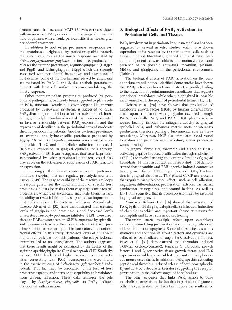
Table 2: Biological effects of PAR1 activation in periodontal cells.
PAR1 Periodontal destruction Periodontal repair/protection
Oral epithelial cells↑ IL-1α, IL-1β, IL-6, TNFα [52]
↑ CXCL 5 [16]
Gingival fibroblasts↑ HGF [50]
↑ endothelin-1 [14]
Osteoblasts↑ COX-2 [51]↑ IL-6 [51]↑ PGE2 [51]
↑ TGF-β [51]↑ FGF-1/FGF-2 [51]
↑ CTFG [51]
Periodontal ligament cells ↑ osteoprotegerin [12]
Monocytic cells ↑ IL-8 [17]
PAR: protease-activated receptor; IL: interleukin; TNF: tumor necrosis factor; CXCL: C-X-C motif chemokine; COX: cyclooxygenase; PGE: prostaglandin E;HGF: hepatocyte growth factor; TGF: transforming growth factor; FGF: fibroblast growth factor; CTFG: connective tissue growth factor.
5Journal of Immunology Research
role by increasing hBD-2 expression in gingival epithelialcells partially through PAR2 receptor signaling pathway. Inaddition, Pereira et al. [60] have showed that in subjects withchronic periodontitis there are significantly higher levels ofPorphyromonas gingivalis associated with increased salivaryhBD-2 levels and gingival crevicular fluid PAR2 mRNAexpression than in healthy subjects and that periodontaltreatment decreases both hBD-2 levels and PAR2 expression.On the other hand, gingipains have also been shown to acti-vate PAR2 on oral epithelial cells leading to the production ofproinflammatory mediators, such as IL-6 [18] and IL-8 [17]that could result in periodontal tissue breakdown. Moreover,Giacaman et al. [52] suggested that gingipains Rgp and Kgpmay cleave and activate PAR2 in oral keratinocytes upregulat-ing the expression of IL-1α, IL-1β, IL-6, and TNF-α. Further-more, a recent study by Tada et al. [61] found that theexpression of IL-33, a cytokine that augments Th2 cytokine-mediated inflammatory responses, is increased during Por-phyromonas gingivalis infection in human gingival epithelialcells via PAR2 through gingipain-dependent activation.
As the bacteria challenge increases, an enhanced perme-ability of the small blood vessels of the subgingival plexusoccurs resulting in an increased neutrophil migration throughthe junctional epithelium and into the gingival sulcus.Interestingly, activated neutrophils may secrete a proteinase(neutrophil proteinase 3) which was shown to activate humanoral epithelial cells through PAR2, inducing IL-8 and mono-cyte chemoattractant protein-1 production [17].
Increased levels of proinflammatory mediators and path-ogenic bacteria in the soft tissues may lead to the disruptionof the epithelial tissue, which in turn facilitates the access ofbacteria and their products to the subepithelial connectivetissue. The exposure of the residing periodontal connectivetissue cells to the bacterial agents may transform them intomajor participants in the pathophysiological process ofperiodontal tissue destruction. The dominant cell type inperiodontal connective tissue is the fibroblast. Interestingly,Uehara et al. [50] demonstrated that human gingival fibro-blasts express PAR2 and that its activation by a syntheticPAR2 agonist peptide (SLIGRL) induces the productionof IL-8 which has the ability to selectively stimulateMMP activity, responsible for collagen destruction withinperiodontitis lesions. Porphyromonas gingivalis may exacer-bate this process since it was demonstrated that gingipainsupregulate PAR2 gene expression in human gingivalfibroblasts [62].
Abraham et al. [21] demonstrated that PAR2 is expressedby osteoblasts and that its activation by a specific syntheticpeptide did not show any effect on osteoblast proliferationor differentiation. In addition, in this study, osteoblast-mediated osteoclast bone resorption was also not stimulatedby PAR2 activation. Furthermore, Smith et al. [63] showedthat PAR2 activation inhibits expression of receptor activatorof nuclear factor kappa-B ligand (RANKL) and suppressedthe RANKL : osteoprotegerin ratio in osteoblasts. However,a study by Amiable et al. [64] showed that PAR2 activationin osteoarthritis subchondral bone osteoblasts induced a sig-nificant upregulation of RANKL and significantly enhancedbone resorptive activity. Interestingly, these findings on theresorptive properties played by PAR2 in osteoblasts are inagreement with data reporting the involvement of PAR2activity in periodontitis [6, 30–33, 65].
Accordingly, it has been shown that a selective PAR2 ago-nist (SLIGRL) causes periodontitis in rats through a mecha-nism involving prostaglandin release and MMP activation[65] and that PAR2-knockout mice infected with Porphyro-monas gingivalis have decreased levels of proinflammatorymediators, such as prostaglandin E2, interferon-gamma, IL-1beta, and IL-6, and less alveolar bone loss when comparedto wild-type animals [6].
Wong et al. [53] also have shown that less alveolar boneresorption occurred in PAR2-knockout mice. In addition,they showed that T-cells from Porphyromonas gingivalis-infected PAR2
−/− mice proliferated less in response to antigenthan those from wild-type mice and that T-cells frominfected or antigen-immunized PAR2-null mice had a signif-icantly different Th1/inflammatory cytokine profile fromwild-type cells such as decreased gamma interferon, ILs(IL-2, IL-3, and IL-17), granulocyte-macrophage colony-stimulating factor, and TNF-alpha than wild-type controls.The absence of PAR2 therefore appears to substantiallydecrease T-cell activation and the Th1/inflammatory res-ponse. In this study [53], increased numbers of mast cellsin the maxillary tissue of infected PAR2
+/+ mice were alsoshown, indicating that PAR2 may also have a role in mast celldifferentiation or infiltration into tissues. Thus, activation ofPAR2 expressed by mast cells by the arginine-specific gingi-pains from Porphyromonas gingivalis may lead to the releaseof inflammatory mediators that are pivotal to early inflam-matory response in chronic periodontitis. It has been shownthat activation of PAR2 leads to degranulation of mast cells,causing the release of proinflammatory compounds that kill
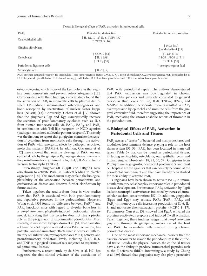
Table 3: Biological effects of PAR2 activation in periodontal cells.
PAR2 Periodontal destruction Periodontal repair/protection
Epithelial cells↑ IL-1α, IL-1β, IL-6, IL-8, TNFα [4, 47]
↑ MCP-1 [17]↑ IL-33 [61]
↑ β-defensin 2 [59]
Fibroblasts ↑ IL-8 [62]
Osteoblasts ↑ RANKL [64]↓ RANKL [63]
↓ RANKL : osteoprotegerin ratio [63]
Monocytic cells ↑ IL-6, IL-8, MCP-1 [17]
IL: interleukin; MCP: monocyte chemoattractant protein; RANKL: receptor activator of nuclear factor kappa-B ligand.
6 Journal of Immunology Research
pathogens and upregulate the immune responses. Inaddition, tryptase, released from the granules of mast cellsupon degranulation, may also activate PAR2, and therefore,these cells could play a role in periodontitis by causing theactivation of the receptor on other cells in the periodontaltissues. Thus, the regulation of such proinflammatory mech-anisms in T-cells and mast cells by PAR2 suggests a pivotalrole in the pathogenesis of the disease (Table 4).
In the gingival crevicular fluid from patients with peri-odontal disease, there are high levels of proteolytic activitycharacterized by a mixture of endogenous and exogenousproteinases which may mediate degradation of connectivetissue [66]. Among these hydrolytic enzymes in the inflamedperiodontal environment, neutrophil proteinase 3, mast celltryptase, and gingipains have been isolated and are known toactivate PAR2. In fact, high levels of proteolytic activityderived from both Porphyromonas gingivalis and neutro-phils are expected to be found in the periodontal pocketof chronic periodontitis, since they are, respectively, themajor periodontal pathogen and the predominant cells(approximately 90%).
PAR2 has been shown to be expressed by cellularelements found in gingival crevicular fluid, which mayinclude epithelial cells, and leukocytes even in clinicallyhealthy human gingival sulci [30]. In addition, PAR2 ex-pression is upregulated in chronic periodontitis patientscompared to that in healthy controls (Table 5). Interestingly,PAR2 upregulation in the inflamed periodontium is asso-ciated with an elevated gingival crevicular fluid trypsin-like activity, probably due to the increased prevalenceof Porphyromonas gingivalis, and expression of neutrophilproteinase 3 [30].
Importantly, proteinase 3 has been shown to activate oralepithelial cells through PAR2 leading to the production ofIL-8 and monocyte chemoattractant protein-1 [17]. Thesefindings clearly suggest that proteinase 3 actively participatesin PAR2-mediated inflammation at periodontal sites.
PAR2 activation results in the synthesis of proinflam-matory mediators including IL-6, IL-8, Il-1, IFN-γ, PGE2,and MMP-9 [6, 67, 68] and activates signaling pathwayssuch as those involving mitogen-activated protein kinaseand nuclear factor-κB, which potentiates inflammatoryresponses [69]. Interestingly, proinflammatory mediatorssuch as TNF-alpha and IL1-beta are reported to increasePAR2 expression [30]. Accordingly, an increased PAR2expression has been demonstrated in deeper periodontalpockets compared to the expression of the receptor inshallower pockets, and it was associated with significantincreased levels of proinflammatory mediators [30]. Inaddition, periodontal treatment statistically reduces PAR2expression [30, 32]. Furthermore, Fagundes et al. [31] dem-onstrated that the presence of Porphyromonas gingivalis isassociated with an elevated expression of PAR2 in humanchronic periodontitis, thus suggesting that Porphyromonasgingivalis may disturb the host inflammatory responses notonly by regulating PAR2 function but also by enhancing itsgenetic expression. Taken together, these findings clearlysuggest that PAR2 overexpression is an essential element ininflammation severity.
Another study by Euzebio Alves et al. [32] showed thatPAR2-positive staining in gingival crevicular fluid cells wasreflective of tissue destruction, and its overexpression waspositively associated to inflammatory clinical parametersand to the levels of the proinflammatory mediators IL-6,IL-8, TNF-alpha, MMP-2, MMP-8, hepatocyte growthfactor, and vascular endothelial growth factor (VEGF).Another interesting finding from this study was that peri-odontally healthy sites from chronic periodontitis individualsshowed a diminished expression of PAR2 mRNA and PAR2protein level, therefore suggesting that PAR2 periodontalexpression is influenced by the presence of infection andnot merely a constitutive characteristic that may favorperiodontal inflammation.
5. Specific Synthetic Agonists
Selective synthetic peptides, corresponding to the tetheredligand sequences, are able to activate selectively the receptorsthrough direct binding to the body of the receptor withoutthe need of proteolysis [22]. With the exception of PAR3, allthe other receptors have their selective agonist peptides. PAR1,PAR2, and PAR4 can be nonenzimatically and selectivelyactivated by TFLLR-NH2, SLIGRL-NH2, and GYPGQVNH2,respectively [1, 2]. Since theirdiscovery, selectivePARagonistshave been used in order to assess the specific impact of PAR1,PAR2, and PAR4 signaling.
Noteworthy, earlier literature in many instances usedthe “TRAP” peptide (SFLLRN) to activate “PAR1” notrealizing that the peptide also coactivates PAR2 [36]. Thus,the earlier use of the “TRAP” peptide may have generatedresults that reflect the coactivation of both PARs 1 and 2.Furthermore, the PAR2-activating peptide (SLIGRL-NH2)can in some settings cross-react with the MAS receptor,whereas the use of a more potent 2-furoyl-LIGRLO-amidecan prevent this issue [36].
6. Recent PAR Antagonists
The newest PAR1 antagonist (Vorapaxar) has been used forthe treatment of cardiovascular diseases. Vorapaxar acts byblocking the docking of the tethered ligand sequencepreventing PAR1 activation, hence platelet aggregation [10].Regarding PAR2 blockage, the antagonist GB88 can blockPAR2 activation by trypsin as well as PAR-activating peptide.GB88 has been shown to inhibit PAR2-induced proinflam-matory cytokine release and attenuate inflammation in a ratmodel of colitis [10]. However, until now, none of theseantagonists were studied in periodontics.
7. PARs: Drug Targets in Inflammation
Therapies focusing on the inhibition of proteinases or, morespecifically, the use of PAR antagonists may constitute animportant approach for the modulation of an infectiouspathology such as periodontal inflammatory disease.
According to Yun et al. [58], PAR2 blockage with the useof antagonists might promote adverse high proteolytic activ-ities in the gingival crevicular fluid. Thus, it seems that the
7Journal of Immunology Research
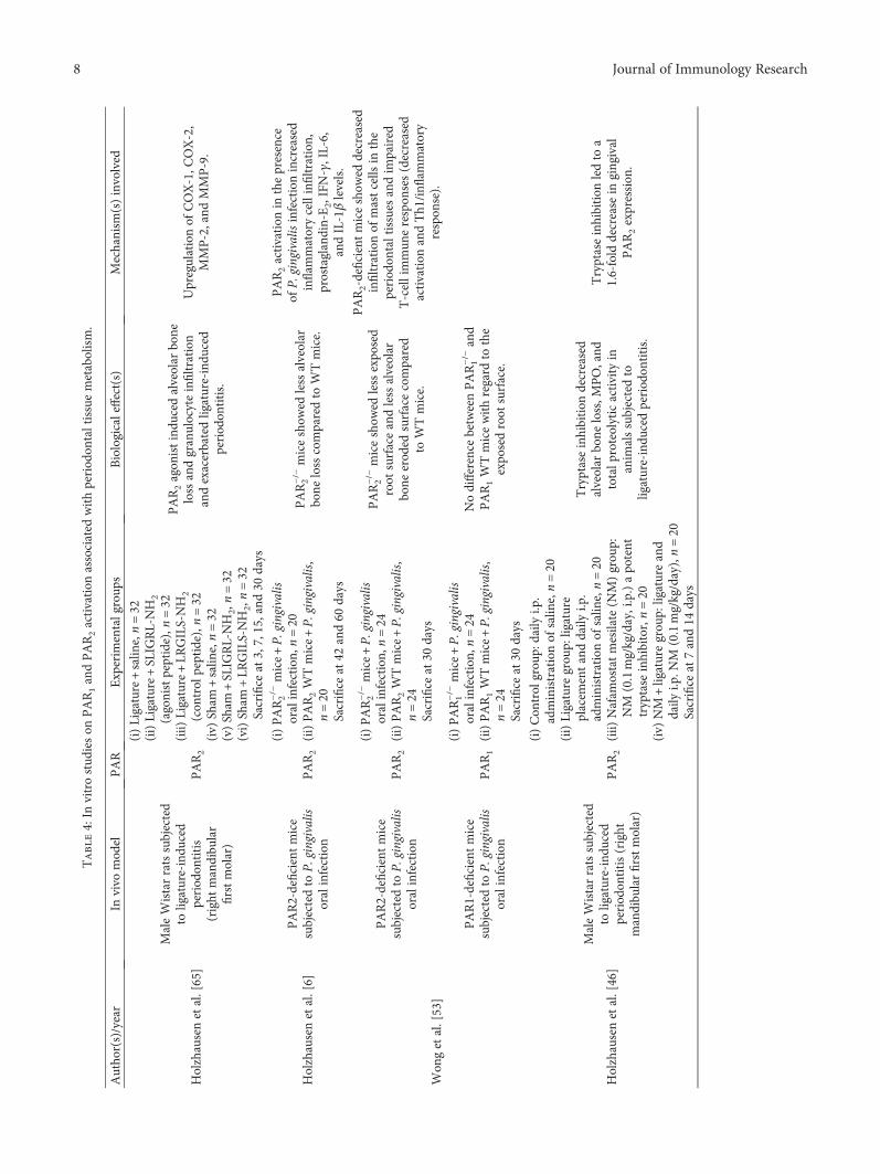
Table4:In
vitrostud
ieson
PAR1andPAR2activation
associated
withperiod
ontaltissuemetabolism.
Autho
r(s)/year
Invivo
mod
elPAR
Experim
entalgroup
sBiologicaleffect(s)
Mechanism
(s)involved
Holzhausenetal.[65]
MaleWistarratssubjected
toligature-indu
ced
period
ontitis
(right
mandibu
lar
firstmolar)
PAR2
(i)Ligature+salin
e,n=32
(ii)Ligature+SLIG
RL-NH
2(agonistpeptide),n
=32
(iii)
Ligature+LR
GILS-NH
2(con
trol
peptide),n
=32
(iv)
Sham
+salin
e,n=32
(v)Sham
+SLIG
RL-NH
2,n=32
(vi)Sham
+LR
GILS-NH
2,n=32
Sacrifice
at3,7,15,and
30days
PAR2agon
istindu
cedalveolar
bone
lossandgranulocyteinfiltration
andexacerbatedligature-indu
ced
period
ontitis.
Upregulationof
COX-1,C
OX-2,
MMP-2,and
MMP-9.
Holzhausenetal.[6]
PAR2-deficientmice
subjectedto
P.gingivalis
oralinfection
PAR2
(i)PAR2−/−mice+P.
gingivalis
oralinfection,
n=20
(ii)PAR2WTmice+P.
gingivalis,
n=20
Sacrifice
at42
and60
days
PAR2−/−miceshow
edless
alveolar
bone
loss
comparedto
WTmice.
PAR2activation
inthepresence
ofP.gingivalis
infectionincreased
inflam
matorycellinfiltration
,prostaglandin-E2,IFN-γ,IL-6,
andIL-1βlevels.
Won
getal.[53]
PAR2-deficientmice
subjectedto
P.gingivalis
oralinfection
PAR2
(i)PAR2−/−mice+P.
gingivalis
oralinfection,
n=24
(ii)PAR2WTmice+P.
gingivalis,
n=24
Sacrifice
at30
days
PAR2−/−miceshow
edlessexpo
sed
root
surfaceandlessalveolar
bone
erod
edsurfacecompared
toWTmice.
PAR2-deficientmiceshow
eddecreased
infiltration
ofmastcells
inthe
period
ontaltissues
andim
paired
T-cellimmun
erespon
ses(decreased
activation
andTh1
/infl
ammatory
respon
se).
PAR1-deficientmice
subjectedto
P.gingivalis
oralinfection
PAR1
(i)PAR1−/−mice+P.
gingivalis
oralinfection,
n=24
(ii)PAR1WTmice+P.
gingivalis,
n=24
Sacrifice
at30
days
Nodifference
betweenPAR1−/−and
PAR1WTmicewithregard
tothe
expo
sedroot
surface.
Holzhausenetal.[46]
MaleWistarratssubjected
toligature-indu
ced
period
ontitis(right
mandibu
larfirstmolar)
PAR2
(i)Con
trol
grou
p:daily
i.p.
administrationof
salin
e,n=20
(ii)Ligature
grou
p:ligature
placem
entanddaily
i.p.
administrationof
salin
e,n=20
(iii)
Nafam
ostatmesilate(N
M)grou
p:NM
(0.1mg/kg/day,i.p.)apo
tent
tryptase
inhibitor,n=20
(iv)
NM
+ligaturegrou
p:ligatureand
daily
i.p.N
M(0.1mg/kg/day),n=20
Sacrifice
at7and14
days
Tryptaseinhibition
decreased
alveolar
bone
loss,M
PO,and
totalp
roteolyticactivity
inanim
alssubjectedto
ligature-indu
cedperiod
ontitis.
Tryptaseinhibition
ledto
a1.6-fold
decrease
ingingival
PAR2expression
.
8 Journal of Immunology Research

Table4:Con
tinu
ed.
Autho
r(s)/year
Invivo
mod
elPAR
Experim
entalgroup
sBiologicaleffect(s)
Mechanism
(s)involved
Spolidorio
etal.[54]
MaleWistarratssubjected
toligature-indu
cedperiod
ontitis
(upp
ersecond
molars)
PAR1
(i)Ligature+intraoral3
μgparstatin,
n=16
(ii)Ligature+intraoralP
BS,n=16
(iii)
Sham
,n=8
Sacrifice
at8and15
days
Parstatin
(peptide
released
upon
PAR1activation
)prevented
period
ontaltissuebreakd
own.
Parstatin
supp
ressed
inflam
matory
cellinfiltration
anddecreased
MPO,IL-1β
,TNF-α,and
IL-6.
Castroetal.[33]
MaleWistarratssubjectedto
ligature-indu
cedperiod
ontitis
(mandibu
larfirstmolars)
PAR2
(i)Noligatureandno
treatm
ent,
n=20
(ii)Ligature+placebo(0,9%
NaC
lsolution),n
=20
(iii)
Ligature+5mgsubantim
icrobial
dose
ofdo
xycycline(SDD)by
daily
gavage,n
=20
Sacrifice
at3and15
days
SDDdo
wnregulated
alveolar
bone
loss.
Dow
nregulationof
PAR2,
IL-17,IL-1β,and
TNF-α.
PAR:protease-activated
receptor;COX:cyclooxygenase;MMP:matrixmetalloproteinase;P.
gingivalis:Po
rphyromonas
gingivalis;WT:wild
-typemice;
IL:interleukin;
IFN-γ:interferon
gamma;
MPO:
myeloperoxidase;P
BS:ph
osph
ate-bu
ffered
salin
e;TNF:
tumor
necrosisfactor.
9Journal of Immunology Research
potential side effects that the concept of PAR2 blockadeencounters do not seem to overcome the beneficial aspectsfor the treatment of periodontal inflammation.
At a certain point in the inflammatory process, PAR 1and 2 blockages may be necessary to prevent increasedinflammation, whereas at later time points, PAR activationmay be required to aid resolution. Further, the use of specificproteinase inhibitors, rather than PARs antagonists, mayresult in a “dual” blockage inhibiting both the degradationof host molecules and activation of PARs. Golub et al. [70]found that tetracyclines can inhibit tissue collagenolytic activ-ity in in vivo, in vitro, and periodontal pockets presented bysubjects with and without diabetes. Castro et al. [33] foundthat administration of a subantimicrobial dose of doxycycline(SDD) in a rat periodontitis model may result in PAR2modulation through a dual mode, downregulating its geneexpression and decreasing its posterior activation by protein-ases. Doxyxycline in a subantimicrobial dose is able to inhibitthe activity of MMPs and thus reduce the degradation of col-lagen, fibronectin, and elastin in the periodontal tissues [71],and its clinical use in the modulation of the immuno-inflammatory host response as coadjuvant to periodontalconventional therapy is approved by the Food and DrugAdministration since 1998. Moreover, in periodontal inflam-mation, the inhibition of MMPs by drugs from the tetracy-cline family may not only attenuate the activation of PARsby MMPs but also can prevent extracellular matrix remodel-ing which can sequester cell-regulating polypeptide that inturn can act together with the PAR- promoting fibrosis [2].However, additional studies are necessary to confirm theclinical benefits of tetracycline family and other highly spe-cific inhibitors on PAR-mediated periodontal inflammation.
8. Concluding Remarks
PARs together with Toll-like receptors and NOD-like recep-tors are pattern recognition receptors that contribute to innateimmunity. A new paradigm in microbial pathogenicity hasbeen established in which bacterial proteinases manipulatehost cell functions through PAR activation. An uncontrolledPAR activation will certainly result in a disruptive localinflammatory response, which can benefit the periodontalmicrobial community.
The in vitro studies described here highlight the differen-tial actions of PARs on periodontal cells, suggesting thatPARs, like other components of the innate immunity, act asa double-edged sword with both protective and destructiveresponses. However, in periodontal tissues, PAR2 seemsto be upregulated during inflammation, where it is believedto be activated by bacterial and host proteinases, whereasPAR1 is upregulated during periodontal tissue repair. Thiscounterregulation of PARs actions was also demonstratedby Xue et al. [72] in rheumatoid arthritis synovial fibroblasts,where PAR2 activation was associated with an elevated TNF-alpha release and PAR1 activation prevented the release ofproinflammatory cytokines. Interestingly, da Silva et al. [47]showed that PAR1 overexpression after periodontal treat-ment was inversely correlated to PAR2 expression in gingivalcrevicular fluid cells. In addition, Zhang et al. [73] observedthat PAR2 is upregulated whereas PAR1 is downregulatedin human gingival epithelial cells during Porphyromonasgingivalis infection. Understanding the mechanisms thatkeep their functions in balance can bring new knowledgeon the role of PARs in the development and treatment ofperiodontal inflammation.
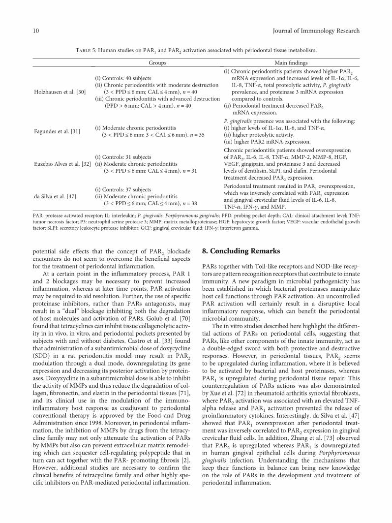
Table 5: Human studies on PAR1 and PAR2 activation associated with periodontal tissue metabolism.
Groups Main findings
Holzhausen et al. [30]
(i) Controls: 40 subjects(ii) Chronic periodontitis with moderate destruction
(3 < PPD≤ 6mm; CAL≤ 4mm), n = 40(iii) Chronic periodontitis with advanced destruction
(PPD > 6mm; CAL > 4mm), n = 40
(i) Chronic periodontitis patients showed higher PAR2mRNA expression and increased levels of IL-1α, IL-6,IL-8, TNF-α, total proteolytic activity, P. gingivalisprevalence, and proteinase 3 mRNA expressioncompared to controls.
(ii) Periodontal treatment decreased PAR2mRNA expression.
Fagundes et al. [31](i) Moderate chronic periodontitis
(3 < PPD≤ 6mm; 3 < CAL≤ 6mm), n = 35
P. gingivalis presence was associated with the following:(i) higher levels of IL-1α, IL-6, and TNF-α,(ii) higher proteolytic activity,(iii) higher PAR2 mRNA expression.
Euzebio Alves et al. [32](i) Controls: 31 subjects(ii) Moderate chronic periodontitis
(3 < PPD≤ 6mm; CAL≤ 4mm), n = 31
Chronic periodontitis patients showed overexpressionof PAR2, IL-6, IL-8, TNF-α, MMP-2, MMP-8, HGF,VEGF, gingipain, and proteinase 3 and decreasedlevels of dentilisin, SLPI, and elafin. Periodontaltreatment decreased PAR2 expression.
da Silva et al. [47](i) Controls: 37 subjects(ii) Moderate chronic periodontitis
(3 < PPD≤ 6mm; CAL≤ 4mm), n = 38
Periodontal treatment resulted in PAR1 overexpression,which was inversely correlated with PAR2 expressionand gingival crevicular fluid levels of IL-6, IL-8,TNF-α, IFN-γ, and MMP.
PAR: protease activated receptor; IL: interleukin; P. gingivalis: Porphyromonas gingivalis; PPD: probing pocket depth; CAL: clinical attachment level; TNF:tumor necrosis factor; P3: neutrophil serine protease 3; MMP: matrix metalloproteinase; HGF: hepatocyte growth factor; VEGF: vascular endothelial growthfactor; SLPI: secretory leukocyte protease inhibitor; GCF: gingival crevicular fluid; IFN-γ: interferon gamma.
10 Journal of Immunology Research
Conflicts of Interest
The authors declare that they have no conflict of interests.
Acknowledgments
This work was supported by grants from the Sao PauloResearch Foundation (FAPESP Grant nos. 2015/07396-2and 2015/11587-8).
References
[1] V. S. Ossovskaya and N. W. Bunnett, “Protease-activatedreceptors: contribution to physiology and disease,” Physiologi-cal Reviews, vol. 84, no. 2, pp. 579–621, 2004.
[2] R. Ramachandran, C. Altier, K. Oikonomopoulou, and M. D.Hollenberg, “Proteinases, their extracellular targets, andinflammatory signaling,” Pharmacological Reviews, vol. 68,no. 4, pp. 1110–1142, 2016.
[3] N.Vergnolle, J. L.Wallace,N.W.Bunnett, andM.D.Hollenberg,“Protease-activated receptors in inflammation, neuronal signal-ing and pain,” Trends in Pharmacological Sciences, vol. 22, no. 3,pp. 146–152, 2001.
[4] A. Lourbakos, J. Potempa, J. Travis et al., “Arginine-specificprotease from Porphyromonas gingivalis activates protease-activated receptors on human oral epithelial cells and inducesinterleukin-6 secretion,” Infection and Immunity, vol. 69,no. 8, pp. 5121–5130, 2001.
[5] M. Holzhausen, L. C. Spolidorio, and N. Vergnolle, “Role ofprotease-activated receptor-2 in inflammation, and its possibleimplications as a putativemediator of periodontitis,”Memóriasdo InstitutoOswaldoCruz, vol. 100, Supplement 1, pp. 177–180,2005.
[6] M. Holzhausen, L. C. Spolidorio, R. P. Ellen et al., “Protease-activated receptor-2 activation: a major role in the pathogene-sis of Porphyromonas gingivalis infection,” American Journalof Pathology, vol. 168, no. 4, pp. 1189–1199, 2006.
[7] O. Déry, C. U. Corvera, M. Steinhoff, and N. W. Bunnett,“Proteinase-activated receptors: novel mechanisms of signal-ing by serine proteases,” American Journal of Physiology,vol. 274, no. 6 pt 1, pp. 1429–1452, 1998.
[8] S. R. Coughlin, “Thrombin signalling and protease-activatedreceptors,” Nature, vol. 407, no. 6801, pp. 258–264, 2000.
[9] P. J. O'Brien, M. Molino, M. Kahn, and L. F. Brass, “Proteaseactivated receptors: theme and variations,” Oncogene, vol. 20,no. 13, pp. 1570–1581, 2001.
[10] R. Ramachandran, F. Noorbakhsh, K. Defea, and M. D.Hollenberg, “Targeting proteinase-activated receptors: thera-peuticpotentialandchallenges,”NatureReviewsDrugDiscovery,vol. 11, no. 1, pp. 69–86, 2012.
[11] S. J. Song, C. N. Pagel, T. M. Campbell, R. N. Pike, and E. J.Mackie, “The role of protease-activated receptor-1 in bonehealing,” American Journal of Pathology, vol. 166, no. 3,pp. 857–868, 2005.
[12] U. Arayatrakoollikit, P. Pavasant, and T. Yongchaitrakul,“Thrombin induces osteoprotegerin synthesis via phos-phatidylinositol 3′-kinase/mammalian target of rapamycinpathway in human periodontal ligament cells,” Journal ofPeriodontal Research, vol. 43, no. 5, pp. 537–543, 2008.
[13] Y. Kanno, A. Ishisaki, E. Kawashita, H. Kuretake, K. Ikeda,and O. Matsuo, “uPA attenuated LPS-induced inflammatory
osteoclastogenesis through the plasmin/PAR-1/Ca(2+)/CaMKK/AMPK axis,” International Journal of BiologicalSciences, vol. 12, no. 1, pp. 63–71, 2016.
[14] N. Ohuchi, K. Hayashi, K. Iwamoto et al., “Thrombin-stimu-lated proliferation is mediated by endothelin-1 in culturedrat gingival fibroblasts,” Fundamental & Clinical Pharmacol-ogy, vol. 24, no. 4, pp. 501–508, 2010.
[15] W. H. Yang, Y. T. Deng, Y. P. Hsieh, K. J. Wu, and M. Y. Kuo,“Thrombin activates latent TGFβ1 via integrin αvβ1 in gingi-val fibroblasts,” Journal of Dental Research, vol. 95, no. 8,pp. 939–945, 2016.
[16] M. G. Rohani, R. P. Beyer, B. M. Hacker, H. Dommisch, B. A.Dale, and W. O. Chung, “Modulation of expression of innateimmunity markers CXCL5/ENA-78 and CCL20/MIP3alphaby protease-activated receptors (PARs) in human gingival epi-thelial cells,” Innate Immunity, vol. 16, no. 2, pp. 104–114,2010.
[17] A. Uehara, T. Imamura, J. Potempa, J. Travis, and H. Takada,“Gingipains from Porphyromonas gingivalis synergisticallyinduce the production of proinflammatory cytokines throughprotease-activated receptors with Toll-like receptor andNOD1/2 ligands in human monocytic cells,” Cellular Microbi-ology, vol. 10, no. 5, pp. 1181–1189, 2008.
[18] A. Lourbakos, Y. P. Yuan, A. L. Jenkins et al., “Activation ofprotease-activated receptors by gingipains from Porphyromo-nas gingivalis leads to platelet aggregation: a new trait inmicrobial pathogenicity,” Blood, vol. 97, no. 12, pp. 3790–3797, 2001.
[19] S. K. Bohm, W. Kong, D. Bromme et al., “Molecular cloning,expression and potential functions of the human proteinase-activated receptor-2,” Biochemical Journal, vol. 314, no. Pt 3,pp. 1009–1016, 1996.
[20] S. Nystedt, V. Ramakrishnan, and J. Sundelin, “Theproteinase-activated receptor 2 is induced by inflammatorymediators in human endothelial cells. Comparison with thethrombin receptor,” The Journal of Biological Chemistry,vol. 271, no. 25, pp. 14910–14915, 1996.
[21] L. A. Abraham, C. Chinni, A. L. Jenkins et al., “Expression ofprotease-activated receptor-2 by osteoblasts,” Bone, vol. 26,no. 1, pp. 7–14, 2000.
[22] T. M. Cocks and J. D. Moffatt, “Protease-activated receptors:sentries for inflammation?” Trends in PharmacologicalSciences, vol. 21, no. 3, pp. 103–108, 2000.
[23] N. Vergnolle, N. W. Bunnett, K. A. Sharkey et al., “Proteinase-activated receptor-2 and hyperalgesia: a novel pain pathway,”Nature Medicine, vol. 7, no. 7, pp. 821–826, 2001.
[24] N. Cenac, A. M. Coelho, C. Nguyen et al., “Induction ofintestinal inflammation in mouse by activation of proteinase-activated receptor-2,” American Journal of Pathology,vol. 161, no. 5, pp. 1903–1915, 2002.
[25] S. R. Coughlin and E. Camerer, “PARticipation in inflam-mation,” Journal of Clinical Investigation, vol. 111, no. 1,pp. 25–27, 2003.
[26] A. M. Coelho, N. Vergnolle, B. Guiard, J. Fioramonti, and L.Bueno, “Proteinases and proteinase-activated receptor 2: apossible role to promote visceral hyperalgesia in rats,” Gastro-enterology, vol. 122, no. 4, pp. 1035–1047, 2002.
[27] F. Schmidlin, S. Amadesi, K. Dabbagh et al., “Protease-acti-vated receptor 2 mediates eosinophil infiltration and hyper-reactivity in allergic inflammation of the airway,” Journal ofImmunology, vol. 169, no. 9, pp. 5315–5321, 2002.
11Journal of Immunology Research
[28] W. R. Ferrell, J. C. Lockhart, E. B. Kelso et al., “Essential rolefor proteinase-activated receptor-2 in arthritis,” Journal ofClinical Investigation, vol. 111, no. 1, pp. 35–41, 2003.
[29] J. R. Lindner, M. L. Kahn, S. R. Coughlin et al., “Delayed onsetof inflammation in protease-activated receptor-2-deficientmice,” Journal of Immunology, vol. 165, no. 11, pp. 6504–6510, 2000.
[30] M. Holzhausen, J. R. Cortelli, V. A. da Silva, G. N. Franco, S. C.Cortelli, and N. Vergnolle, “Protease-activated receptor-2(PAR(2)) in human periodontitis,” Journal of Dental Research,vol. 89, no. 9, pp. 948–953, 2010.
[31] J. A. Fagundes, L. D. Monoo, V. T. Euzébio Alves et al.,“Porphyromonas gingivalis is associated with protease-activated receptor-2 upregulation in chronic periodontitis,”Journal of Periodontology, vol. 82, no. 11, pp. 1596–1601, 2011.
[32] V. T. Euzebio Alves, H. A. Bueno da Silva, B. N. de Françaet al., “Periodontal treatment downregulates protease-activated receptor 2 in human gingival crevicular fluid cells,”Infection and Immunity, vol. 81, no. 12, pp. 4399–4407, 2013.
[33] M. L. Castro, G. C. Franco, L. S. Branco-de-Almeida et al.,“Downregulation of proteinase-activated receptor-2, interleu-kin-17, and other proinflammatory genes by subantimicrobialdoxycycline dose in a rat periodontitis model,” Journal ofPeriodontology, vol. 87, no. 2, pp. 203–210, 2016.
[34] E. Csernok, M. Ernst, W. Schmitt, D. F. Bainton, and W. L.Gross, “Activated neutrophils express proteinase 3 on theirplasmamembrane invitro and invivo,”Clinical&ExperimentalImmunology, vol. 95, no. 2, pp. 244–250, 1994.
[35] P. Zhao, M. Metcalf, and N. W. Bunnett, “Biased signaling ofprotease-activated receptors,” Frontiers in Endocrinology(Lausanne), vol. 5, p. 67, 2014.
[36] M. D. Hollenberg, K. Mihara, D. Polley et al., “Biased signal-ling and proteinase-activated receptors (PARs): targetinginflammatory disease,” British Journal of Pharmacology,vol. 171, no. 5, pp. 1180–1194, 2014.
[37] J. Romer, T. H. Bugge, C. Fyke et al., “Impaired wound healingin mice with a disrupted plasminogen gene,” Nature Medicine,vol. 2, no. 3, pp. 287–292, 1996.
[38] V. A. Ploplis, E. L. French, P. Carmeliet, D. Collen, and E. F.Plow, “Plasminogen deficiency differentially affects recruit-ment of inflammatory cell populations in mice,” Blood,vol. 91, no. 6, pp. 2005–2009, 1998.
[39] D. Collen, “Ham-Wasserman lecture: role of the plasminogensystem in fibrin-homeostasis and tissue remodeling,” Hema-tology / the Education Program of the American Society ofHematology. American Society of Hematology. EducationProgram, vol. 2001, no. 1, pp. 1–91, 2001.
[40] R. Sulniute, T. Lindh, M. Wilczynska, J. Li, and T. Ny,“Plasmin is essential in preventing periodontitis in mice,”American Journal of Pathology, vol. 179, no. 2, pp. 819–828,2011.
[41] S. Steinsvoll, K. Helgeland, and K. Schenck, “Mast cells–a rolein periodontal diseases?” Journal of Clinical Periodontology,vol. 31, no. 6, pp. 413–419, 2004.
[42] B. M. Eley and S. W. Cox, “Cathepsin B/L-, elastase-, tryptase-,trypsin- and dipeptidyl peptidase IV-like activities in gingivalcrevicular fluid: a comparison of levels before and after peri-odontal surgery in chronic periodontitis patients,” Journal ofPeriodontology, vol. 63, no. 5, pp. 412–417, 1992.
[43] S. W. Cox and B. M. Eley, “Cathepsin B/L-, elastase-, tryptase-,trypsin- and dipeptidyl peptidase IV-like activities in gingival
crevicular fluid. A comparison of levels before and afterbasic periodontal treatment of chronic periodontitispatients,” Journal of Clinincal Periodontology, vol. 19, no. 5,pp. 333–339, 1992.
[44] B. M. Eley and S. W. Cox, “Correlation of gingival crevicularfluid proteases with clinical and radiological measurementsof periodontal attachment loss,” Journal of Dentistry, vol. 20,no. 2, pp. 90–99, 1992.
[45] B. M. Eley and S. W. Cox, “Cathepsin B/L-, elastase-, tryptase-,trypsin- and dipeptidyl peptidase IV-like activities in gingivalcrevicular fluid: correlation with clinical parameters inuntreated chronic periodontitis patients,” Journal of Periodon-tal Research, vol. 27, no. 1, pp. 62–69, 1992.
[46] M. Holzhausen, R. D. Balejo, G. M. Lara, S. C. Cortelli, W. A.Saad, and J. R. Cortelli, “Nafamostat mesilate, a potent tryptaseinhibitor, modulates periodontitis in rats,” Clinincal OralInvestigation, vol. 15, no. 6, pp. 967–973, 2011.
[47] H. A. da Silva, V. T. Euzebio Alves, L. C. Spolidório et al.,“Expression of protease activated receptor-1 in chronicperiodontitis,” Journal of Periodontology, vol. 85, no. 12,pp. 1763–1769, 2014.
[48] T. Shimada, N. Sugano, K. Ikeda, K. Shimada, T. Iizuka, andK. Ito, “Protease-activated receptor 2 mediates interleukin-8and intercellular adhesion molecule-1 expression in responseto Aggregatibacter actinomycetemcomitans,” Oral Microbiol-ogy and Immunology, vol. 24, no. 4, pp. 285–291, 2009.
[49] J. Travis and J. Potempa, “Bacterial proteinases as targets forthe development of second-generation antibiotics,” Biochimicaet Biophysica Acta, vol. 1477, no. 1-2, pp. 35–50, 2000.
[50] A. Uehara, K. Muramoto, T. Imamura et al., “Arginine-specificgingipains from Porphyromonas gingivalis stimulate produc-tion of hepatocyte growth factor (scatter factor) throughprotease-activated receptors in human gingival fibroblasts inculture,” Journal of Immunology, vol. 175, no. 9, pp. 6076–6084, 2005.
[51] C. N. Pagel, S. J. Song, L. H. Loh et al., “Thrombin-stimulatedgrowth factor and cytokine expression in osteoblasts is medi-ated by protease-activated receptor-1 and prostanoids,” Bone,vol. 44, no. 5, pp. 813–821, 2009.
[52] R. A. Giacaman, A. C. Asrani, K. F. Ross, and M. C. Herzberg,“Cleavage of protease-activated receptors on an immortalizedoral epithelial cell line by Porphyromonas gingivalis gingi-pains,” Microbiology, vol. 155, no. 10, pp. 3238–3246, 2009.
[53] D. M. Wong, V. Tam, R. Lam et al., “Protease-activated recep-tor 2 has pivotal roles in cellular mechanisms involved inexperimental periodontitis,” Infection and Immunity, vol. 78,no. 2, pp. 629–638, 2010.
[54] L. C. Spolidorio, P. D. Lucas, J. P. Steffens et al., “Impact of par-statin on experimental periodontal disease and repair in rats,”Journal of Periodontology, vol. 85, no. 9, pp. 1266–1274, 2014.
[55] N. Vergnolle, “Protease-activated receptors as drug targets ininflammation and pain,” Pharmacology & Therapeutics,vol. 123, no. 3, pp. 292–309, 2009.
[56] V. Shpacovitch, M. Feld, N. W. Bunnett, and M. Steinhoff,“Protease-activated receptors: novel PARtners in innateimmunity,” Trends in Immunology, vol. 28, no. 12, pp. 541–550, 2007.
[57] A. Lourbakos, C. Chinni, P. Thompson et al., “Cleavage andactivation of proteinase-activated receptor-2 on humanneutrophils by gingipain-R from Porphyromonas gingivalis,”FEBS Letters, vol. 435, no. 1, pp. 45–48, 1998.
12 Journal of Immunology Research
[58] L. W. Yun, A. A. Decarlo, and N. Hunter, “Blockade ofprotease-activated receptors on T cells correlates with alteredproteolysis of CD27 by gingipains of Porphyromonas gingiva-lis,” Clinical & Experimental Immunology, vol. 150, no. 2,pp. 217–229, 2007.
[59] W. O. Chung, S. R. Hansen, D. Rao, and B. A. Dale, “Protease-activated receptor signaling increases epithelial antimicrobialpeptide expression,” Journal of Immunology, vol. 173, no. 8,pp. 5165–5170, 2004.
[60] A. L. Pereira, M. Holzhausen, G. C. Franco, S. C. Cortelli, andJ. R. Cortelli, “Human β-defensin 2 and protease activatedreceptor-2 expression in patients with chronic periodontitis,”Archives of Oral Biology, vol. 57, no. 12, pp. 1609–1614, 2012.
[61] H. Tada, T. Matsuyama, T. Nishioka et al., “Porphyromonasgingivalis gingipain-dependently enhances IL-33 productionin human gingival epithelial cells,” PLoS One, vol. 11, no. 4,article e0152794, 2016.
[62] G. N. Belibasakis, N. Bostanci, and D. Reddi, “Regulation ofprotease-activated receptor-2 expression in gingival fibroblastsand Jurkat T cells by Porphyromonas gingivalis,” Cell BiologyInternational, vol. 34, no. 3, pp. 287–292, 2010.
[63] R. Smith, M. Ransjö, L. Tatarczuch et al., “Activation ofprotease-activated receptor-2 leads to inhibition of osteoclastdifferentiation,” Journal of Bone and Mineral Research,vol. 19, no. 3, pp. 507–516, 2004.
[64] N. Amiable, S. K. Tat, D. Lajeunesse et al., “Proteinase-activated receptor (PAR)-2 activation impacts bone resorp-tive properties of human osteoarthritic subchondral boneosteoblasts,” Bone, vol. 44, no. 6, pp. 1143–1150, 2009.
[65] M. Holzhausen, L. C. Spolidorio, and N. Vergnolle, “Protein-ase-activated receptor-2 (PAR2) agonist causes periodontitisin rats,” Journal of Dental Research, vol. 84, no. 2, pp. 154–159, 2005.
[66] J. J. Reynolds and M. C. Meikle, “Mechanisms of connectivetissue matrix destruction in periodontitis,” Periodontology2000, vol. 14, no. 1, pp. 144–157, 1997.
[67] T. J. Moraes, R. Martin, J. D. Plumb et al., “Role of PAR2 inmurine pulmonary pseudomonal infection,” American Journalof Physiology Lung Cellular and Molecular Physiology, vol. 294,no. 4, pp. 368–377, 2008.
[68] H. M. Lee, H. Y. Kim, H. J. Kang et al., “Up-regulationof protease-activated receptor 2 in allergic rhinitis,”Annals of Otology, Rhinology & Laryngology, vol. 116, no. 7,pp. 554–558, 2007.
[69] S. M. McFarlane, M. J. Seatter, T. Kanke, G. D. Hunter, and R.Plevin, “Proteinase-activated receptors,” PharmacologicalReviews, vol. 53, no. 2, pp. 245–282, 2001.
[70] L. M. Golub, N. Ramamurthy, T. F. McNamara et al., “Tetra-cyclines inhibit tissue collagenase activity. A new mechanismin the treatment of periodontal disease,” Journal of PeriodontalResearch, vol. 19, no. 6, pp. 651–655, 1984.
[71] H. M. Lee, S. G. Ciancio, G. Tüter, M. E. Ryan, E. Komaroff,and L. M. Golub, “Subantimicrobial dose doxycycline efficacyas a matrix metalloproteinase inhibitor in chronic periodonti-tis patients is enhanced when combined with a non-steroidalanti-inflammatory drug,” Journal of Periodontology, vol. 75,no. 3, pp. 453–463, 2004.
[72] M. Xue, Y. K. Chan, K. Shen et al., “Protease-activated receptor2, rather than protease-activated receptor 1, contributes to theaggressive properties of synovial fibroblasts in rheumatoidarthritis,” Arthritis & Rheumatology, vol. 64, no. 1, pp. 88–98, 2012.
[73] D. Zhang, S. Li, L. Hu et al., “Protease-activated receptorsexpression in gingiva in periodontal health and disease,”Archives of Oral Biology, vol. 59, no. 4, pp. 393–399, 2014.
13Journal of Immunology Research

Submit your manuscripts athttps://www.hindawi.com
Stem CellsInternational
Hindawi Publishing Corporationhttp://www.hindawi.com Volume 2014
Hindawi Publishing Corporationhttp://www.hindawi.com Volume 2014
MEDIATORSINFLAMMATION
of
Hindawi Publishing Corporationhttp://www.hindawi.com Volume 2014
Behavioural Neurology
EndocrinologyInternational Journal of
Hindawi Publishing Corporationhttp://www.hindawi.com Volume 2014
Hindawi Publishing Corporationhttp://www.hindawi.com Volume 2014
Disease Markers
Hindawi Publishing Corporationhttp://www.hindawi.com Volume 2014
BioMed Research International
OncologyJournal of
Hindawi Publishing Corporationhttp://www.hindawi.com Volume 2014
Hindawi Publishing Corporationhttp://www.hindawi.com Volume 2014
Oxidative Medicine and Cellular Longevity
Hindawi Publishing Corporationhttp://www.hindawi.com Volume 2014
PPAR Research
The Scientific World JournalHindawi Publishing Corporation http://www.hindawi.com Volume 2014
Immunology ResearchHindawi Publishing Corporationhttp://www.hindawi.com Volume 2014
Journal of
ObesityJournal of
Hindawi Publishing Corporationhttp://www.hindawi.com Volume 2014
Hindawi Publishing Corporationhttp://www.hindawi.com Volume 2014
Computational and Mathematical Methods in Medicine
OphthalmologyJournal of
Hindawi Publishing Corporationhttp://www.hindawi.com Volume 2014
Diabetes ResearchJournal of
Hindawi Publishing Corporationhttp://www.hindawi.com Volume 2014
Hindawi Publishing Corporationhttp://www.hindawi.com Volume 2014
Research and TreatmentAIDS
Hindawi Publishing Corporationhttp://www.hindawi.com Volume 2014
Gastroenterology Research and Practice
Hindawi Publishing Corporationhttp://www.hindawi.com Volume 2014
Parkinson’s Disease
Evidence-Based Complementary and Alternative Medicine
Volume 2014Hindawi Publishing Corporationhttp://www.hindawi.com