Embed Size (px)
Citation preview
2089
The Simultaneous Occurrence of Variant Hairy Cell Leukemia and Chronic-Phase Chronic Myelogenous Leukemia A Case Report
James E. Reeves, M.D.," Bruce A. Robbins, M.D.,t Lee R. Pankey, M.D.,S Abdalla L. Elias, M.D.,S and William F. Anderson, M.D."
Background. Coexistence of second malignancies in patients with hairy cell leukemia is not uncommon. Most second malignancies are solid tumors or other lympho- proliferative disorders. In this study, a case of a tartrate- resistant acid phosphatase (TRAP)-positive hairy cell leukemia variant with the subsequent development of Philadelphia chromosome-positive chronic myelogenous leukemia (CML) is reported.
Methods. Routine morphology was performed on the peripheral blood, bone marrow and spleen. Peripheral smears were stained for TRAP. Peripheral blood was studied by two-color flow cytometry for a panel of lym- phocytic markers including CD11, CD25, and CD103. Cy- togenetic studies were performed on a bone marrow aspi- rate.
Results. A unique case of a hairy cell leukemia vari- ant and CML in a patient who responded to the new pu- rine analog 2-chlorodeoxyadenosine (2-CdA) is pre- sented.
Conclusions. The first case of concurrent hairy cell leukemia with CML is reported. Cancer 1995;75:2089-92.
Key words: hairy cell leukemia, chronic myelogenous leukemia, tartrate-resistant acid phosphatase, 2-CdA.
Hairy cell leukemia (HCL) is a rare chronic malignant lymphoproliferative disorder with well defined clinical and pathologic characteristics consisting of pancytope- nia, splenomegaly, and typical hairy cells in the periph- eral blood and bone marrow. The diagnosis is usually
From the *Hematology/Oncology Clinic, and the $Glenwood Regional Medical Center, West Monroe, Louisiana and the tHead Di- vision of Laboratory Medicine, Department of Pathology, Scripps Clinic and Research Foundation, La Jolla, California.
Address for reprints: James E. Reeves, M.D., Hematology/On- cology Clinic, 109 Regency Place, West Monroe, LA 71291.
Received September 16, 1994; revision received December 29, 1994; accepted December 29, 1994.
confirmed by positive tartrate-resistant acid phospha- tase and specific patterns of immun~reactivity.'-~ A wide variety of second malignancies have been noted in HCL with an incidence of 8.7Y0.~ A recent report re- vealed an unexpectedly high incidence of secondary neoplasms in patients after treatment with interferon for HCL.5 Most of the second malignancies have been solid tumors or other lymphoproliferative disorders. At least one case of a myeloproliferative disorder was found in a patient with HCL.6 However, Philadelphia chromosome-positive chronic myelogenous leukemia (CML) has never been reported in a patient with HCL to the authors' knowledge. We report a case of an el- derly man treated with splenectomy for an HCL vari- ant, who subsequently developed CML within 1 year. To our knowledge, these two diseases never have been reported to occur in one patient. Our patient also re- sponded to 2-chlorodeoxyadenosine (2-CdA), a rela- tively new purine analog that almost has replaced inter- feron as the primary treatment for HCL.
Case Report
An 83-year-old male who presented to his primary physician with abdominal pain in June 1992 is described. Results of a physical examination revealed a significantly enlarged spleen. A computed tomographic scan of the abdomen confirmed marked splenomegaly with no lymphadenopathy. Peripheral counts were as follows: hemoglobin level, 10.7 grams/dl; platelet count, 136,OOO/cu mm; and leukocyte count, 24,0001 cu mm, with 12% segmented neutrophils, 85% lymphocytes, and 3% monocytes. The peripheral blood smear revealed lym- phocytes with morphologic features of hairy cells, i.e., abun- dant agranular cytoplasms, irregular cytoplasmic projections, and oval to slightly indented nuclei with dispersed chromatin and inconspicuous nucleoli. However, additional lympho- cytes showed higher nuclear/cytoplasmic ratios and small nucleoli. Erythrocyte morphology and platelets appeared nor-

2090 CANCER April 25,1995, Volume 75, No. 8
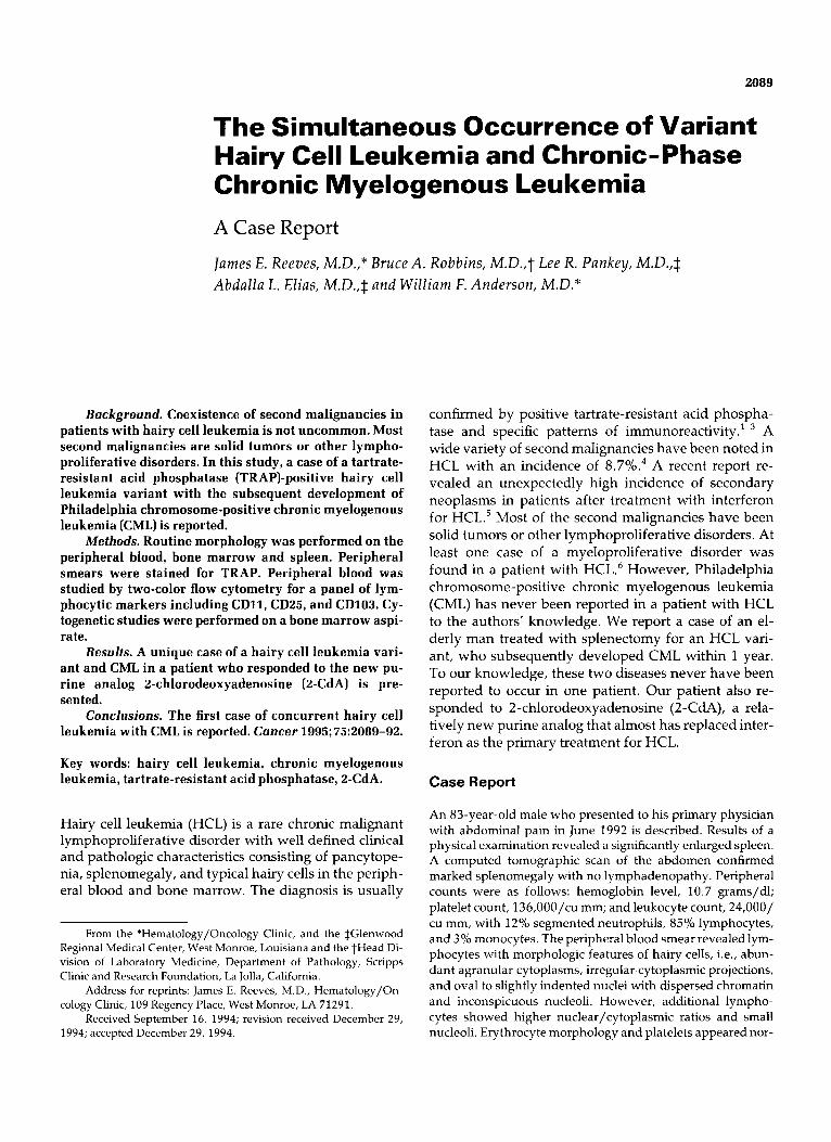
Figure 1 . Peripheral blood smear from October 1993 shows features of hairy cell leukemia and chronic myelogenous leukemia. Three hairy cells are seen along with immature myeloid cells and a basophil (original magnification XlOOO).
mal. The bone marrow aspiration and biopsy were 50% cellu- lar with an atypical lymphoid infiltrate interspersed through- out the sections. Some areas showed a typical honeycomb pat- tern as seen in HCL. The patient was diagnosed as having HCL. Due to the patient’s symptoms of hypersplenism, a sple- nectomy was performed. The patient tolerated the procedure well, and his platelet count increased after surgery. His hemo- globin level remained stable, and the lymphocytosis also showed little change. The spleen weighed 3200 grams. There was a diffuse lymphoid infiltrate involving the red pulp but also exhibiting a nodular pattern of infiltration in the white pulp. The lymphoid cells were of medium size and had higher
U
.. . .
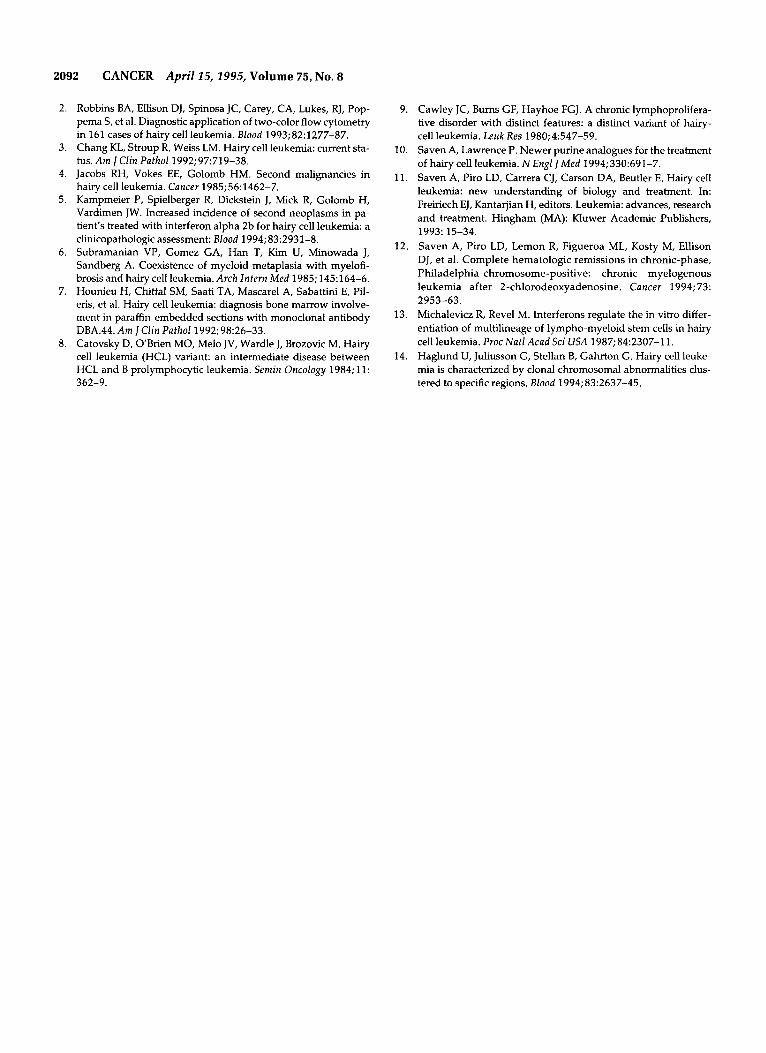
CD103 Figure 2. Two-color flow cytometry shows simultaneous expression of CD19 and CD103 on the hairy cell population from March 1994.
i
Figure 3. A section of spleen shows red pulp involvement by hairy cell leukemia variant. The leukemic cells show rounded, vesicular nuclei and more prominent nucleoli than typical HCL (PAS stain, original magnification X640).
nuclear/cytoplasmic ratios and more prominent nucleoli than typical HCL.
The patient was subsequently lost to follow-up until Oc- tober 1993, when he again presented to his primary physician with pneumonia and a pleural effusion. He was noted to have a marked leukocytosis (leukocyte count of 168,OOO/cu mm), with a hemoglobin level of 10.5 gm/dl and a platelet count of 232,OOO/cu mm. The peripheral blood smear was suggestive of myelophthisis with a predominance of myelocytes and metamyelocytes. Peripheral blood differential was as follows: 22% segmented neutrophils, 9% lymphocytes, 5% mono- cytes, 4% eosinophils, 2% basophils, 19% neutrophilic bands, 9% metamyelocytes, 19% myelocytes, 7% promyelocytes, and 4% myeloblasts. A small percentage of lymphocytes with features of HCL were still seen (Fig. 1). The bone marrow bi- opsy was 100% cellular with a marked myeloid hyperplasia and no evidence of myelofibrosis. Blasts comprised less than 10% of the marrow elements. Hairy cells were recognized on the aspirate smears. Cytogenetic studies were performed by Medical Genetic Consultants, Inc., Ocean Springs, Missis- sippi, and revealed t(9; 22)(q34; q l 1) Philadelphia chromo- some positivity in 20/20 metaphase spreads (100%). No other cytogenetic abnormalities were noted. tartrate-resistant acid phosphatase stains on peripheral smears showed strong cyto- plasmic staining of mononuclear cells with HCL morphology.
Two-color flow cytometry was performed on peripheral blood from March 1994 (Fig. 2) and exhibited a monoclonal population of B cells expressing surface immunoglobulins M, D, G, and kappa light chains. There was also strong staining for CDllc and positive staining for CD103, which is a typical feature for HCL. However, staining for CD25 was negative. Immunohistochemical staining of the spleen showed the lym- phoid population to stain positively for CD45(LCA) and pan 8-cell marker CD20(L26), but negative for hairy cell-associ- ated marker DBA.44 and T-cell marker CD43(Leu22).
The leukocytosis was controlled initially with hydroxy- urea, followed by periods of relapse requiring cytarabine che- motherapy. However, a hematologic remission was not ob-

Case Report of HCL and CML in a Single Patient/Reeves et al. 2091
tained. Treatment was subsequently changed to 2-CdA. We initially attempted an outpatient intravenous piggyback regi- men, but still failed to obtain adequate control. He then re- ceived two cycles of 2-CdA with a dosage of 0.1 mg/kg per day by continuous infusion for 7 days for a total of two courses. At the time of the first infusion of 2-CdA, his leuko- cyte count had risen to 62,1OO/cu mm with a hemoglobin level of 13.4 gm/dl and platelets 620,OOO/cu mm. The pe- ripheral blood smear was compatible with CML with only oc- casional atypical lymphocytes. Eleven days into his first cycle, his leukocyte count decreased to 6,8OO/cu mm, and there was no morphologic evidence of the CML nor HCL. The second course of 2-CdA began as his leukocyte count rose to 25,100/ cu mm with still no definite evidence of CML nor HCL. His platelet count was 368,OOO/cu mm. He subsequently devel- oped prolonged thrombocytopenia with a platelet count of lO,OOO/cu mm and required multiple prophylactic platelet transfusions. His leukocyte count decreased to a low of 2,400/ cu mm. However, the patient never had a fever and did not develop any evidence of infection. His hemoglobin level de- creased to 10.9 gm/dl and he received packed erythrocyte transfusions. Twenty-eight days into his second treatment, a bone marrow biopsy and aspiration were obtained. The bone marrow biopsy was hypocellular and showed a complete morphologic remission of the HCL and CML. He did not, however, obtain suppression of the Philadelphia chromo- some.
Discussion
Hairy cell leukemia is a rare chronic lymphoprolifera- tive disorder with characteristic clinical and pathologic findings. Classically, patients present with splenomeg- aly and peripheral cytopenias. Although the pancyto- penia may be variable, most patients do have monocy- topenia. Although our patient was tartrate-resistant acid phosphatase stain-positive, he initially presented with atypical features including an elevated leukocyte count and absence of monocytopenia. In addition, many of the initial cells exhibited larger nuclei, higher nuclear/cytoplasmic ratios, and more prominent nucleoli than are present in the typical hairy cell. The splenic infiltrate also exhibited unusual features includ- ing nodular white pulp involvement, lymphoid cells with vesicular nuclei, and frequent nucleoli (Fig. 3). The flow cytometry was also atypical because the CD25 stain was negative, although the CDllc and CD103 were strongly positive.
Typical HCL has been shown to stain positively for CD25, CDllc, and CD103 in 99% of cases.' Tartrate- resistant acid phosphatase stain is also positive in 99% of most reported ~ e r i e s . ~ The DBA.44, however, was also negative in our patient and is usually positive in HCL.7
The CML was diagnosed in October 1993 approxi- mately 17 months after the diagnosis of HCL. This was
confirmed by the presence of the Philadelphia chromo- some. Unfortunately, no cytogenetics were performed on the original bone marrow biopsy in June 1992.
This case most likely represents an unusual variant of HCL such as described by Catovsky' or Cawley.' These typically present with higher lymphocyte counts and atypical cells with morphologic features intermedi- ate between HCL and prolymphocytic leukemia. This form of HCL variant typically stains negative for CD25. We believe that this patient has two independent dis- ease processes: HCL variant and Philadelphia chromo- some-positive CML. A search of the literature reveals no reported cases of these two diseases occurring simul- taneously in the same patient.
Treatment with 2-CdA has proven to be effective in the majority of patients with HCL.'of" 2-chlorodeoxy- adenosine is effective against resting and dividing lym- phocytes, and immature myeloid cells also have been found to be inhibited markedly in in vitro studies. A recent report also found 2-CdA to induce hematologic responses in CML without suppression of the Philadel- phia chromosome.'2 Although patients obtain complete hematologic and morphologic responses, there is no suppression of the Philadelphia chromosome. This also was the case in our patient. He received 2-CdA to con- trol the leukocytosis and resulted in a morphologic re- mission of the HCL variant along with the CML. The Philadelphia chromosome, however, persists.
Although the origin of the hairy cell remains to be determined, most studies suggest the hairy cell repre- sents a late stage of B-cell ontogeny.6 A pluripotent lym- phomyeloid stem cell was demonstrated in patients with HCL, however, these colony-forming cells are dis- tinct from the hairy ~e1ls. l~ Chromosomal aberrations are thought to exist in most patients with HCL. A recent report showed cytogenetic analysis revealing a high fre- quency of clonal abnormalities in HCL patients (67%) with chromosomes 1, 2, and 5 the most ~ignificant.'~ One could speculate that the chromosomal changes that are present in the majority of HCL patients may have led to the evolution of the BCR-ABL oncogene. It would be interesting to see if fluorescent in situ hybridization could detect the chromosomal translocation in the orig- inal splenic tissue. To our knowledge, however, the Philadelphia chromosome never has been reported in a patient with HCL. The authors believed that the devel- opment of CML in this patient with HCL variant prob- ably was related to the patient's age and possibly the immunocompromised state secondary to the HCL as suggested by Kampmeier et aL5
References
1. Golomb HM, Catovsky D, Golde D. Hairy cell leukemia: a clini- cal review based on 71 cases. Ann Intern Med 1978;89:677-83.
2092 CANCER April 15,1995, Volume 75, No. 8
2. Robbins BA, Ellison DJ, Spinosa JC, Carey, CA, Lukes, RJ, Pop- pema s, et al. Diagnostic application of two-color flow cytometry in 161 cases of hairy cell leukemia. Blood 1993;82:1277-87.
3. Chang KL, Stroup R, Weiss LM. Hairy cell leukemia: current sta- tus. A m JClin Pathol 1992;97:719-38.
4. Jacobs RH, Vokes EE, Golomb HM. Second malignancies in hairy cell leukemia. Cancer 1985;56:1462-7.
5. Kampmeier P, Spielberger R, Dickstein J, Mick R, Golomb H, Vardimen JW. Increased incidence of second neoplasms in pa- tient’s treated with interferon alpha 2b for hairy cell leukemia: a clinicopathologic assessment: Blood 1994; 832931-8.
6. Subramanian VP, Gomez GA, Han T, Kim U, Minowada J, Sandberg A. Coexistence of myeloid metaplasia with myelofi- brosis and hairy cell leukemia. Arch Intern Med 1985; 145:164-6. Hounieu H, Chittal SM, Saati TA, Mascarel A, Sabattini E, Pil- ens, et al. Hairy cell leukemia: diagnosis bone marrow involve- ment in paraffin-embedded sections with monoclonal antibody DBA.44. A m J Clin Pathol 1992;98:26-33.
8. Catovsky D, OBrien MO, Melo JV, Wardle J, Brozovic M. Hairy cell leukemia (HCL) variant: an intermediate disease between HCL and B prolymphocytic leukemia. Semin Oncology 1984; 11:
7.
362-9.
9. Cawley JC, Burns GF, Hayhoe FGJ. A chronic lymphoprolifera- tive disorder with distinct features: a distinct variant of hairy- cell leukemia. Leuk Res 1980;4:547-59.
10. Saven A, Lawrence P. Newer purine analogues for the treatment of hairy cell leukemia. N Engl J M e d 1994;330:691-7.
11. Saven A, Piro LD, Carrera CJ, Carson DA, Beutler E. Hairy cell leukemia: new understanding of biology and treatment. In: Freiriech EJ, Kantarjian H, editors. Leukemia: advances, research and treatment. Hingham (MA): Kluwer Academic Publishers,
12. Saven A, Piro LD, Lemon R, Figueroa ML, Kosty M, Ellison DJ, et al. Complete hematologic remissions in chronic-phase, Philadelphia-chromosome-positive: chronic myelogenous leukemia after 2-chlorodeoxyadenosine. Cancer 1994; 73:
13. Michalevicz R, Revel M. Interferons regulate the in vitro differ- entiation of multilineage of lympho-myeloid stem cells in hairy cell leukemia. Proc Natl Acad Sci USA 1987; 842307-1 1. Haglund U, Juliusson G, Stellan B, Gahrton G. Hairy cell leuke- mia is characterized by clonal chromosomal abnormalities clus- tered to specific regions. Blood 1994; 832637-45.
1993: 15-34.
2953-63.
14.


![[Ghiduri][Cancer]Chronic Myelogenous Leukemia](https://img.pdfslide.net/doc/110x75/577cc6ea1a28aba7119f80de/ghiduricancerchronic-myelogenous-leukemia.jpg)

