Embed Size (px)
Citation preview

AAN EM P R A C T I C E G U I D E L I N E
Utility and practice of electrodiagnostic testing in the pediatricpopulation: An AANEM consensus statement
Peter B. Kang MD1,2 | Hugh J. McMillan MD, MSc3 | Nancy L. Kuntz MD4 |
Tanya J. Lehky MD5 | Katharine E. Alter MD6 | Kevin F. Fitzpatrick MD7 |
Charbel El Kosseifi MD8 | Susana Quijano-Roy MD8 | The Professional Practice
Committee of the American Association of Neuromuscular & Electrodiagnostic Medicine
1Division of Pediatric Neurology, Department
of Pediatrics, University of Florida College of
Medicine, Gainesville, Florida
2Department of Neurology, University of
Florida College of Medicine, Gainesville,
Florida
3Department of Pediatrics, University of
Ottawa and Children's Hospital of Eastern
Ontario, Ottawa, Ontario, Canada
4Department of Pediatrics, Northwestern
University Feinberg School of Medicine and
Lurie Children's Hospital, Chicago, Illinois
5National Institute of Neurological Disorders
and Stroke, Bethesda, Maryland
6Functional and Applied Biomechanics Section,
Rehabilitation Medicine, National Institutes of
Health Clinical Center, Bethesda, Maryland
7Inova Neuroscience and Spine Institute, Inova
Fairfax Hospital, Falls Church, Virginia
8Centre de Référence Maladies
Neuromusculaires, Service de Neurologie,
Réanimation et Réeducation Pédiatriques,
Hôpital Raymond Poincaré, Garches, France
Correspondence
American Association of Neuromuscular &
Electrodiagnostic Medicine (AANEM), 2621
Superior Drive NW, Rochester, MN 55901.
Email: [email protected]
Funding information
This research was supported in part by the
Intramural Research Program at the National
Institute of Neurological Disorders and Stroke,
National Institutes of Health
Abstract
Nerve conduction studies and needle electromyography, collectively known as elec-
trodiagnostic (EDX) studies, have been available for pediatric patients for decades,
but the accessibility of this diagnostic modality and the approach to testing vary sig-
nificantly depending on the physician and institution. The maturation of molecular
diagnostic approaches and other diagnostic technologies such as neuromuscular
ultrasound indicate that an analysis of current needs and practices for EDX studies in
the pediatric population is warranted. The American Association of Neuromuscular &
Electrodiagnostic Medicine convened a consensus panel to perform literature
searches, share collective experiences, and develop a consensus statement. The panel
found that electrodiagnostic studies continue to have high utility for the diagnosis of
numerous childhood neuromuscular disorders, and that standardized approaches
along with the use of high-quality reference values are important to maximize the
diagnostic yield of these tests in infants, children, and adolescents.
K E YWORD S
adolescents, children, electrodiagnostic medicine, electromyography, infants, neuromuscular
disorders, pediatric
1 | INTRODUCTION
Electrodiagnostic (EDX) testing has been used to evaluate children with
suspected neuromuscular disorders for decades. With the widespread
adoption of genetic testing in the 1990s and 2000s, questions arose
regarding whether the utility of EDX testing in the pediatric population
Abbreviations: AANEM, American Association of Neuromuscular & Electrodiagnostic
Medicine; CCM, corneal confocal microscopy; CMAP, compound muscle action potential;
CMS, congenital myasthenic syndrome; EDX, electrodiagnostic; EMG, electromyography;
LEMS, Lambert-Eaton myasthenic syndrome; MCD, mean consecutive difference; MEP,
motor evoked potential; MUAP, motor unit action potential; NCS, nerve conduction study;
RNS, repetitive nerve stimulation; SFEMG, single-fiber electromyography; SMA, spinal
muscular atrophy; SPACE, stimulation potential analysis with concentric electrodes; SSEP,
somatosensory evoked potential; tcMEP, transcranial motor evoked potential.
Received: 6 November 2019 Accepted: 6 November 2019
DOI: 10.1002/mus.26752
Muscle & Nerve. 2020;61:143–155. wileyonlinelibrary.com/journal/mus © 2020 Wiley Periodicals, Inc. 143

would decline to obsolescence.1 On the contrary, EDX tests have con-
tinued to show utility for the diagnostic evaluation of children, and
patient referrals for these studies have remained at least stable overall,
and have even been documented to be increasing in some centers.2
The mix of referral questions has changed, however; fewer patients
with suspected muscular dystrophy are being referred for EDX testing,
while evaluations for traumatic and other acquired injuries have
increased.2 Additionally, selected standard electrophysiologic measures,
such as compound muscle action potentials (CMAPs), are being used in
pediatric clinical trials, along with specialized electrophysiologic mea-
sures such as motor unit number estimation.3 Despite this stable and
perhaps increasing demand for pediatric EDX testing, the availability of
these tests varies from center to center, along with the approaches to
performing the studies. For these reasons, the American Association of
Neuromuscular & Electrodiagnostic Medicine (AANEM) convened an
expert panel to review the literature and discuss their collective experi-
ences to provide some guidance to performing these diagnostic tests in
the pediatric population.
2 | METHODS
Literature searches were performed on PubMed (www.ncbi.nlm.nih.
gov/pubmed) for keywords relevant to the following subjects: (1) indi-
cations for EDX testing; (2) sedation and consent issues, parental
presence during needle electromyography (EMG); (3) technical issues
including stimulator size; (4) normative data including late responses;
(5) specialized nerve conduction studies (NCSs), including repetitive
stimulation and blink reflexes; (6) needle EMG issues; (7) single-fiber
EMG (SFEMG); (8) small nerve fiber testing; and (9) evoked potentials.
The literature searches included the keywords “pediatric” or
“children,” or alternatively were restricted to an age range of
0-18 years. Details of individual literature searches for the individual
topics may be found in Supporting Information Supplemental
Methods, which is available online. The retrieved abstracts were
screened manually by individual panel members and relevant articles
identified for detailed analysis. Whenever possible, articles focusing
primarily on the pediatric population were used. Review articles and
single case reports were generally excluded. Panel members also con-
tributed previously known articles relevant to these topics in cases
where the literature search did not yield any relevant articles, or to
supplement the results of the literature searches. The selected articles
were then analyzed for contributory data; these data are summarized
and interpreted below.
3 | RESULTS
3.1 | Indications for EDX testing
The indications for ordering EDX testing in children have evolved over
the decades. In 1983, Bady et al. published a series of 1624 studies in
1385 children performed over a 3-year period.4 Within a subset of
122 children with hypotonia and weakness, 48% of studies were
abnormal, and the most common diagnoses were spinal muscular atro-
phy (SMA), congenital myopathy, and peripheral neuropathy. In 2000,
Darras and Jones published a review that addressed this issue, and
recommended that EDX studies be ordered as an early diagnostic
study when the diagnoses of congenital myopathy, indeterminate
proximal weakness, inflammatory neuropathy, inherited neuropathy,
and disorders of the neuromuscular junction were under consider-
ation.1 They recommended that EDX studies be used as a second tier
test for other diagnoses such as facioscapulohumeral muscular dystro-
phy, myotonic dystrophy, periodic paralysis, and SMA. They noted
that at a major pediatric referral center, the volume of studies hovered
in the 100-150 range annually from 1979 to 1999, and noted no per-
ceptible decrease in patient volume, despite the growing availability
of diagnostic genetic testing during that period.
A follow-up article from the same center in 2014 noted that EDX
testing volume hovered around 150 cases per year from 2001 to
2007, then increased significantly to 200-250 annually from 2008 to
2011, suggesting that EDX testing continues to be useful, despite
ongoing advances in genetic testing.2 That study found that common
indications for ordering an EDX study in children include evaluations
for suspected polyneuropathy, mononeuropathy, and various focal
neurological symptoms in one or more extremities.
Mononeuropathies are less common in the pediatric population
than in adults, yet they constitute a significant percentage of nerve
injuries in this age group, and EDX studies play a critical role in the
diagnosis and monitoring of such injuries. This was recognized at least
as far back as the 1980s.5
In the upper limb, carpal tunnel syndrome, the most common
mononeuropathy in adults, is relatively rare in children, but this disease
does occur, often in the setting of mucopolysaccharidosis,6,7 congenital
bone abnormalities in the wrist,8 as a manifestation of hereditary neu-
ropathy with liability to pressure palsies,9,10 or as an isolated hereditary
disease,7,9,10 and less commonly in the context of Schwartz-Jampel syn-
drome.9 Proximal median neuropathies comprise a more significant
proportion of median neuropathies in children compared with adults.11
Ulnar neuropathy, the most common upper limb mononeuropathy
in childhood, is associated with both axonal and demyelinating
physiology,12 and is often caused by trauma, compression, and entrap-
ment.12,13 The final major upper limb mononeuropathy, radial neuropa-
thy, has similar common etiologies of trauma, compression, and
entrapment, generally with a good prognosis.14 In contrast to adults,
radial neuropathy in childhood does not typically localize to the spiral
groove; the posterior interosseous nerve and distal main radial trunk are
more common sites of injury.15
In the lower limbs, sciatic neuropathy in childhood has been asso-
ciated with trauma,16 iatrogenic causes,16 compression,16 various
tumors,16,17 including perineurioma,18 and sometimes has vascular
causes,16 including occlusion of the inferior gluteal artery.19 The other
major mononeuropathy in the lower limbs, fibular (peroneal) neuropa-
thy, has been associated with compression, trauma, and entrapment.20
One cause of compression of the fibular nerve is osteochondroma
at or near the fibular head.21 As in adults, fibular neuropathy in chil-
dren is frequently associated with axonal and fascicular injury.22
144 KANG ET AL.

Clubfoot has on occasion been associated with EDX abnormalities,
most commonly fibular neuropathy.23,24
There is ongoing debate in the literature regarding the utility of
EDX studies in the setting of neonatal brachial plexus palsy. Some sur-
geons argue that EDX data do not add meaningfully to information
obtained by means of history and physical examination, or may under-
estimate the severity of some lesions.25 However, a survey of periph-
eral nerve surgeons found that a majority request EDX studies before
surgical intervention.26 Many surgeons and EDX physicians would
likely agree that EDX studies play a role in the evaluation of neonatal
brachial plexus palsy, but that EDX data should be placed in the con-
text of the entire clinical evaluation and not interpreted in isolation.27
EDX studies have been found to be useful in the evaluation of
outcomes in pediatric facial reanimation procedures.28
3.2 | Special considerations, including consent andoptimization of patient comfort
Neurophysiologic studies of the motor unit produce valuable informa-
tion to guide diagnosis and treatment planning. However, the
approach to performing studies in infants, children, and adolescents
needs to be individualized to the age and developmental stage of the
patient to obtain maximal data. Most of the “art” of pediatric EDX
testing has been shared through mentoring and common experiences;
however, well-written summaries discuss methods and technique.29,30
It is important for the physician to understand methods of obtaining
informed consent for EDX testing. One practice has the referring physi-
cian discuss the clinical need (potential benefits) of the testing while
deferring a more detailed discussion of the actual test process to the
physician performing the study. This clinician can describe the proposed
testing (potential risks) using optimum vocabulary: “electrical pulses”
rather than “shocks,” “wire probes” rather than “needles.” While there
was individual variability, a formal prospective study of pediatric
patients at Great Ormond Street Hospital in London demonstrated that
children over the age of 4 years objectively graded discomfort from
needle EMG in the same range as that of venipuncture.31
Minimizing pain and discomfort for the patient is of critical impor-
tance in pediatric EDX testing. Protocols for EDX testing in awake
children are individualized to the diagnostic question with consider-
ation for the child's developmental/emotional state. In most cases,
starting with the appropriate sensory NCSs would be optimal: sensory
nerves frequently require lower levels of stimulation and pure sensory
nerve stimulation does not produce a muscle twitch. Longer intervals
between each individual nerve stimulus (even when building toward
supramaximal stimulus) help children tolerate NCSs. In small children,
toddlers, and infants, stimulation above 50 mA is rarely needed.
Increases in stimulus intensity from one stimulation to the next should
be limited to approximately 5 mA.32 Minimizing the number of supra-
maximal stimulations is also helpful. The full complement of nerve
testing is accomplished more quickly while administering each stimu-
lus at longer intervals in children, with fewer premature refusals to
continue. Completion of the EMG before allowing the child to exam-
ine the needle electrode is usually helpful.
A survey of EDX practitioners in 1992 identified younger children,
children who had been uncooperative with previous painful proce-
dures and those whose mothers reported fear and anxiety about
undergoing EDX testing as factors predicting behavioral distress dur-
ing EDX tests.33 Avoiding unneeded anxiety-provoking explanations,
providing support from Child Life professionals and appropriately sup-
portive parents, and providing pleasant distraction in terms of music/
videos/playful interaction all help many children tolerate the proce-
dures without requiring pharmacologic interventions.
Distraction (ideally with the assistance of a Child Life specialist) has
been found to be effective at reducing pain and should be used when-
ever practical. Several pharmacologic options also exist to accomplish
this goal.29 Topical anesthetic preparations, subcutaneous lidocaine
injections, vapocoolant sprays, and needle-free lidocaine jet injection
systems have been used at various centers. Another alternative pain
reduction method is a commercially available device that combines an
ice pack with mechanical vibration. This device has been reported to
reduce pain from needle insertions including intramuscular injections
and other procedures.34 It is widely used by pediatricians for immuniza-
tions and venipuncture in children. This device is used by some pediat-
ric EDX physicians for EMG electrode insertions as well as for other
injections, including botulinum toxin (K.E.A., personal communication).
The degree of distress or pain tolerated by infants and children for
elective medical tests in an outpatient setting varies throughout the
world; thus, it is critically important to match appropriate patients
with the ideal pain-minimizing procedures. In addition, the ages
between 18 months and 5 years are frequently ones when non-
compliant behavior is at its peak. In consideration of the above, con-
scious sedation and/or general anesthesia are used in some centers to
facilitate neurophysiologic testing at these ages. While many anes-
thetic agents depress central activity and reflexes (eg, electroencepha-
lographic background rhythms, cortical evoked potentials, blink
reflexes, H reflexes, and F responses), they do not impair peripheral
motor and sensory nerve conduction. Deeper levels of anesthesia
clearly inhibit voluntary movement; however, use of combinations
such as ketamine and midazolam or nitrous oxide and midazolam
allow children to move intermittently or in response to reflex activa-
tion allowing analysis of motor unit recruitment and characteristics.
Tetanic stimulation at 10 Hz can be used to simulate exercise needed
during testing of neuromuscular transmission.
3.3 | Technical issues
A variety of technical factors impact the performance and interpreta-
tion of pediatric NCS and EMG. It is important to appreciate these
concepts to provide appropriate interpretation of data obtained and
arrive at a correct diagnosis.
Standard, adult nerve stimulators generally have a distance of
3 cm between the anode and cathode. These are generally acceptable
for patients above the age of 18-24 months. In younger patients,
stimulators with shorter distances between anode and cathode
(as little as 0.5 cm) are preferable.32,35 For NCSs performed on
children, it is also preferable to use a stimulating device that allows
KANG ET AL. 145

the examiner's hand to encircle the limb being studied while simulta-
neously holding the stimulating electrode. Small, bar-style stimulating
electrodes are ideal for this purpose.30
Due to the shorter limb lengths in pediatric patients as compared
to adults, it is not always possible or preferable to use stimulator-to-
recording electrode distances that have been standardized for adult
patients. For these reasons, it is often necessary to use anatomic land-
marks, and not standardized distances, to determine stimulation
sites.32 For the same reason, recording-to-reference electrode dis-
tance (which is standardized to at least 3 cm in adult sensory NCSs)
may need to be modified for pediatric patients. Thus, adult bar-style
recording electrodes with fixed interelectrode distances are not
practical for some pediatric NCSs.
Standard-sized reusable electrodes are insufficient for recording
over very small surfaces, as is often required in pediatric NCSs. Most
disposable, stick-on electrodes can be cut to allow for smaller record-
ing surfaces, making them a necessity for pediatric NCSs.35
To minimize pain and discomfort during needle electrode examina-
tion, the smallest possible needle should be used. Concentric, 30-gauge
(0.30 mm diameter), 1-inch-long EMG needles (sometimes referred to
as “facial” needles) should be adequate for most needle electrode exam-
inations in pediatric patients, although it is important that the length of
the needle be adequate to pass through subcutaneous fat, which may
be more prominent in children, especially in infants and toddlers.30 Fur-
thermore, minimizing the number of muscles to be studied is critical to
the success of a pediatric EMG. In various institutions, some studies are
completed with needle electrode examination of only a single muscle
based on considerations of expected additional diagnostic data to be
obtained with further study weighed against the goal of maximizing
patient comfort.30 Of course, the number of muscles to be tested is
dependent on the suspected diagnosis and clinical presentation of the
patient, but should be minimized in all pediatric tests.
3.4 | Normative data including late responses
Normative data for NCSs from infancy to adolescence has become
increasingly available in recent years. Interpreting NCS data for young
children must take age into consideration as axon diameter and mye-
lination gradually increase from birth until adult values are attained at
4-5 years of age.36,37 Whereas a healthy adult is predicted to have an
ulnar motor nerve conduction velocity > 52 m/s,38 ulnar motor nerve
conduction velocities for a healthy full-term newborn are expected to
be 20-36 m/s36,39 and a premature infant at 25 weeks gestational age
would be expected to have velocities <15 m/s.40 Correspondingly,
serial NCSs for preterm infants show gradual increases in conduction
velocities toward term values during the third trimester.40
Neurophysiologists have established ranges of normal values for
sensory and motor amplitudes, distal latencies, and conduction veloci-
ties for children. Awareness of the physiological changes that occur
during infancy and early childhood is essential to avoid misinterpreta-
tion of normal NCSs. The AANEM's Normative Data Taskforce
established criteria for evaluating articles under consideration as nor-
mative standards.41 Applying those criteria to published pediatric
studies yielded seven articles published in peer-reviewed journals
since 1980 that provide high-quality normative data, from preterm
infants42 to older children43-48 (Table 1). One additional article with a
cohort of 92 children was included as it approached the 100-patient
threshold established by the AANEM task force49 (Table 1).
The oldest study in the modern era was published by Lang et al.48
Of particular interest is this study's compilation of data separated by
sex. Nerve conduction velocities tended to be higher in girls compared
with boys, but statistical analyses were not conducted for those differ-
ences. Parano et al.47 provided normative data on 155 children segre-
gated by various age groups. Cai and Zhang45 included a large cohort of
168 children, the majority of whom (N = 85) were less than 1 year old.
This study, therefore, provides useful data for the incremental increases
in conduction velocity and amplitude that occur with the physiological
increase in axon diameter (thereby increasing conduction velocity) and
in the case of motor studies, physiological muscle fiber hypertrophy
(reflected in increased CMAP amplitudes) in early childhood. Garcia
et al.49 provided values for 92 children aged 1 week (born full-term) to
6 years. Wang et al.44 is unique in that it provides data for femoral
sensory and motor NCSs in 163 healthy children, including F-wave
latencies and detection rates for proximal F-wave responses (see
below). Although femoral NCSs are not routinely performed in most
children, they may nevertheless be useful for those with quadriceps
weakness and/or suspected femoral neuropathy.
The most recent study, with by far the largest cohort, is Ryan
et al.,43 which includes data from a large set of 1,918 NCSs performed
on 1,849 unique pediatric patients, stratified into multiple age groups.
Lower limits for amplitude and conduction velocity, and upper limits for
distal latency, were provided for values at the 5th percentile or 95th
percentile, respectively (ie, 2 SD below or above the mean). In light of
the comprehensive nature of the data and the extensive set of tables,
this article provides high-quality data that may be used in pediatric EDX
laboratories on a daily basis. A limitation of this study was the low sam-
ple size for infants. Data from this single study should be compared
with those from other studies as needed, especially for infants.
Smit et al.42 was the only large study of 200 preterm infants aged
25-30 weeks gestational age. Motor nerve conduction velocities were
provided for median and tibial nerves with normative data provided
including thresholds of 2 SD below the mean for each week of gesta-
tional age achieved. The gradual improvement of conduction velocity in
this preterm cohort is similar to that reported in a smaller study.40
Advances in statistical and computational resources now allow
neurophysiologists to calculate reference ranges for individual EDX
laboratories given an adequate amount of data from those laborato-
ries. The analyses from one such approach are referred to as e-
norms.50 It is important to ensure that robust sample sizes are used
(>100 patients) and that developmental factors are considered (physi-
ologically small axon diameters and incomplete myelination for chil-
dren less than 5 years) to place these calculations into the proper
developmental context.
Height should be taken into account for children as nerve conduc-
tion velocity is inversely proportional to an individual's height.48
146 KANG ET AL.

This may also be an important consideration for late responses (H-reflex)
performed in rapidly growing adolescents.
Subcutaneous fat, particularly in younger children (<2 years old),
can pose technical challenges and can make it difficult to elicit sural or
superficial peroneal sensory nerve responses consistently in this age
group. Medial and lateral plantar responses may be measured
orthodromically in such younger children to overcome this challenge.
As in adults, effects of temperature must be carefully considered
in the pediatric age group. In normal subjects, within a range that
excludes very high or very low temperatures, a 1�C decrease in local
temperature results in a 1.2 to 2.4 m/s decrease in motor and sensory
conduction velocities.51 Infants may be more susceptible to cooling
(due to their large surface area) and overly anxious children may have
cooler limbs due to the autonomic nervous system shunting blood
TABLE 1 Publications providing pediatric (<18 years of age) normal values over the past 35 years
Study Cohort Age stratifications Sensory nerves Motor nerves
Lang et al., 198548 129 children, ages
3-19 years
Stratified by sex rather
than age groups
Radial
Sural
Superficial fibular (peroneal)
Median
Fibular (peroneal)
Parano et al., 199347 155 children, ages 1 week to
14 years
1 week to 1 month
1 to 6 months
6 to 12 months
1 to 2 years
2 to 4 years
4 to 6 years
6 to 14 years
Median
Sural
Median
Fibular (peroneal)
Hyllienmark et al., 199546 128 children, ages 6 to
20 years
Median
Superficial fibular (peroneal)
Sural
Median
Fibular (peroneal)
Cai and Zhang, 199745 168 children and adults, ages
0 to 30 years
24 to 72 hours
0 to 3 months
4 to 6 months
7 to 12 months
1 to 3 years
4 to 6 years
7 to 14 years
18 to 30 years
Median
Ulnar
Sural
Median
Ulnar
Tibial
Fibular (peroneal)
F responses for all
Smit et al., 199942 200 preterm infants, ages
25 to 30 weeks gestation
0 to 3 days
14 to 17 days
Term
6 months corrected age
Not examined Ulnar
Tibial
Garcia et al., 200049 92 children, ages 1 week to
6 years
<1 month
1 to 6 months
6 to 12 months
1 to 2 years
2 to 4 years
4 to 6 years
Median
Medial plantar
Median
Ulnar
Fibular (peroneal)
Tibial
F responses for all
Wang et al., 201444 163 children, ages 0 to
14 years
0 to 3 months
3 to 6 months
6 to 12 months
1 to 3 years
3 to 6 years
6 to 14 years
Femoral Femoral
Ryan et al., 201943 1,918 sets of NCSs on 1,849
children, ages 0 to
18 years
<1 month
1 to 6 months
6 to 12 months
1 to 2 years
2 to 3 years
3 to 4 years
4 to 5 years
5 to 10 years
10 to 15 years
15 to 18 years
Median
Ulnar
Radial
Lateral antebrachial cutaneous
Medial antebrachial cutaneous
Sural
Superficial fibular (peroneal)
Saphenous
Lateral plantar
Medial plantar
Median
Ulnar
Radial
Spinal accessory
Femoral
Fibular (peroneal)
Tibial
KANG ET AL. 147
away from extremities. Standardization of temperature during testing
is necessary to obviate this potential artifact. Upper limb tempera-
tures should be maintained at 34-36�C and lower limbs at 32-34�C).
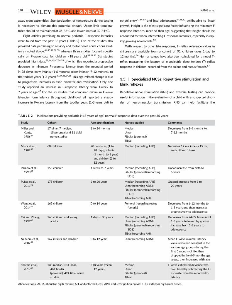
Eight articles pertaining to normal pediatric F response latencies
were found from the past 35 years (Table 2). Five of the studies also
provided data pertaining to sensory and motor nerve conductions stud-
ies as noted above,44,45,47,52,53 whereas three studies focused specifi-
cally on F-wave data for children <18 years old.39,54,55 Six studies
provided infant data,39,44,45,47,54,55 of which five reported a progressive
decrease in minimum F-response latency from the neonatal period
(< 28 days), early infancy (1-6 months), older infancy (7-12 months), to
the toddler years (1-3 years).39,44,45,54,55 This age-related change is due
to progressive increases in axon diameter and myelination. Only one
study reported an increase in F-response latency from 1 week to
7 years of age.47 For the six studies that compared minimum F-wave
latencies form infancy throughout childhood, all reported a steady
increase in F-wave latency from the toddler years (1-3 years old) to
school entry47,54,55 and into adolescence,44,45,52 attributable to linear
growth. Height is the most significant factor influencing the minimum F
response latencies, more so than age, suggesting that height should be
accounted for when interpreting F response latencies, especially in rap-
idly growing adolescents.52
With respect to other late responses, H-reflex reference values in
children are available from a cohort of 91 children (ages 1 day to
12 months).56 Normal values have also been calculated for a novel T-
reflex measuring the latency of myoelectric deep tendon (T) reflex
response in children, recorded from the soleus and rectus femoris.57
3.5 | Specialized NCSs: Repetitive stimulation andblink reflexes
Repetitive nerve stimulation (RNS) and exercise testing can provide
useful information in the evaluation of a child with a suspected disor-
der of neuromuscular transmission. RNS can help facilitate the
TABLE 2 Publications providing pediatric (<18 years of age) normal F-response data over the past 35 years
Study Cohort Age stratifications Nerves studied Comments
Miller and
Kuntz,
198639
17 ulnar, 7 median,
15 peroneal and 11 tibial
nerve studies
1 to 24 months Median
Ulnar
Fibular (peroneal)
Tibial
Decreases from 1-6 months to
7-12 months
Misra et al.,
19895560 children 20 neonates, (1 to
28 days), infants
(1 month to 1 year)
and children (2 to
12 years)
Median (recording APB) Neonates 17 ms, infants 15 ms,
and children 16 ms
Parano et al.,
199347155 children 1 week to 7 years Median (recording APB)
Fibular (peroneal) (recording
EDB)
Linear increase from birth to
6 years
Puksa et al.,
201152175 children 3 to 20 years Median (recording APB)
Ulnar (recording ADM)
Fibular (peroneal) (recording
EDB)
Tibial (recording AH)
Gradual increase from 3 to
20 years
Wang et al.,
201444163 children 0 to 14 years Femoral (recording rectus
femoris)
Decreases from 6-12 months to
1-3 years and then increases
progressively to adolescence
Cai and Zhang,
199745168 children and young
adults
1 day to 30 years Median (recording APB)
Ulnar (recording ADM)
Fibular (peroneal) (recording
EDB)
Tibial (recording AH)
Decreases from 24-72 hours until
1-3 years, followed by gradual
increase from 1-3 years to
adolescence
Nadeem et al.,
200254167 infants and children 0 to 12 years Ulnar (recording ADM) Mean F-wave minimal latency
value remained constant in the
various age groups during the
first 6 months of life, then
dropped in the 6-9 months age
group, then increased with age
Sharma et al.,
201953138 median, 384 ulnar,
461 fibular
(peroneal), 424 tibial nerve
studies
<18 years (mean
12 years)
Median
Ulnar
Fibular (peroneal)
Tibial
F-wave estimated deviance was
calculated by subtracting the F-
estimate from the recorded F-
latency
Abbreviations: ADM, abductor digiti minimi; AH, abductor halluces; APB, abductor pollicis brevis; EDB, extensor digitorum brevis.
148 KANG ET AL.

diagnosis of disorders such as transient neonatal myasthenia gravis or
congenital myasthenic syndrome (CMS). Many infants and children
with disorders of neuromuscular transmission may present with weak-
ness and breathing or feeding difficulty, which can show clinical and
electrophysiological overlap with myopathies,58,59 including several
inherited neuromuscular diseases.60 As such, a careful interrogation of
the function of the neuromuscular junction is an important consider-
ation. RNS is commonly used after routine NCSs have been com-
pleted, but it is important to note that, as in adults, important
information can be obtained from routine motor studies. Routine
motor NCSs may elicit “double” CMAP responses, which can be a clue
to the presence of certain types of CMS, including slow-channel CMS
or acetylcholinesterase deficiency.61 While this can be a helpful clue,
the sensitivity of this electrophysiological finding is not known. More-
over, it is not specific to CMS as it can also be seen with organophos-
phate exposure.62 A failure of neuromuscular transmission may lead
to reduced CMAP amplitudes as well.
Slow RNS (2 to 3 Hz) in children is performed in a similar manner
as in adults. Supramaximal stimulation is delivered as a train of 5 or
10 pulses. The CMAP amplitudes of the first and fourth (or fifth)
responses are compared, and a CMAP decrement of >10% is consid-
ered abnormal. While this test can provide important information, it is
uncomfortable and can be difficult for young children or those with
cognitive impairment to tolerate and cooperate. At least 30-65% of
children with juvenile myasthenia gravis may initially present with ocu-
lar symptoms,63,64 and the likelihood of a child presenting with ocular
(rather than generalized) myasthenia gravis symptoms is reportedly
higher in prepubertal (vs postpubertal) children.65 This must be consid-
ered because RNS of the facial nerve (stimulating anterior to the tragus
and recording from the nasalis or orbicularis oculi) is particularly chal-
lenging for younger children and less likely to be performed success-
fully without sedation or general anesthesia. Adult studies have found
SFEMG to be more sensitive than RNS at diagnosing a disorder of neu-
romuscular transmission.66,67 Although these data are lacking in chil-
dren, many pediatric neurologists and neurophysiologists would concur
that SFEMG or even stimulated SFEMG is a more sensitive tool.68
SFEMG may also detect abnormalities in clinically unaffected forearm
muscles in adult patients presenting with ocular forms of the disease.69
Transient neonatal myasthenia gravis is commonly diagnosed with
RNS because this tends to be a more generalized disorder as a result
of the passive transfer of maternal antibodies.70
Fast RNS (20 to 50 Hz) is needed to test for possible facilitation or
increment in CMAP amplitude in presynaptic disorders of neuromus-
cular transmission such as infantile botulism, presynaptic CMS71,72
and pediatric Lambert-Eaton myasthenic syndrome (LEMS). There are
case reports of infants with CMS (including choline acetyltransferase
deficiency caused by CHAT mutations) who have subsequently been
found to have abnormalities on fast or prolonged RNS.71,73 Although
these diagnoses are individually rare, all have potential clinical implica-
tions if overlooked, including apnea (botulism or CHAT mutations) or
paraneoplastic causes (LEMS).74
Exercise testing has diagnostic value for patients with suspected
muscle channelopathies as it may document abnormal membrane
excitability. Standardized protocols comprising the short and long
exercise tests have been developed to differentiate among these dis-
orders during neurophysiological testing and different patterns of
abnormality can provide evidence for a potential sodium or chloride
channelopathy.75 Limb cooling may also help to differentiate the
two.76 Of note, these tests require cooperation on the part of the
patient and are, therefore, more likely to be of value in older children
and adolescents.
Facial nerve motor NCSs, blink reflex studies, and facial muscle
EMG have roles in evaluating infants and children with facial or bulbar
weakness. They may assist with localizing the site of injury, which can
include: muscle (eg, nemaline rod myopathy), peripheral nerve (eg,
birth trauma), or brainstem nuclei (eg, brainstem dysgenesis). Normal
values for facial nerve motor NCSs are available for age ranges
throughout childhood.77
Blink reflex testing evaluates integrity of the trigeminal and facial
nerves as well as the pathways between their cranial nuclei. A supra-
maximal shock is applied to the supraorbital branch of the trigeminal
nerve resulting in a direct response that has two components: R1
(rapid and brief) and R2 (delayed and prolonged). The R1 response is
present from birth and achieves normal adult values (range, 9-13 ms)
at 4 weeks of life for a term infant.78 The ipsilateral R2 (R2i) can be
elicited in most term newborns (latency range, 34-43 ms); however,
the contralateral R2 (R2c) is not consistently obtained before the age
of 9 months old.79 The initial absence of an R2c is thought to be due
to lack of normal myelination of the brainstem, facial, and trigeminal
pathways in very young children.80 Latency differences between R2i
and R2c have also been reported in normal infants <12 months old,
with physiological differences in latencies lessening after that age, so
that the latencies are identical to adults by 3 years old.81
Interpretation of blink reflex findings in children is otherwise simi-
lar to that of adults. Abnormality of the blink reflex has been reported
as an early electrophysiological sign of acute inflammatory demyelin-
ating polyneuropathy82 as well as in children with
myelomeningocele,83 children with Tourette syndrome,84 or adoles-
cents and adults with migraine,80 although practically it is not used for
any of these conditions.
While electrophysiological testing can reliably elicit R1 and R2i
responses from birth, observational studies have noted that administer-
ing a shock to the glabellum reliably elicits a blink response in 100% of
term infants,85 whereas handheld esthiometers elicited a blink in only
25% of infants at 1 week old, slowly increasing to 100% of infants by
12 weeks of age.86 An intact blink reflex has been consistently shown
to be a good predictor of clinical outcome for idiopathic facial palsy
(Bell's palsy) in adults87,88 as well as in children.89
Blink reflex is abnormal in children with Moebius syndrome and
other disorders of brainstem dysgenesis, a finding that can aid in the
diagnosis of these conditions.90,91
For infants with facial or bulbar dysfunction and concern regarding
potential feeding difficulty, simultaneous needle EMG of the gen-
ioglossus and thyrohyoid have been used at specialized centers to
permit electrophysiological evaluation of infants clinically suspected
to have dyscoordinated sucking and swallowing.77
KANG ET AL. 149
3.6 | Needle EMG issues
EMG is generally a necessary and valuable complement to NCSs.2,92
In studies addressing an isolated traumatic nerve injury, the focus of
the needle EMG study is directed by clinical examination of weakness
and likely nerve(s) involved. In diagnostic studies that assess general-
ized neuromuscular disorders, the choice and number of muscles to
study is more complicated. The aim is to optimize the diagnostic value
of the study while minimizing discomfort to the child. The sensitivity
of EMG in neurogenic diagnoses can be as high as 99.5%.93 Myogenic
disorders are less sensitive to diagnosis by EMG in ages under 2 years
old,1,93-95 although sensitivity improves in the older age groups.
Review of the literature suggests that EMG of at least one arm and
leg is recommended when feasible, with four muscles being optimal for
making a diagnosis.94 This allows for sampling muscles in a wide ana-
tomic distribution but may be limited by patient cooperation; thus, in
many cases the clinical context and the results of the NCSs indicate
that sampling one muscle is most prudent, as noted above. Pediatric
studies looking at pain perception equate the EMG with venipunc-
ture.31 There is increased perception of pain when more than one
muscle is sampled and in the upper extremities (including genioglossus
in this study). Another consideration is the effect of using smaller pedi-
atric concentric needles instead of standard concentric needles, which
may lower motor unit action potential (MUAP) amplitude without a sig-
nificant effect on MUAP duration.96 With these considerations, it is
judicious to initially choose the most appropriate muscle to sample.
Neurological exam cannot be overemphasized as well as knowledge
of the suspected type of disorder. Muscle ultrasound before needle
EMG can scan several muscles quickly and is painless.97 Using ultra-
sound to identify abnormal-appearing muscles and to estimate the
severity of abnormalities increases the likelihood of choosing muscles
for needle EMG that will display clear diagnostic findings. Sedation in
performing EMG studies is used to varying degrees in different institu-
tions, some places perform sedated procedures routinely94 and others
more rarely.1 Sedation has the advantage of permitting the study to be
performed without upsetting the child. The limitation is a reduced ability
to volitionally activate many muscles to evaluate motor unit activity.
The risk of anesthesia in a child also needs to be considered, particularly
if there is severe pulmonary or cardiac involvement.98
One representative EDX laboratory familiar to two of the authors
(T.J.L. and K.E.A.) is the EMG Laboratory at the National Institutes of
Health, where both awake and sedated pediatric studies are per-
formed. In complicated situations where multiple diagnostic tests are
needed, sedated studies are usually performed in a multidisciplinary
facility that also includes specialized personnel for MRI imaging, audi-
ology, dental, ophthalmology, cerebrospinal fluid studies (lumbar
puncture), and skin biopsy. In sedated studies and in addition to stan-
dard NCSs, these two authors sample two muscles in the leg (tibialis
anterior and internal hamstrings) and one muscle in the arm (biceps).
These muscles are chosen because they are easiest to activate in a
lightly sedated patient while others, muscles particularly in the hand,
are more difficult to activate. Other studies such as RNS or SFEMG
may be performed if clinically indicated. In awake patients, the EDX
physician is more dependent on clinical and ultrasound examination to
choose the muscle most likely to be abnormal for needle EMG. For
cooperative children, more than one muscle is examined.
3.7 | Jitter analysis for disorders of theneuromuscular junction: SFEMG and stimulationpotential analysis with concentric electrodes
Analysis of jitter is the most sensitive study of the neuromuscular
junction.99,100 This is an important diagnostic study for CMS, botu-
lism, and neonatal myasthenia gravis. In the pediatric population, tra-
ditional stimulated SFEMG techniques have been used.73 In addition,
an adaptation of the stimulated SFEMG technique, using a concentric
needle electrode to record from the orbicularis oculi, has been used
successfully by several groups. This adapted technique is known vari-
ously as SPACE (stimulation potential analysis with concentric
electrodes),101-103 stimulated jitter analysis,100,104 or concentric nee-
dle jitter.105 The zygomatic branch of the facial nerve is stimulated by
a monopolar needle. In SPACE and stimulated jitter studies, a concen-
tric facial needle is used to collect a sufficient number of single motor
fiber unit potentials. The advantage of these adapted techniques is
that they can be performed sedated or with local anesthesia and
within approximately 15-20 min. The disadvantage is that there may
be biasing of the potentials leading to a decreased jitter.106 Normative
values in the pediatric population are limited by the ability to perform
studies on normal children. Using an analytic technique of e-norms, it
was determined that children over the age 2 years have jitter values
(mean consecutive difference [MCD], 22 μs) approaching adult levels
(MCD, 21 μs).50 Children younger than 2 years old have slightly longer
jitter values; with 1- to 2-year-olds having an MCD of 33 μs, and
those under 1 year having an MCD of 45 μs.107
The diagnostic value of jitter in the orbicularis oculi varies in the
different CMS.59 Patients with DOK7 CMS are less likely to have an
abnormal jitter. Also, the possibility of a neurogenic disorder needs to
be ruled out because it can cause a false positive result.
3.8 | Small nerve fiber testing
Small nerve fibers are thinly myelinated A delta and unmyelinated C
fibers that mediate the sensations of pain and temperature and
autonomic function. Autonomic symptoms that can be produced by
dysfunction of autonomic small nerve fibers include: dry eyes, dry
mouth, orthostatic symptoms (from lightheadedness through syn-
cope), palpitations, variations in blood pressure, abnormal sweating,
overheating, erectile dysfunction, nausea, vomiting, constipation, early
satiety, urinary frequency, nocturia and others.
Evaluation of small fiber function is important in children as there
are several disorders associated with dysfunction limited to these
fiber types in which standard NCSs are unrevealing. These include
erythromelalgia,108 Fabry disease,109 diffuse widespread pain,110-112
and familial dysautonomia, and other hereditary neuropathies.113,114
Sympathetic skin response testing is a simple technique that can
be performed using standard EDX equipment. Surface electrodes are
150 KANG ET AL.

placed over the palm and sole of the feet and can record sympathetic
responses to a variety of stimuli: tactile, auditory, or electrical
pulses.115 However, this response habituates quickly to repeated
stimuli and the response is poorly graded and so usually assessed
qualitatively as present or absent. Standard motor and sensory NCSs
do not evaluate small nerve fibers.
Patient-reported outcome surveys have been developed and vali-
dated for adults with small fiber polyneuropathies.116 These surveys
have not been validated in children or adolescents; however, they have
been used informally with perceived benefit (Oaklander A.L., personal
communication).
Computer-assisted quantitative sensory testing can be used to
assess temperature and pain perception and has demonstrated repro-
ducible results in children as young as 6 years of age.117 Values of
cold sensation, warm sensation, cold pain, heat pain and vibration sen-
sation detection thresholds were determined in the hand and foot for
healthy children between 6 and 17 years of age.
Intraepidermal nerve fiber measurements have been used to quan-
tify the status of small nerve fibers in humans.118 The invasiveness of
performing one to several 3-mm full-thickness skin punch biopsies
and the absence of normative values has limited use of this technique
in children. However, recent modifications to technique (obtaining
suction blister specimens on extremities) have been correlated to
values measured on full-thickness skin punch biopsies and quantified
in 20 healthy children between 7 and 17 years of age.119
Corneal confocal microscopy (CCM) is a noninvasive method for
measuring small nerve fiber branching, density, and length. CCM can
be scored in an automated manner and does not require undue
degrees of cooperation so would appear to be a promising technique
for quantifying small nerve fiber function in children.120
Contact heat evoked potentials require a heated foil thermode to
apply cycles of heating and cooling on an extremity while the evoked
potential is recovered over a CZ scalp electrode. The technique does
not cause tissue injury and is considered noninvasive. There are no
published reports using this technique in children.121
Measurements of the extent of surface area involved and the vol-
ume of sweat generated by standard heat stresses have been used
with several techniques (thermoregulatory sweat testing and quantita-
tive sudomotor axon reflex testing) to evaluate small fiber func-
tion.122,123 These techniques have been used and demonstrated to
contribute significantly to diagnosis and treatment planning in various
disorders in children and adolescents.123
Other autonomic tests evaluating vagal, sympathetic, and gastro-
intestinal function have been demonstrated to be useful while con-
tributing to diagnosis, prognosis, and treatment planning.123
In conclusion, evaluating small nerve fiber function is important
for all who care for children and adolescents. However, the tech-
niques for this testing reach beyond standard NCSs and needle EMG.
Techniques such as thermoregulatory sweat testing can be used at
present with good validity and interpretability. However, validation of
reproducibility and knowledge of age appropriate normal values for
younger children do not yet exist for other commonly used autonomic
tests, such as the Valsalva ratio and heart rate response to deep
breathing,
3.9 | Evoked potentials
There is an extensive literature on the use of evoked potentials in chil-
dren. However, the bulk of the articles focus on audiologic, ophthalmo-
logic, or central nervous system applications of this test modality,
which are not the focus of the current discussion. With respect to
lower motor neuron / peripheral nervous system applications, somato-
sensory evoked potentials (SSEPs) and motor evoked potentials (MEPs)
were historically used for diagnostic purposes,124 but those applica-
tions have become less common with the wide availability of spine
MRI technology and other diagnostic tests. Currently, evoked poten-
tials frequently have utility in children as intraoperative monitoring
modalities during procedures such as spine surgeries.124 Intraoperative
neurophysiologic monitoring using SSEPs, MEPs, and/or EMG (ideally
all three simultaneously) is useful whenever nervous system tracts are
at risk during operative procedures, so that any potential damage to
such tissues may be prevented or mitigated. MEPs are often generated
using transcranial electrical stimulation, yielding transcranial MEPs, also
known as tcMEPs. Technical details of tcMEPs in children such as the
optimal number of pulses during spine surgeries have been studied.125
The interactions between MEPs and various anesthetic agents have
been studied, and MEPs are preserved in children who are adminis-
tered either propofol or desflurane.126 In contrast, dexmedetomidine
tends to attenuate tcMEP signals.127
4 | DISCUSSION
Literature review revealed that a significant proportion of the literature
on the use of EDX testing in pediatrics consists of articles published
before 2000; however, it is notable that there continues to be a steady
stream of papers that continue to the current day on this topic. This
reflects the stable and even increasing volume of children who undergo
these studies and demonstrates the continued utility of EDX testing in
children, albeit with a shift in the proportions of various indications and
diagnoses. Most notably, certain categories of inherited diseases such
as muscular dystrophy and SMA do not routinely require EMG as part
of the diagnostic evaluation. However, in atypical cases EDX testing can
provide critical assistance with narrowing of the differential diagnosis.
Data on normative values are difficult to gather in large quantities,
especially for children. It is heartening to see that seven articles met
rigorous criteria to help determine what normative values to use on a
routine basis. It is also encouraging to see that one of these articles
was published recently, in 2019.
It is important for any medical center that provides care for a signifi-
cant number of children to provide this service to patients who would
benefit from it, performed by trained EDX physicians who should have
a strong background in pediatrics.
The panel concludes that the use of EDX testing in children will
thrive for some time to come, and that techniques and practice
KANG ET AL. 151

patterns for this important diagnostic test modality will continue to
evolve in the future. EDX testing in children will continue to comple-
ment other diagnostic test modalities such as various serum tests,
muscle biopsy, imaging, and genetic testing.
ACKNOWLEDGMENTS
The authors thank the AANEM and Millie Suk, JD, for their support
for this project.
CONFLICT OF INTEREST
P.B.K. reports consulting for AveXis (Novartis) and ChromaDex, advi-
sory board activities for Sarepta Therapeutics, and honoraria from
Springer (co-editor for textbook on pediatric electromyography),
Wiley (for editorial work on Muscle & Nerve), and Wolters Kluwer
(authorship for topics in UpToDate). H.J.M. reports consulting for
Avexis (Novartis) and has received honoraria from Springer (co-editor
for textbook on pediatric electromyography). N.L.K. reports advisory
board service for Audentes Therapeutics, AveXis (Novartis), Biogen,
Cytokinetics, Roche, and Sarepta Therapeutics. T.J.L., K.E.A., K.F.F.,
and C.E. report no disclosures. S.Q.-R. reports advisory board activi-
ties for AveXis (Novartis), Biogen, and Roche.
ETHICAL PUBLICATION STATEMENT
The authors confirm that they have read Muscle & Nerve's position on
issues involved in ethical publication and affirm that this report is con-
sistent with those guidelines. This article underwent review by the
AANEM Professional Practice Committee and by the Editor-in-Chief,
but did not undergo external peer review.
ORCID
Peter B. Kang https://orcid.org/0000-0002-4270-7325
REFERENCES
1. Darras BT, Jones HR. Diagnosis of pediatric neuromuscular disorders
in the era of DNA analysis. Pediatr Neurol. 2000;23(4):289-300.
2. Karakis I, Liew W, Darras BT, Jones HR, Kang PB. Referral and diag-
nostic trends in pediatric electromyography in the molecular era.
Muscle Nerve. 2014;50(2):244-249.
3. Kang PB, Gooch CL, McDermott MP, et al. The motor neuron
response to SMN1 deficiency in spinal muscular atrophy. Muscle
Nerve. 2014;49(5):636-644.
4. Bady B, Vila A, Boulliat G, et al. Value of electromyography in the child.
Apropos of 1,624 examinations performed over a 3 year period. Rev
Electroencephalogr Neurophysiol Clin. 1983;13(3):282-288.
5. Jones HR Jr. Compressive neuropathy in childhood: a report of
14 cases. Muscle Nerve. 1986;9(8):720-723.
6. Gschwind C, Tonkin MA. Carpal tunnel syndrome in children with
mucopolysaccharidosis and related disorders. J Hand Surg Am. 1992;
17(1):44-47.
7. Van Meir N, De Smet L. Carpal tunnel syndrome in children. J Pediatr
Orthop B. 2005;14(1):42-45.
8. Potulska-Chromik A, Lipowska M, Gawel M, Ryniewicz B, Maj E,
Kostera-Pruszczyk A. Carpal tunnel syndrome in children. J Child
Neurol. 2014;29(2):227-231.
9. Cruz Martinez A, Arpa J. Carpal tunnel syndrome in childhood: study
of 6 cases. Electroencephalogr Clin Neurophysiol. 1998;109(4):
304-308.
10. Davis L, Vedanarayanan VV. Carpal tunnel syndrome in children.
Pediatr Neurol. 2014;50(1):57-59.
11. Deymeer F, Jones HR Jr. Pediatric median mononeuropathies: a clin-
ical and electromyographic study. Muscle Nerve. 1994;17(7):
755-762.
12. Karakis I, Liew W, Fournier HS, Jones HR Jr, Darras BT, Kang PB.
Electrophysiologic features of ulnar neuropathy in childhood and
adolescence. Clin Neurophysiol. 2017;128(5):751-755.
13. Felice KJ, Royden Jones H Jr. Pediatric ulnar mononeuropathy:
report of 21 electromyography-documented cases and review of the
literature. J Child Neurol. 1996;11(2):116-120.
14. Escolar DM, Jones HR Jr. Pediatric radial mononeuropathies: a clini-
cal and electromyographic study of sixteen children with review of
the literature. Muscle Nerve. 1996;19(7):876-883.
15. Karakis I, Georghiou S, Jones HR, Darras BT, Kang PB. Electrophysi-
ologic features of radial neuropathy in childhood and adolescence.
Pediatr Neurol. 2018;81:14-18.
16. Srinivasan J, Ryan MM, Escolar DM, Darras B, Jones HR. Pediatric
sciatic neuropathies: a 30-year prospective study. Neurology. 2011;
76(11):976-980.
17. McMillan HJ, Srinivasan J, Darras BT, et al. Pediatric sciatic neuropa-
thy associated with neoplasms. Muscle Nerve. 2011;43(2):183-188.
18. Ferraresi S, Garozzo D, Bianchini E, Gasparotti R. Perineurioma of
the sciatic nerve: a possible cause of idiopathic foot drop in children:
report of 4 cases. J Neurosurg Pediatr. 2010;6(5):506-510.
19. Srinivasan J, Escolar D, Ryan M, Darras B, Jones HR. Pediatric sci-
atic neuropathies due to unusual vascular causes. J Child Neurol.
2008;23(7):738-741.
20. Jones HR Jr, Felice KJ, Gross PT. Pediatric peroneal mononeuropathy:
a clinical and electromyographic study. Muscle Nerve. 1993;16(11):
1167-1173.
21. Levin KH, Wilbourn AJ, Jones HR Jr. Childhood peroneal neuropathy
from bone tumors. Pediatr Neurol. 1991;7(4):308-309.
22. Karakis I, Khoshnoodi M, Liew W, et al. Electrophysiologic features
of fibular neuropathy in childhood and adolescence. Muscle Nerve.
2017;55(5):693-697.
23. Song KS, Kang CH, Min BW, Bae GC, Cho CH, Lee JH. Congenital
clubfoot with concomitant peroneal nerve palsy in children. J Pediatr
Orthop B. 2008;17(2):85-89.
24. Thometz J, Sathoff L, Liu XC, Jacobson R, Tassone JC. Electromyog-
raphy nerve conduction velocity evaluation of children with club-
feet. Am J Orthop (Belle Mead NJ). 2011;40(2):84-86.
25. Abid A. Brachial plexus birth palsy: management during the first year
of life. Orthop Traumatol Surg Res. 2016;102(1 Suppl):S125-S132.
26. Belzberg AJ, Dorsi MJ, Storm PB, Moriarity JL. Surgical repair of bra-
chial plexus injury: a multinational survey of experienced peripheral
nerve surgeons. J Neurosurg. 2004;101(3):365-376.
27. Vanderhave KL, Bovid K, Alpert H, et al. Utility of electrodiagnostic
testing and computed tomography myelography in the preoperative
evaluation of neonatal brachial plexus palsy. J Neurosurg Pediatr.
2012;9(3):283-289.
28. Terzis JK, Karypidis D. Outcomes of direct muscle neurotization in
pediatric patients with facial paralysis. Plast Reconstr Surg. 2009;
124(5):1486-1498.
29. Kang PB. Approach to electrodiagnostic testing in children. In:
McMillan HJ, Kang PB, eds. Pediatric Electromyography: Concepts and
Clinical Applications. Cham, Switzerland: Springer; 2017:23-27.
30. Pitt M. Paediatric Electromyography. Oxford: Oxford University
Press; 2018. 352 p.
152 KANG ET AL.

31. Alshaikh NM, Martinez JP, Pitt MC. Perception of pain during
electromyography in children: a prospective study. Muscle Nerve.
2016;54(3):422-426.
32. McHugh JC. Sensory studies. In: McMillan HJ, Kang PB, eds. Pediat-
ric Electromyography: Concepts and Clinical Applications. Cham,
Switzerland: Springer; 2017:31-48.
33. Hays RM, Hackworth SR, Speltz ML, Weinstein P. Exploration of vari-
ables related to children's behavioral distress during electrodiagnosis.
Arch Phys Med Rehabil. 1992;73(12):1160-1162.
34. Ballard A, Khadra C, Adler S, Trottier ED, Le May S. Efficacy of
the buzzy device for pain management during needle-related pro-
cedures: a systematic review and meta-analysis. Clin J Pain. 2019;
35(6):532-543.
35. Pitt MC. Nerve conduction studies and needle EMG in very small
children. Eur J Paediatr Neurol. 2012;16(3):285-291.
36. Thomas JE, Lambert EH. Ulnar nerve conduction velocity and H-
reflex in infants and children. J Appl Physiol. 1960;15:1-9.
37. Wagner AL, Buchthal F. Motor and sensory conduction in infancy
and childhood: reappraisal. Dev Med Child Neurol. 1972;14(2):
189-216.
38. Chen S, Andary M, Buschbacher R, et al. Electrodiagnostic reference
values for upper and lower limb nerve conduction studies in adult
populations. Muscle Nerve. 2016;54(3):371-377.
39. Miller RG, Kuntz NL. Nerve conduction studies in infants and chil-
dren. J Child Neurol. 1986;1(1):19-26.
40. Lori S, Bertini G, Bastianelli M, et al. Peripheral nervous system mat-
uration in preterm infants: longitudinal motor and sensory nerve
conduction studies. Childs Nerv Syst. 2018;34(6):1145-1152.
41. Dillingham T, Chen S, Andary M, et al. Establishing high-quality ref-
erence values for nerve conduction studies: a report from the
normative data task force of the American Association of Neuro-
muscular & Electrodiagnostic Medicine. Muscle Nerve. 2016;54(3):
366-370.
42. Smit BJ, Kok JH, De Vries LS, Dekker FW. Ongerboer de Visser
BW. Motor nerve conduction velocity in very preterm infants.
Muscle Nerve. 1999;22(3):372-377.
43. Ryan CS, Conlee EM, Sharma R, Sorenson EJ, Boon AJ, Laughlin RS.
Nerve conduction normal values for electrodiagnosis in pediatric
patients. Muscle Nerve. 2019;60(2):155-160.
44. Wang L, Hu Y, Jiang L, Zhong X, Chen J, Xu N. Electrophysiological
characteristics of the pediatric femoral nerve and their use in clinical
diagnosis. Pediatr Neurol. 2014;50(2):149-157.
45. Cai F, Zhang J. Study of nerve conduction and late responses in nor-
mal Chinese infants, children, and adults. J Child Neurol. 1997;12(1):
13-18.
46. Hyllienmark L, Ludvigsson J, Brismar T. Normal values of nerve con-
duction in children and adolescents. Electroencephalogr Clin
Neurophysiol. 1995;97(5):208-214.
47. Parano E, Uncini A, De Vivo DC, Lovelace RE. Electrophysiologic
correlates of peripheral nervous system maturation in infancy and
childhood. J Child Neurol. 1993;8(4):336-338.
48. Lang HA, Puusa A, Hynninen P, Kuusela V, Jantti V, Sillanpaa M.
Evolution of nerve conduction velocity in later childhood and ado-
lescence. Muscle Nerve. 1985;8(1):38-43.
49. Garcia A, Calleja J, Antolin FM, Berciano J. Peripheral motor and
sensory nerve conduction studies in normal infants and children. Clin
Neurophysiol. 2000;111(3):513-520.
50. Jabre JF, Pitt MC, Deeb J, Chui KK. E-norms: a method to extrapolate
reference values from a laboratory population. J Clin Neurophysiol.
2015;32(3):265-270.
51. Dioszeghy P, Stalberg E. Changes in motor and sensory nerve con-
duction parameters with temperature in normal and diseased nerve.
Electroencephalogr Clin Neurophysiol. 1992;85(4):229-235.
52. Puksa L, Eeg-Olofsson KE, Stalberg E, Falck B. Reference values for
F wave parameters in healthy 3-20 year old subjects. Clin
Neurophysiol. 2011;122(1):199-204.
53. Sharma R, Sorenson EJ, Ryan CS, Conlee EM, Boon AJ, Laughlin RS.
Establishing normal values for F-Wave latencies in diagnosing
peripheral neuropathies for the pediatric population. J Clin
Neurophysiol. 2019. https://doi.org/10.1097/WNP.
0000000000000000609 [Epub ahead of print].
54. Nadeem AS, El-Yassin DI, Al-Ani F. Analysis of F-wave parameters in
normal infants and children. Ann Saudi Med. 2002;22(3–4):181-185.55. Misra UK, Tiwari S, Shukla N, Nishith SD, Malik GK, Nag D. F-
response studies in neonates, infants and children. Electromyogr Clin
Neurophysiol. 1989;29(4):251-254.
56. Mayer RF, Mosser RS. Excitability of motoneurons in infants. Neurol-
ogy. 1969;19(10):932-945.
57. Pereon Y. Nguyen The Tich S, Fournier E, Genet R, Guiheneuc
P. Electrophysiological recording of deep tendon reflexes: normative
data in children and in adults. Neurophysiol Clin. 2004;34(3–4):
131-139.
58. Mongiovi PC, Elsheikh B, Lawson VH, Kissel JT, Arnold WD. Neuro-
muscular junction disorders mimicking myopathy. Muscle Nerve.
2014;50(5):854-856.
59. Kinali M, Beeson D, Pitt MC, et al. Congenital myasthenic syn-
dromes in childhood: diagnostic and management challenges.
J Neuroimmunol. 2008;201–202:6–12.60. Nicolau S, Kao JC, Liewluck T. Trouble at the junction: when myopa-
thy and myasthenia overlap. Muscle Nerve. 2019;60(6):648–657.61. Kumar RS, Kuruvilla A. Repetitive compound muscle action poten-
tials in electrophysiological diagnosis of congenital myasthenic syn-
dromes: a case report and review of literature. Ann Indian Acad
Neurol. 2010;13(2):139-141.
62. van Dijk JG, Lammers GJ, Wintzen AR, Molenaar PC. Repetitive
CMAPs: mechanisms of neural and synaptic genesis. Muscle Nerve.
1996;19(9):1127-1133.
63. Ashraf VV, Taly AB, Veerendrakumar M, Rao S. Myasthenia gravis in
children: a longitudinal study. Acta Neurol Scand. 2006;114(2):
119-123.
64. Snead OC III, Benton JW, Dwyer D, et al. Juvenile myasthenia gravis.
Neurology. 1980;30(7 Pt 1):732-739.
65. Batocchi AP, Evoli A, Palmisani MT, Lo Monaco M, Bartoccioni M,
Tonali P. Early-onset myasthenia gravis: clinical characteristics and
response to therapy. Eur J Pediatr. 1990;150(1):66-68.
66. Gilchrist JM, Massey JM, Sanders DB. Single fiber EMG and repeti-
tive stimulation of the same muscle in myasthenia gravis. Muscle
Nerve. 1994;17(2):171-175.
67. Sanders DB, Howard JF Jr, Johns TR. Single-fiber electromyography
in myasthenia gravis. Neurology. 1979;29(1):68-76.
68. Pitt M. Neurophysiological assessment of abnormalities of the neu-
romuscular junction in children. Int J Mol Sci. 2018;19(2).
69. Guan YZ, Cui LY, Liu MS, Niu JW. Single-fiber electromyography in
the extensor digitorum communis for the predictive prognosis of
ocular myasthenia gravis: a retrospective study of 102 cases. Chin
Med J (Engl). 2015;128(20):2783-2786.
70. Hays RM, Michaud LJ. Neonatal myasthenia gravis: specific advan-
tages of repetitive stimulation over edrophonium testing. Pediatr
Neurol. 1988;4(4):245-247.
71. Dilena R, Abicht A, Sergi P, et al. Congenital myasthenic syndrome
due to choline acetyltransferase mutations in infants: clinical suspi-
cion and comprehensive electrophysiological assessment are impor-
tant for early diagnosis. J Child Neurol. 2014;29(3):389-393.
72. Gurnett CA, Bodnar JA, Neil J, Connolly AM. Congenital myasthenic
syndrome: presentation, electrodiagnosis, and muscle biopsy. J Child
Neurol. 2004;19(3):175-182.
KANG ET AL. 153

73. Zafeiriou DI, Pitt M, de Sousa C. Clinical and neurophysiological
characteristics of congenital myasthenic syndromes presenting in
early infancy. Brain Dev. 2004;26(1):47-52.
74. Morgan-Followell B, de Los Reyes E. Child neurology: diagnosis of
Lambert-Eaton myasthenic syndrome in children. Neurology. 2013;
80(21):e220-e222.
75. Fournier E, Arzel M, Sternberg D, et al. Electromyography guides
toward subgroups of mutations in muscle channelopathies. Ann Neu-
rol. 2004;56(5):650-661.
76. Michel P, Sternberg D, Jeannet PY, et al. Comparative efficacy of
repetitive nerve stimulation, exercise, and cold in differentiating
myotonic disorders. Muscle Nerve. 2007;36(5):643-650.
77. Renault F. Pediatric cranial neuropathies. In: McMillan HJ, Kang PB,
eds. Pediatric Electromyography: Concepts and Clinical Applications.
Cham, Switzerland: Springer; 2017:181-198.
78. Renault F. Facial electromyography in newborn and young infants
with congenital facial weakness. Dev Med Child Neurol. 2001;43(6):
421-427.
79. Vecchierini-Blineau MF, Guiheneuc P. Maturation of the blink reflex
in infants. Eur Neurol. 1984;23(6):449-458.
80. Blank A, Ferber I, Shapira Y, Fast A. Electrically elicited blink reflex in
children. Arch Phys Med Rehabil. 1983;64(11):558-559.
81. Tomita Y, Shichida K, Takeshita K, Takashima S. Maturation of blink
reflex in children. Brain Dev. 1989;11(6):389-393.
82. Vucic S, Cairns KD, Black KR, Chong PS, Cros D. Neurophysiologic find-
ings in early acute inflammatory demyelinating polyradiculoneuropathy.
Clin Neurophysiol. 2004;115(10):2329-2335.
83. Koehler J, Schwarz M, Urban PP, Voth D, Holker C, Hopf HC. Mas-
seter reflex and blink reflex abnormalities in Chiari II malformation.
Muscle Nerve. 2001;24(3):425-427.
84. Raffaele R, Rampello L, Vecchio I, et al. Blink reflex abnormalities in
children with Tourette syndrome. Eur J Neurol. 2006;13(8):869-873.
85. Anday EK, Cohen ME, Hoffman HS. The blink reflex: maturation and
modification in the neonate. Dev Med Child Neurol. 1990;32(2):
142-150.
86. Snir M, Axer-Siegel R, Bourla D, Kremer I, Benjamini Y,
Weinberger D. Tactile corneal reflex development in full-term
babies. Ophthalmology. 2002;109(3):526-529.
87. Heath JP, Cull RE, Smith IM, Murray JA. The neurophysiological
investigation of Bell's palsy and the predictive value of the blink
reflex. Clin Otolaryngol Allied Sci. 1988;13(2):85-92.
88. Dumitru D, Walsh NE, Porter LD. Electrophysiologic evaluation of
the facial nerve in Bell's palsy. A review. Am J Phys Med Rehabil.
1988;67(4):137-144.
89. Wong V. Outcome of facial nerve palsy in 24 children. Brain Dev.
1995;17(4):294-296.
90. Jaradeh S, D'Cruz O, Howard JF Jr, Haberkamp TJ, Konkol RJ.
Mobius syndrome: electrophysiologic studies in seven cases. Muscle
Nerve. 1996;19(9):1148-1153.
91. Hatanaka T, Yoshijima S, Hayashi N, Owa K, Suehiro Y, Shinomiya K.
Electrophysiologic studies in an infant with Mobius syndrome.
J Child Neurol. 1993;8(2):182-185.
92. Orhan EK, Kirac LB, Dikmen PY, et al. Electromyography in Pediatric
Population. Noro Psikiyatr Ars. 2018;55(1):36-39.
93. Hellmann M, von Kleist-Retzow JC, Haupt WF, Herkenrath P,
Schauseil-Zipf U. Diagnostic value of electromyography in children
and adolescents. J Clin Neurophysiol. 2005;22(1):43-48.
94. Ghosh PS, Sorenson EJ. Diagnostic yield of electromyography in chil-
dren with myopathic disorders. Pediatr Neurol. 2014;51(2):215-219.
95. Rabie M, Jossiphov J, Nevo Y. Electromyography (EMG) accuracy
compared to muscle biopsy in childhood. J Child Neurol. 2007;22(7):
803-808.
96. Brownell AA, Bromberg MB. Comparison of standard and pediatric
size concentric needle EMG electrodes. Clin Neurophysiol. 2007;
118(5):1162-1165.
97. Zaidman CM, van Alfen N. Ultrasound in the assessment of myo-
pathic disorders. J Clin Neurophysiol. 2016;33(2):103-111.
98. Racca F, Mongini T, Wolfler A, et al. Recommendations for anesthe-
sia and perioperative management of patients with neuromuscular
disorders. Minerva Anestesiol. 2013;79(4):419-433.
99. Massey JM. Electromyography in disorders of neuromuscular trans-
mission. Semin Neurol. 1990;10(1):6-11.
100. Bhatia S, Quinlan H, McCracken C, Price EW, Guglani L, Verma S.
Serial stimulated jitter analysis in juvenile myasthenia gravis. Muscle
Nerve. 2018;58(5):729-732.
101. Pitt MC. Use of stimulated electromyography in the analysis of the
neuromuscular junction in children. Muscle Nerve. 2017;56(5):
841-847.
102. Pitt MC, McHugh JC, Deeb J, Smith RA. Assessing neuromuscular
junction stability from stimulated EMG in children. Clin Neurophysiol.
2017;128(2):290-296.
103. Tidswell T, Pitt MC. A new analytical method to diagnose congenital
myasthenia with stimulated single-fiber electromyography. Muscle
Nerve. 2007;35(1):107-110.
104. Verma S, Lin J. Stimulated jitter analysis for the evaluation of neuro-
muscular junction disorders in children. Muscle Nerve. 2016;53(3):
471-472.
105. Sanders DB, Arimura K, Cui L, et al. Guidelines for single fiber EMG.
Clin Neurophysiol. 2019;130(8):1417-1439.
106. Stalberg E, Kouyoumdjian JA, Paiva GP, Yanaze LL, Sanders DB.
Problems in comparing jitter values obtained with voluntary activa-
tion and electrical stimulation. J Neuromuscul Dis. 2018;5(2):
225-230.
107. Pitt MC, Jabre JF. Determining jitter values in the very young by use
of the e-norms methodology. Muscle Nerve. 2017;55(1):51-54.
108. Cook-Norris RH, Tollefson MM, Cruz-Inigo AE, Sandroni P, Davis MD,
Davis DM. Pediatric erythromelalgia: a retrospective review of
32 cases evaluated at Mayo Clinic over a 37-year period. J Am Acad
Dermatol. 2012;66(3):416-423.
109. Biegstraaten M, Binder A, Maag R, Hollak CE, Baron R, van
Schaik IN. The relation between small nerve fibre function, age, dis-
ease severity and pain in Fabry disease. Eur J Pain. 2011;15(8):
822-829.
110. Hoeijmakers JG, Faber CG, Miedema CJ, Merkies IS, Vles JS. Small
fiber neuropathy in children: two case reports illustrating the impor-
tance of recognition. Pediatrics. 2016;138(4).
111. Oaklander AL, Klein MM. Evidence of small-fiber polyneuropathy in
unexplained, juvenile-onset, widespread pain syndromes. Pediatrics.
2013;131(4):e1091-e1100.
112. Kafaie J, Al Balushi A, Kim M, Pestronk A. Clinical and laboratory
profiles of idiopathic small fiber neuropathy in children: case series.
J Clin Neuromuscul Dis. 2017;19(1):31-37.
113. Hilz MJ, Axelrod FB. Quantitative sensory testing of thermal and
vibratory perception in familial dysautonomia. Clin Auton Res. 2000;
10(4):177-183.
114. Kuntz NL. Diagnosis and evaluation of small fiber peripheral neurop-
athy in children. In: McMillan HJ, Kang PB, eds. Pediatric Electromy-
ography: Concepts and Clinical Applications. Cham, Switzerland:
Springer; 2017:265-280.
115. Shahani BT, Halperin JJ, Boulu P, Cohen J. Sympathetic skin response–a method of assessing unmyelinated axon dysfunction in peripheral
neuropathies. J Neurol Neurosurg Psychiatry. 1984;47(5):536-542.
116. Treister R, Lodahl M, Lang M, Tworoger SS, Sawilowsky S,
Oaklander AL. Initial development and validation of a patient-reported
symptom survey for small-fiber polyneuropathy. J Pain. 2017;18(5):
556-563.
117. Meier PM, Berde CB, DiCanzio J, Zurakowski D, Sethna NF. Quanti-
tative assessment of cutaneous thermal and vibration sensation and
thermal pain detection thresholds in healthy children and adoles-
cents. Muscle Nerve. 2001;24(10):1339-1345.
154 KANG ET AL.

118. Loseth S, Lindal S, Stalberg E, Mellgren SI. Intraepidermal nerve fibre
density, quantitative sensory testing and nerve conduction studies
in a patient material with symptoms and signs of sensory poly-
neuropathy. Eur J Neurol. 2006;13(2):105-111.
119. Panoutsopoulou IG, Luciano CA, Wendelschafer-Crabb G,
Hodges JS, Kennedy WR. Epidermal innervation in healthy children
and adolescents. Muscle Nerve. 2015;51(3):378-384.
120. Schneider C, Bucher F, Cursiefen C, Fink GR, Heindl LM,
Lehmann HC. Corneal confocal microscopy detects small fiber dam-
age in chronic inflammatory demyelinating polyneuropathy (CIDP).
J Peripher Nerv Syst. 2014;19(4):322-327.
121. Lagerburg V, Bakkers M, Bouwhuis A, et al. Contact heat evoked
potentials: normal values and use in small-fiber neuropathy. Muscle
Nerve. 2015;51(5):743-749.
122. Fealey RD. Thermoregulatory sweat test. In: Low PA, ed. Clinical
Autonomic Disorders. 3rd ed. Baltimore, MD: Lippincott Williams &
Wilkins; 2008:244-263.
123. Kuntz NL, Patwari PP. Laboratory evaluation of pediatric autonomic
disorders. Semin Pediatr Neurol. 2013;20(1):35-43.
124. Fagan ER, Taylor MJ, Logan WJ. Somatosensory evoked potentials:
part II. A review of the clinical applications in pediatric neurology.
Pediatr Neurol. 1987;3(5):249-255.
125. Azabou E, Manel V, Andre-obadia N, et al. Optimal parameters of
transcranial electrical stimulation for intraoperative monitoring of
motor evoked potentials of the tibialis anterior muscle during pedi-
atric scoliosis surgery. Neurophysiol Clin. 2013;43(4):243-250.
126. Holdefer RN, Anderson C, Furman M, Sangare Y, Slimp JC. A com-
parison of the effects of desflurane versus propofol on transcranial
motor-evoked potentials in pediatric patients. Childs Nerv Syst.
2014;30(12):2103-2108.
127. Mahmoud M, Sadhasivam S, Sestokas AK, Samuels P, McAuliffe J.
Loss of transcranial electric motor evoked potentials during pediatric
spine surgery with dexmedetomidine. Anesthesiology. 2007;106(2):
393-396.
SUPPORTING INFORMATION
Additional supporting information may be found online in the
Supporting Information section at the end of this article.
How to cite this article: Kang PB, McMillan HJ, Kuntz NL,
et al. Utility and practice of electrodiagnostic testing in the
pediatric population: An AANEM consensus statement. Muscle
Nerve. 2020;61:143–155. https://doi.org/10.1002/mus.
26752
KANG ET AL. 155