Embed Size (px)
Citation preview

The Vomiting Child A crash course in Pediatric Abdominal Pain

Objectives
Develop age appropriate differential diagnosis for abdominal pain and vomiting
Identify red flags on history and physical exam that prompt further evaluation.
Review guidelines for the management of common conditions including gastroenteritis, GERD and
constipation

Case # 1
A 1 month old female presents to your office with a 3 day history of vomiting. The vomit appears to be
milk. The infant feeds vigorously at the breast, requires frequent burping, spits up multiple times with
each feed since the first week of life and sometimes arches and cries during feeding. She is afebrile. She
has 2 -3 yellow seedy stools each day.
PMHx: 37 and 2 days, SVD, birth weight 3.9 kg
P/E: Well appearing infant. Exam is limited by crying but patient consoles in parent arms. Soft abdomen.
No masses palpable.
HR 154, RR 34, T 36.8 rectal, BP 80/58
Weight = 3.72 kg

Which of the following features from history and physical make you more worried about this infant? a) Non bilious vomits b) Frequent burping/ spit ups c) 150 g weight loss d) She is a full term infant
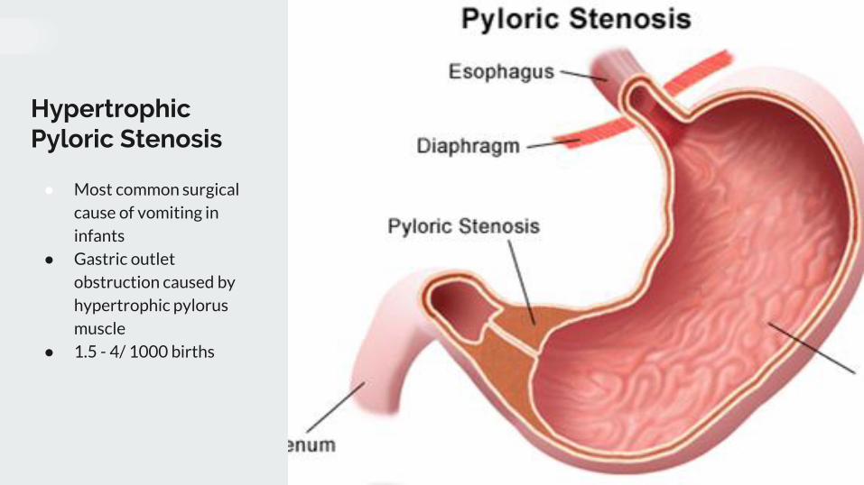
Hypertrophic Pyloric Stenosis
● Most common surgical
cause of vomiting in
infants
● Gastric outlet
obstruction caused by
hypertrophic pylorus
muscle
● 1.5 - 4/ 1000 births

Presentation of HPS
● 2 -4 weeks of life → typically full term infants
● Forcefull, nonbilious emesis 10 - 30 mins after feeding ○ Can be coffee grounds if gastritis or esophagitis is present
● Vigorous feeders
● Spectrum from very well appearing to severely dehydrated and in shock
● “Olive mass”
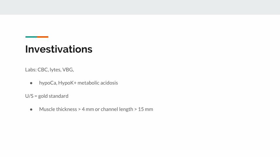
Investivations
Labs: CBC, lytes, VBG,
● hypoCa, HypoK+ metabolic acidosis
U/S = gold standard
● Muscle thickness > 4 mm or channel length > 15 mm

Management
Replace fluids with 20/ kg NS bolus then 5% dextrose at 1.5 x maintenance
Electrolyte correction
NG decompression
Surgical pylorotomy → longitudinal through hypertrophied muscle (but sparing mucosa)
Nonsurgical management → generally only for nonsurgical candidates in North America
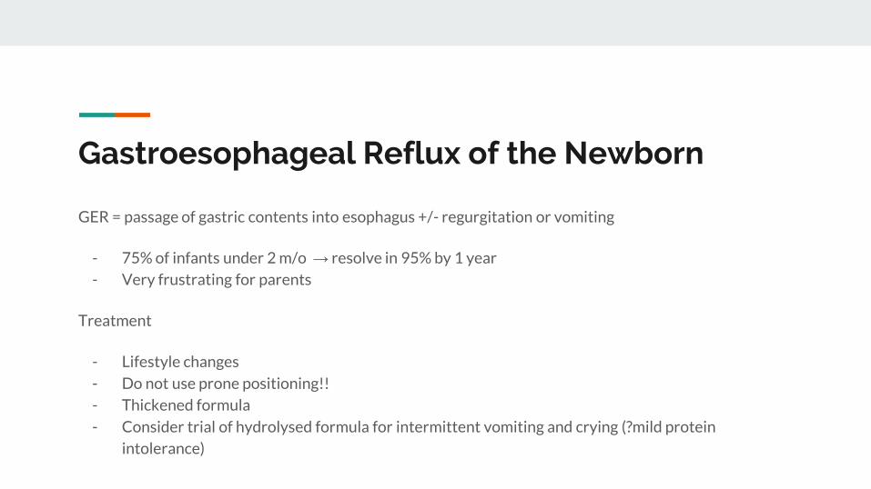
Gastroesophageal Reflux of the Newborn
GER = passage of gastric contents into esophagus +/- regurgitation or vomiting
- 75% of infants under 2 m/o → resolve in 95% by 1 year
- Very frustrating for parents
Treatment
- Lifestyle changes
- Do not use prone positioning!!
- Thickened formula
- Consider trial of hydrolysed formula for intermittent vomiting and crying (?mild protein
intolerance)
Gastroesophageal Reflux Disease GERD = when reflux causes symptoms/ complications (nutritional issues, esophagitis)
- ?Hx and P/E sufficient for diagnosis
- ? PPI trial
Treatment:
- Lifestyle changes
- Antisecretory Agents
● H2RAs (ranitidine)
- Issue of tolerance
● PPIs (omeprazole)
- Not approved/ well validated for infants
- Delay to onset of optimal action (consider bridging older children)

So what do I do?

Let’s imagine mom comes in and shows you the most recent vomit….
Malrotation
Malrotation = abnormal peritoneal
adhesive bands (Ladd bands) form
between cecum and right abdominal
wall which mechanically obstruct the
duodenum
● 1/500 live births
● 50% present first month of
life, 90% within first year
Midgut Volvulus Volvulus = small bowel twists around this Ladd band
compressing the superior mesenteric artery and
causing small bowel necrosis
● Occurs in 70% of infants with malrotation
Presentation
First month of life:
● Bilious emesis (77 - 100% of cases)
● Sudden onset of inconsolable crying
● Feeding intolerance
● Abdominal exam is NORMAL in about 60% of cases
Late Findings:
● Distension
● Abdominal pain
● Infarcted bowel: erythema and edema of the abdo wall, fever, dehydration, cardiovascular collapse
Investigations
Labs - nonspecific
Plain films - can be normal, can show distension of the stomach or duodenum +/- distal intraluminal air
Limited upper GI contrast study = gold standard
Malrotation - ligament of treitz does not cross left of midline on contrast study
● U/S also useful for non fixed cecum or abnormal oriented SMA

Management
Emergency
● Fluid resuscitation
● Gastric decompression via NG
● IV antibiotics (*)
Surgical
● Ladd procedure

Intussusception
Classically episodic abdo pain +/- vomiting with
currant jelly stools, ‘sausage mass’ on exam
Reality:
- intermittently inconsolable infant -
toddler
- Must be on your differential for lethargy
Ultrasound for Diagnosis
Case # 2
6 y/o M with periumbilical abdominal pain starting this morning (6 hours ago). He vomited food contents
2 x after eating lunch and has had 2 loose bowel movements. There is tactile warmth but no documented
fever. He presents with his mom who is concerned about dehydration as he is refusing everything but
small sips of water.
P/E:
Well appearing child. HR 120, RR 24, T 37.4.
Appendicitis
● Lifetime risk of 7 - 9
● Can occur at any age but most common 10 - 17 y/o → less common under 4 y/o (2/10 000)but
young (preverbal) children have the highest perforation rate (80 - 100%)
● One of the most common causes of malpractice litigation
Presentation
● Classic: periumbilical abdo pain → anorexia, nausea, vomiting → RLQ pain, +/- fever
● Reality : ○ Frequent loose stools (usually small volume)
○ Dysuria +/- hematuria (if appendix is overlaying a ureter)
○ Flank pain (retrocecal appendix)
○ Hesitant to move around (rebound tenderness) → increases likeliness of appendicitis 3x
○ Fever?
■ High grade more associated with perforation
**perforation can cloud the physical exam
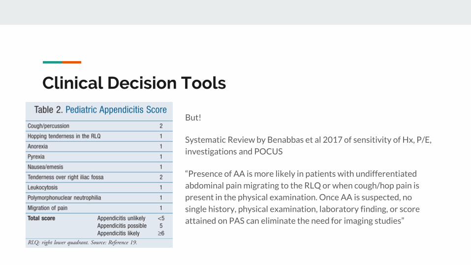
Clinical Decision Tools
But!
Systematic Review by Benabbas et al 2017 of sensitivity of Hx, P/E,
investigations and POCUS
“Presence of AA is more likely in patients with undifferentiated
abdominal pain migrating to the RLQ or when cough/hop pain is
present in the physical examination. Once AA is suspected, no
single history, physical examination, laboratory finding, or score
attained on PAS can eliminate the need for imaging studies”
Investigations
Ultrasound is gold standard
Plain Film?
● Evidence in about 10% of cases of visible fecalith
● Only to r/o other indications
Bloodwork?
● Varies a lot by practise setting
● When I do it? ○ Order if high pretest probability to avoid treatment delays but not so much to guide diagnosis
Management
Pain Control
● acetaminophen +/- ibuprofen in the office (does not mask diagnosis)
● NPO
● Fluid resuscitation
● Surgical consultation
● Broad spectrum Abx ○ Simple - setting variation
○ Perforated - triple therapy (amp, gent, flagyl)
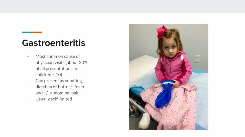
Gastroenteritis - Most common cause of
physician visits (about 20%
of all presentations for
children < 10)
- Can present as vomiting,
diarrhea or both +/- fever
and +/- abdominal pain
- Usually self limited
Gastroenteritis
Investigations
- When to stool Cx?
- Urine
- Blood pressure
Management:
Oral ondansetron if available decreases need for IV and hospital admission (as per CPS guideline)
● 8 kg to 15 kg: 2 mg
● 15 kg to 30 kg: 4 mg
● Greater than 30 kg: 6 mg to 8 mg

Empiric Treatment for Bacterial Gastroenteritis

Case # 3
14 y/o M presents to your office with a 2 day history of vomiting and diarrhea with epigastric and right
upper quadrant abdominal pain. He has an 8 month history of intermittent epigastric abdominal pain
with vomiting last 1 -2 days usually 2 -3 episodes per month and is well in between with occasional
postprandial epigastric discomfort. He is afebrile. Emesis is nonbilious. Prior to this episode his stool
habits were small lumps, every second day.
P/E: uncomfortable appearing, diffusely tender, focal RUQ tenderness, -ve Murphy’s and McBurney’s, +
stool palpable LLQ
Habitus - 78 kg
Differential Diagnosis?
Which of the following is least likely to be part of your differential diagnosis for this case?
a) Functional constipation
b) Cyclic vomiting syndrome
c) Biliary Colic
d) Gastroenteritis
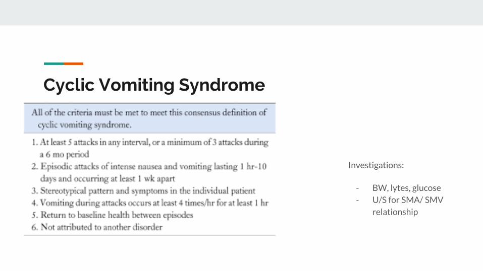
Cyclic Vomiting Syndrome
Investigations:
- BW, lytes, glucose
- U/S for SMA/ SMV
relationship
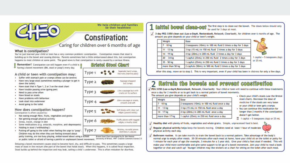
Constipation
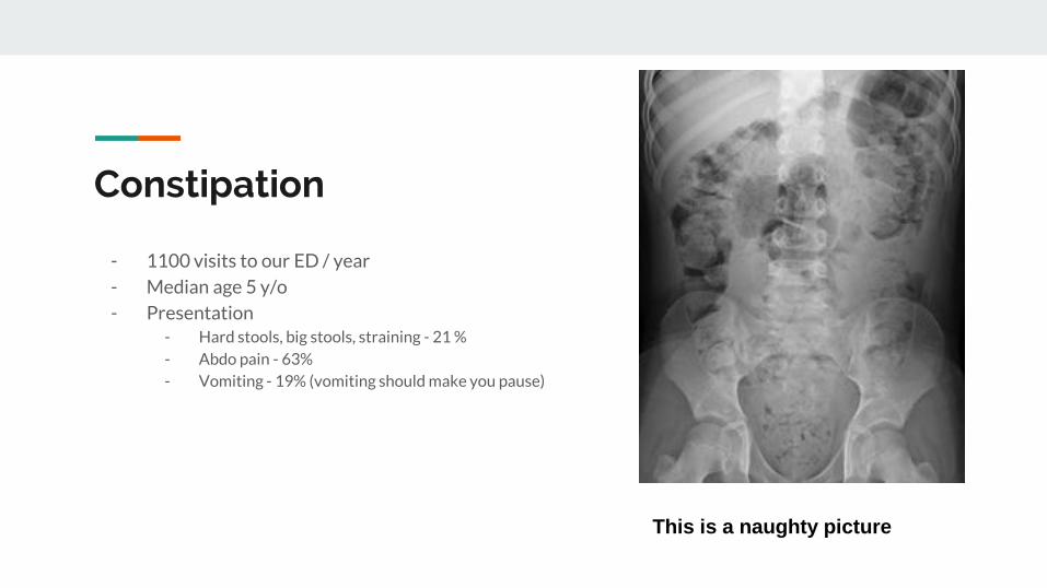
- 1100 visits to our ED / year
- Median age 5 y/o
- Presentation - Hard stools, big stools, straining - 21 %
- Abdo pain - 63%
- Vomiting - 19% (vomiting should make you pause)
This is a naughty picture
Which of the following would not be a ‘red flag’ for an organic cause of constipation in an infant? a) Passage of first meconium at 36 hours of life
b) Explosive stool passage after rectal exam
c) Failure to thrive
d) Abdominal distension

Red Flags for Organic Cause
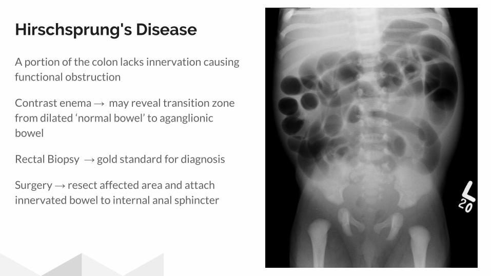
Hirschsprung's Disease
A portion of the colon lacks innervation causing
functional obstruction
Contrast enema → may reveal transition zone
from dilated ‘normal bowel’ to aganglionic
bowel
Rectal Biopsy → gold standard for diagnosis
Surgery → resect affected area and attach
innervated bowel to internal anal sphincter

Do a neuro
exam!
+/- a rectal
exam
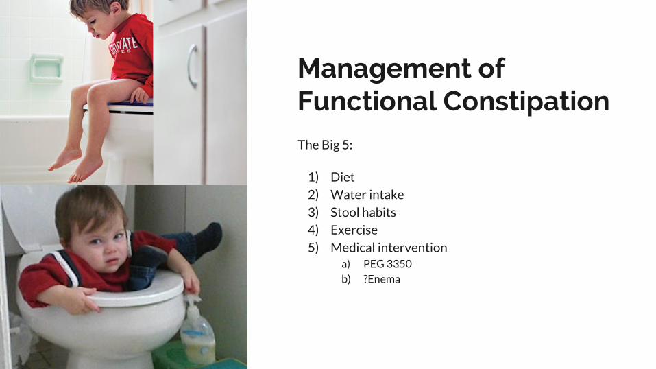
Management of Functional Constipation The Big 5:
1) Diet
2) Water intake
3) Stool habits
4) Exercise
5) Medical intervention a) PEG 3350
b) ?Enema
Back to Our Case
Investigations:
Labs:
- AST = 310, ALT = 455, GGT = 294
- Amylase normal
U/S:
- CBD 12 mm, dilated intra and extrahepatic ducts, multiple gallstones
ERCP removing stone from distal CBD → asymptomatic at follow up
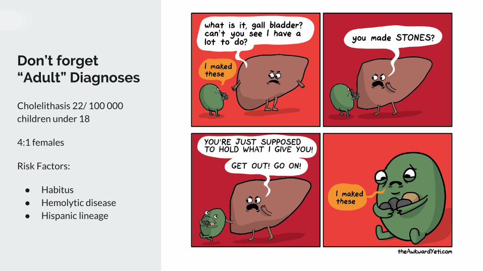
Don’t forget “Adult” Diagnoses
Cholelithasis 22/ 100 000
children under 18
4:1 females
Risk Factors:
● Habitus
● Hemolytic disease
● Hispanic lineage
So What Should I Remember?
1) Bilious emesis in an infant is malrotation until proven otherwise
2) Convince yourself the vomiting infant doesn’t have pyloric stenosis,
not that they do have GER (and be judicious with pharmalogical
treatment of GER/D
3) Acute abdominal pain + RLQ tenderness or rebound = imaging
4) Do a neuro exam for all constipation presentations
5) Enema sparingly for functional constipation, X-ray even less
6) Any case could be your most interesting case today!

Questions?