Embed Size (px)
Citation preview
Thyroid Nodules:Practical and Molecular Approaches to a
Common Problem
Bryan R. Haugen, MDUniversity of Colorado, School of Medicine
University of Colorado Cancer CenterSurgery Grand Rounds
May 9, 2011
Disclosure
Research support – Veracyte, Inc
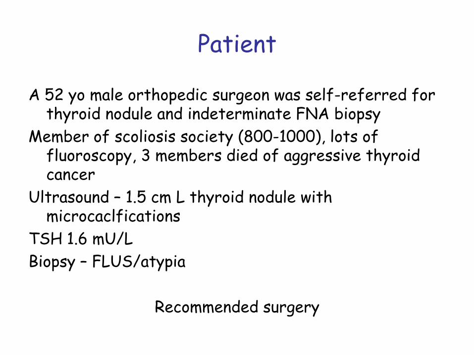
Patient
A 52 yo male orthopedic surgeon was self-referred for thyroid nodule and indeterminate FNA biopsy
Member of scoliosis society (800-1000), lots of fluoroscopy, 3 members died of aggressive thyroid cancer
Ultrasound – 1.5 cm L thyroid nodule with microcaclfications
TSH 1.6 mU/LBiopsy – FLUS/atypia
Recommended surgery
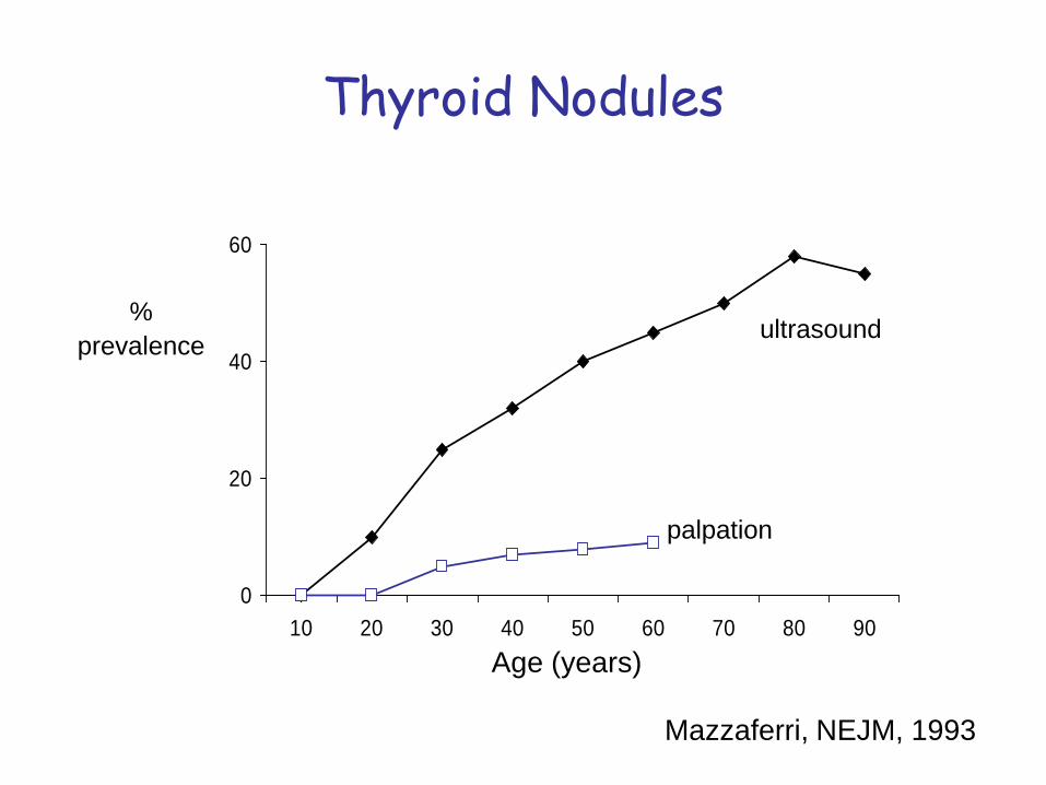
Thyroid Nodules
0
20
40
60
10 20 30 40 50 60 70 80 90
%prevalence
Age (years)
ultrasound
palpation
Mazzaferri, NEJM, 1993
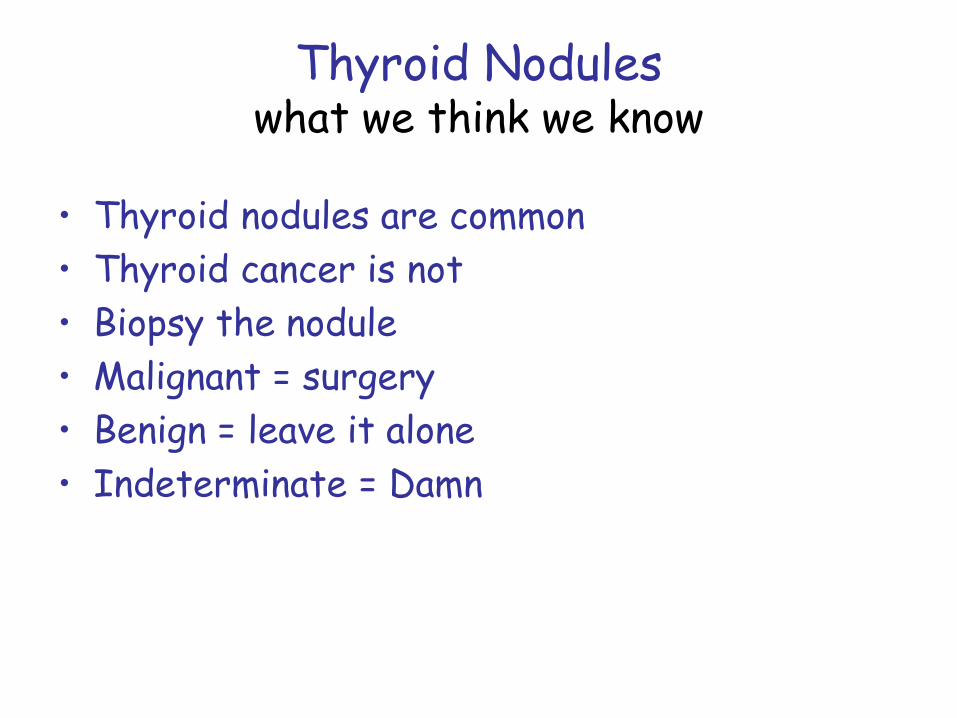
Thyroid Noduleswhat we think we know
• Thyroid nodules are common• Thyroid cancer is not • Biopsy the nodule• Malignant = surgery• Benign = leave it alone• Indeterminate = Damn
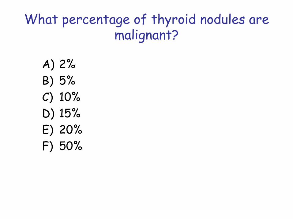
What percentage of thyroid nodules are malignant?
A) 2%B) 5%C) 10%D) 15%E) 20%F) 50%
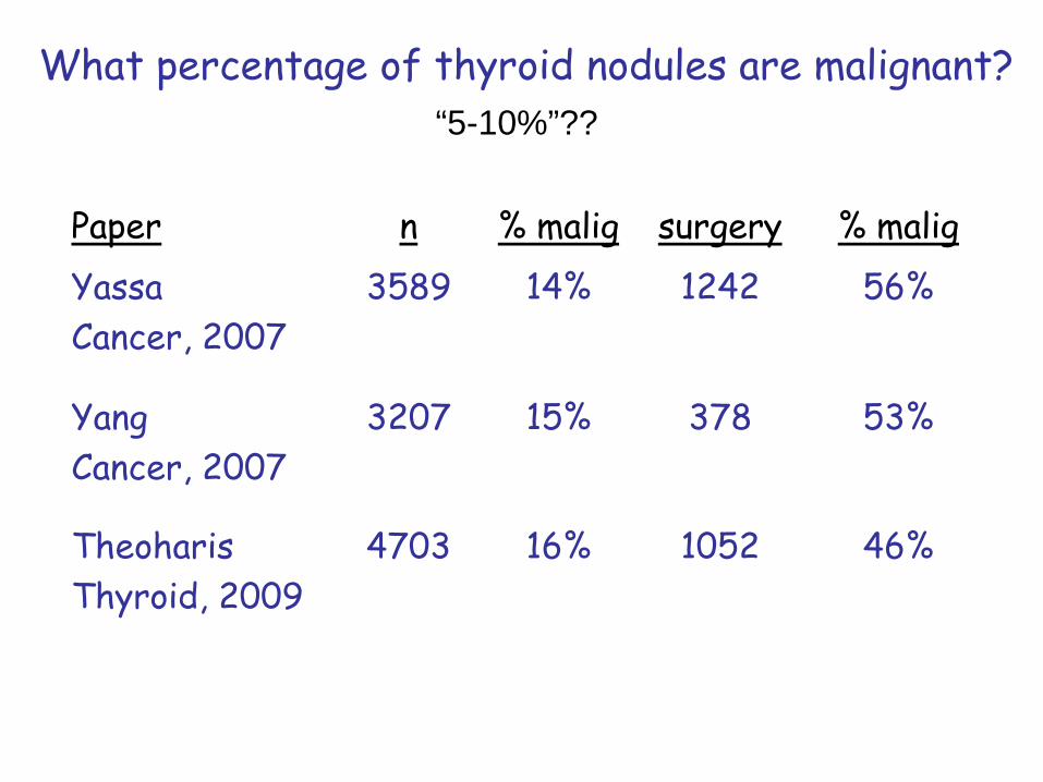
What percentage of thyroid nodules are malignant?
Paper n % malig surgery % maligYassaCancer, 2007
3589 14% 1242 56%
YangCancer, 2007
3207 15% 378 53%
TheoharisThyroid, 2009
4703 16% 1052 46%
“5-10%”??
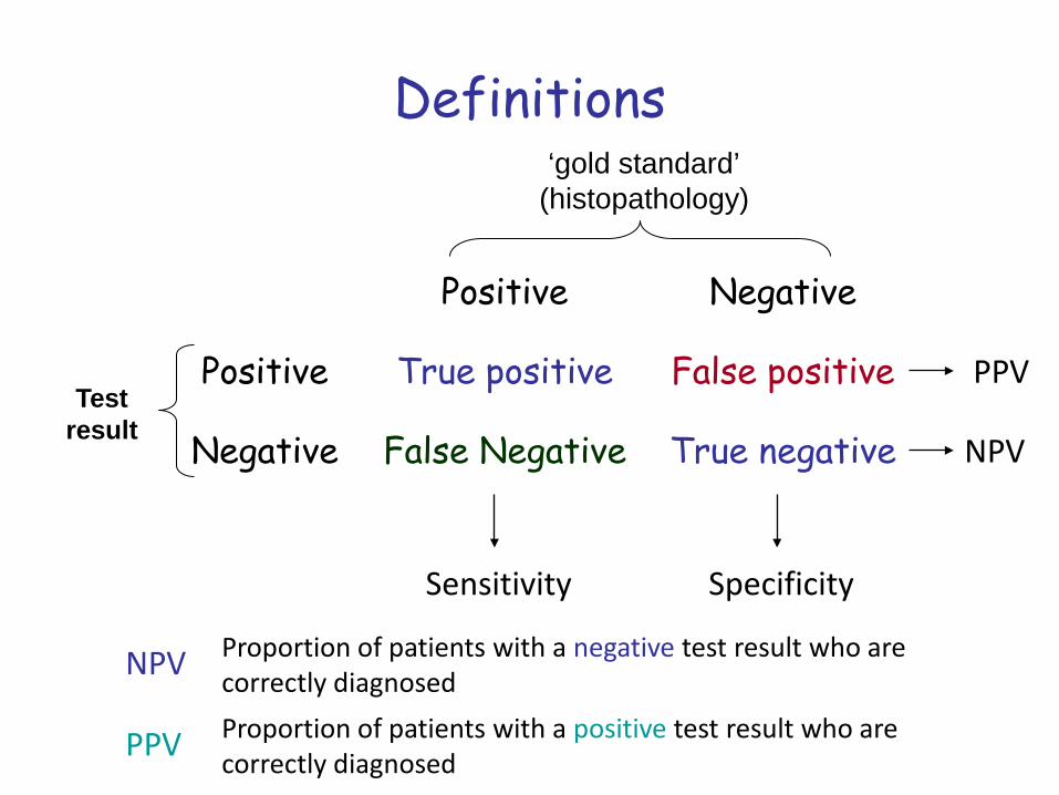
Definitions
Positive Negative
Positive True positive False positive
Negative False Negative True negativeTest
result
‘gold standard’(histopathology)
PPV
NPV
Sensitivity Specificity
NPV Proportion of patients with a negative test result who are correctly diagnosed
PPV Proportion of patients with a positive test result who are correctly diagnosed
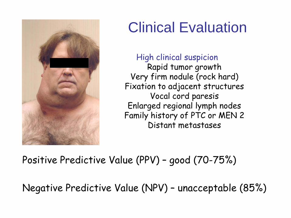
Clinical Evaluation
Positive Predictive Value (PPV) – good (70-75%)
Negative Predictive Value (NPV) – unacceptable (85%)
High clinical suspicionRapid tumor growth
Very firm nodule (rock hard)Fixation to adjacent structures
Vocal cord paresisEnlarged regional lymph nodes
Family history of PTC or MEN 2Distant metastases
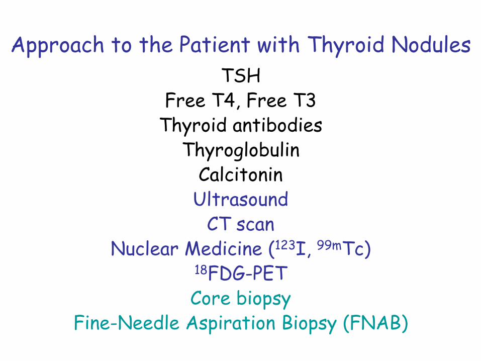
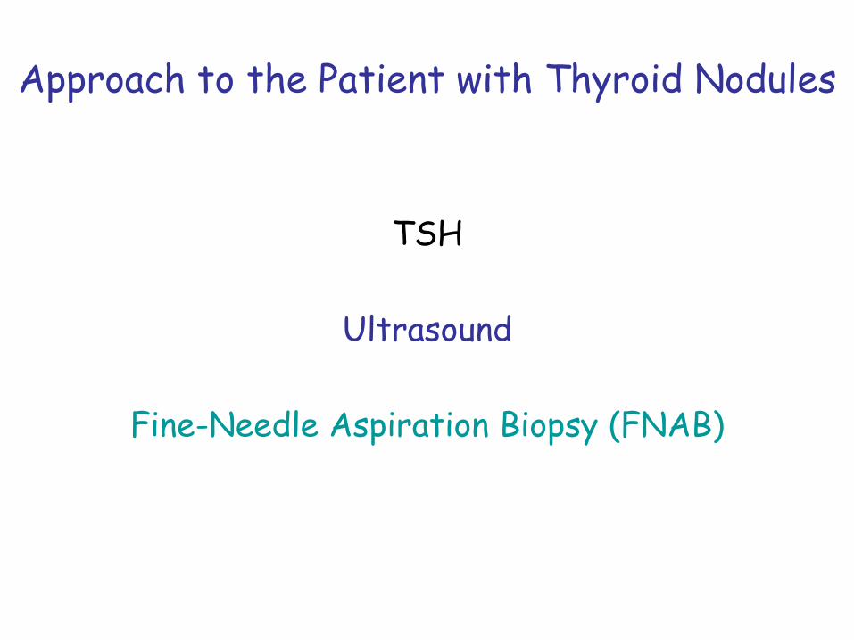
Approach to the Patient with Thyroid NodulesTSH
Free T4, Free T3Thyroid antibodies
ThyroglobulinCalcitonin
UltrasoundCT scan
Nuclear Medicine (123I, 99mTc)18FDG-PETCore biopsy
Fine-Needle Aspiration Biopsy (FNAB)
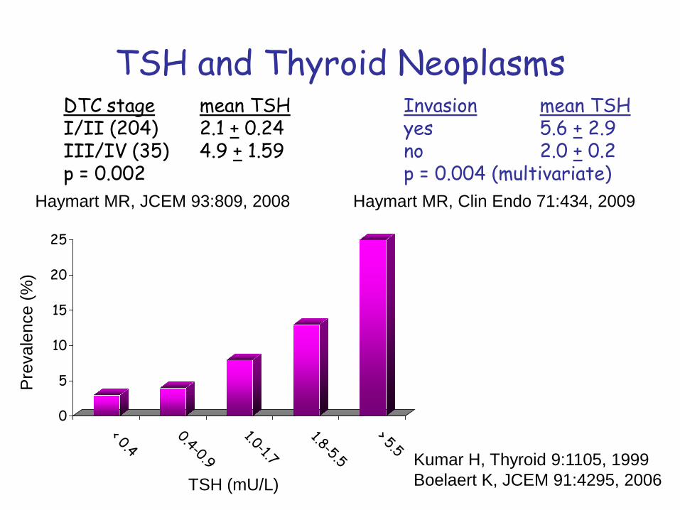
TSH and Thyroid Neoplasms
0
5
10
15
20
25
< 0.40.4-0.9
1.0-1.71.8-5.5
> 5.5
Pre
vale
nce
(%)
TSH (mU/L)
DTC stage mean TSHI/II (204) 2.1 + 0.24III/IV (35) 4.9 + 1.59p = 0.002
Kumar H, Thyroid 9:1105, 1999Boelaert K, JCEM 91:4295, 2006
Invasion mean TSHyes 5.6 + 2.9no 2.0 + 0.2p = 0.004 (multivariate)
Haymart MR, JCEM 93:809, 2008 Haymart MR, Clin Endo 71:434, 2009
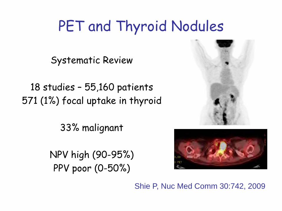
PET and Thyroid Nodules
Systematic Review
18 studies – 55,160 patients571 (1%) focal uptake in thyroid
33% malignant
NPV high (90-95%)PPV poor (0-50%)
Shie P, Nuc Med Comm 30:742, 2009
Approach to the Patient with Thyroid Nodules
TSH
Ultrasound
Fine-Needle Aspiration Biopsy (FNAB)
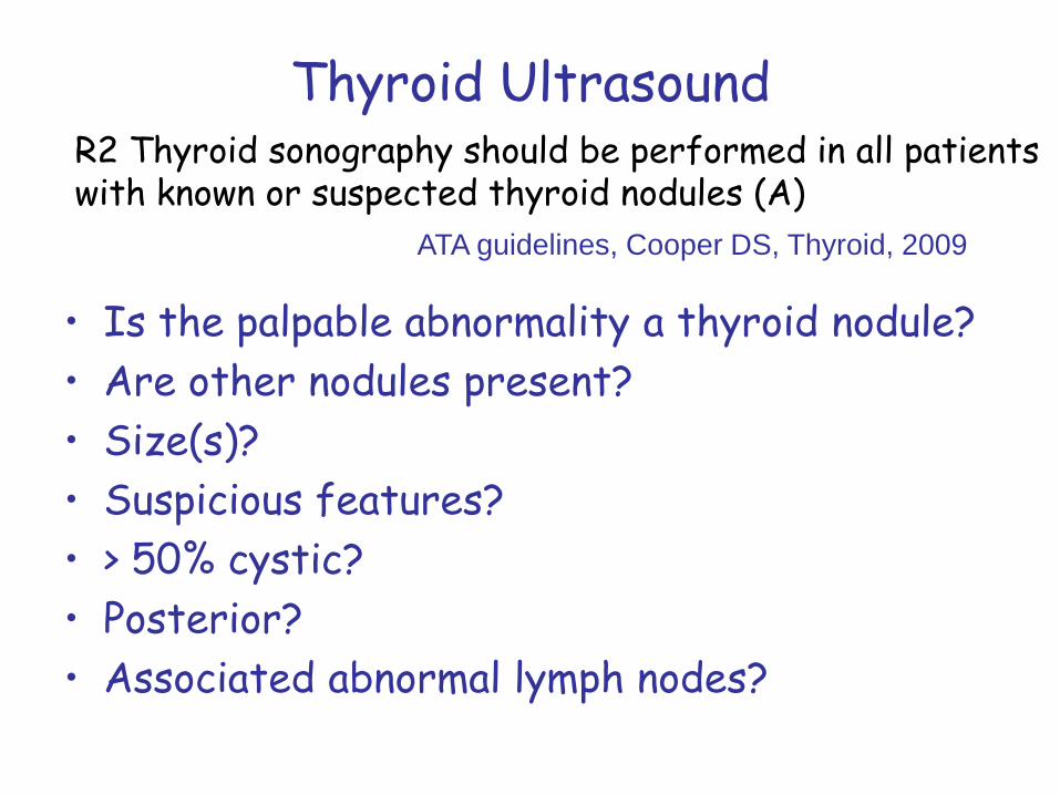
Thyroid Ultrasound
• Is the palpable abnormality a thyroid nodule?• Are other nodules present?• Size(s)?• Suspicious features?• > 50% cystic?• Posterior?• Associated abnormal lymph nodes?
R2 Thyroid sonography should be performed in all patients with known or suspected thyroid nodules (A)
ATA guidelines, Cooper DS, Thyroid, 2009
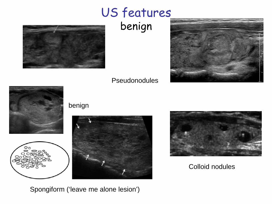
US featuresbenign
Spongiform (‘leave me alone lesion’)
benign
Colloid nodules
Pseudonodules
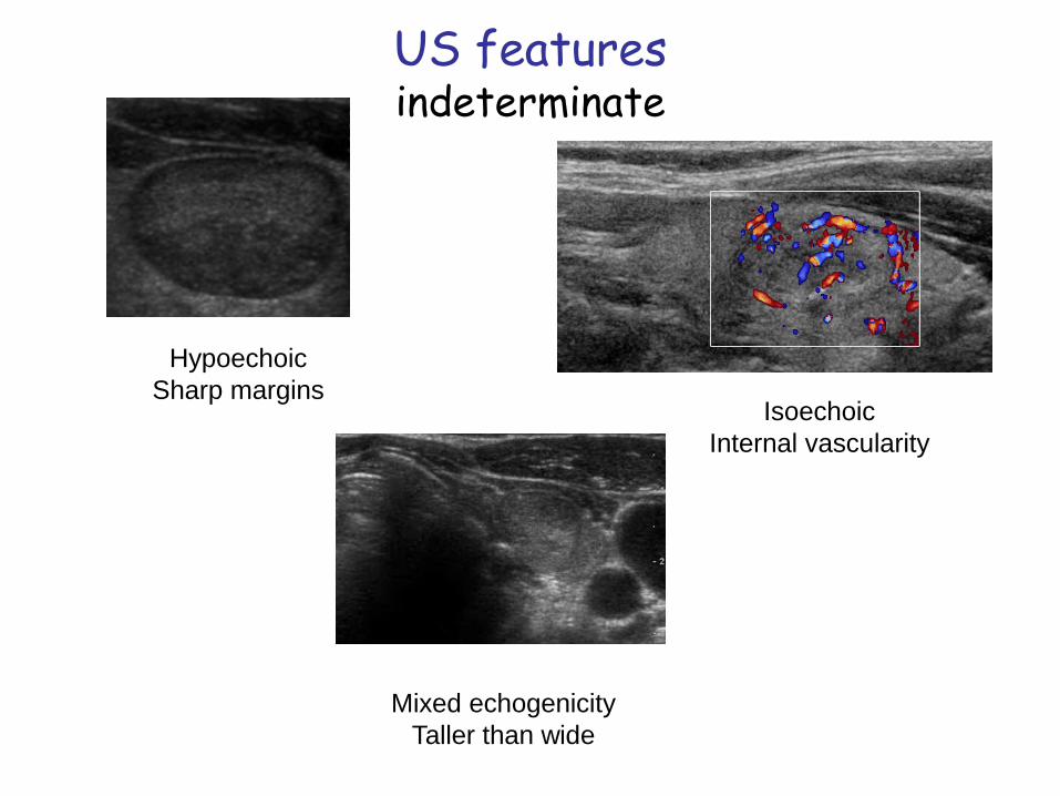
US featuresindeterminate
HypoechoicSharp margins
IsoechoicInternal vascularity
Mixed echogenicityTaller than wide
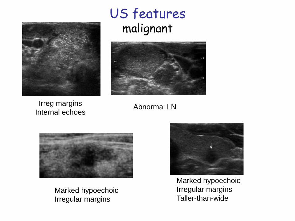
US featuresmalignant
Marked hypoechoicIrregular marginsTaller-than-wide
Irreg marginsInternal echoes
Abnormal LN
Marked hypoechoicIrregular margins
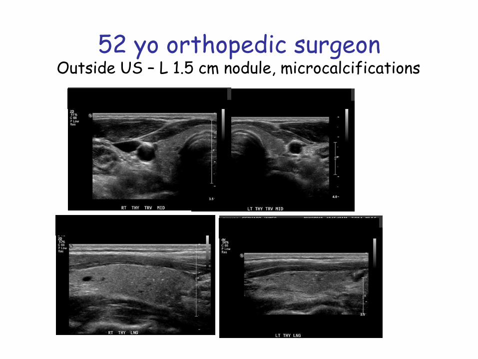
52 yo orthopedic surgeonOutside US – L 1.5 cm nodule, microcalcifications
Which nodule(s) should I biopsy?
Which nodule(s) don’t need a biopsy?
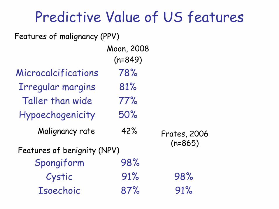
Predictive Value of US features
Moon, 2008(n=849)
Microcalcifications 78%Irregular margins 81%Taller than wide 77%
Hypoechogenicity 50%
Spongiform 98%Cystic 91% 98%
Isoechoic 87% 91%
Features of malignancy (PPV)
Features of benignity (NPV)
Malignancy rate 42% Frates, 2006(n=865)
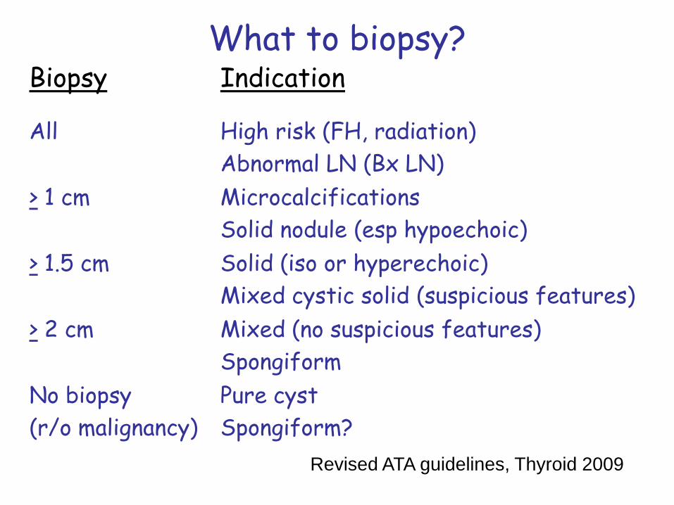
What to biopsy?Biopsy Indication
All High risk (FH, radiation)Abnormal LN (Bx LN)
> 1 cm MicrocalcificationsSolid nodule (esp hypoechoic)
> 1.5 cm Solid (iso or hyperechoic)Mixed cystic solid (suspicious features)
> 2 cm Mixed (no suspicious features)Spongiform
No biopsy(r/o malignancy)
Pure cystSpongiform?
Revised ATA guidelines, Thyroid 2009
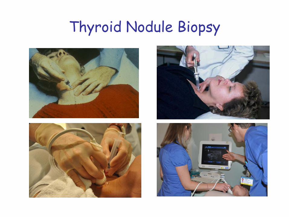
Thyroid Nodule Biopsy
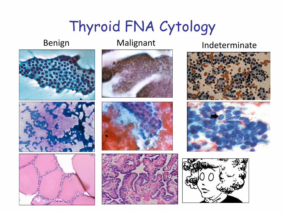
Thyroid FNA CytologyBenign Malignant Indeterminate
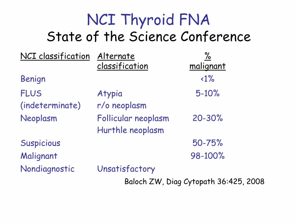
NCI Thyroid FNAState of the Science Conference
NCI classification Alternate classification
% malignant
Benign <1%FLUS(indeterminate)
Atypiar/o neoplasm
5-10%
Neoplasm Follicular neoplasmHurthle neoplasm
20-30%
Suspicious 50-75%Malignant 98-100%Nondiagnostic Unsatisfactory
Baloch ZW, Diag Cytopath 36:425, 2008
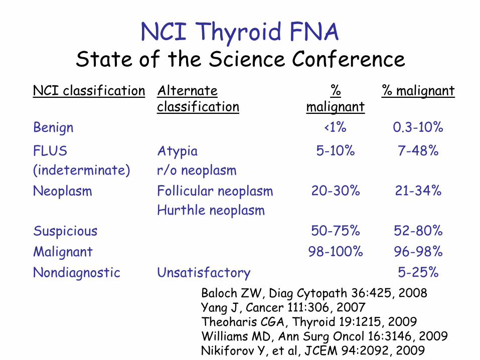
NCI Thyroid FNAState of the Science Conference
NCI classification Alternate classification
% malignant
% malignant
Benign <1% 0.3-10%FLUS(indeterminate)
Atypiar/o neoplasm
5-10% 7-48%
Neoplasm Follicular neoplasmHurthle neoplasm
20-30% 21-34%
Suspicious 50-75% 52-80%Malignant 98-100% 96-98%Nondiagnostic Unsatisfactory 5-25%
Baloch ZW, Diag Cytopath 36:425, 2008Yang J, Cancer 111:306, 2007Theoharis CGA, Thyroid 19:1215, 2009Williams MD, Ann Surg Oncol 16:3146, 2009Nikiforov Y, et al, JCEM 94:2092, 2009
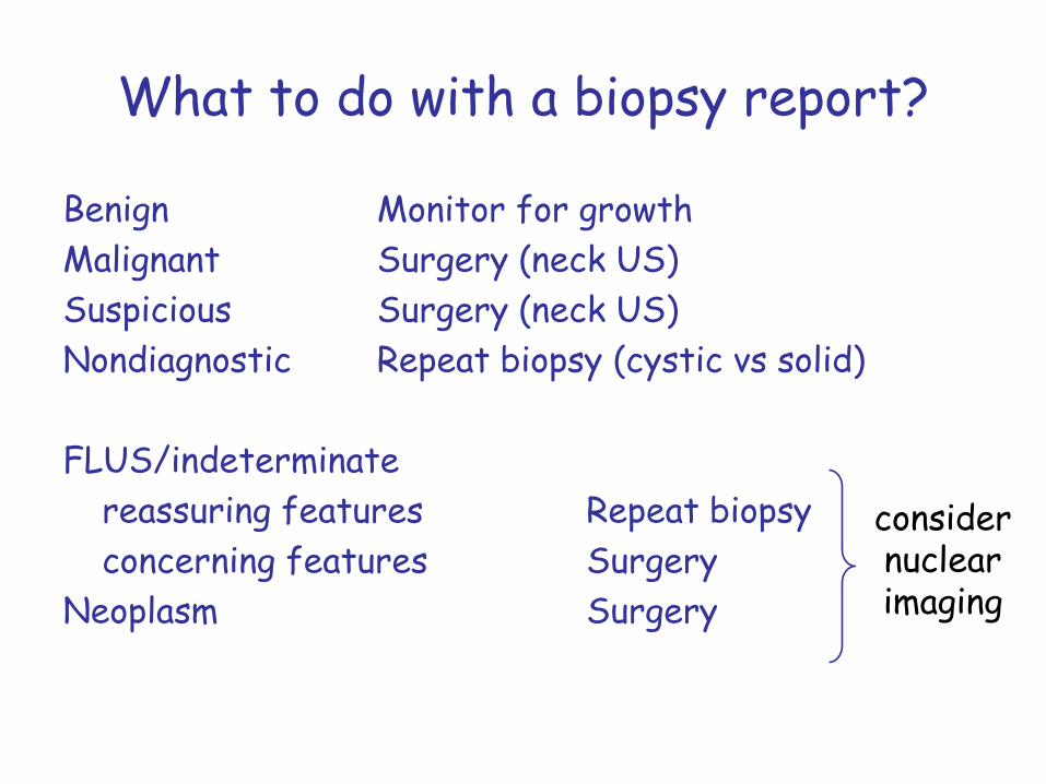
What to do with a biopsy report?
Benign Monitor for growthMalignant Surgery (neck US)Suspicious Surgery (neck US)Nondiagnostic Repeat biopsy (cystic vs solid)
FLUS/indeterminatereassuring features Repeat biopsyconcerning features Surgery
Neoplasm Surgery
considernuclearimaging
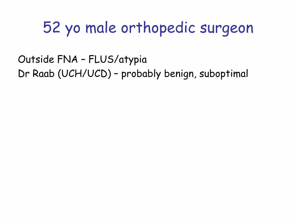
52 yo male orthopedic surgeon
Outside FNA – FLUS/atypiaDr Raab (UCH/UCD) – probably benign, suboptimal
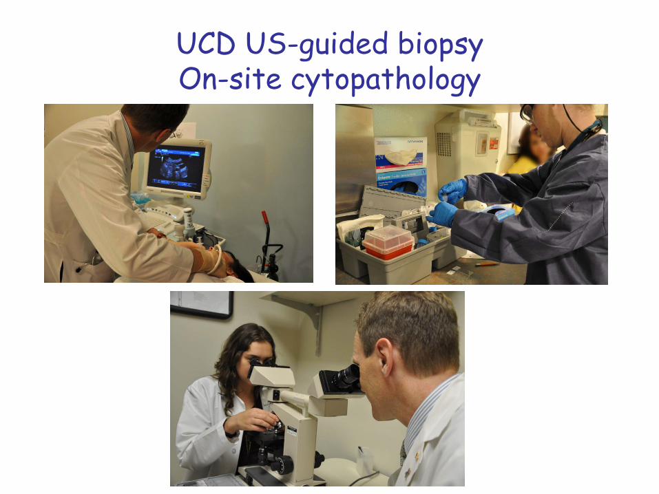
UCD US-guided biopsyOn-site cytopathology
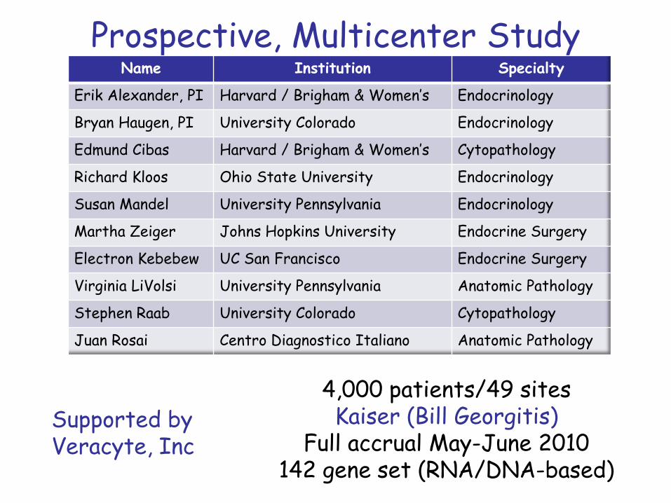
Prospective, Multicenter StudyName Institution Specialty
Erik Alexander, PI Harvard / Brigham & Women’s Endocrinology
Bryan Haugen, PI University Colorado Endocrinology
Edmund Cibas Harvard / Brigham & Women’s Cytopathology
Richard Kloos Ohio State University Endocrinology
Susan Mandel University Pennsylvania Endocrinology
Martha Zeiger Johns Hopkins University Endocrine Surgery
Electron Kebebew UC San Francisco Endocrine Surgery
Virginia LiVolsi University Pennsylvania Anatomic Pathology
Stephen Raab University Colorado Cytopathology
Juan Rosai Centro Diagnostico Italiano Anatomic Pathology
4,000 patients/49 sitesKaiser (Bill Georgitis)
Full accrual May-June 2010142 gene set (RNA/DNA-based)
Supported byVeracyte, Inc
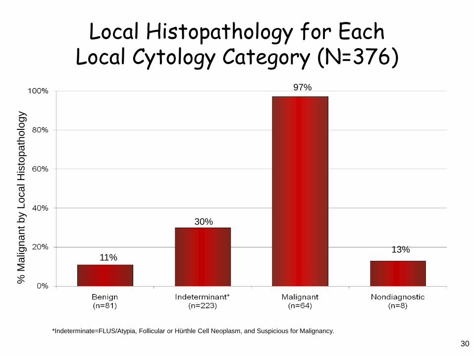
Local Histopathology for Each Local Cytology Category (N=376)
30
11%
% M
alig
nant
by
Loca
l His
topa
thol
ogy
97%
30%
13%
*Indeterminate=FLUS/Atypia, Follicular or Hürthle Cell Neoplasm, and Suspicious for Malignancy.
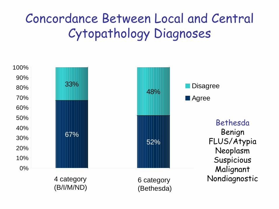
67%
33%
52%
48%
0%
10%20%
30%40%
50%
60%70%
80%90%
100%
Disagree
Agree
4 category(B/I/M/ND)
6 category(Bethesda)
BethesdaBenign
FLUS/AtypiaNeoplasmSuspiciousMalignant
Nondiagnostic
Concordance Between Local and CentralCytopathology Diagnoses
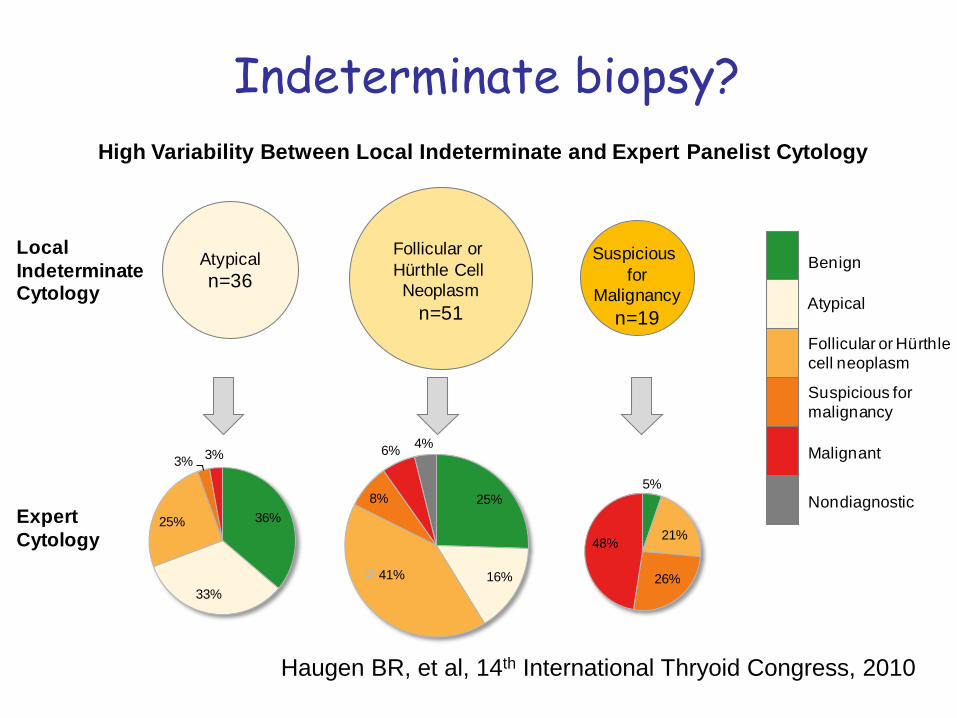
Indeterminate biopsy?
Atypicaln=36
Follicular or Hürthle Cell Neoplasm
n=51
Suspicious for
Malignancyn=19
36%
33%
25%
3% 3%
5%
21%
26%
48%
25%
16%41%
8%
6% 4%
High Variability Between Local Indeterminate and Expert Panelist Cytology
Local Indeterminate Cytology
Expert Cytology
Benign
Atypical
Follicular or Hürthle cell neoplasm
Suspicious for malignancy
Nondiagnostic
Malignant
Haugen BR, et al, 14th International Thryoid Congress, 2010
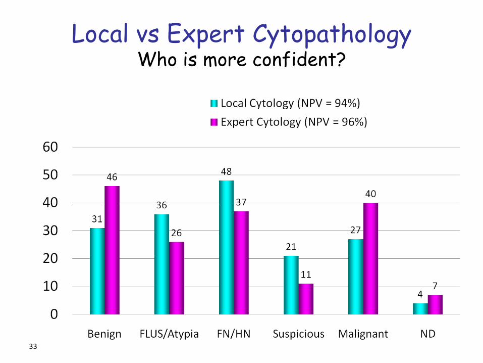
Local vs Expert CytopathologyWho is more confident?
33
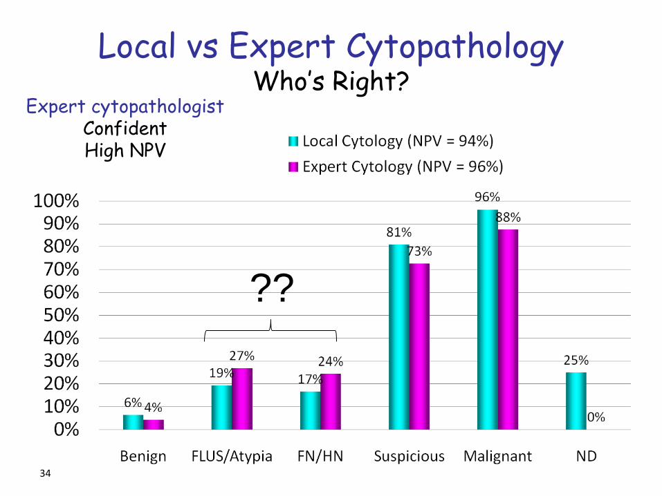
Local vs Expert CytopathologyWho’s Right?
34
Expert cytopathologistConfidentHigh NPV
??
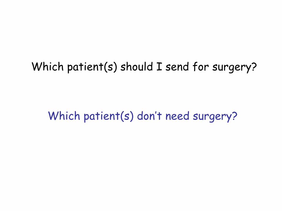
Which patient(s) should I send for surgery?
Which patient(s) don’t need surgery?
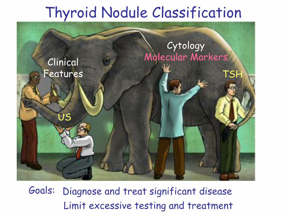
Thyroid Nodule Classification
CytologyMolecular Markers
TSHClinical
Features
US
Goals: Diagnose and treat significant diseaseLimit excessive testing and treatment
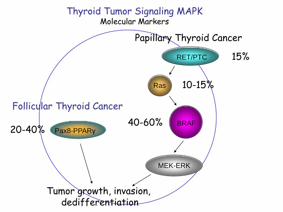
Thyroid Tumor Signaling MAPKMolecular Markers
Papillary Thyroid Cancer
Tumor growth, invasion,dedifferentiation
Ras
RET/PTC
BRAF
MEK-ERK
15%
10-15%
40-60%Pax8-PPARγ20-40%
Follicular Thyroid Cancer
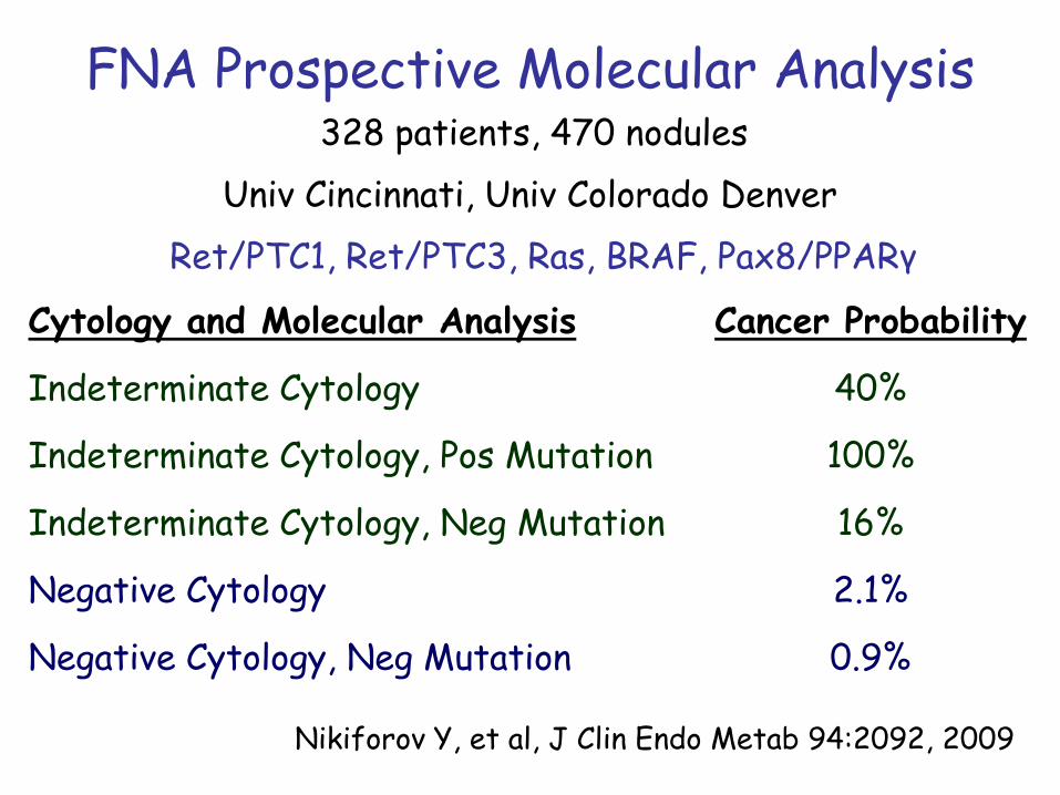
FNA Prospective Molecular Analysis
Nikiforov Y, et al, J Clin Endo Metab 94:2092, 2009
328 patients, 470 nodulesUniv Cincinnati, Univ Colorado Denver
Ret/PTC1, Ret/PTC3, Ras, BRAF, Pax8/PPARγ
Cytology and Molecular Analysis Cancer Probability
Indeterminate Cytology 40%
Indeterminate Cytology, Pos Mutation 100%
Indeterminate Cytology, Neg Mutation 16%
Negative Cytology 2.1%
Negative Cytology, Neg Mutation 0.9%
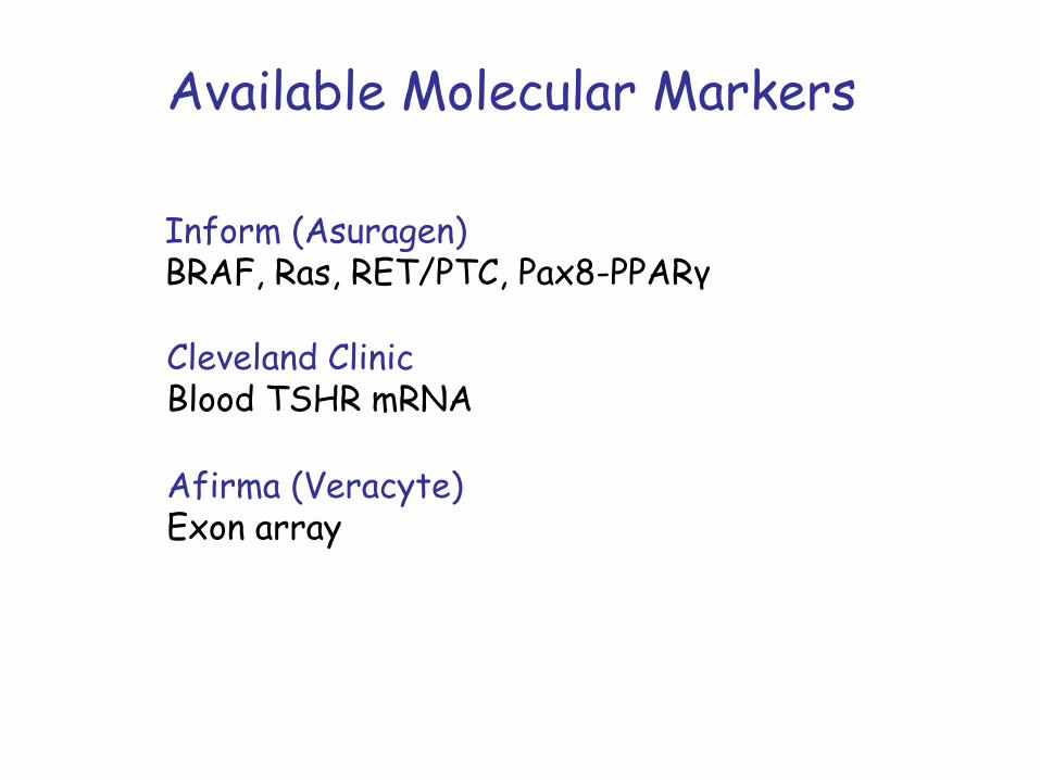
Available Molecular Markers
Inform (Asuragen)BRAF, Ras, RET/PTC, Pax8-PPARγ
Cleveland ClinicBlood TSHR mRNA
Afirma (Veracyte)Exon array
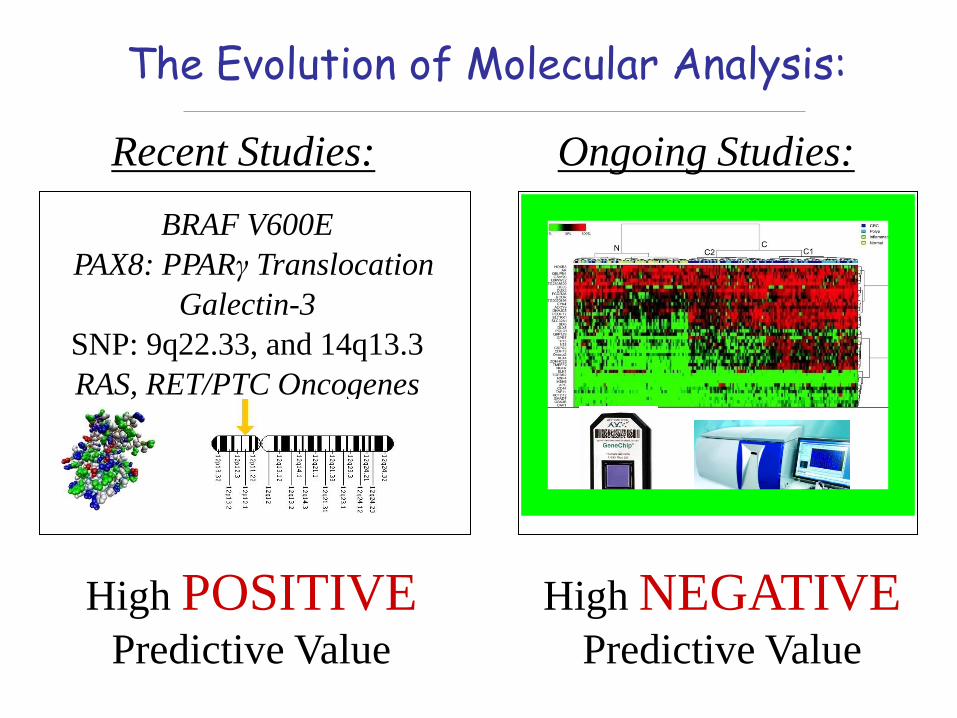
The Evolution of Molecular Analysis:
PAX8: PPARγ TranslocationBRAF V600E
Galectin-3SNP: 9q22.33, and 14q13.3RAS, RET/PTC Oncogenes
Recent Studies: Ongoing Studies:
High POSITIVEPredictive Value
High NEGATIVEPredictive Value
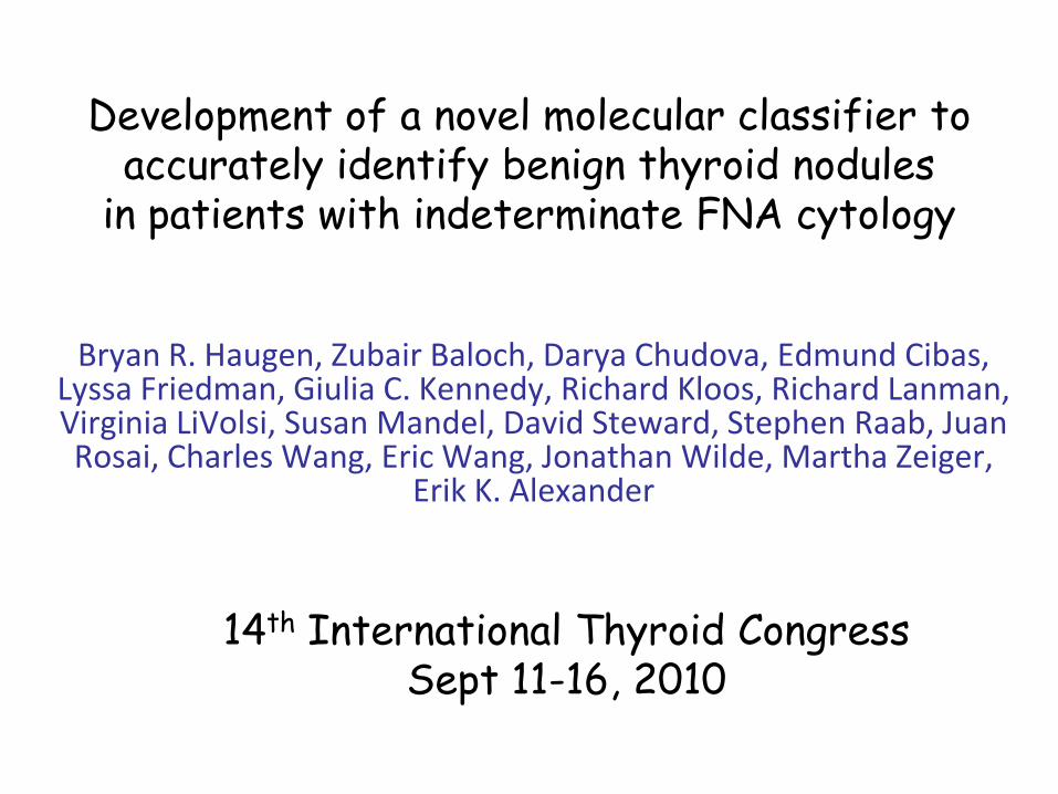
Development of a novel molecular classifier to accurately identify benign thyroid nodules
in patients with indeterminate FNA cytology
Bryan R. Haugen, Zubair Baloch, Darya Chudova, Edmund Cibas, Lyssa Friedman, Giulia C. Kennedy, Richard Kloos, Richard Lanman, Virginia LiVolsi, Susan Mandel, David Steward, Stephen Raab, Juan Rosai, Charles Wang, Eric Wang, Jonathan Wilde, Martha Zeiger,
Erik K. Alexander
14th International Thyroid CongressSept 11-16, 2010
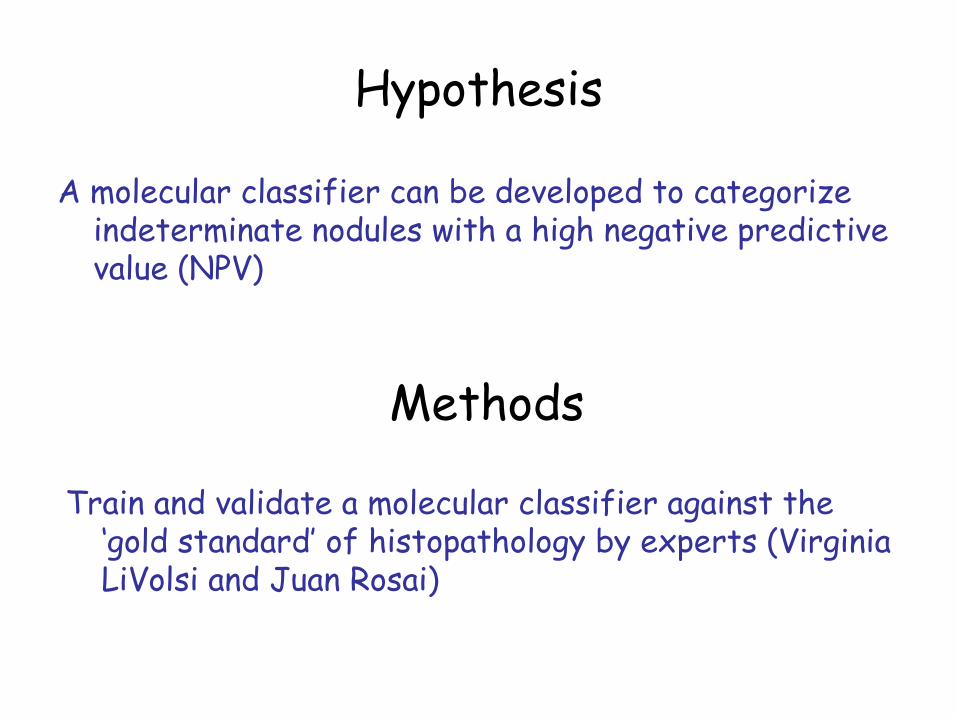
Hypothesis
A molecular classifier can be developed to categorize indeterminate nodules with a high negative predictive value (NPV)
Methods
Train and validate a molecular classifier against the ‘gold standard’ of histopathology by experts (Virginia LiVolsi and Juan Rosai)
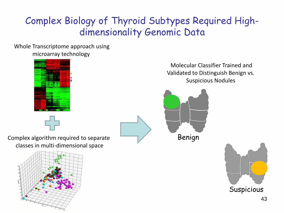
Complex Biology of Thyroid Subtypes Required High-dimensionality Genomic Data
Complex algorithm required to separate classes in multi-dimensional space
Whole Transcriptome approach using microarray technology
Molecular Classifier Trained andValidated to Distinguish Benign vs.
Suspicious Nodules
Benign
Suspicious43
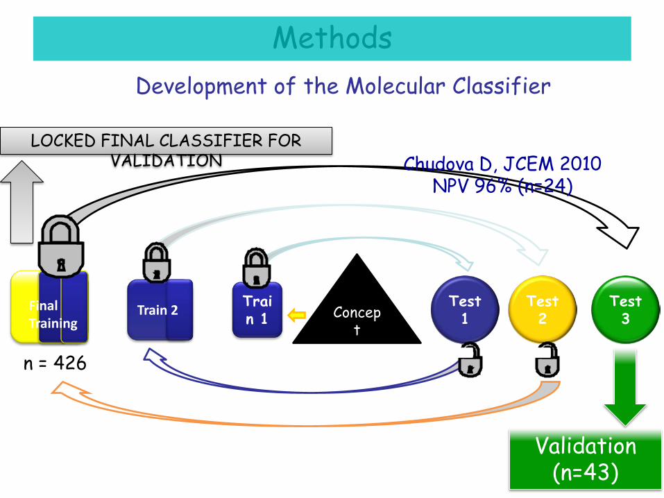
Development of the Molecular Classifier
Concept
Validation (n=43)
Test 1
Test 2
Test 3
Train 1Train 2Final
Training
LOCKED FINAL CLASSIFIER FOR VALIDATION Chudova D, JCEM 2010
NPV 96% (n=24)
Methods
n = 426
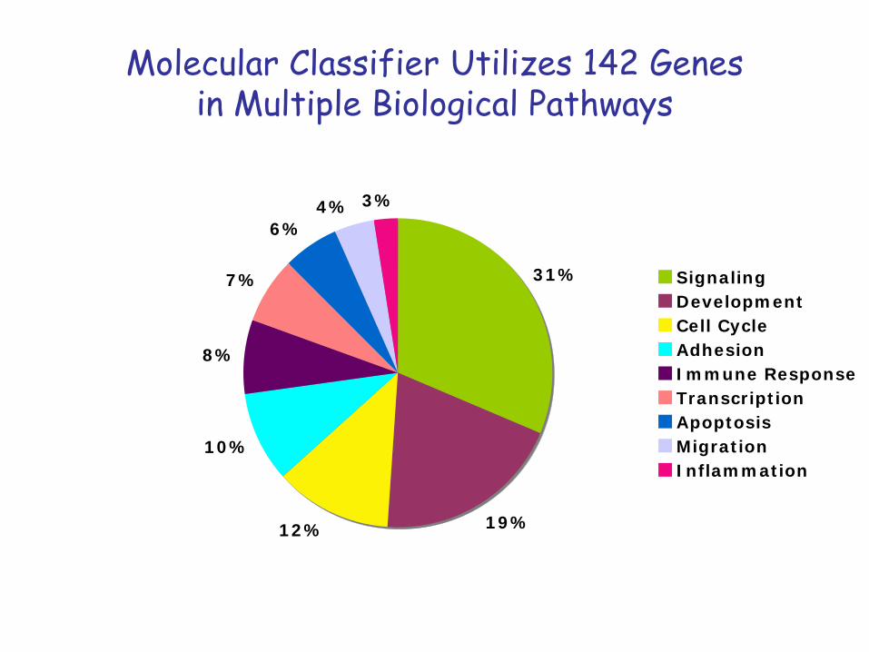
Molecular Classifier Utilizes 142 Genesin Multiple Biological Pathways
31%
19%12%
10%
8%
7%
6%4% 3%
SignalingDevelopmentCell CycleAdhesionImmune ResponseTranscriptionApoptosisMigrationInflammation
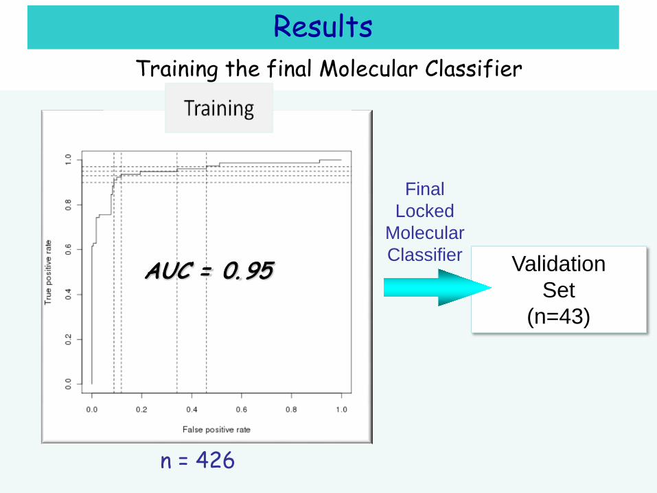
Training the final Molecular Classifier
ValidationSet
(n=43)
AUC = 0. 95
Results
n = 426
FinalLocked
MolecularClassifier
Sensitivity 97%Specificity 64%PPV 58%NPV 98%
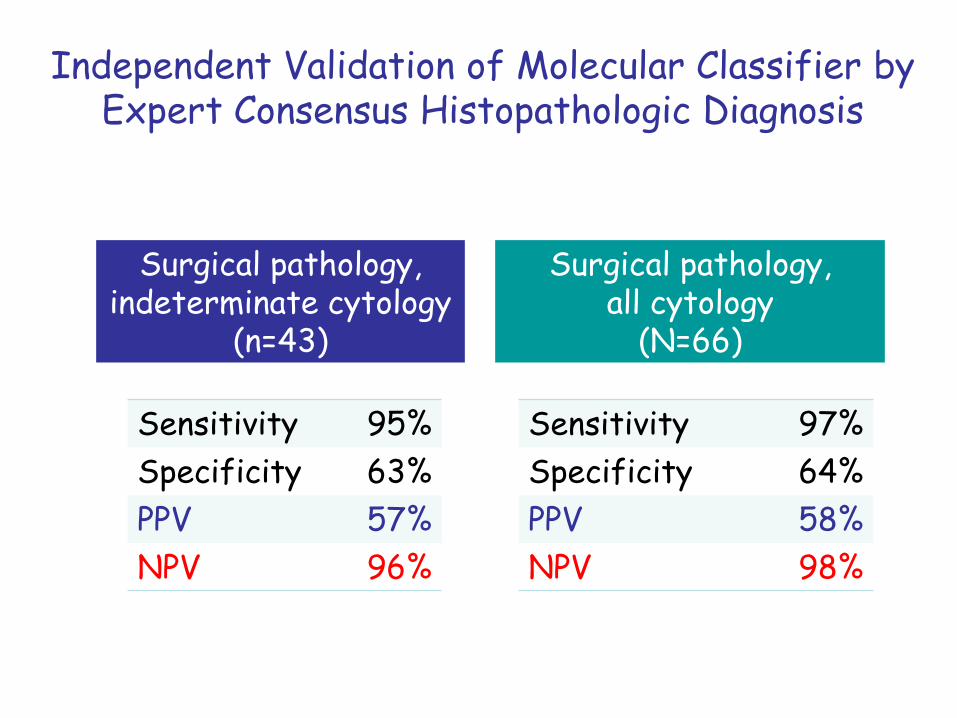
Independent Validation of Molecular Classifier by Expert Consensus Histopathologic Diagnosis
Surgical pathology, indeterminate cytology
(n=43)
Sensitivity 95%Specificity 63%PPV 57%NPV 96%
Surgical pathology,all cytology
(N=66)
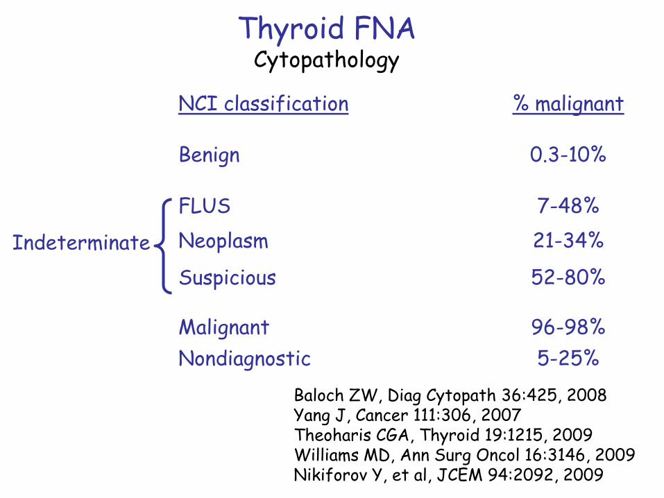
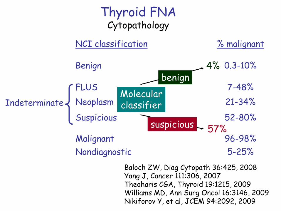
Thyroid FNACytopathology
NCI classification % malignant
Benign 0.3-10%
FLUS 7-48%Neoplasm 21-34%
Suspicious 52-80%
Malignant 96-98%Nondiagnostic 5-25%
Baloch ZW, Diag Cytopath 36:425, 2008Yang J, Cancer 111:306, 2007Theoharis CGA, Thyroid 19:1215, 2009Williams MD, Ann Surg Oncol 16:3146, 2009Nikiforov Y, et al, JCEM 94:2092, 2009
Indeterminate
Thyroid FNACytopathology
NCI classification % malignant
Benign 0.3-10%
FLUS 7-48%Neoplasm 21-34%
Suspicious 52-80%
Malignant 96-98%Nondiagnostic 5-25%
Baloch ZW, Diag Cytopath 36:425, 2008Yang J, Cancer 111:306, 2007Theoharis CGA, Thyroid 19:1215, 2009Williams MD, Ann Surg Oncol 16:3146, 2009Nikiforov Y, et al, JCEM 94:2092, 2009
IndeterminateMolecularclassifier
4%
57%
benign
suspicious
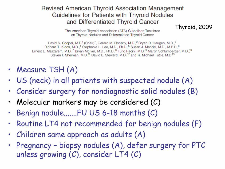
Thyroid, 2009
• Measure TSH (A)• US (neck) in all patients with suspected nodule (A)• Consider surgery for nondiagnostic solid nodules (B)• Molecular markers may be considered (C)• Benign nodule.......FU US 6-18 months (C)• Routine LT4 not recommended for benign nodules (F)• Children same approach as adults (A)• Pregnancy – biopsy nodules (A), defer surgery for PTC
unless growing (C), consider LT4 (C)
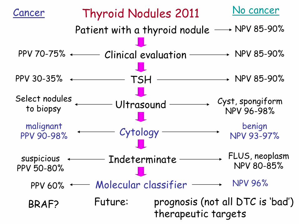
Patient with a thyroid nodule
No cancerCancerNPV 85-90%
Clinical evaluation NPV 85-90%PPV 70-75%
TSH NPV 85-90%PPV 30-35%
Ultrasound Cyst, spongiformNPV 96-98%
Select nodulesto biopsy
Cytology benignNPV 93-97%
malignantPPV 90-98%
Indeterminate FLUS, neoplasmNPV 80-85%
suspiciousPPV 50-80%
Molecular classifier NPV 96%PPV 60%
Future: prognosis (not all DTC is ‘bad’)therapeutic targets
BRAF?
Thyroid Nodules 2011