Embed Size (px)
Citation preview

i
TITLE: In vivo gamma scintigraphy comparison of inhaled corticosteroid monotherapy delivered by pressurised metered dose inhaler with and without a spacer in adolescents with asthma
CANDIDATE:
Natalie Johnson BSc
MEDICAL SCHOOL DIVISION OF PAEDIATRICS
University of Western Australia
This Thesis is presented in partial fulfilment of the requirements for the degree
of Master of Child Health Research of the University of Western Australia.
2017

ii
THESIS DECLARATION
I, Natalie Johnson, certify that:
This thesis has been substantially accomplished during enrolment in the
degree.
This thesis does not contain material which has been accepted for the award of
any other degree or diploma in my name, in any university or other tertiary
institution.
No part of this work will, in the future, be used in a submission in my name, for
any other degree or diploma in any university or other tertiary institution without
the prior approval of The University of Western Australia and where applicable,
any partner institution responsible for the joint-award of this degree.
This thesis does not contain any material previously published or written by
another person, except where due reference has been made in the text.
The work(s) are not in any way a violation or infringement of any copyright,
trademark, patent, or other rights whatsoever of any person.
Written patient consent has been received and archived for the research
involving patient data reported in this thesis.
The following approvals were obtained prior to commencing the relevant work
described in this thesis: Ethics ID 2013105EP
The work described in this thesis was funded by [PMH (now PCH)
Foundation].
Technical assistance was kindly provided by Joyce Wilson and Karen Hindley
to perform the Nuclear Medicine scans described in 2.2.6.3.
This thesis does not contain work that I have published, nor work under
review for publication.
Signature: Date: 1-12-17

iii
ABSTRACT
Spacer use is considered essential for children using inhaled corticosteroids
(ICS) delivered via pressurised metered dose inhaler (pMDI) and recommended
for all ages by the Asthma Foundation of Australia. Spacer recommendation is
primarily to reduce oropharyngeal deposition and associated corticosteroid-
induced side effects. Additionally, spacers minimise the effects of incorrect
inhaler use, which commonly occurs and is associated with reduced medication
compliance. However, anecdotal evidence indicates that spacers are seldom
used as recommended, particularly by adolescents.
The recommendation for spacer use with a pMDI may not be necessary, with
some asthma medications available in Australia generating fine and extrafine
aerosols (fine= <2.5 μm mass median aerodynamic diameter (MMAD),
extrafine= <2.0 μm). These aerosols result in less oropharyngeal deposition
compared to coarse aerosols (>2.5–10 μm MMAD). Thus, the aims of this study
were 1) to establish if spacers are required for use with a pMDI in adolescents
and 2) investigate the effect of pMDI aerosol MMAD on lung distribution, both
with and without a spacer.
To investigate these aims, this thesis evaluates in vivo lung deposition of two
ICS pMDI produced aerosols, one coarse, one fine, each delivered either with
or without a spacer, utilising a radiolabelling technique validated in vitro.
Retrospective analysis to confirm the integrity of previously used radiolabelling
methodology for the drugs (fluticasone propionate formulated as Flixotide®
(coarse, 2.8 μm MMAD) and beclomethasone dipropionate (BDP) formulated as
QVAR® (extrafine, 1.2 μm MMAD)), as used in this study, was also reported on.
We recruited fourteen adolescents aged 13–17 years (6 male, 8 female) with
mild stable asthma for a randomised crossover study on the use of one of the
two pMDIs mentioned above, with or without a spacer. On the study day, after
validation of the radiolabelled drug was confirmed successful, radiolabelled drug
was inhaled by the participant using a single maximal inhalation and breath-hold
technique. Dose of drug deposited was quantified immediately with 2D gamma
scintigraphy. Drug deposition was compared in adolescents inhaling the same

iv
drug with and without a spacer, then further analysed with multivariate statistical
modelling.
Univariate analysis showed no significant difference in total lung deposition with
the coarse aerosol when a spacer was used, compared to pMDI use alone
(p=0.31), or in the group inhaling the extrafine aerosol (p=0.52). There was a
mean decrease of 32% in oropharyngeal deposition when using a coarse
aerosol with a spacer, compared to a decrease of only 10% when using an
extrafine aerosol. The mean ratio of peripheral to central lung deposition
achieved with the extrafine aerosol was not significantly different with and
without spacer use (p=0.82), and similar results were seen in users of the
coarse aerosol (p=0.15), although a more peripheral deposition was seen
visually with the extrafine aerosol. After multivariate analysis, deposition in the
actuator (p=<0.0001), sex, age and BMI were significantly associated with lung
deposition with the extrafine aerosol only.
Dependent on further investigation in a larger cohort we recommend that BDP
as QVAR™ (MMAD 1.2 μm) may be used without a spacer in adolescents,
assuming regular inhaler technique training (every 30 days) with an
appropriately trained clinical professional. Spacer use should still be
recommended for FP as Flixotide®.

v
TABLE OF CONTENTS
1 CHAPTER I: INTRODUCTION .................................................................... 1
1.1 ASTHMA OVERVIEW............................................................................ 1
1.2 DISEASE PROCESS AND PRESENTATION ....................................... 2
1.2.1 DISEASE PROCESS IN ALLERGIC ASTHMA ............................... 2
1.3 TREATMENT OF ASTHMA ................................................................... 3
1.4 MEDICATION FOR ASTHMA TREATMENT ......................................... 4
1.4.1 RELIEVER MEDICATION ............................................................... 4
1.4.1.1 Short acting beta 2 agonists ..................................................... 5
1.4.1.2 Long acting beta 2 agonists ...................................................... 5
1.4.2 PREVENTER MEDICATION ........................................................... 5
1.4.2.1 Non-corticosteroid therapy ........................................................ 5
1.4.2.2 Oral corticosteroids ................................................................... 6
1.4.2.3 Inhaled corticosteroids .............................................................. 6
1.5 ORALLY INHALED DRUG THERAPIES: ADVANTAGES AND
DISADVANTAGES .......................................................................................... 9
1.5.1 NEBULISERS ................................................................................. 9
1.5.2 DRY POWDER INHALERS ............................................................. 9
1.5.3 BREATH ACTUATED INHALERS ................................................. 10
1.5.4 PRESSURISED METERED DOSE INHALERS ............................ 10
1.5.4.1 PMDI device compliance ........................................................ 11
1.5.4.2 Spacer use.............................................................................. 11
1.5.4.3 Spacer non-compliance .......................................................... 12
1.5.4.4 Drug formulations available by pMDI ...................................... 12
1.5.4.5 Inhaled corticosteroids delivered by pressurised metered dose
inhaler (pMDI) in Australia ...................................................................... 14
1.6 ADHERENCE TO MEDICATION ......................................................... 14

vi
1.6.1 NEAR FATAL ASTHMA AND NON-COMPLIANCE ...................... 15
1.6.2 MEDICATION COMPLIANCE IN ADOLESCENT PATIENTS ....... 15
1.7 METHODS OF ASSESSMENT OF INHALER USE AND EFFICACY . 17
1.7.1 THEORETICAL IN VITRO ASSESSMENT (MATHEMATICAL
MODELLING) ............................................................................................ 17
1.7.2 IN VITRO LABORATORY STUDIES ............................................. 18
1.7.3 IN VIVO DRUG DELIVERY STUDIES .......................................... 19
1.7.4 IN VIVO LUNG DEPOSITION STUDIES ...................................... 19
1.7.4.1 Lung deposition studies using radiolabelled Technetium........ 20
1.7.4.2 Validation of drug radiolabelling techniques ........................... 20
1.7.4.3 International guidelines for interpreting successful
radiolabelling of inhaled drugs ............................................................... 21
1.7.4.4 Importance of international standards for inhaled radiolabelled
preparations ........................................................................................... 21
1.7.4.5 Gamma scintigraphy to assess in vivo lung deposition........... 23
1.7.4.6 Single photon emission computed tomography 3D Imaging ... 24
1.7.4.7 Positron Emission Tomography Imaging ................................ 24
1.7.5 CLINICAL OBSERVATION OF INHALER TECHNIQUE ............... 24
1.7.6 CLINICAL TRIALS ........................................................................ 24
1.8 SUMMARY AND THESIS AIMS .......................................................... 25
1.8.1 BACKGROUND SUMMARY ......................................................... 25
1.8.2 PRIMARY STUDY OBJECTIVES ................................................. 25
1.8.3 PRIMARY AIMS AND HYPOTHESES .......................................... 26
1.8.3.1 Aims ........................................................................................ 26
1.8.3.2 Hypotheses ............................................................................. 26
1.8.4 SECONDARY STUDY OBJECTIVE ............................................. 27
2 CHAPTER II: MATERIALS AND METHODS ............................................. 28
2.1 MATERIALS ........................................................................................ 28
2.1.1 MATERIALS AND EQUIPMENT ................................................... 28
vii
2.1.2 MATERIALS AND EQUIPMENT PREPARATION EACH STUDY
DAY 28
2.1.2.1 Reagents preparation (Appendices 5.1) ................................. 28
2.1.2.2 Preparation of spacers for study day use with radiolabelled
pMDI 28
2.1.2.3 Pneumotachometer calibration ............................................... 29
2.1.2.4 Airflow calibration, Anderson Cascade Impactor (ACI) ........... 29
2.2 METHODS ........................................................................................... 29
2.2.1 RADIOLABELLING METHOD ....................................................... 29
2.2.1.1 Total radiation dose given to participants ............................... 30
2.2.1.2 Radiation clearance ................................................................ 30
2.2.1.3 Determination of radioactivity dispensed per pMDI actuation . 30
2.2.1.4 Radiation dose accountability ................................................. 30
2.2.2 PARTICLE SIZE DISTRIBUTION ASSESSMENT OF LABELLED
AND UNLABELLED DRUG AND DISTRIBUTION OF RADIOTRACER .... 31
2.2.2.1 Ultraviolet visible spectrophotometry to interpolate drug
concentrations ........................................................................................ 32
2.2.2.2 Standard curve generation to interpolate drug concentrations 32
2.2.3 PRE-STUDY QUALITY CHECK OF RADIOLABELLING METHOD
(VALIDATION) ........................................................................................... 34
2.2.3.1 In vitro validation analysis using international guidelines as
quality criteria ......................................................................................... 34
2.2.4 RETROSPECTIVE ANALYSIS OF PAST RADIOLABELLED
VALIDATION FOR DRUGS AS USED IN THIS STUDY............................ 35
2.2.5 IN VIVO AEROSOL DEPOSITION STUDY METHODS ................ 35
2.2.5.1 Study Design .......................................................................... 35
2.2.5.2 Participants ............................................................................. 37
2.2.5.3 Participant randomisation and allocation ................................ 37
2.2.6 ASSESSMENTS ........................................................................... 38

viii
2.2.6.1 Screening questionnaire ......................................................... 38
2.2.6.2 Inhalation technique................................................................ 40
2.2.6.3 Gamma scintigraphy to assess drug deposition in vivo .......... 40
2.2.6.4 Gamma scintigraphy image acquisition and analysis ............. 40
2.2.6.5 Using gamma scintigraphy data to calculate an attenuation
corrected interpretation of emitted radiation in each region of interest
(ROI) 42
2.2.6.6 Camera efficiency ................................................................... 44
2.2.7 STATISTICAL ANALYSIS ............................................................. 44
2.2.7.1 Power calculations .................................................................. 45
3 CHAPTER III: RESULTS ........................................................................... 46
3.1 IN VITRO VALIDATION OF RADIOLABELLED DRUG PREPARATION
TO DETERMINE IN VIVO REPORTING ....................................................... 46
3.2 RETROSPECTIVE ANALYSIS OF RADIOLABELLED FP AND BDP . 49
3.3 IN VIVO DRUG DEPOSITION STUDY RESULTS .............................. 54
3.3.1 STUDY POPULATION .................................................................. 54
3.3.1.1 Screening questionnaire data ................................................. 56
3.3.2 SHOT WEIGHT DELIVERED BY PMDI ........................................ 57
3.3.3 GAMMA SCINTIGRAPHY IN VIVO DEPOSITION AFTER
INHALATION OF RADIOLABELLED DRUG ............................................. 57
3.3.4 STATISTICAL ANALYSIS OF GAMMA SCINTIGRAPHY
DEPOSITION DATA AFTER TRANSFORMATION ................................... 61
4 CHAPTER IV: DISCUSSION ..................................................................... 62
4.1 CONCLUSION ..................................................................................... 67
BIBLIOGRAPHY ............................................................................................... 68
5 APPENDICES ........................................................................................... 78
5.1 Materials and equipment for radiolabelled drug preparation,
administration and gamma scintigraphy ........................................................ 78

ix
5.2 Materials and equipment for assessment of drug particle size
distribution ..................................................................................................... 79
5.3 Consumables for radiolabelling and gamma scintigraphy on study day
80
5.4 Parent information and consent form ................................................... 81
5.5 Physical examination ........................................................................... 87
5.6 Vital sign measurements ...................................................................... 88
5.7 Screening questionnaire ...................................................................... 89
5.8 Look up table ....................................................................................... 90

x
LIST OF TABLES
Table 1-1 A Reflection on Guidelines for Validation Practices. Table
shows key developments of the validation guidelines since their inception. ..... 22
Table 2-1 User interpretation of adherence to medication survey
responses as described in 2.2.6.1. ................................................................ 39
Table 3-1 Fluticasone propionate particle size distribution assessment
comparisons. Ratios confirmed that validation was successful when within the
range of 0.85–1.18 or ±2% for the groups that had under 10% of the total drug.
.......................................................................................................................... 47
Table 3-2 Beclomethasone di-propionate particle size distribution
assessment comparison. Ratios confirmed that validation was successful
when within the range of 0.85–1.18 or ±2% for the groups that had under 10%
of the total drug. ................................................................................................ 48
Table 3-3 Beclomethasone di-propionate particle size distribution
assessment comparison. Ratios confirmed that validation was successful
when within the range of 0.85–1.18 or ±2% for the groups which had under 10%
of the total drug. ................................................................................................ 53
Table 3-4 Full cohort (n=14) demographics ...................................................... 54
Table 3-5 Group demographics separated for BDP and FP ........................ 55
Table 3-6 Comparison of mean (SD) deposition data for purpose of cross
study comparison. Deposition values are displayed for each region of interest
as percent of ex-valve or ex-actuator ................................................................ 59
Table 3-7 Analysis of regional deposition data after transformation. Lung
deposition from each drug was analysed: with a single variable (Spacer etc.), in
a full model to adjust for confounders, then variables were eliminated via
backward selection at 0.05 level of significance, until the final model was left
(exit point p-values displayed). ......................................................................... 61
xi
LIST OF FIGURES
Figure 1-1 Classical disease model of Th2 mediated asthma. Aeroallergen
incites Th2 cytokine response, causing inflammation, damage and airway
remodelling, airways become hyperresponsive and broncho-constriction results.
Picture retrieved from; (Anderson, 2008). ........................................................... 2
Figure 1-2 Presentation of regional airway deposition relative to Anderson
Cascade Impactor stage size range (28.3 LPM) adapted from (Dunbar &
Mitchell, 2005). Showing approximate cut offs for Anderson Cascade Impactor
stages Throat, Jet, and stages 0–Filter. ............................................................ 13
Figure 2-1 Comparison of original and improved assay for a standard
curve generated for fluticasone propionate drug interpolation on semi log
(x) scale. Data show absorbance of light at λ=247 nm of fluticasone propionate
standards prepared for the original assay, for each concentration (inverted
triangles); 1 μg/mL, 2 μg/mL, 5 μg/mL, 10 μg/mL and 20 μg/mL, and with
optimised detection limits (circles); 0.625 μg/mL, 1.25 μg/mL, 2.5 μg/mL, 5
μg/mL,10 μg/mL, 20 μg/mL and 40 μg/ mL. ...................................................... 33
Figure 2-2 Comparison of original and improved assay for a standard
curve generated for beclomethasone diproprionate drug interpolation on a
semi log (x) scale. Data show absorbance of light at λ=238 nm of
beclomethasone diproprionate standards prepared for the original assay, for
each concentration (inverted triangles); 1 μg/mL, 2 μg/mL, 5 μg/mL, 10 μg/mL,
and 20 μg/mL, and with optimised detection limits (circles); 0.125 μg/mL, 0.25
μg/mL, 0.5 μg/mL, 1 μg/mL, 2 μg/mL, 5 μg/mL, 10 μg/mL, 20 μg/mL 30 μg/mL
and 40 μg/mL. ................................................................................................... 33
Figure 2-3 A randomised cross over design study, where each participant
completed one of two possible arms. .......................................................... 36
Figure 2-4 Subjective delineation of the lungs depicting method for
quantifying central and peripheral regions as described in 2.2.6.4. The total
region of the lung (orange/red) is drawn around the whole lung (green) in order
to measure a third of the length and half the width (blue box). ......................... 41
Figure 2-5 Depiction of peripheral to central ratios. Central region of lung
depicted inside blue box. Anything not inside this region, but within the lung
region (red/orange delineation) was considered peripheral deposition. ............ 41

xii
ACKNOWLEDGEMENTS
Thank-you to the participants, their parents and to principal investigator Ms
Charlotte Allen for organising their participation and generously giving time to
facilitate the study to completion. Thank-you Associate Professor Sunalene
Devadason for acquiring funding to support the study, and to the Princess
Margaret Hospital Foundation for providing it. This research was supported by
an Australian Government Research Training Program (RTP) Scholarship.
Thank you to my supervisors Dr William Ditcham, Dr Stephanie Trend and
Associate Professor Sunalene Devadason for guiding, advising and providing
editorial assistance throughout my Thesis writing process. Additional thanks
firstly to Sunalene for helping me navigate through my course easily, and
advising me with her extensive knowledge of the field in the international and
national environments. Secondly, thanks to Dr Stephanie Trend for role
modelling and encouraging my involvement in the larger scientific community,
and for her counsel in times of need. Finally, thanks to Dr William Ditcham for
his consistently generous advice, counsel, encouragement and support, without
which this Thesis would not be possible.
I’d like to thank my colleagues in the Epithelial and Vaccine Trials Research
groups for including me in their groups and providing opportunity for, and
collegial advice when presenting. Thank-you particularly to Dr Anthony Kicic, Dr
Liz Starcevich and Ms Tabitha Woodman for providing moral support and
encouragement and to Tabitha for her editorial assistance and guidance.
Thanks to my colleague and friend Ms Leesa Harris, for thesis and presentation
proofing, support and encouragement.
Finally and most importantly, thank-you to my husband, parents, and friends
Mrs Danielle Newman and Ms Claire Stade for listening to me and believing in
me. Thank-you to my father Mr Philip Johnson for his mathematical assistance
throughout my statistics units and to Mrs Danielle Newman for editorial
assistance on my final Thesis version. Thank-you to my father, my cousin Dr
Mark Kitchen, my late aunt Ms Janice Kitchen, and uncle Dr Alan Kitchen for
role modelling and encouraging continued education in medical science and
engineering.

xiii
STATEMENT OF CANDIDATE CONTRIBUTION
The candidate performed all of the work described in this thesis, except where
contributions of other individuals have been acknowledged below. Estimated
percentages of contributions (where not 100%) are shown below.
Funding for the study was obtained from the PMH foundation by Associate
Professor Devadason.
Recruitment of participants was completed, and consent obtained, by Ms
Charlotte Allen
Validation of radiolabelled preparations, and preparation of the radiolabelled
study drug each day was completed by the candidate, with assistance from Dr
Ditcham (45%)
Participant technique training and clinical assessment was performed by Ms
Charlotte Allen
Inhalation patterns were obtained from participants by the candidate with some
initial assistance from Dr Ditcham (10%)
Participants were instructed how to inhale the study drug by Dr Ditcham (95%)
Gamma scintigraphy imaging was performed by Ms Joyce Wilson, Ms Karen
Hindley, Ms Jill Summers, and Mr Simon Ferrero.
Experimental data obtained from gamma scintigraphy was processed by the
candidate with assistance and guidance from Dr Ditcham (30%)
Data analysis was carried out by the candidate with assistance from Ms Charley
Budgeon at the Centre for Applied Statistics, University of Western Australia.
The candidate wrote all sections of the thesis, with critical feedback and editing
provided by Associate Professor Devadason (35%) and Drs Trend (35%) and
Ditcham (30%).
Critical feedback that contributed to the discussion and conclusion was given by
examiners, Drs Fink, and Shah.

xiv
PUBLICATIONS ARISING FROM THIS THESIS
Publications in preparation
Does improved delivery of ICS monotherapy to adolescents with asthma still
require spacer use?
Oral Presentations/Abstracts
Annual Rottnest Respiratory Symposium, New Investigator Award
Session Nov 2016: Spacer use in asthmatic adolescents
Annual Scientific Meeting, Thoracic Society of Australia and New Zealand
March 2017: The effect of particle size delivered by pressurised meter dose
inhaler (pMDI) in asthmatic adolescents

xv
DEFINITIONS
Adherence; adhering to the recommended regimen and use of the prescribed
medication
Coarse aerosol; an aerosol containing a mass median aerodynamic particle
size distribution of ≥2.5 μm–10 μm
Compliance; complying with the intended use of a device
Extrafine aerosol; an aerosol containing a mass median aerodynamic particle
size distribution of ≤2 μm but not ≤ 0.1 μm
Fine aerosol; an aerosol containing a mass median aerodynamic particle size
distribution of ≤2.5 μm but not ≤ 0.1 μm
Ultrafine aerosol; an aerosol containing a mass median aerodynamic particle
size distribution of ≤0.1 μm

xvi
ABBREVIATIONS
ACI Anderson cascade impactor
APSD Aerodynamic particle size distribution
ARPANSA Australian Radiation Protection and Nuclear Safety Agency
BDP Beclomethasone diproprionate
FP Fluticasone propionate
LUT Look up table
PBS Pharmaceutical Benefits Scheme
pMDI Pressurised metered dose inhaler
PMH Princess Margaret Hospital
PSD Particle size distribution
ROI Region of interest
99mTc Technetium99m
TPA Tetraphenylarsonium chloride
TGA Therapeutic Goods Administration
WHO The World Health Organisation
GINA The Global Initiative for Asthma

xvii
UNITS
LPM Litres per minute
MBq Megabecquerel
mSv Millisievert
cps Counts per second
rpm revolutions per minute
s seconds
w/v weight for volume
FEV1 one second forced expiratory volume
BMI body mass index


1
1 CHAPTER I: INTRODUCTION
1.1 ASTHMA OVERVIEW
Asthma is defined as a single disease with variable phenotypes, characterised
by chronic airway inflammation and respiratory symptoms which have multiple
triggers and vary in severity and frequency (Program., 2017). The variable
presentation of symptoms give rise to asthma being under or over diagnosed,
although when appropriately diagnosed and treated, symptoms and severity
can be reduced with medication in most individuals (Aaron et al., 2017; Boulet,
FitzGerald, & Reddel, 2015; Rabe et al., 2004). According to prescription
dispensing data, in Australia, asthma is over diagnosed, thus over treated in
these individuals, and in those with correct diagnosis, undertreated due to lack
of adherence to medication regime (AIHW: Correll PK, 2015). Asthma is
estimated to contribute to one in every 250 deaths worldwide, but deaths
resulting from exacerbation can be preventable in most individuals when
treatment is adhered to (Masoli, Fabian, Holt, Beasley, & Global Initiative for
Asthma, 2004). Higher levels of adherence to medication are associated with
decreased risk of exacerbations, however medication adherence is generally
low (Engelkes, Janssens, de Jongste, Sturkenboom, & Verhamme, 2015). Low
adherence to medication leads to decreased asthma control levels, the latter
which are already low and likely to be over-estimated (AIHW: Correll PK, 2015),
so it is critical to find effective treatment that is easy to implement in practice.
The prevalence of asthma in Australia is high compared to international rates;
one in ten adult Australians have asthma, and the direct health costs of asthma
to the Australian economy were estimated to be AUD 1.2 billion in 2015
(Australian Centre for Asthma Monitoring, 2011; National Asthma Council
Australia, 2016a). In 2015 there were 421 asthma-related deaths in Australia
although the mortality rate has decreased over the last 10 years as has the
prevalence of asthma in children and young adults (15–34), but remained stable
in adults over 35 (Australian Centre for Asthma Monitoring, 2011). Childhood
asthma is the leading non-communicable disease worldwide and prevalence in
Australian children and adolescents (ages 5–17) still remains high compared to
other age groups (Australian Centre for Asthma Monitoring, 2011; World Health

2
Organisation, 2013). In Australia, more males than females have asthma during
childhood (ages 0–14), similar levels of both sexes experience asthma during
adolescence, and from adulthood onwards more females than males have
asthma (ages 25+) (Australian Centre for Asthma Monitoring, 2011).
1.2 DISEASE PROCESS AND PRESENTATION
The lack of consensus on a single, environmental or genetic, pathophysiological
aetiology for asthma has led to reassessment of this condition as a syndrome
(Lötvall et al., 2011; Program., 2017). Asthma disease phenotypes are
important clinically, reduce complexity of diagnosis, and improve asthma
management (Agache, Akdis, Jutel, & Virchow, 2012), however it is now
understood that phenotypes are not directly related to disease process, and
disease variants exist (Lötvall et al., 2011). Common clinically identifiable
phenotypes include; allergic asthma, non-allergic asthma, late-onset asthma,
asthma with fixed airflow limitation, and asthma with obesity (Program., 2017). It
is estimated that approximately 50% of asthma cases involve the allergic
asthma phenotype which is considered an early onset, or childhood, asthma,
that can persist into adulthood (Haldar et al., 2008; Wenzel, 2012; Woodruff et
al., 2009).
1.2.1 DISEASE PROCESS IN ALLERGIC ASTHMA
The T-helper type two (Th2) response (Figure 1-1) appropriately describes a
proposed biological mechanism of the allergic asthma phenotype in adults and
children (Anderson, 2008; Wenzel, 2012).
Figure 1-1 Classical disease model of Th2 mediated asthma.
Aeroallergen incites Th2 cytokine response, causing inflammation, damage and airway remodelling,
airways become hyperresponsive and broncho-constriction results. Picture retrieved from; (Anderson,
2008).
3
A typical immune response to aeroallergen results in Th2 specific cytokine
production; IL-4, IL-5, and IL-13 (Grünig et al., 1998; Watanabe et al., 1997).
These inflammatory cytokines induce various forms of inflammation; recruitment
of eosinophils to the airways, B-cell class switching to produce IgE, and mucus
production in airway epithelium via goblet cells. Damage, resulting from
inflammation, leads to airway hyperresponsiveness and bronchoconstriction,
and once established, airway hyperresponsiveness can be worsened by
inflammation (Denham et al., 2008; Henderson et al., 2003; Winkler & Venegas,
2007). Long term inflammation can lead to airway remodelling, and thickening
of the sub-epithelial basement membrane that contributes significantly to
asthma pathophysiology (Brusselle, Kips, Joos, Bluethmann, & Pauwels, 1995;
Dabbagh et al., 1999; Manetsch et al., 2012; Snapper, Finkelman, & Paul,
1988). Individuals with an allergic asthma phenotype, defined as having
eosinophilic airway inflammation, usually with an early onset, respond well to
inhaled corticosteroid (Lötvall et al., 2011; Program., 2017; Woodruff et al.,
2009).
1.3 TREATMENT OF ASTHMA
Initial treatment recommendations for those not already using preventer
treatment are determined by disease presentation. For children aged six and
over, disease presentation is initially classed as: infrequent intermittent,
frequent intermittent, or persistent asthma (mild, moderate or severe) (National
Asthma Council Australia, 2016a; Van Asperen PP, Mellis CM, Sly PD, & C.,
2010). These categories are defined by the pattern and intensity of the
presenting symptoms. Persistent asthma is scaled by percent predicted FEV1
score: ≥80%=mild, ≥60% to <80%=moderate, and ≤60%=severe. It is estimated
that 74% of young children have infrequent intermittent asthma, which does not
require inhaled corticosteroid (ICS) use (Van Asperen PP et al., 2010). Initial
treatment recommended for frequent intermittent and mild persistent asthma, is
daily low dose ICS monotherapy, if no clinical response is seen after an initial
four week trial of non-corticosteroid therapy. Moderate to severe asthma
symptoms are initially recommended a low dose inhaled corticosteroid, to be
reviewed after four weeks (Van Asperen PP et al., 2010).
Adolescents from 14–16 years onwards are recommended to be treated as
adults for diagnosis and medical management purposes, with additional
4
consideration for confidentiality and psychosocial status (National Asthma
Council Australia, 2016a). Acknowledged difficulties exist however, when
investigating symptoms in adolescents due to their denial or overplay of
symptoms (National Asthma Council Australia, 2016a). Additionally this
population may be less likely to adhere to medication regimens (Dima et al.,
2015) and special consideration in treatment and diagnosis must therefore be
taken. A particular consideration for mental health issues, increased likelihood
of risk-taking behaviours, cigarette smoking and/or exposure to second hand
smoke is recommended in the diagnostic approach (National Asthma Council
Australia, 2016a). Spirometry can be used to confirm a diagnosis even if
asthma was experienced during childhood (National Asthma Council Australia,
2016a).
Adults presenting with asthma symptoms are prescribed medication according
to symptom frequency (National Asthma Council Australia, 2016a). The initial
treatment for those experiencing symptoms more than twice per month is to
prescribe a short acting beta-2 agonist and regular, low dose, ICS
monotherapy. Combination ICS/Long acting beta-2 agonists are optionally
recommended for those additionally suffering persistent daytime symptoms. A
regular high dose ICS monotherapy is recommended for very uncontrolled
asthma (National Asthma Council Australia, 2016a).
1.4 MEDICATION FOR ASTHMA TREATMENT
The mainstay of treatment for asthma is drug therapy consisting of preventative
medication to decrease the airway inflammation, and reliever medication to
relieve airway constriction (National Asthma Council Australia, 2016a).
Medication can be delivered by oral tablet, or as orally inhaled drug direct to the
lungs, with dose and frequency adjusted according to individual symptoms and
severity (National Asthma Council Australia, 2016a). Nasal inhalation therapy is
not used for asthma although those also experiencing allergic rhinitis do receive
some improvement of asthma specific outcomes (Lohia, Schlosser, & Soler,
2013).
1.4.1 RELIEVER MEDICATION
Reliever medication is given to reduce airway constriction, which causes
wheezing and breathlessness during an asthma attack. Short acting beta-2
5
agonists are the main bronchodilators prescribed to treat this condition in
Australia, on a taken as-needed basis (National Asthma Council Australia,
2016a).
1.4.1.1 Short acting beta 2 agonists
The most commonly recommended beta-2 agonists in Australia are salbutamol
delivered by pMDI, suitable for all ages, and terbutaline delivered by breath
actuated inhaler, suitable for all ages over six years. Terbutaline, due to the
type of delivery device, requires a threshold acceleration for activation that is
not possible for many small children to achieve (National Asthma Council
Australia, 2016a).
1.4.1.2 Long acting beta 2 agonists
Long acting beta-2 agonists (LABA) are usually formulated in combination with
inhaled corticosteroids and not recommended at all for treatment of asthma in
children 0–5 years, where studies are absent (Program., 2017; Van Asperen PP
et al., 2010). In adults and adolescents ICS/LABA combination is generally
recommended as a step up treatment for those whose symptoms both persist
despite daily low-dose ICSs, and are not due to lack of adherence or poor
technique (Program., 2017). Certain combination LABAs have been shown
effective in adults, but not in children, to reduce the risk of flare ups compared
with inhaled corticosteroid treatment alone (Chauhan, Chartrand, Ni Chroinin,
Milan, & Ducharme, 2015; Ducharme, Ni Chroinin, Greenstone, & Lasserson,
2010).
1.4.2 PREVENTER MEDICATION
There are two main types of preventer medications, non-corticosteroid therapy
and corticosteroid therapy (CS), which can be oral, or inhaled (ICS). Preventer
medication acts to target airway inflammation, and with regular, correct use,
reduces the risk of exacerbations and hospitalisation.
1.4.2.1 Non-corticosteroid therapy
Non-steroid preventer therapy is more commonly used in children or can be
used to prevent exercise-induced bronchoconstriction, although pre-dosing
before exercise with a short acting beta-2 agonist is the preferred treatment
(National Asthma Council Australia, 2016a). In children, non-steroidal therapy is
initially prescribed for 2–4 weeks and if no clinical response is seen, it is

6
recommended to proceed to inhaled corticosteroid monotherapy (National
Asthma Council Australia, 2016a; Van Asperen PP et al., 2010).
Anti-IgE monoclonal antibody therapy (Omalizumab, Xolair®) has only recently
been made available by prescription in Australia for children aged 6–12 years
(2016), although was previously available to adolescents and adults. It is
recommended as an add-on therapy in those who have documented
exacerbations despite daily high dose inhaled corticosteroid treatment, often
those with moderate-severe persistent allergic asthma (AIHW: Correll PK,
2015).
1.4.2.2 Oral corticosteroids
Oral corticosteroids are currently used only in short courses (less than two
weeks) for acute asthma management, as this duration is considered safe and
effective, excluding children aged 0–5 years where efficacy has not been
validated (National Asthma Council Australia, 2016a). Oral corticosteroid
treatment with prednisolone is recommended in Australia for 5–10 day courses,
for acute asthma flare-ups, usually after presentation at an emergency
department (National Asthma Council Australia, 2016a). Due to the benefits of
ICS, they largely replaced oral corticosteroids as a mainstream treatment in the
80’s (Petrisko, Skoner, & Skoner, 2008). Limitations of oral corticosteroids
include greater systemic side effects, and higher doses required to be effective
compared to ICS (Davies, Stampone, & O'Connor, 1998).
1.4.2.3 Inhaled corticosteroids
Inhaled corticosteroids (ICS) are the mainstay of asthma treatment in Australia
and in 2013 they were dispensed to 6.9% of the population (AIHW: Correll PK,
2015) (National Asthma Council Australia, 2016a). If optimally administered,
some orally inhaled therapies, usually those with a fine (<2.5 μm mass median
aerodynamic diameter, MMAD) aerosol, can deliver a higher fraction of total
drug dose directly to the lungs, and a correspondingly lower fraction to the
oropharyngeal (C. L. Leach, Kuehl, Chand, & McDonald, 2015). ICSs therefore
require a smaller therapeutic dose to be effective when compared to oral
corticosteroids. Glucocorticoid receptors are present in almost every cell type in
the body and so the targeted delivery of inhaled drugs to these receptors in lung
epithelium reduces systemic side effects caused by non-specific exposure

7
(Barnes, 2004). Additionally when using a drug with a fine (<2.5 µm) MMAD, the
theoretical assumption is a higher fraction of total dose is depositing in the
peripheral airways (<2 µm diameter), the main site of airway inflammation in
asthma (Hamid et al., 1997; Tsuda, Henry, & Butler, 2011).
Various corticosteroids drugs are available in Australia for inhalation and are
prescribed either as monotherapy, or as combined therapy with LABA in more
severe persistent cases. Equivalence has been shown therapeutically between
drug variations, although sub-optimal delivery affects therapeutic efficacy, and
side effects may differ between drugs (Chet Leach, Colice, & Luskin, 2009;
Lipworth, 1999).
LOCAL SIDE EFFECTS
A recognised local side effect of ICS is oral thrush, which can be avoided
through mouth rinsing immediately after administration. Hoarseness can be
experienced in adults, dependent on device and formulation used, but rarely
reported in children using a spacer and pMDI (National Asthma Council
Australia, 2016a). Aerosol formulations with a pH under 5.5 pH, usually dry
powders, can dissolve tooth enamel, particularly in children (National Asthma
Council Australia, 2016a).
SYSTEMIC SIDE EFFECTS
Systemic side effects can occur due to gastrointestinal or lung absorption and
are dose, treatment, and treatment duration dependent (Lipworth, 1999; Loke,
Blanco, Thavarajah, & Wilson, 2015; National Asthma Council Australia, 2016a;
Petrisko et al., 2008; Zhang, Prietsch, & Ducharme, 2014). If the duration of a
low to medium daily dose ICS treatment continues for a year, growth can be
reduced at a rate of 0.48 cm during that year, however growth suppression
decreases in the subsequent years if treatment continues, and there is little
effect on final adult height of children (Loke et al., 2015; Zhang et al., 2014).
Upper dose limits in children for ICS in Australia are 500 μg for fluticasone
propionate (FP), and 400 μg for beclomethasone di-propionate (BDP), much
lower than the daily dose that causes marked adrenal suppression (750 μg FP,
or 1500 μg BDP) (Lipworth, 1999; National Asthma Council Australia, 2016a;
Program., 2017). Doses above these limits do not equate to increased

8
therapeutic effect, are not prescribed, and adhering to prescribed dose limits
strongly encouraged, so adrenal suppression due to high daily dosing is unlikely
to occur (National Asthma Council Australia, 2016a; Randell et al., 2003; Van
Asperen PP et al., 2010).
To reduce potential side effects some investigations are focusing on increasing
drug receptor site affinity and absorption time in the lung epithelium (Salter et
al., 2007; Van Den Berge et al., 2010). An ideal delivery is where action is
exerted in the peripheral airways <2 μm (target region) and excess drug does
not move into the systemic circulation where side effects can potentiate (Hamid
et al., 1997; Van Asperen PP et al., 2010).
CORTICOSTEROID INSENSITIVITY
There are small subsets of the asthmatic population, usually with severe
asthma, who are corticosteroid insensitive (Adcock & Lane, 2003). Investigating
the mechanism of action of corticosteroids has been a focus for this reason
(Kobayashi, Mercado, Barnes, & Ito, 2011; Nair et al., 2017; Quante et al.,
2008). The mechanism of action by which corticosteroids treat airway
inflammation is not yet fully understood, although recent studies have
elucidated some mechanisms. Corticosteroids act, in part, by decreasing
transcription of inflammatory cytokines, including IL-4, IL-5 and IL-13 (Barnes,
1998). Furthermore, they supress inflammation in airway smooth muscle, by up-
regulation of an endogenous mitogen-activated protein kinase (MAPK) inhibitor,
which contributes to the repression of IL-6 secretion (Manetsch et al., 2012;
Quante et al., 2008). Glucocorticoid activation of Kruppel-like factor 15
represses airway smooth muscle hypertrophy (Sasse et al., 2017).
Severe asthma can be corticosteroid insensitive, which similarly manifests in
Chronic Obstructive Pulmonary Disease and may be reflective of a different
disease variant entirely (Barnes, 2013). Increasing evidence shows that true
severe asthma is distinct in pathophysiological presentation to mild-moderate
asthma, and most likely established in early life, remaining unchanged over time
(Anderson, 2008; Phelan, Robertson, & Olinsky, 2002; Wenzel et al., 1999).
Despite true severe asthma being pathologically different in presentation, mild
to moderate asthma can deteriorate to present as severe when prescribed

9
treatment is not followed, and severe-untreated asthma also exists in some
populations (Chung et al., 2014; Romagnoli et al., 2007).
1.5 ORALLY INHALED DRUG THERAPIES: ADVANTAGES AND
DISADVANTAGES
Due to the lung site specificity and minimised side effects, delivery via oral
inhalation is most common for both preventative and reliever medications.
There are various delivery devices available, each with their benefits and
limitations. Children under five years of age, who have trouble coordinating
inhalation instructions, are recommended to use a facemask in addition to an
inhaled therapy (National Asthma Council Australia, 2016a).
1.5.1 NEBULISERS
Nebulisers are devices that produce inhalable drug as a “wet’’ aerosol,
administered via either the mouth or nose. The aerosol is commonly produced
by a jet nebuliser, where drug solution is drawn through an orifice by a pressure
drop, generated by a venturi and a compressor, to provide the driving airflow.
Alternatively, pressure drop generation can be via a vibrating mesh nebuliser,
where the aerosol is generated by the vibration of a thin flexible piece of metal
perforated by many tiny holes, and driven by the vibration of a piezo-electric
crystal. Most machines are either costly, in the case of vibrating mesh
nebulisers, or noisy and time consuming to use, in the case of jet nebulisers.
Additionally, those devices that deliver drug via the nose are often
uncomfortable to administer, especially with young children.
Nebulisers are not recommended as an initial treatment for asthma in Australia,
only to be considered if the child cannot be taught to use an inhaler, usually
relevant to children under five years (National Asthma Council Australia,
2016a).
1.5.2 DRY POWDER INHALERS
Dry powder inhalers (DPIs) are devices that aerosolise particles, after actuation
by a deep in-breath, and cannot be used by young children (under 5 years) or in
those who cannot generate adequate inspiratory flow to de-aggregate the
powder, activating the device (Federico Lavorini, 2013). They are
recommended as a secondary option, after pMDI’s, for children over 6 years

10
(National Asthma Council Australia, 2016a). Similar to pMDIs, the lung
deposition of a DPI depends on inhalation parameters and device design. DPIs
exhibit similar issues to pMDIs, with oropharyngeal deposition although unlike a
pMDI these cannot be avoided with addition of a spacer device and instead
would require a slower inhalation flow for activation if to reduce the risk of
oropharyngeal deposition (Federico Lavorini, 2013). Another disadvantage is
that not all DPIs provide multiple dose capacity. Those that do are either
designed as multiple dose reservoirs (i.e. Turbuhaler®) or as multiple dose units
(i.e. Accuhaler™, Elliptor®). The former do not always provide consistency of
dosage, and the latter do but are more expensive (Federico Lavorini, 2013).
1.5.3 BREATH ACTUATED INHALERS
Breath actuated inhalers require a threshold inspiratory flow to activate them,
and therefore may not use be used optimally or consistently by young children,
whom are instead prescribed pMDIs with spacers (National Asthma Council
Australia, 2016a). They are recommended for patients who have difficulty using
their hands to connect spacer devices to their pMDI (National Asthma Council
Australia, 2016a). Breath actuated inhalers can be used with a spacer, however
a much larger inspiratory flow must be used, to adequately draw airflow through
the additional spacer device, and thus limits it general use and recommendation
(National Asthma Council Australia, 2016a). Additionally, problems potentiate
with non-autonomous operation; increased oropharyngeal deposition occurs if
breathing ceases after device activation usually if the aerosol plume is
distasteful or cold when hitting the back of the throat.
1.5.4 PRESSURISED METERED DOSE INHALERS
The most commonly used device to administer orally inhaled products is the
pressurised metered dose inhaler (pMDI). It is cheap and portable, and reported
to have equivalent or improved delivery to other available devices, factors that
contribute to their higher prescription rate (AIHW: Correll PK, 2015;
Brocklebank, Wright, & Cates, 2001; Labiris & Dolovich, 2003; F. Lavorini et al.,
2011). PMDIs aerosolise medication into carrier particles ranging in size
between
1.1–10 μm in diameter. The contents of the pMDI consist of a drug in
suspension or a solution, propellant, dissolved excipients, and a co-solvent
(Stein, Sheth, Younis, Mogalian, & Myrdal, 2015). A pMDI’s ability to target

11
therapy to the lung is dependent on formulation parameters, device design,
clinical factors and particle size distribution (Stein et al., 2015).
1.5.4.1 PMDI device compliance
Coordinating timing of actuation with inhalation is required for correct
administration of aerosolised drug from a pMDI. Inhaler misuse is demonstrated
regardless of age, which compounds already low, adherence-related levels of
asthma control, by sub-optimal use of prescribed devices (Brennan, Osman,
Graham, Critchlow, & Everard, 2005; Desai & Oppenheimer, 2011; Levy,
Hardwell, McKnight, & Holmes, 2013)(Sanchis, Gich, & Pedersen, 2016). Sub-
optimal use may be avoided with repetitive training, focused at the stage of
initial diagnosis, as compliance is highest after visiting a clinical professional
and subsequently decreases (Haahtela et al., 2006; Kamps, Ewijk, Roorda, &
Brand, 2000). For those who aren’t coordinated, particularly children under five,
spacer use is recommended with a pMDI to minimise oropharyngeal deposition
and associated side effects (National Asthma Council Australia, 2016a).
1.5.4.2 Spacer use
Due to the limitations of pMDIs, spacers are currently recommended in Australia
as an accessory device for anyone prescribed ICS delivered by a pMDI, and for
those who cannot coordinate, with any pMDI (National Asthma Council
Australia, 2016a). The concept of using a pMDI with a spacer, was introduced in
the 1950’s with the primary aim to minimise oropharyngeal deposition, a result
achieved when larger particles impact in the spacer first rather than in the
mouth or throat (Newman, Woodman, Clarke, & Sackner, 1986; Stein & Thiel,
2017; Toogood, Baskerville, Jennings, Lefcoe, & Johansson, 1984). Spacers
are essential in groups who either cannot perform a breath hold considered to
be adequate (>5 s), or cannot coordinate timing actuation with inhalation
(National Asthma Council Australia, 2016a). These groups are usually young
children who lack these skills due to developmental age, but can also be elderly
or disabled patients (Brennan, Osman, Graham, Critchlow, & Everard, 2005).
Spacers require cleaning between use with warm soapy water and drip-drying
to avoid electrostatic build up (Wildhaber et al., 1996).
12
1.5.4.3 Spacer non-compliance
Spacer devices are bulky and time consuming to use, as opposed to a pMDI
alone, and non-compliance with use is frequently reported, (Brennan et al.,
2005) and observed at Princess Margaret hospital for Children. Even in studies
where patients can demonstrate effective technique with a spacer, they can still
use their device incorrectly (Brennan et al., 2005). If the subject perceives use
as time-consuming or conspicuous, they may release multiple actuations into
the spacer device before inhalation, to speed up the process (Barry &
O'Callaghan, 1995). Others can delay inhalation in error. Both result in a
decrease in respirable fraction available for inhalation (Barry & O'Callaghan,
1995; Nikander, Nicholls, Denyer, & Pritchard, 2014; Rubin & Fink, 2003). The
inconvenience of the device, to use and transport, often results in disuse (Shim,
2000). The outcome when a spacer device is prescribed to those who do not
comply with use, may be a decrease in asthma control, or reversion to pMDI
use alone (Brennan et al., 2005). To address the reality and consequences of
this common problem, pMDI drug formulations must be considered for use
without a spacer.
1.5.4.4 Drug formulations available by pMDI
Chlorofluorocarbon (CFC) propellants, implicated in ozone depletion, were
phased out with the ratification of the Montreal Protocol of 1987, and replaced
with hydrofluoroalkane propellants (HFAs) (The Secretariat for The Vienna
Convention for the Protection of the Ozone Layer & The Montreal Protocol on
Substances that Deplete the Ozone Layer, 2003). The required reformulation of
pMDIs with HFA propellant provided the opportunity for other desired changes
to formulation and devices, such as formulations with a fine aerosol (<2.5μm
MMAD) or extrafine aerosol (<2 μm MMAD), or devices producing a softer
spray force (Gabrio, Stein, & Velasquez, 1999; Gentile & Skoner, 2010;
Secretariat, 2003). A softer spray force results in less oropharyngeal deposition
as the decreased velocity of particles released causes less impaction in the
upper respiratory tract, and leaves a larger drug fraction available for lung
deposition (Figure 1-2). Fine aerosols (≤2.5 μm MMAD) give increased lung
deposition when compared to coarse aerosols (MMAD >2.5–10 μm) (Australian
Government, 2004; Davies et al., 1998). Additionally, fine aerosols can achieve
equivalent lung deposition to coarse aerosols, with smaller total doses (Davies

13
et al., 1998). Two pMDIs producing fine aerosol, HFA, formulations, were
approved by the Therapeutic Goods Association (TGA) Australia in early 2000,
however few studies have re-assessed associated clinical practices, such as
spacer use (C. L. Leach & Colice, 2010).
Evidence shows that adults can achieve equivalent lung deposition using
extrafine aerosols with or without a spacer (C. L. Leach & Colice, 2010).
Additionally, when using a pMDI producing an extrafine aerosol, adolescents
with a spacer can achieve lung deposition consistent to the adult amounts
without a spacer (Devadason, Huang, Walker, Troedson, & Le Souëf, 2003; C.
M. Roller, Zhang, Troedson, Leach, Le Souef, et al., 2007).
Figure 1-2 Presentation of regional airway deposition relative to Anderson Cascade Impactor stage
size range (28.3 LPM) adapted from (Dunbar & Mitchell, 2005). Showing approximate cut offs for
Anderson Cascade Impactor stages Throat, Jet, and stages 0–Filter.
0
1
2
3
4
5
6
7
8
F
J
T
smaller airways <2μm
secondary bronchi >2 μm
primary bronchi >3 μm
mouth and throat >4 μm
14
1.5.4.5 Inhaled corticosteroids delivered by pressurised metered dose
inhaler (pMDI) in Australia
In Australia there are three inhaled corticosteroids delivered via pressurised
metered dose inhaler (pMDI) that do not include other drugs in combination
(monotherapies), a small subset of the orally inhaled asthma medication
available in Australia (National Asthma Council Australia, 2016a). Drug choice
by consumer or recommending clinician may be affected by whether that drug is
listed on the Pharmaceutical Benefits Scheme (PBS), which subsidises
medications to all Australian permanent residents. Fluticasone propionate (FP)
formulated as Flixotide®, a coarse aerosol delivered via pMDI (2.8 µm MMAD),
is the most widely used of the monotherapies according to 2013–2015 PBS
data. Ciclesonide formulated as Alvesco® and beclomethasone dipropionate
(BDP) formulated as QVAR®, are both extrafine aerosols (1.2 µm MMAD), and
lesser used than Flixotide®.
1.6 ADHERENCE TO MEDICATION
Adherence to medication usage regimes was recognised as an issue and
formally addressed in Australia and worldwide nearly 20 years ago, with the aim
of improving asthma management and subsequent long term health benefits for
affected individuals (Haahtela et al., 2006; National Asthma Council Australia,
1999). According to the Australian Institute for Health and Welfare, in 2013, only
17 out of 100 people received and used their prescribed inhaled asthma drugs
correctly (Figure 1-3).

15
P e o p le d is p e n s e d in h a le r s in 2 0 1 3
M e d ic a t io n h is to ry s u g g e s ts u n l ik e ly to h a v e c h ro n ic re s p ira to r y d is e a s e
M e d ic a t io n h is to ry s u g g e s ts d is p e n s in g in c o n s is te n t w i th in f re q u e n t u s e
M e d ic a t io n h is to r y s u g g e s ts d is p e n s in g c o n s is te n t w i th re g u la r u s e
Figure 1-3 People dispensed preventer inhalers in 2013, adapted from:
Source: AIHW: Correll PK, Poulos LM, Ampon R, Reddel HK & Marks GB 2015. Respiratory medication
use in Australia 2003–2013: treatment of asthma and COPD. Cat. no. ACM 31. Canberra: AIHW.
1.6.1 NEAR FATAL ASTHMA AND NON-COMPLIANCE
Near-fatal asthma is defined as the most severe clinical presentation of asthma,
usually requiring intensive care unit admission (Restrepo & Peters, 2008). It
may be possible to characterise a more exacerbation-prone asthmatic type,
however increased exacerbation episodes are not necessarily an indication of
an individual having a severe asthmatic phenotype (Restrepo & Peters, 2008;
Romagnoli et al., 2007). A near-fatal asthma phenotype is described as having
mild-moderate asthma with reduced medication compliance, therefore regular
use of prescribed ICS medications can protect against near-fatal asthma and
fatal asthma exacerbations (Restrepo & Peters, 2008; Romagnoli et al., 2007).
1.6.2 MEDICATION COMPLIANCE IN ADOLESCENT PATIENTS
Adolescents (12–18 years) are considered a special population in health care
and increased sensitivity in the approach with health care, and thus when
treating asthma is advised (Michaud, 2007; Program., 2017). Management
advice addresses the adolescents privacy and their transitioning to more
autonomous functioning, with raised awareness on the increased risk taking
behaviours exhibited by this age group, such as smoking, which is a known
trigger for exacerbation (National Asthma Council Australia, 2016a; Program.,
2017).

16
There are a number of psychosocial and socioenvironmental barriers to
medication adherence in adolescents that can negatively influence symptom
control, thus increase morbidity, and risk of hospitalisation (Chen, Bloomberg,
Fisher Jr, & Strunk, 2003). Good family psychosocial functioning (support) is a
particularly important to reduce these barriers, and a reduction can lead to
improved asthma control, and increased quality of life scores (improvements in
emotional functioning, symptom reduction and decreased limitations on daily
activity) (Rhee, Belyea, & Brasch, 2010). Barriers that exist are both general
and specific - those experienced by all chronic disease sufferers, and those
specific to the adolescent population with asthma.
Particularly in the adolescent age group, it is reported that forgetting to take
medication, or not wanting to when at school, are contributors to non-adherence
(Koster, Philbert, de Vries, van Dijk, & Bouvy, 2015). During adolescence, the
responsibility for taking their medication becomes theirs, including getting their
own prescriptions (Orrell-Valente, Jarlsberg, Hill, & Cabana, 2008). This
responsibility comes at a transitional time, and it has been identified that key
factors, such as forgetfulness, social pressures, and conspicuousness of taking
medication can influence both medication adherence, or incorrect prescribed
use, and even shared non-prescription use of inhalers (Boyd, McCabe, & Teter,
2006; Desai & Oppenheimer, 2011; Rhee et al., 2010). These issues are
anecdotally evident in focus group interviews (Koster et al., 2015), where
individuals describe not taking their medication until they have symptoms and
feeling embarrassed about taking their medication. A good relationship with the
family medical practitioner (Haskard Zolnierek & DiMatteo, 2009) and parent or
guardian is important to improving medication adherence during this time. It has
been shown that increased adherence to medication results if there is
continuing and repeated instruction for use of inhaler device (every 30 days),
and high level supervision by a care giver (Park et al., 2015). Whilst it is clear
there are a multitude of reasons effecting adherence to medication in
adolescents, some are clearly more difficult to address than others, such as
family psychosocial functioning. Furthermore, it is unclear whether the
conspicuousness associated with spacer use is the primary reason, as this is
difficult to study directly without risk of bias. Regardless, there are obvious
inefficiencies in the current delivery methods of ICS medication, so reducing

17
these, by re-visiting the requirement for spacer use will ultimately benefit this
population.
Non-adherence is associated with exacerbation, and hospitalization, and
potentially life threatening (Milgrom et al., 1996). It is increasingly critical to find
ways to encourage adherence, particularly in this population. If it is apparent
that adolescents may be less inclined to adhere to their treatment regimen due
to psychosocial, and cognitive changes at this time, then it is evident that
making treatment more effective, less conspicuous, and easier to implement in
practice will benefit this population group.
1.7 METHODS OF ASSESSMENT OF INHALER USE AND
EFFICACY
1.7.1 THEORETICAL IN VITRO ASSESSMENT (MATHEMATICAL
MODELLING)
Mathematical modelling of the lung deposition of inhaled particles can be used
to support lung deposition measurements obtained from investigational studies
by describing underlying physical mechanisms (Asgharian, Price, & Hofmann,
2006). Considering this is important, as the lung geometry of an adult is not
proportionately smaller for a child (Yu & Xu, 1987) and lung deposition studies
in children are few. A realistic representation of particle deposition in the lungs
via a mathematical model is dependent on accurate lung geometry and
ventilation, and aerosol-particle transport models. Lung geometry can be
obtained with models or with computed tomography lung geometries Lung
ventilation can be uniform or non-uniform, depending on the lung geometries
(Asgharian et al., 2006). Aerosol-particle transport models can be determined
mathematically using the aerodynamic diameter and density of the spherical
envelope of a particle. These parameters effect the deposition efficiency that
occurs via impaction and sedimentation, interception and diffusion (Cai & Yu,
1988; Yu & Xu, 1987). However, mathematical models cannot predict
accurately a particles trajectory, as most models use a typical–path, symmetric,
airway geometry, and multiple-path, asymmetric, airway geometry makes
determining lung ventilation more difficult, and most cannot consider the
turbulence of the upper airway (Asgharian, Hofmann, & Bergmann, 2001;
Chang, 1999; R Lambert, O'Shaughnessy, Tawhai, Hoffman, & Lin, 2011).

18
Computational fluid dynamic (CFD) models of lung deposition can however,
with the use of 3D technologies, particularly those that use accurate whole
airway ventilation models and are able to create moving lung geometries in
three dimensions, more reflective of a realistic airway. Flow is typically
described using Reynolds Averaged Navier–Stokes, an equation of movement
for fluid flow, or Large Eddy Simulation, a mathematical model for turbulence
Flow under normal conditions in the lung is considered laminar and in the upper
airway, turbulent (Cai & Yu, 1988; Mead-Hunter, King, Larcombe, & Mullins,
2013). CFD has largely been performed with static lung models, due to the
complexity of the computational design in creating a moving lung, although
(Mead-Hunter et al., 2013) have considered both static and moving lung
geometries.
1.7.2 IN VITRO LABORATORY STUDIES
In vitro studies are used to estimate lung deposition of an aerosol based on its
aerosol particle size distribution. This measurement can then theoretically
estimate what should be received regionally in the mouth, throat, conducting
airways and peripheral regions of the lung by an individual who uses that drug.
Testing is carried out with a particle-sizing device, (or flow domain), a device
designed to separate aerosol particle sizes within fractions, simulating the lungs
of a breathing individual, with deposition in the device or domain mimicking that
within the lungs. To administer drug into the particle-sizing device, either a
single rate of flow, a simulated breath pattern, or a recorded breath pattern from
a patient can be used, although the rate of flow through the particle-sizing
device must remain constant. These devices are unable to simulate the
expansion of lungs, thus do not mimic accurate lung ventilation, and can
consequently overestimate lung deposition. They also do not take into account
user variability, and are not generalisable of administration in practice. In vitro
studies are good however, at accounting for total body dose, and measuring the
MMAD of an aerosolised drug. For this reason they are a required result when
assessing therapeutic efficacy of an inhaled drug (Australian Government
Department of Health Therapeutic Goods Administration, 2013; European
Medicines Agency Committee for Medicinal Products for Human Use, 2009).
19
1.7.3 IN VIVO DRUG DELIVERY STUDIES
Drug delivery to inspiratory filters in studies is designed to measure the amount
of drug emitted by a device that will potentially enter the airways, or the
estimate of total body dose. They are useful for studies in small children where
ethical approval for studies involving administration of drugs is difficult to obtain.
A limitation of filter studies is that no information is obtained about the dose that
would be delivered to the lungs.
1.7.4 IN VIVO LUNG DEPOSITION STUDIES
In vivo testing is required to estimate airway deposition, as no current in vitro
study is able to accurately predict patterns of airway deposition in vivo, although
considerable progress has been made with CFD (Stephen P. Newman & Chan,
2008). Deposition studies allow us to understand the characteristics of an
aerosolised test drug in vivo, and attempt to relate this to its in vitro particle size
distribution (Stephen P. Newman & Chan, 2008). Lung deposition studies have
an advantage over other studies because they account for an individuals
variability. Variability in lung deposition is dependent on patient factors, such as
peak inspiratory flow, whether the participant completes a full exhalation before
inhalation, and airway diameter and characteristics, which vary according to the
individual (Labiris & Dolovich, 2003). As such, lung deposition in studies is
shown to decrease with inhalation errors such as decreased breath hold time
and delaying actuation after inhalation (C. L. Leach, Davidson, Hasselquist, &
Boudreau, 2005; Newman, Pavia, Garland, & Clarke, 1982). The mechanisms
by which drug deposits, both in the lung and higher in the respiratory tract,
varies according to aerosol particle size distribution. Theoretically, particles (>5
μm) impact by inertia and do not easily pass through the oropharyngeal tract to
the lungs, where the drug is required, especially in children (Xu & Yu, 1986).
Small particles (<2 μm) theoretically deposit by gravitational sedimentation and
were thought to be exhaled after an inadequate breath hold, however studies
have since disproved this although the mechanism is not fully understood (C. L.
Leach et al., 2015; Rubin & Fink, 2003). Combining the theoretical deposition of
a particle based on its size, with an aerosol particle size distribution
measurement obtained with in vitro laboratory studies, and then evaluating the
same aerosol via lung deposition studies provides a complete assessment of

20
most factors contributing to lung deposition, excluding transport or absorption at
a cellular level.
1.7.4.1 Lung deposition studies using radiolabelled Technetium
Deposition studies using a gamma emitter, Technetium (99mTc) as a radiotracer,
with 2D gamma scintigraphy imaging is the current best practice for assessing
drug deposition patterns in vivo (Adams et al., 2010). As there is no way to
chemically combine the drug and a radioisotope with suitable decay
requirements, (i.e. long enough to complete the study requirements, and short
enough to minimise radiation exposure to the individual), indirect labelling with
99mTc (half-life of 6 hours) is used with aerosolised drugs (S. P. Newman, 1996).
Indirect labelling creates a physiochemical-association between label and drug
(Farr, 1996). After inhalation of the aerosolised radiolabelled drug by the study
participant, the radioactivity deposited in the lungs and elsewhere can be
measured to estimate aerosol deposition with a gamma scintigraphy scan. Due
to this indirect assessment, validation that the labelling method does not change
the characteristics of the aerosol or drug, when compared to the reference drug,
is essential to ensure quality control (Corcoran, Devadason, & Kuehl, 2012)
1.7.4.2 Validation of drug radiolabelling techniques
The validation process for radiolabelled drugs requires that two separate testing
processes be carried out. One is to characterise the reference drug, and the
other is to characterise the radiolabelled drug. The reference drug is a canister
of commercially available drug, unaltered. Characterisation is carried out using
an in vitro particle size testing device, usually a multistage cascade impactor,
and the resulting data is presented as three sets of information; percentages of
total recovery for; labelled drug, radiation, and commercial drug, for each
impactor stage.
These data are displayed mathematically as ratios of labelled to unlabelled
drug, of radiotracer to labelled drug and of unlabelled drug to radioactivity for
each stage individually, or as combined stages with a minimum of four
groupings. With most particle-sizing devices, the information from stages three
through to filter, <3 μm, (Figure 1-2) can be interpreted in combination as the
fine particle fraction, and is considered respirable (not swallowed) (Ditcham et
al., 2014).

21
The validation process involves in vitro testing of the radiolabelled drug and if
carried out successfully it will ensure that:
1. The radiolabelled drug formulation is an accurate representation of the
commercial drug formulation;
2. That the radiotracer Technetium (99mTc) has created a physiochemical
association with the test drug and;
3. That the radiolabelling process has not altered the aerodynamic
characteristics of the labelled aerosol from that of the reference drug
aerosol.
1.7.4.3 International guidelines for interpreting successful radiolabelling
of inhaled drugs
Point one of the international guidelines for radiolabelling of inhaled drugs,
Devadason et al. (2012), suggests that when reported mathematically, the
acceptability range for the mean ratio of drug dose for the labelled to unlabelled
drug be between 0.85–1.18. If regional (inner and outer lung regions) lung
deposition is being measured, this ratio is obtained per impactor stage or as
grouped stages (min. four groups) (point four). Point two suggests that the ratio
of labelled drug in the fine particle fraction, to that of the radiotracer itself also
falls within 0.85–1.18 for measurement of total lung deposition, and that ratios
are measured per impactor stage when regional deposition is of interest (point
three) (Devadason et al., 2012). Point five requires that 90 percent confidence
intervals are calculated for all of the above points, and point six requires that
total emitted dose be measured prior to in vivo testing. Point seven
recommends that data for the aerodynamic particle size distribution (drug on
each impactor stage) be presented visually with a histogram or similar
(Fig. 1.6.2) (Devadason et al., 2012).
1.7.4.4 Importance of international standards for inhaled radiolabelled
preparations
Changes in the validation process for inhaled radiolabelled preparations have
been reflective of field developments in methodology, device design, and
reporting guidelines over time, with notable developments displayed in
Table 1-1.
22
Table 1-1 A Reflection on Guidelines for Validation Practices. Table shows key developments of the validation guidelines since their inception.
Practices as published in
1996 (Dolovich, 1996)
Guidelines as published in 1999
(Snell & Ganderton, 1999)
Proposed practice standards (Devadason et
al., 2012)
Data reporting No standard for data reporting
discussed
No standard for data reporting discussed Minimum criteria for reporting data, histogram
recommended for validation data display
Particle-sizing
devices
Recommended that the entry for the
sizing device be standardized.
Particle-sizing methods not
routinely used. Some devices with
glass throats in use (Clarke et al.,
1992)
Throat used, Variable number of sizing
device stages
Throat used, Minimum of five stages
Impactor flow
rate
Particle-sizing methods not
routinely used. Inhalation rate
limited to device design, if used.
Inhalation rate limited to device design Inhalation rate must be an accurate representation of the
study population
Number of
replicates in
validation phase
Not discussed Testing to be carried out prior to dosing
participant, on study day, a minimum of five
unlabelled and five labelled inhalers be
tested prior to study day.
Minimum of six inhalers to be tested (labelled and
unlabelled comparisons) before study day, as well as study
day testing of inhaler, If time elapses (>3 months) between
testing and study day, two acceptable tests must be
conducted additionally prior to study day.
Validation
acceptability
range
No acceptability range mentioned Three key points to determine acceptability.
Ratio of labelled to unlabelled to fall within
0.8–1.2
Seven key points to determine acceptability. Ratio of
labelled to unlabelled to fall within 0.85–1.18
23
The currently proposed international guidelines for radiolabelled drug
preparations offer improvement on previous practices (Table 1-1). Minimum
requirements for data reporting allow easy comparison of methodology, and
provide ability for study replication or improvement. Reporting of the particle-
sizing device used, for example, is essential, as validation data characterised
on a particle-sizing device without a throat would give readings of higher
proportions and with a larger particle size than are representative of the in vivo
deposited drug. Furthermore, a larger range of stage cut-offs in the particle-
sizing device allows particle size distribution to be determined with greater
resolution, which could affect quality of results negatively if not reported, but if
reported could be taken into consideration. A particular point to note is the
requirement for number of replicates involved in validation, which allows future
meta/collaborative studies to have consistent base numbers to work with. An
extension of the acceptability range criteria to seven key points (1.7.5.3) allows
ease of interpretation that has never previously been established, and should
provide improved consistency of reporting and thus more easily comparable
results. Retrospective analysis of previous validation data, using current
international guidelines can provide confirmation that data obtained with earlier
versions of the guidelines are still relevant, and can be used for future
comparison. Additionally, assessment will support the integrity of the
radiolabelling method used, for use in current and future studies.
1.7.4.5 Gamma scintigraphy to assess in vivo lung deposition
Gamma scintigraphy is a 2D imaging process where gamma radiation from a
known source (i.e. inhaled radiolabelled (Tc99m) drug), released by
disintegration of the radioactive nucleus, is captured when the camera detects
this release, as disintegrations (counts) per second, or Becquerels. To quantify
the radiation emitted from the deposited radiolabelled drug, simultaneous
anterior and posterior images are taken. Some activity is not detected as
emitted rays not entering the collimators are not counted. Depending on the
size of the individual the amount lost will vary, but is estimated to be minimal
(C. L. Leach et al., 2015).

24
1.7.4.6 Single photon emission computed tomography 3D Imaging
Single photon emission computed tomography utilises a 360º rotating camera,
taking longer to acquire than the 2D gamma scintigraphy planar image, and
thus requiring a higher radiation dose. For this reason 3D imaging is rarely used
in lung deposition studies. Recent studies have used this method to compare
the 2D and 3D imaging, which confirmed that only small amounts of radiation
are lost to the cameras with 2D, and that 3D imaging provides a more accurate
estimate of regional lung deposition when estimating peripheral and central
deposition ratio (C. L. Leach et al., 2006).
1.7.4.7 Positron Emission Tomography Imaging
Positron Emission Tomography is where a radiotracer can be directly
incorporated into a drug molecule, usually with a positron emitter such as 11C or
18F. The method is expensive and the radioisotopes required do not have
suitable decay requirements for the experimental process involved in lung
deposition studies.
1.7.5 CLINICAL OBSERVATION OF INHALER TECHNIQUE
Inhaler competence according to the Asthma Council Australia is defined
according to device used (National Asthma Council Australia, 2016a). It is
understood that failure to utilise a device with correct technique leads to poor
asthma control (Engelkes et al., 2015; Levy, Hardwell, McKnight, & Holmes,
2013). Furthermore, it is evident that clinically observing technique is not
necessarily a reliable measure (Biswas, Patel, Mohsin, Hanania, & Sabharwal,
2016).
1.7.6 CLINICAL TRIALS
Clinical trials are used to assess drug therapeutic efficacy via improvement in
symptom related outcomes, and can also be used to compare drug efficacy via
these means (Lasserson, Cates, Lasserson, & White, 2010). They can also be
used for investigating adherence to medication regimens (Rand et al., 1992).
Trials provide evidence that asthma can be controlled in the majority of
individuals when treatment is both adhered and delivered correctly (de Groot,
Kreggemeijer, & Brand, 2015; Engelkes et al., 2015). While symptom
presentation and asthma control are important clinically, clinical studies
investigating these are limited in their ability to determine that the prescribed

25
dose reaches its destination in the lungs, and how efficiently. Furthermore, if a
participant is using incorrect technique, this may not always be observable
clinically and drug loss to incorrect technique, results in decreased asthma
control and an unnecessary increase of treatment regimen (Biswas et al., 2016;
Bousquet et al., 2010). Thus, clinical trials, in combination with in vitro particle
size distribution studies, are used to provide therapeutic efficacy of an orally
inhaled product by the Australian Therapeutic Goods Administration (Australian
Government Department of Health Therapeutic Goods Administration, 2013).
1.8 SUMMARY AND THESIS AIMS
1.8.1 BACKGROUND SUMMARY
Asthma is a major cause of morbidity worldwide with the highest prevalence in
children and adolescents, making it critical to find effective methods of
treatment in these age groups. There is a distinction between those who suffer
from exacerbations and severe asthmatics. Exacerbation and near-fatal asthma
is often an indicator of non-adherence to medication, and individuals who suffer
from exacerbations can be mild asthmatics. There is evidence that
exacerbations and hospitalisations can be prevented in many individuals, when
preventative treatment is adhered to. A spacer is an additional device used with
pMDIs to maximise therapeutic benefit, although this device may be
unnecessary in the adolescent age group with the invention of fine and extrafine
aerosol formulations. Furthermore, adolescence is recognised as a challenging
environment, a time of complex cognitive and social change, and so it is
increasingly critical to find new treatments with improved efficacy that are easy
to implement in practice, encouraging both adherence and compliance in this
age group. If a spacer was not required with use of a pMDI producing an
extrafine aerosol, it would highlight the need of regular user training (every 30
days), with an appropriately trained clinical professional, for correct
administration of a pMDI (Park et al., 2015).
1.8.2 PRIMARY STUDY OBJECTIVES
The primary objective of this study is to quantify total drug deposition and
distribution in the airways of a group of adolescents, following inhalation of
radiolabelled aerosols with different particle size distributions. Both an extrafine
aerosol; beclomethasone diproprionate (BDP) as QVAR, MMAD=1.2 µm, and
26
a coarse aerosol; fluticasone proprionate (FP) as Flixotide, MMAD=2.8 µm will
be used to represent a range of the ICS monotherapies available. Inhalation of
aerosol via a single maximal inhalation and breath-hold technique will be used.
Oropharyngeal and gastrointestinal deposition will be assessed to identify if
decreased deposition in these regions occurs when using an extrafine aerosol
with a spacer/pMDI combination with breath hold technique, as previously
reported (Devadason et al., 2003). If use of the pMDI producing an extrafine
aerosol without a spacer can achieve both similar levels of oropharyngeal
deposition and lung deposition to that achieved by the same aerosol with a
spacer, it may suggest a spacer is not necessary. Future studies would be
required to confirm this finding in larger cohorts in order to impact on clinical
recommendation to use a spacer alone.
1.8.3 PRIMARY AIMS AND HYPOTHESES
1.8.3.1 Aims
1. To assess if equivalent total lung deposition can be achieved with an
adolescent participant using an extrafine aerosol with or without a
spacer, whom has been trained to use the device correctly immediately
prior to use, and witnessed doing so by a trained professional during
administration in the study.
2. To assess if equivalent total lung deposition can be achieved with an
adolescent participant, using a coarse aerosol with or without a spacer
whom has been trained to use the device correctly immediately prior to
use, and witnessed doing so by a trained professional during
administration in the study.
3. To assess decrease in oropharyngeal deposition of a extrafine aerosol
with and without a spacer
4. To assess decrease in oropharyngeal deposition of a coarse aerosol
with and without a spacer
1.8.3.2 Hypotheses
1. Adolescents with good technique who use extrafine aerosolised drugs
will achieve equivalent lung deposition of drug using a pMDI with or
without a spacer
27
2. The mean difference in oropharyngeal drug deposition with and without a
spacer will be lower with an extrafine aerosol than a coarse aerosol.
1.8.4 SECONDARY STUDY OBJECTIVE
To retrospectively analyse the validation data from two previous radiolabelling
studies completed in our laboratory, using current validation guidelines for
inhaled radiolabelled products. If data analysed falls within the current
guidelines, this will provide confirmation of the integrity of previously obtained
data, the radiolabelling methodology, and thus results obtained in this study.

28
2 CHAPTER II: MATERIALS AND METHODS
All experiments were initially conducted by the author under the direct
supervision of a senior member of laboratory staff with an appropriate level of
experience in the methods described here. After deemed competent by the
supervisor, methods were completed by the author unsupervised. A radiation
handling and safety course was attended by the author to enable the
completion of methods described here.
2.1 MATERIALS
2.1.1 MATERIALS AND EQUIPMENT
Materials and equipment used for gamma scintigraphy are described in
appendix 5-1. Materials used for radiolabelling and particle size distribution
assessment are described in Appendix 5-1 and 5-2 respectively. Drugs
(Appendix 5-3) were radiolabelled with the gamma radiation emitter,
99mTechnetium (Tc99m Global Medical Solutions, Perth Radiopharmacy,
Australia).
2.1.2 MATERIALS AND EQUIPMENT PREPARATION EACH STUDY DAY
A study day was any day where a recruited participant completed a visit that
contributed to the result of the study.
2.1.2.1 Reagents preparation (Appendices 5.1)
A fresh 10 mg/mL solution of tetraphenylarsonium chloride (Sigma Aldrich) was
prepared with room temperature ultrapure water (Milli-Q, Ultrapure Water
System, Millipore Australia) each study day.
A stock solution of either fluticasone propionate (FP, Sigma Aldrich) or
beclomethasone di-propionate (BDP, Sigma Aldrich) at a concentration of
100μg/mL in methanol (Thermofischer Scientific, Australia) was prepared and
stored at 4 °C until required.
2.1.2.2 Preparation of spacers for study day use with radiolabelled pMDI
To reduce electrostatic effects (Wildhaber et al., 1996), the spacers (Compact
Space Chamber Plus®, Allersearch, VIC, Australia) were left to soak for a
minimum of one hour in a 1:5000 w/v detergent solution (Pyroneg,
JohnsonDiversey Pty Ltd, Australia), then left to dry without rinsing in an

29
incubator at 37 °C. A control spacer was soaked in the same water as the study
spacer.
2.1.2.3 Pneumotachometer calibration
A 160 litres per minute (LPM) pneumotachometer head was attached directly to
a Research Pneumotach Systems 3 L syringe, and calibrated using Research
Pneumotach Systems software (Hans Rudolph), to detect a flow (inspiratory
and expiratory) of 3 L ± 30 mL, with a flow rate of 80 LPM ± 8 LPM.
2.1.2.4 Airflow calibration, Anderson Cascade Impactor (ACI)
The Anderson Cascade Impactor (ACI) was attached to the vacuum pump, and
the pneumotachometer head was placed in the ACI throat using an actuator
mouthpiece adaptor and airtight laboratory film. The airflow drawn through the
Anderson Cascade Impactor (ACI) was checked with Research Pneumotach
Systems software and adjusted on each study day to be 28.3 LPM.
2.2 METHODS
2.2.1 RADIOLABELLING METHOD
The radiolabelling technique used has been described previously (CL Leach,
Davidson, & Boudreau, 1998) and validated in our laboratory prior to the project
(Christina M. Roller, Schaefer, Zhang, & Devadason, 2006; C. M. Roller, Zhang,
Troedson, Leach, Le Souëf, et al., 2007). All radiolabelling procedures carried
out on a study day were undertaken in a class II laminar flow hood
(Standardised Protection Pty Ltd). Each radiolabelled canister contained
500–600 MBq of 99mTc as sodium pertechnetate (Na99mTcO4). Na99mTcO4
(2 mL) in 0.9% sodium chloride was mixed with 6 μL tetraphenylarsonium
chloride (10 mg/mL in ultrapure water) (ICN Biomedicals, Ohio, USA) and 30 μL
ammonium hydroxide solution (28–30% NH3), (Sigma Aldrich, Australia).
Butanone (6 mL) was added and 99mTc was extracted as tetraphenylarsonium
pertechnetate by shaking. The emulsion was transferred into a glass separating
funnel, allowed to separate and the aqueous components run to waste. The
butanone, containing the radiotracer, was transferred into a new aluminium
pMDI canister and evaporated to dryness with a gentle stream of nitrogen. A
commercial pMDI (Appendix 2-1) was supercooled in liquid nitrogen for
50 seconds, the valve removed with a pipe cutter, and the entire contents
transferred into the 99mTc−coated dried canister. For the BDP radiolabelled
30
canister, 80 μL of ethanol was then added. The vial was re-crimped with a new
valve (manual crimper, Pamasol, Willi Mader, AG) and weighed to ensure that
95% of the contents of the original pMDI by mass had been transferred. The
newly crimped canister was placed on a shaker at 200 rpm for 30 minutes then
re-weighed to ensure the seal was maintained.
2.2.1.1 Total radiation dose given to participants
Ethical approval for the radiation dose delivered to adolescents (aged 11–17
years) was given by the Princess Margaret Hospital (PMH) Ethics Committee.
Data obtained from the International Commission on Radiological Protection,
(International Commission on Radiological Protection, 1988) for
99mTc pertechnetate was used to calculate dose estimates by Clinical Physics,
Department of Medical, Engineering and Physics at Royal Perth Hospital
(appendix, Item 2). The maximum radiation dose estimates for
99mTc pertechnetate (as a radiolabelled preparation) in the protocol for children
between 11–17 years, (up to six MBq dispensed in total) range from
approximately 0.04 to 0.4 mSv. Using Australian Radiation Protection and
Nuclear Safety Agency guidelines, the risk associated with this level of activity
is believed to be ‘very low’ (dose range, 0.02 mSv ≤ 2 mSv).
2.2.1.2 Radiation clearance
Dose estimates calculated by Clinical Physics, Department of Medical
Engineering and Physics at Royal Perth Hospital (appendix, Item 3) were based
on both quick−clearing and slow−clearing aerosols, with lung clearance rates
(biological half-life times) of 1 hr for QVAR™ and 24 hrs for Flixotide®.
2.2.1.3 Determination of radioactivity dispensed per pMDI actuation
Radioactivity emitted per actuation was measured by using a glass wool filter
connected to the vacuum pump drawing air through the filter at 28.3 L/min, and
actuating the pMDI five times into the filter. The canister was weighed pre- and
post- five actuations, and the radioactivity of the canister and the glass filter
were measured before and after the actuations, and mean activity per actuation
calculated.
2.2.1.4 Radiation dose accountability
The total radioactivity of the labelled canister was measured immediately prior
to study drug inhalation (Atomlab 200 dose calibrator, Gammasonics, Sydney,

31
Australia). A single dose was emitted to waste then the activity of the
radiolabelled pMDI immediately measured, to ensure radiation emitted did not
exceed the approved dose. If the activity/actuation exceeded two MBq, but was
not greater than three MBq, then a single actuation of the pMDI was
administered to the participant. If the dose measured was less than two MBq,
but not less than one MBq, then two actuations of the pMDI were administered
to the patient.
2.2.2 PARTICLE SIZE DISTRIBUTION ASSESSMENT OF LABELLED AND
UNLABELLED DRUG AND DISTRIBUTION OF RADIOTRACER
Particle size distribution (PSD) of study drugs were assessed before and after
radiolabelling, using an eight stage non−viable Anderson Cascade Impactor
(ACI) (Copley Scientific, Nottingham UK) at a continuous flow of 28.3 L/min.
The measurement was taken by firing ten actuations into the entraining airflow
at the entrance of the ACI throat, with ~5 seconds of shaking in between firing.
Once complete, the flow was stopped and the ACI was dismantled. The
actuator, throat, and each stage of the ACI was then held above a funnel resting
on a 25 mL volumetric vial with clamp tongs, and rinsed back and front with
~22 mL methanol, with a further ~3 mL used to rinse the funnel of residual drug.
The concentration of drug rinsed from each item or stage (each vial) was then
measured using ultraviolet spectrophotometry. Associated radioactivity of each
25 mL vial (radiotracer) was measured separately, prior to spectrophotometry,
in an ionization chamber (Atomlab 200 dose calibrator, Gammasonics). The
particle size distribution was considered the staging of drug content from each
vial, as percent of sum-total recovery of drug from all vials. The distribution of
the radiotracer was assessed similarly.

32
2.2.2.1 Ultraviolet visible spectrophotometry to interpolate drug
concentrations
Spectrophotometry measurements were taken either after 24–48 hrs decay time
for a radiolabelled pMDI, or immediately after particle size distribution
assessment for unlabelled drug. Prior to measurement on the
spectrophotometer (Shimadzu, Model 1601) a zero reading was taken with
methanol, for all the samples except the spacer, where the zero was a methanol
rinse of a detergent coated spacer. Each 25 mL volumetric flask was inverted
2–3 times, then 2–3 mL were transferred, by pouring into a quartz cuvette, and
the absorbance at λ=238 nm for BDP or λ=247 nm for FP was measured for
each sample in duplicate. The quantity of drug in tested samples was
determined by interpolation of these absorbance readings, using a standard
curve generated on the same day (Figures 2-1–2-2).
2.2.2.2 Standard curve generation to interpolate drug concentrations
The lower limits of detection for the concentration of drug standards used
previously in our laboratory (range 1–20 μg/mL) were insufficient for this study.
Therefore, the method was optimised to decrease the lowest detectable
concentrations of drug in samples. An assay with a range of standards (range
0.625–40 μg/mL) was prepared by diluting a stock solution of 100 µg/mL in
10 mL for both FP and BDP (Figures 2-1 and 2-2 respectively). A standard
curve was created from the absorbance reading of each standard at wavelength
λ=238 (BDP) or λ=247 (FP). The correlation, of absorbance to μg/mL, for BDP
and FP was linear and repeatable, r2 0.997 (n=5), r2 0.999 (n=5), respectively.

33
0 .1 1 1 0 1 0 0
0 .0
0 .5
1 .0
1 .5
F lu t ic a s o n e p ro p io n a te (u g /m L )
u g / m L f lu tic a s o n e p ro p io n a te in m e th a n o l
Ab
so
rb
an
ce
at
24
7n
m
o rig in a l a s s a y
im p ro v e d
d e te c tio n
lim its
Figure 2-1 Comparison of original and improved assay for a standard curve generated for
fluticasone propionate drug interpolation on semi log (x) scale. Data show absorbance of light at
λ=247 nm of fluticasone propionate standards prepared for the original assay, for each concentration
(inverted triangles); 1 μg/mL, 2 μg/mL, 5 μg/mL, 10 μg/mL and 20 μg/mL, and with optimised detection
limits (circles); 0.625 μg/mL, 1.25 μg/mL, 2.5 μg/mL, 5 μg/mL,10 μg/mL, 20 μg/mL and 40 μg/ mL.
0 .1 1 1 0 1 0 0
0 .0
0 .5
1 .0
1 .5
B e c lo m e th a s o n e d i-p ro p io n a te (u g /m L )
u g / m L b e c lo m e th a s o n e d i-p ro p io n a te in m e th a n o l
Ab
so
rb
an
ce
at
23
8n
m
o rg in a l a s s a y
im p ro v e d
d e te c tio n
lim its
Figure 2-2 Comparison of original and improved assay for a standard curve generated for
beclomethasone diproprionate drug interpolation on a semi log (x) scale. Data show absorbance of
light at λ=238 nm of beclomethasone diproprionate standards prepared for the original assay, for each
concentration (inverted triangles); 1 μg/mL, 2 μg/mL, 5 μg/mL, 10 μg/mL, and 20 μg/mL, and with
optimised detection limits (circles); 0.125 μg/mL, 0.25 μg/mL, 0.5 μg/mL,
1 μg/mL, 2 μg/mL, 5 μg/mL, 10 μg/mL, 20 μg/mL 30 μg/mL and 40 μg/mL.

34
2.2.3 PRE-STUDY QUALITY CHECK OF RADIOLABELLING METHOD
(VALIDATION)
Validation of a radiolabelled inhaled drug is essential to ensure quality control
and standardised reporting within the field (1.7.3.2). The process was
determined by assessing the PSD (2.2.2) before and after labelling, and the
distribution of the radiotracer (2.2.2). A total of six PSD assessments were
completed and analysed using the international guidelines as quality criteria,
prior to administering the study drug preparation to participants (Devadason et
al., 2012).
2.2.3.1 In vitro validation analysis using international guidelines as
quality criteria
Pre-study quality checks were done using international guidelines as quality
criteria, obtained from (Devadason et al., 2012) (1.7.4.2–1.7.4.3):
1. The ratio of total drug recovery (µg) of the labelled drug and reference
drug was within the range 0.85–1.18.
2. The ratio of drug recovery of the labelled drug’s respirable fraction as
percent of total drug recovery, when compared to its reference drug
counterpart, was within the range 0.85–1.18.
3. The ratio of radioactivity recovery for the labelled drug’s respirable
fraction as percent of total radioactivity recovery, when compared to its
reference drug counterpart was within the range 0.85–1.18.
4. The ratio of the amount of drug recovered from each stage obtained from
the PSD assessment, as a percentage of total drug recovery, for labelled
drug and radiotracer, to the reference drug, was to be within the range of
0.85–1.18. If amount of drug in any one group was under 10% of the total
recovered dose, an absolute difference of both the labelled and
radiotracer to the reference drug of ±2 percent was deemed allowable.
5. The respirable fraction as described above is obtained from the PSD
assessment as percent of total of the combined ACI stages three to filter
(2.2.2).
6. Minor deviations from the ratio were considered acceptable (≤±4).

35
To verify the integrity of radiolabelling (2.2.2) after validation (2.2.3), on each
study day, the PSD was reassessed. If a radiolabelling failure was suspected,
due to the ratios described above not falling within acceptable guidelines, (n=2,
FP), PSD testing was repeated, and only accepted if the ratio was within limits
on the repeat (n=1). Failure was confirmed if repeat testing was not acceptable
(n=1), and another radiolabelled canister was prepared (n=1).
2.2.4 RETROSPECTIVE ANALYSIS OF PAST RADIOLABELLED
VALIDATION FOR DRUGS AS USED IN THIS STUDY
Radiolabelling methodology used previously in our labs, as described in (C.
Roller, 2012) for FP and in (C. M. Roller, Zhang, Troedson, Leach, Le Souef, et
al., 2007) for BDP, had been validated using now superseded guidelines. To
verify the integrity of the radiolabelling method, and the data obtained, we
obtained previous abovementioned raw data and re-analysed this using the
current guidelines for validation of radiolabelled preparations (1.7.4.2–1.7.4.3).
We then compared this studies radiolabelling validation results for each drug
with previous radiolabelling results for each drug to assess the robustness of
the methods.
2.2.5 IN VIVO AEROSOL DEPOSITION STUDY METHODS
2.2.5.1 Study Design
In order to investigate the requirement for a spacer, and the effect of aerosol
particle size distribution on this recommendation, our study assessed lung
deposition of two ICS drugs, with and without a spacer. The study was designed
as a randomised cross-over trial (Figure 2-3), where participants completed two
visits, with a washout period of 3–7 days between each visit. The asthma ICS
pMDIs used in this study, did not include other drugs in combination, and were
chosen from those available in Australia, based on their particle size distribution
comparison, and whether they had been previously used in similar studies
within our laboratory. PMDIs used were; fluticasone propionate (FP), formulated
as Flixotide®, (MMAD 3.5 μm), and beclomethasone diproprionate (BDP)
formulated as QVAR® (MMAD of 1.1 μm).

36
Figure 2-3 A randomised cross over design study, where each participant completed one of two
possible arms.
Assessed for eligibility (n=14)
Excluded (n=0)
Allocated to pMDI+Spacer (n=7)
Received allocated intervention (n=7)
Allocated to pMDI+Spacer (n=7)
Received allocated intervention (n=7)
Allocated to Flixotide (n=7)
Received allocated intervention (n=7)
Allocated to pMDI (n=7)
Received allocated intervention (n=7)
Allocated to QVAR (n=7)
Received allocated intervention
(n=7)
Allocated to pMDI (n=7)
Received allocated intervention (n=7)
Allocation
Cross-over
Randomisation
Enrolment
Allocation
37
2.2.5.2 Participants
Adolescent participants, between ages 11–17, with mild stable asthma were
recruited from a general paediatrics outpatient clinic at PMH, Perth Western
Australia (n=14). Participants and their parents gave informed consent (see
appendices 5.4), and participants were screened for clinical wellness by the
respiratory clinical nurse specialist at PMH. Screening for clinical wellness
included a physical examination and obtaining vital signs (oxygen levels,
respiratory rate, heart rate, blood pressure, temperature) (see appendices 5.5
and 5.6). All participants were within normal ranges of clinical wellness and
included in the study. Participants were also interviewed at the screening visit to
assess asthma medication use and compliance. No participant had any of the
following exclusion criteria: 1) previous involvement in a radiolabelling
deposition study for research purposes; 2) previous involvement in a clinical trial
within the past four weeks; 3) a known previous adverse reaction to
99mTechnetium (Tc) pertechnetate, beclomethasone diproprionate or fluticasone
propionate or; 4) a past or present diagnosis of cardiovascular, renal or liver
disease.
2.2.5.3 Participant randomisation and allocation
Prior to their first study day visit, participants were randomised to device
according to their recruitment number and a sequence of 28 binary integers
(1=pMDI or 2=spacer) generated by an internet application (random.org). Drug
group was allocated on the day according to which drug had been radiolabelled.
The complexity and lengthiness of the laboratory process in combination with
the requirements of validation verification meant that it was not feasible to label
two drugs on one day. Each participant in the randomised cross-over study
received the same drug at both visit one and two (Figure 2-3). However, on visit
two, each participate used the device they were not randomised to receive on
their first visit. Clinical cases were prioritised over research access to the
gamma camera, and so, to maximise time, multiple participants were scheduled
on a single study day, hence receiving the same drug.
38
2.2.6 ASSESSMENTS
2.2.6.1 Screening questionnaire
Participants were required to complete a screening questionnaire, similar to that
used clinically at PMH, concerning adherence to medication and related issues
(see appendices item 5.7). The survey was administered with an open, non-
judgemental approach, by the clinical nurse asthma specialist at PMH. Survey
response data was then interpreted by a single user according to Table 2-1.
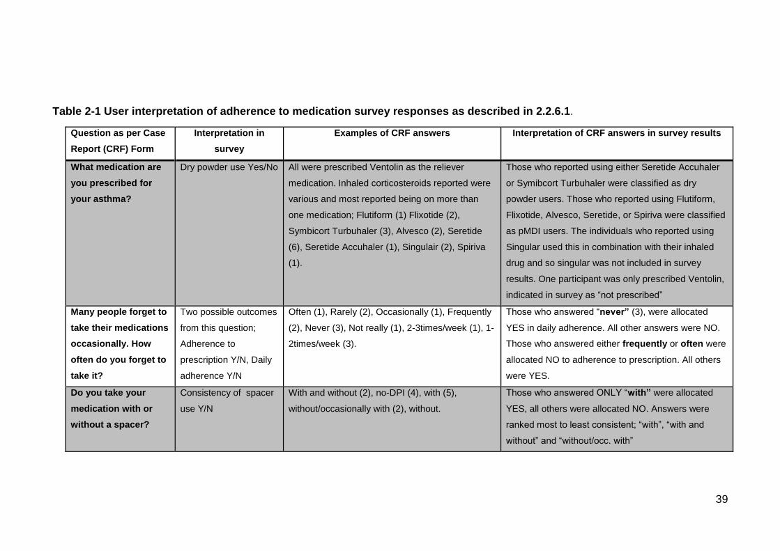
39
Table 2-1 User interpretation of adherence to medication survey responses as described in 2.2.6.1.
Question as per Case
Report (CRF) Form
Interpretation in
survey
Examples of CRF answers Interpretation of CRF answers in survey results
What medication are
you prescribed for
your asthma?
Dry powder use Yes/No
All were prescribed Ventolin as the reliever
medication. Inhaled corticosteroids reported were
various and most reported being on more than
one medication; Flutiform (1) Flixotide (2),
Symbicort Turbuhaler (3), Alvesco (2), Seretide
(6), Seretide Accuhaler (1), Singulair (2), Spiriva
(1).
Those who reported using either Seretide Accuhaler
or Symibcort Turbuhaler were classified as dry
powder users. Those who reported using Flutiform,
Flixotide, Alvesco, Seretide, or Spiriva were classified
as pMDI users. The individuals who reported using
Singular used this in combination with their inhaled
drug and so singular was not included in survey
results. One participant was only prescribed Ventolin,
indicated in survey as “not prescribed”
Many people forget to
take their medications
occasionally. How
often do you forget to
take it?
Two possible outcomes
from this question;
Adherence to
prescription Y/N, Daily
adherence Y/N
Often (1), Rarely (2), Occasionally (1), Frequently
(2), Never (3), Not really (1), 2-3times/week (1), 1-
2times/week (3).
Those who answered “never” (3), were allocated
YES in daily adherence. All other answers were NO.
Those who answered either frequently or often were
allocated NO to adherence to prescription. All others
were YES.
Do you take your
medication with or
without a spacer?
Consistency of spacer
use Y/N
With and without (2), no-DPI (4), with (5),
without/occasionally with (2), without.
Those who answered ONLY “with” were allocated
YES, all others were allocated NO. Answers were
ranked most to least consistent; “with”, “with and
without” and “without/occ. with”

40
2.2.6.2 Inhalation technique
On study day, immediately prior to inhalation of the study drug, all participants
(n=14) were trained by the clinical nurse asthma specialist to use their pMDI or
pMDI/spacer combination. PMDI training was in accordance with National
Asthma Council guidelines for inhalation technique using a single maximal
inhalation and breath-hold, either with or without a spacer (National Asthma
Council Australia, 2016b). Training involved the nurse specialist first
demonstrating the technique required, then asking the adolescent to
demonstrate this, and training was repeated until correct demonstration was
observed. All participants were assessed as competent and included in the
study.
2.2.6.3 Gamma scintigraphy to assess drug deposition in vivo
Four gamma scintigraphy scans were successively taken using the 2D gamma
camera (Ecam, Siemens), at PMH Nuclear Medicine department, all with a
120 second acquisition time. Firstly, prior to inhalation of the study drug, a scan
was taken of a planar flood source containing ~37 MBq Tc99m. Secondly, a
transmission scan was taken of the participant using the same planar flood
source to allow for assessment of attenuation of body tissues by gamma
radiation (Macey & Marshall, 1982). Thirdly, immediately after the participant
had inhaled the study drug, simultaneous anterior and posterior planar
scintigraphy scans were taken of the chest and abdomen with lateral positioning
of the head and neck. Lastly, Device components (pMDI actuator, spacer,
exhalation filters) were scanned immediately after the participant. Times
between all abovementioned scans were recorded in order to calculate decay
correction in analysis stages. There was not more than a 15 minute duration
between the initial scan and the final scan.
2.2.6.4 Gamma scintigraphy image acquisition and analysis
Images were acquired using Syngo VE31F software. Each participant had
regions of interest (ROI) subjectively delineated for their transmission, anterior
and posterior scan, by a single trained user. ROI were each whole lung, central
and peripheral lung regions, oropharyngeal, oesophagus, and stomach. The
central lung region was defined as a rectangle one third of the length and half

41
the width of a rectangular ROI drawn to just contain the entire lung, centred on
the proximal boundary of the rectangle. The lung ROI, (Figure 2-4) except for
the central box, was defined as peripheral lung (Figure 2-5). Total counts and
the area in pixels were obtained for each ROI, the flood source, and a
background count for transmission, device component, anterior and posterior
scans.
Figure 2-4 Subjective delineation of the lungs depicting method for quantifying central and
peripheral regions as described in 2.2.6.4. The total region of the lung (orange/red) is drawn around the
whole lung (green) in order to measure a third of the length and half the width (blue box).
Figure 2-5 Depiction of peripheral to central ratios. Central region of lung depicted inside blue box.
Anything not inside this region, but within the lung region (red/orange delineation) was considered
peripheral deposition.
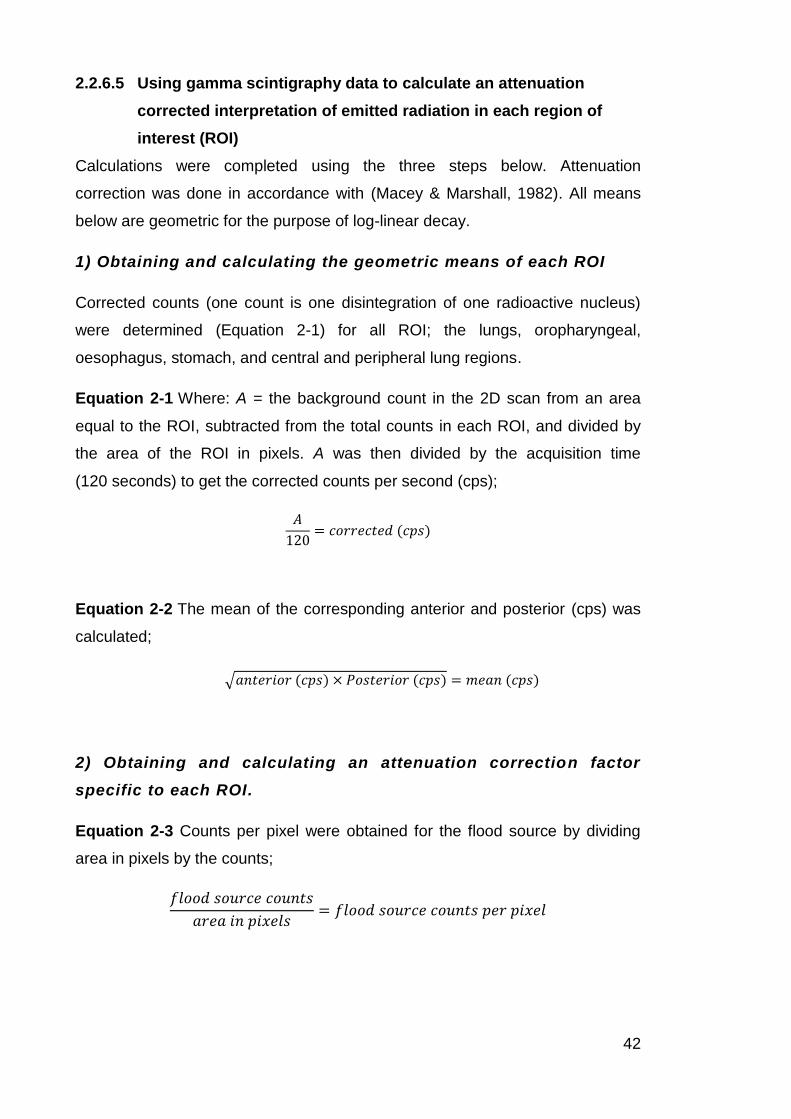
42
2.2.6.5 Using gamma scintigraphy data to calculate an attenuation
corrected interpretation of emitted radiation in each region of
interest (ROI)
Calculations were completed using the three steps below. Attenuation
correction was done in accordance with (Macey & Marshall, 1982). All means
below are geometric for the purpose of log-linear decay.
1) Obtaining and calculating the geometric means of each ROI
Corrected counts (one count is one disintegration of one radioactive nucleus)
were determined (Equation 2-1) for all ROI; the lungs, oropharyngeal,
oesophagus, stomach, and central and peripheral lung regions.
Equation 2-1 Where: A = the background count in the 2D scan from an area
equal to the ROI, subtracted from the total counts in each ROI, and divided by
the area of the ROI in pixels. A was then divided by the acquisition time
(120 seconds) to get the corrected counts per second (cps);
𝐴
120= 𝑐𝑜𝑟𝑟𝑒𝑐𝑡𝑒𝑑 (𝑐𝑝𝑠)
Equation 2-2 The mean of the corresponding anterior and posterior (cps) was
calculated;
√𝑎𝑛𝑡𝑒𝑟𝑖𝑜𝑟 (𝑐𝑝𝑠) × 𝑃𝑜𝑠𝑡𝑒𝑟𝑖𝑜𝑟 (𝑐𝑝𝑠) = 𝑚𝑒𝑎𝑛 (𝑐𝑝𝑠)
2) Obtaining and calculating an attenuation correction factor
specific to each ROI.
Equation 2-3 Counts per pixel were obtained for the flood source by dividing
area in pixels by the counts;
𝑓𝑙𝑜𝑜𝑑 𝑠𝑜𝑢𝑟𝑐𝑒 𝑐𝑜𝑢𝑛𝑡𝑠
𝑎𝑟𝑒𝑎 𝑖𝑛 𝑝𝑖𝑥𝑒𝑙𝑠= 𝑓𝑙𝑜𝑜𝑑 𝑠𝑜𝑢𝑟𝑐𝑒 𝑐𝑜𝑢𝑛𝑡𝑠 𝑝𝑒𝑟 𝑝𝑖𝑥𝑒𝑙

43
Equation 2-4 Using the decay correction formula the flood source counts per
pixel were adjusted to the time of the patient scan. Where A=the negative time
from the patient scan to the flood source scan divide by the half-life of
technetium in minutes;
𝑓𝑙𝑜𝑜𝑑 𝑠𝑜𝑢𝑟𝑐𝑒 𝑐𝑜𝑢𝑛𝑡𝑠 𝑝𝑒𝑟 𝑝𝑖𝑥𝑒𝑙 × 2(𝐴)
= 𝑑𝑒𝑐𝑎𝑦 𝑐𝑜𝑟𝑟𝑒𝑐𝑡𝑒𝑑 𝑓𝑙𝑜𝑜𝑑 𝑠𝑜𝑢𝑟𝑐𝑒 𝑐𝑜𝑢𝑛𝑡𝑠 𝑝𝑒𝑟 𝑝𝑖𝑥𝑒𝑙
Equation 2-5 Transmission counts per pixel were also corrected to the time of
the patient scan using the decay correction formula. Where A=the negative time
from the patient scan to the transmission scan divide by the half-life of
technetium;
𝑡𝑟𝑎𝑛𝑠𝑚𝑖𝑠𝑠𝑖𝑜𝑛 𝑐𝑜𝑢𝑛𝑡𝑠 𝑝𝑒𝑟 𝑝𝑖𝑥𝑒𝑙 × 2(𝐴)
= 𝑑𝑒𝑐𝑎𝑦 𝑐𝑜𝑟𝑟𝑒𝑐𝑡𝑒𝑑 𝑡𝑟𝑎𝑛𝑠𝑚𝑖𝑠𝑠𝑖𝑜𝑛 𝑐𝑜𝑢𝑛𝑡𝑠 𝑝𝑒𝑟 𝑝𝑖𝑥𝑒𝑙
Equation 2-6 Transmission counts per pixel were then calculated by;
𝑑𝑒𝑐𝑎𝑦 𝑐𝑜𝑟𝑟𝑒𝑐𝑡𝑒𝑑 𝑡𝑟𝑎𝑛𝑠𝑚𝑖𝑠𝑠𝑖𝑜𝑛 𝑐𝑜𝑢𝑛𝑡𝑠
𝑎𝑟𝑒𝑎 𝑖𝑛 𝑝𝑖𝑥𝑒𝑙𝑠= 𝑡𝑟𝑎𝑛𝑠𝑚𝑖𝑠𝑠𝑖𝑜𝑛 𝑐𝑜𝑢𝑛𝑡𝑠 𝑝𝑒𝑟 𝑝𝑖𝑥𝑒𝑙
Equation 2-7 The attenuation of activity of the flood source counts per pixel by
body tissues are then calculated for each ROI;
√𝑑𝑒𝑐𝑎𝑦 𝑐𝑜𝑟𝑟𝑒𝑐𝑡𝑒𝑑 𝑓𝑙𝑜𝑜𝑑 𝑠𝑜𝑢𝑟𝑐𝑒 𝑐𝑜𝑢𝑛𝑡𝑠
𝑡𝑟𝑎𝑛𝑠𝑚𝑖𝑠𝑠𝑖𝑜𝑛 𝑐𝑜𝑢𝑛𝑡𝑠 𝑝𝑒𝑟 𝑝𝑖𝑥𝑒𝑙= 𝑎𝑡𝑡𝑒𝑛𝑢𝑎𝑡𝑖𝑜𝑛 𝑓𝑎𝑐𝑡𝑜𝑟
Equation 2-8 The mean (cps) is then multiplied by the attenuation correction factor to obtain attenuation corrected (cps) in a region of interest;
𝑚𝑒𝑎𝑛 (𝑐𝑝𝑠) 𝑖𝑛 𝑟𝑒𝑔𝑖𝑜𝑛 𝑜𝑓 𝑖𝑛𝑡𝑒𝑟𝑒𝑠𝑡
× 𝑎𝑡𝑡𝑒𝑛𝑢𝑎𝑡𝑖𝑜𝑛 𝑐𝑜𝑟𝑟𝑒𝑐𝑡𝑖𝑜𝑛 𝑓𝑎𝑐𝑡𝑜𝑟
= 𝑎𝑡𝑡𝑒𝑛𝑢𝑎𝑡𝑖𝑜𝑛 𝑐𝑜𝑟𝑟𝑒𝑐𝑡𝑒𝑑 𝑚𝑒𝑎𝑛 𝑐𝑝𝑠

44
3) Using the attenuation corrected means to interpret radiation
per ROI.
Equation 2-9 Attenuation corrected (cps) of the means were converted to a
reading of MBq in each ROI;
𝑚𝑒𝑎𝑛 (𝑐𝑝𝑠) × 𝑎𝑡𝑡𝑒𝑛𝑢𝑎𝑡𝑖𝑜𝑛 𝑓𝑎𝑐𝑡𝑜𝑟
𝑐𝑎𝑚𝑒𝑟𝑎 𝑒𝑓𝑓𝑖𝑐𝑖𝑒𝑛𝑐𝑦= 𝑀𝑏𝑞 𝑖𝑛 𝑅𝑂𝐼
This MBq reading in each ROI is defined as the percent of total detected dose.
2.2.6.6 Camera efficiency
Camera efficiency was measured as cps per MBq, and this measurement was
taken on the study day by using a uniform flood source of known concentration
with a lookup table (Radpharm®). The lookup table was based on known
constant decay rates for a Cobalt-57 flood source (see appendices 5.8). The
average of measurements taken (n=6) was used to interpret all participant data
(105 cps/MBq). The efficiency was confirmed by camera servicing history to
have little inter-day variation.
2.2.7 STATISTICAL ANALYSIS
Gamma scintigraphy deposition data was preliminarily assessed via t-test in
paired analysis with and without a spacer for each drug group using GraphPad
Prism. Group demographics for FP and BDP were assessed with a Chi-Square
test (SAS) for categorical variables; sex, age, height, weight and BMI.
To complete multivariate analysis, drug and actuator deposition variables were
transformed using the arcsine square root transformation for analysis with SAS
ver. 9.4. The BMI variable was used to simplify variables of weight and height.
Univariate assessment first looked at the relationship on total lung deposition
with each variable separately, in each drug group; age, sex, body mass index
(BMI), visit order, sequence, actuator deposition, and spacer. Multivariate
assessment was then employed to look at total lung deposition in each drug
group (proc mixed, SAS) in a full model including variables: age, sex, body
mass index (BMI), visit order, sequence, actuator deposition, spacer and
random effect (a compulsory variable for mixed models). Multivariate analysis
with backward selection (significance level of 0.05) further assessed the
relationship.

45
All statistical analyses using SAS version 9.4 for Windows (SAS Institute Inc.,
USA) were performed with assistance from the Centre for Applied Statistics, at
the University of Western Australia. Analysis done with GraphPad Prism v. 7.02
(GraphPad Software, Inc.) were performed unassisted.
2.2.7.1 Power calculations
A pre-study calculation based on previous data within our group (C. M. Roller,
Zhang, Troedson, Leach, Le Souef, et al., 2007) showed that with a sample size
of (n=28), 14 in each of the experimental groups, the study had 80% power to
detect a 10% difference in lung dose at alpha=0.5.
46
3 CHAPTER III: RESULTS
3.1 IN VITRO VALIDATION OF RADIOLABELLED DRUG
PREPARATION TO DETERMINE IN VIVO REPORTING
To confirm successful validation, particle size distributions (with stages divided
into four groups), are shown for fluticasone propionate (FP) and
beclomethasone diproprionate (BDP) in Figure 3-1 and 3-2 respectively, with
the corresponding ratio comparison displayed in table 3-1 and 3-2 respectively.
Additional confirmation that radiolabelling the commercial drug has not
significantly altered the aerodynamic diameter of the labelled drug is shown via
total recoveries of mass (Figure 3-1, A, C FP, and Figure 3-2, A, C, BDP) for
pre-study and study day reference and labelled drugs.

47
A BP re -S tu d y (n = 6 )
S tu d y d a y (n = 1 2 )
Refe
ren
ce
Lab
elled
0
5 0
1 0 0
M e a n + /-S D flu tic a s o n e p ro p io n a te
1 2 5 u g a c tu a tio n p re -s tu d y v a lid a tio n (n = 6 )
% o
f T
ota
l re
co
ve
ry
Actu
ato
r -T
hro
at
Jet-
Zero
On
e-T
wo
Th
ree-F
ilte
r
0
2 0
4 0
6 0
8 0
M e a n (9 5 % C I) p re -s tu d y v a lid a tio n
o f ra d io la b e llin g m e th o d (n = 6 ) 2 0 1 6
% o
f T
ota
l re
co
ve
ry
R e fe re n c e
L a b e lled
R a d io tra c e r
Refe
ren
ce
Lab
elled
0
5 0
1 0 0
M e a n + /-S D flu tic a s o n e p ro p io n a te
1 2 5 u g a c tu a tio n s tu d y v a lid a tio n (n = 1 2 )
% o
f T
ota
l re
co
ve
ry
Ac
tua
tor -
Th
roa
t
Je
t-Z
ero
On
e-T
wo
Th
ree
-Fil
ter
0
2 0
4 0
6 0
8 0
M e a n (9 5 % C I) s tu d y v a lid a tio n
o f ra d io la b e llin g m e th o d (n = 1 2 ) 2 0 1 6
% o
f T
ota
l re
co
ve
ry
L a b e lled
R a d io tra c e r
R e fe re n c e
C D
Figure 3-1 (A, C) Recovery of FP 125 μg per actuation, comparison of reference to labelled drug,
The corresponding ratios of reference to labelled drug (A, C) were 0.89 and 0.97 respectively. Particle size
distribution was divided into four groups, to determine if the labelled drug and radiotracer counterpart fall
within the 95% CI of the reference drug (B, D).
Table 3-1 Fluticasone propionate particle size distribution assessment comparisons. Ratios
confirmed that validation was successful when within the range of 0.85–1.18 or ±2% for the groups that
had under 10% of the total drug.
Stage of
ACI:
Actuator to
throat
Jet to 0
(difference
from
reference)
One to two
(difference from
reference)
Three to filter
Ratio
obtained
from
Pre-
study
Study
day
Pre-
study
Study
day
Pre-
study
Study
day
Pre-
study
Study
day
Reference
/labelled
canister
1.12 1.06 ±2% ±2% ±2% ±2% 0.83 0.85
Reference/
radiotracer
0.95 1.07 ±2% ±2% ±2% ±2.746% 0.86 0.81

48
A BP re -S tu d y (n = 6 )
S tu d y d a y (n = 8 )C D
Refe
ren
ce
Lab
elled
0
5 0
1 0 0
M e a n + /- S D p re -s tu d y v a lid a t io n (n = 6 ) 2 0 1 6
% o
f T
ota
l re
co
ve
ry
Actu
ato
r -T
hro
at
Jet-
Zero
On
e-T
wo
Th
ree-F
ilte
r
0
2 0
4 0
6 0
8 0
M e a n (9 5 % C I) p re -s tu d y v a lid a tio n
o f ra d io la b e llin g m e th o d (n = 6 ) 2 0 1 6
% o
f T
ota
l re
co
ve
ry
R e fe re n c e
L a be lled
R a d io tra c e r
Refe
ren
ce
Lab
elled
0
5 0
1 0 0
M e a n + /- S D s tu d y v a lid a t io n (n = 8 ) 2 0 1 6
% o
f T
ota
l re
co
ve
ry
Actu
ato
r -T
hro
at
Jet-
Zero
On
e-T
wo
Th
ree-F
ilte
r
0
2 0
4 0
6 0
8 0
M e a n (9 5 % C I) s tu d y v a lid a tio n
o f ra d io la b e llin g m e th o d (n = 8 ) 2 0 1 6%
of
To
tal
re
co
ve
ry
L a be lled
R e fe re n c e
R a d io tra c e r
Figure 3-2 (A, C) Recovery of BDP 100 μg per actuation, comparison of reference to labelled, The
corresponding ratios of reference to labelled drug (A,C) were 1.03, and 0.97 respectively. Particle size
distribution was divided into four groups, to determine if the labelled drug and radiotracer counterpart fall
within the 95% CI of the reference drug (B, D).
Table 3-2 Beclomethasone di-propionate particle size distribution assessment comparison. Ratios
confirmed that validation was successful when within the range of 0.85–1.18 or ±2% for the groups that
had under 10% of the total drug.
Stage of ACI Actuator to
throat
Jet to 0
(difference
from
reference)
One to two
(difference from
reference)
Three to filter
Ratio
obtained
from
Pre-
study
Study
day
Pre-
study
Study
day
Pre-
study
Study
day
Pre-
study
Study
day
Reference
/labelled
canister
1.08 1.11 ±2% ±2% ±2% ±2% 0.87 0.88
Reference/
radiotracer
1.07 1.04 ±2% ±2% ±2% ±2% 0.90 0.92

49
3.2 RETROSPECTIVE ANALYSIS OF RADIOLABELLED FP AND
BDP
Confirmation of successful validation of previous data, as described in C. Roller
(2012) for FP and C. M. Roller, Zhang, Troedson, Leach, Le Souef, et al. (2007)
for BDP, particle size distributions (with stages divided into four groups), is
shown, reanalysed, for fluticasone propionate (FP) and beclomethasone
diproprionate (BDP) in Figure 3-4 and 3-6 respectively. Corresponding ratio
comparisons are displayed in table 3-3 and 3-4 respectively. Additional
confirmation that radiolabelling the commercial drug has not significantly altered
the aerodynamic diameter is shown via total recoveries of mass (Figure 3-4, A,
C, FP, and Figure 3-6, A, C, BDP) for pre-study and study day reference and
labelled drugs. Radiolabelling validation results for FP from this study (3.1,
Figure 3-1) were compared to retrospectively analysed results (3.2, Figure 3-4)
confirming that the radiolabelling method is robust over time. FP particle size
distribution varied with each study due to the difference in mass delivered per
actuation (125 and 250 μg), Figure 3-3, which was not the case for BDP (Figure
3-5) as both studies used 100 μg per actuation. The lower dose used for FP in
this study resulted in a higher respirable fraction (Three to Filter), and lower
Actuator to Throat fraction. Radiolabelling validation results for BDP from this
study (3.1, Figure 3-2) were compared to retrospectively analysed results (3.2,
Figure 3-6) confirming that the radiolabelling method is robust over time.

50
Actu
ato
r -T
hro
at
Jet-
Zero
On
e-T
wo
Th
ree-F
ilte
r
0
2 0
4 0
6 0
8 0
1 0 0
G ro u p e d fra c t io n s o f p a r t ic le s iz e d is tr ib u t io n
fro m a n d e rs o n c a s c a d e im p a c to r s ta g e s fo r F P
A n d e rs o n c a s c a d e im p a c to r s ta g e g ro u p in g s
Pe
rc
en
t o
f to
tal
re
co
ve
ry
Figure 3-3 Grouped comparison of two different radiolabelled validation results for FP, 125 μg
(hatched) and 250 μg per actuation. Grouped comparison of mean radiotracer and labelled to mean
reference (95% CI) for FP radiolabelling method shows little variation over time or between users. Labelled
drug (Dark Grey) and Radiotracer (Black) were marginally outside the 95% CI of the Reference (Light
Grey) drug, similar over time from 2006 (filled columns) to 2016 (hatched columns).
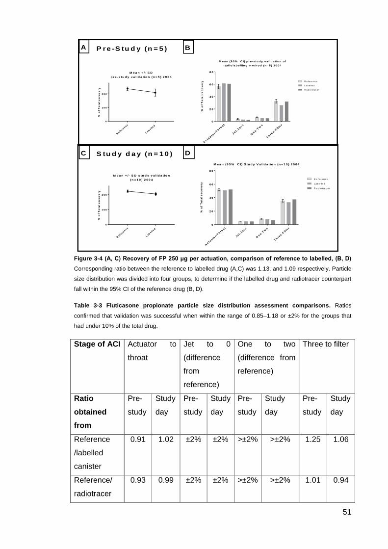
51
A BP re -S tu d y (n = 5 )
S tu d y d a y (n = 1 0 )C D
Refe
ren
ce
Lab
elled
0
1 0 0
2 0 0
M e a n + /- S D
p re -s tu d y v a lid a t io n (n = 5 ) 2 0 0 4
% o
f T
ota
l re
co
ve
ry
Actu
ato
r -T
hro
at
Jet-
Zero
On
e-T
wo
Th
ree-F
ilte
r
0
2 0
4 0
6 0
8 0
M e a n (9 5 % C I) p re -s tu d y v a lid a tio n o f
ra d io la b e llin g m e th o d (n = 5 ) 2 0 0 4
% o
f T
ota
l re
co
ve
ry
R e fe re n c e
L a be lled
R a d io tra c e r
Refe
ren
ce
Lab
elled
0
1 0 0
2 0 0
M e a n + /- S D s tu d y v a lid a t io n
(n = 1 0 ) 2 0 0 4
% o
f T
ota
l re
co
ve
ry
Ac
tua
tor -
Th
roa
t
Je
t-Z
ero
On
e-T
wo
Th
ree
-Fil
ter
0
2 0
4 0
6 0
8 0
M e a n (9 5 % C I) S tu d y V a lid a tio n (n = 1 0 ) 2 0 0 4
% o
f T
ota
l re
co
ve
ry
R e fe re n c e
L a be lled
R a d io tra c e r
Figure 3-4 (A, C) Recovery of FP 250 μg per actuation, comparison of reference to labelled, (B, D)
Corresponding ratio between the reference to labelled drug (A,C) was 1.13, and 1.09 respectively. Particle
size distribution was divided into four groups, to determine if the labelled drug and radiotracer counterpart
fall within the 95% CI of the reference drug (B, D).
Table 3-3 Fluticasone propionate particle size distribution assessment comparisons. Ratios
confirmed that validation was successful when within the range of 0.85–1.18 or ±2% for the groups that
had under 10% of the total drug.
Stage of ACI Actuator to
throat
Jet to 0
(difference
from
reference)
One to two
(difference from
reference)
Three to filter
Ratio
obtained
from
Pre-
study
Study
day
Pre-
study
Study
day
Pre-
study
Study
day
Pre-
study
Study
day
Reference
/labelled
canister
0.91 1.02 ±2% ±2% >±2% >±2% 1.25 1.06
Reference/
radiotracer
0.93 0.99 ±2% ±2% >±2% >±2% 1.01 0.94

52
Actu
ato
r -T
hro
at
Jet-
Zero
On
e-T
wo
Th
ree-F
ilte
r
0
2 0
4 0
6 0
8 0
1 0 0
G ro u p e d fra c t io n s o f p a r t ic le s iz e d is tr ib u t io n f ro m a n d e rs o n
c a s c a d e im p a c to r s ta g e s fo r B D P
Pe
rc
en
t o
f to
tal
re
co
ve
ry
A n d e rs o n c a s c a d e im p a c to r s ta g e g ro u p in g s
Figure 3-5 Grouped fraction comparison of two different radiolabelled validation results for BDP,
both used 100 μg per actuation. Grouped comparison of mean radiotracer and labelled to mean
reference (95% CI) for BDP radiolabelling method shows no variation over time or between users.
Labelled drug (Dark Grey) and Radiotracer (Black) were comparable to the 95% CI of the Reference (Light
Grey) drug, the integrity of the radiolabelling method unchanging over time from 2006 (filled columns) to
2016 (hatched columns).

53
A BP re -S tu d y (n = 1 0 )
S tu d y d a y (n = 1 5 )C D
Re
fere
nc
e
La
be
lle
d
0
5 0
1 0 0
M e a n + /- S D p re -s tu d y v a lid a t io n
(n = 1 0 ) 2 0 0 4
% o
f T
ota
l re
co
ve
ry
Ac
tua
tor -
Th
roa
t
Je
t-Z
ero
On
e-T
wo
Th
ree
-Fil
ter
0
2 0
4 0
6 0
8 0
M e a n (9 5 % C I) p re -s tu d y v a lid a tio n o f
ra d io la b e llin g m e th o d (n = 1 0 ) 2 0 0 4
% o
f T
ota
l re
co
ve
ry
R e fe re n c e
L a be lled
R a d io tra c e r
Re
fere
nc
e
La
be
lle
d
0
5 0
1 0 0
M e a n + /- S D s tu d y v a lid a t io n
(n = 1 5 ) 2 0 0 4
% o
f T
ota
l re
co
ve
ry
Ac
tua
tor -
Th
roa
t
Je
t-Z
ero
On
e-T
wo
Th
ree
-Fil
ter
0
2 0
4 0
6 0
8 0
M e a n (9 5 % C I) s tu d y v a lid a tio n
o f ra d io la b e llin g m e th o d (n = 1 5 ) 2 0 0 4
% o
f T
ota
l re
co
ve
ry
R e fe re n c e
L a be lled
R a d io tra c e r
Figure 3-6 (A, C) Recovery of BDP 100 μg per actuation, comparison of reference to labelled.
Corresponding ratio between the reference to labelled drug (A,C) was 0.93, 0.94 respectively. Particle size
distribution was divided into four groups, to determine if the labelled drug and radiotracer counterpart fall
within the 95% CI of the reference drug (B, D).
Table 3-3 Beclomethasone di-propionate particle size distribution assessment comparison. Ratios
confirmed that validation was successful when within the range of 0.85–1.18 or ±2% for the groups which
had under 10% of the total drug.
Stage of ACI Actuator to
throat
Jet to zero
(difference
from
reference)
One to two
(difference from
reference)
Three to filter
Ratio
obtained
from
Pre-
study
Study
day
Pre-
study
Study
day
Pre-
study
Study
day
Pre-
study
Study
day
Reference
/labelled
canister
0.98 0.97 ±2% ±2% ±2% ±2% 1.05 0.97
Reference/
radiotracer
0.90 0.93 ±2% ±2% ±2% ±2% 1.09 0.98

54
3.3 IN VIVO DRUG DEPOSITION STUDY RESULTS
3.3.1 STUDY POPULATION
The demographic information for the cohort is displayed in Table 3-5 for the full
cohort. The mean demographics assessed were similar within drug groups
(Table 3-6) and to the full cohort demographic means (Table 3-5). There was no
significant difference between groups in any variable (Table 3-6).
Table 3-4 Full cohort (n=14) demographics
Variable Mean, Range
Sex (n) 6 Male, 8 Female
Age (yrs) 15 y, 13–17 years
Height (cm) 169, 158–179
Weight (kg) 67, 46–110
BMI (kg/m2) 23.4, 16–40
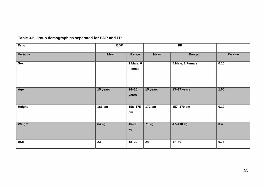
55
Table 3-5 Group demographics separated for BDP and FP
Drug BDP FP
Variable Mean Range Mean Range P-value
Sex 1 Male, 6
Female
5 Male, 2 Female 0.10
Age 15 years 14–16
years
15 years 13–17 years 1.00
Height 166 cm 158–175
cm
172 cm 157–179 cm 0.19
Weight 64 kg 46–89
kg
71 kg 47–110 kg 0.49
BMI 23 18–29 24 17–40 0.76

56
3.3.1.1 Screening questionnaire data
All participants (n=14) completed a screening questionnaire (Figure 3-7),
involving adherence to medication and related issues. Approximately 50% of
participants who required a spacer used it regularly. Over 75% of participants
who were prescribed inhaled corticosteroids for their asthma reported regularly
taking them, but only 25% of those participants said they take them every day
(Figure 3-7).
Figure 3-7 Survey of adherence to pMDI delivered corticosteroid asthma drugs and related questions in mildly asthmatic adolescents. Nearly 70 % of participants did not adhere to their
prescribed daily puffer medication, and of those who did approximately half used their spacers.
0.00%
10.00%
20.00%
30.00%
40.00%
50.00%
60.00%
70.00%
80.00%
90.00%
100.00%
Dry powder use Consistency tospacer use
Adherance toprescription
Daily adherence
Survey of adherence to use of asthma drugs by study participants (n=14)
Not prescribed
Yes
No

57
3.3.2 SHOT WEIGHT DELIVERED BY PMDI
The dose of drug delivered to each participant, measured via mass, is shown in
Figure 3-8. Two significant outliers were observed with BDP, and two with FP,
both confirmed to be due to inhaler variability per shot (Figure 3-8).
n=2
n=5
n=10
0 .0 5
0 .0 6
0 .0 7
0 .0 8
0 .0 9
S h o t w e ig h t (m e a n S D , F P la b e lle d )
M e a n o f a c u ta t io n s p e r d a ta p o in t
ma
ss
pe
r a
ctu
ati
on
dis
pe
ns
ed
(g
m)
n=2
n=5
n=10
0 .0 5
0 .0 6
0 .0 7
0 .0 8
0 .0 9
S h o t w e ig h t (m e a n S D , B D P la b e lle d )
M e a n o f a c u ta t io n s p e r d a ta p o in t
ma
ss
pe
r a
ctu
ati
on
dis
pe
ns
ed
(g
m)
Figure 3-8 Mass of drug delivered to each participant on study day reduced in variability with
multiple shots per mean. Variability of the same labelled canister reduced when obtaining the mean of 5
and 10 actuations, showing natural variability, and not an effect of a flawed radiolabelled drug. Additionally,
inter user variability was present at n=2.Timepoints were obtained successively for n=5,10 and 2.
3.3.3 GAMMA SCINTIGRAPHY IN VIVO DEPOSITION AFTER INHALATION
OF RADIOLABELLED DRUG
Gamma scintigraphy images were taken, obtained, and analysed, according to
methods sections 2.2.6.3, 2.2.6.4, and 2.3.6.5, respectively. Results of gamma
scintigraphy deposition data for FP and BDP are displayed regionally in Figure
3-9 and Table 3-7. Deposition data was defined as percentage of total ex-valve
dose as detected via gamma camera (MBq). Data show a fivefold decrease in
oropharyngeal deposition with a spacer with FP, (p= 0.001), although only
twofold decrease was seen with BDP (p=0.03) (Figure 3-9, A). Lung deposition
was higher with a spacer, although not significantly higher with FP (p=0.31), or
BDP (p=0.52) (Figure 3-9, B). Mean peripheral to central ratio with was higher
with a spacer (FP p=0.16, BDP p=0.81).

58
F P B D P
A
B
C
Sp
acer
+ p
MD
I
pM
DI o
nly
0
2 0
4 0
6 0
8 0
O ro p h a ry n g e a l d e p o s it io n (% e x -v a lv e )
* *
Sp
acer
+ p
MD
I
pM
DI o
nly
0
2 0
4 0
6 0
8 0
O ro p h a ry n g e a l d e p o s it io n (% e x -v a lv e )
*
Sp
acer
+ p
MD
I
pM
DI o
nly
0
2 0
4 0
6 0
8 0
L u n g d e p o s it io n (% e x -v a lv e )
p = 0 .3 1
Sp
acer
+ p
MD
I
pM
DI o
nly
0
2 0
4 0
6 0
8 0
L u n g d e p o s it io n (% e x -v a lv e )
p = 0 .5 2
Sp
acer
+ p
MD
I
pM
DI o
nly
0
1
2
3
R e g io n a l lu n g d e p o s it io n
Pe
rip
he
ra
l to
ce
ntr
al
ra
tio
(P
:C)
p = 0 .1 6
Sp
acer
+ p
MD
I
pM
DI o
nly
0
1
2
3
R e g io n a l lu n g d e p o s it io n
Pe
rip
he
ra
l to
ce
ntr
al
ra
tio
(P
:C)
p = 0 .8 1
Figure 3-9 Univariate analysis after transformation of gamma scintigraphy deposition data, as
percentage of ex-valve, for FP (n=7) and BDP (n=7): A) mean±SD paired oropharyngeal deposition,
after inhalation with radiolabelled drug, B) mean±SD paired lung deposition after inhalation of radiolabelled
drug C) mean±SD regional lung deposition after inhalation of radiolabelled drug as a peripheral to central
ratio.
59
Table 3-6 Comparison of mean (SD) deposition data for purpose of cross study comparison. Deposition values are displayed for each region of interest as percent of ex-valve
or ex-actuator
FP BDP
Spacer
pMDI only Spacer
pMDI only
Ex-valve
(%)
Ex-actuator (%) Ex-valve
(%)
Ex-actuator
(%)
Ex-valve
(%)
Ex-actuator
(%)
Ex-valve
(%)
Ex-actuator
(%)
Actuator 20±9 - 18±8 - 40±27 - 52±31 -
Lung 37±6 46±5 33±9 40±13 34±19 56±14 29±22 54±14
OP 9±7 11±8 49±12 59±13 4±5 9±5 14±8 32±9
Spacer 33±9 41±11 - - 13±9 21±9 - -
Exhaled 1±2 2±2 <0.5±1 <1±1 7±6 13±10 5±4 11±9
60
Visual review of the gamma scintigraphy images, shown for FP in Figure 3-10,
revealed higher oropharyngeal deposition (OP) and deposition around the major
bifurcations, with hot spots in the lungs, particularly when drug was
administered via a pMDI without a spacer. BDP deposition visually showed an
even spread in the lungs with either device, and a slight decrease in OP
deposition when a spacer was used (Figure 3-11). Images were not altered
after obtaining from software.
Figure 3-10 Qualitative analysis of gamma scintigraphy lung deposition images (FP). Image data
show paired data; (A) Deposition with pMDI alone, (B) Deposition with pMDI/spacer combination.
Figure 3-11 Qualitative analysis of gamma scintigraphy lung deposition images (BDP). Image data
show paired data; (A) Deposition with pMDI alone, (B) Deposition with pMDI/spacer combination.
A B
A B

61
3.3.4 STATISTICAL ANALYSIS OF GAMMA SCINTIGRAPHY DEPOSITION
DATA AFTER TRANSFORMATION
When each variable was analysed separately with drug with BDP, percent
deposition in the actuator significantly reduced lung deposition (Table 3-8). After
adjustment in a full model, age, sex and BMI additionally affected lung
deposition with BDP only, and remained significant after backward elimination
at significance level alpha 0.05 (Table 3-8).
Table 3-7 Analysis of regional deposition data after transformation. Lung deposition from each drug
was analysed: with a single variable (Spacer etc.), in a full model to adjust for confounders, then variables
were eliminated via backward selection at 0.05 level of significance, until the final model was left (exit point
p-values displayed).
Univariate Full Model
(adjusted)
Final Model
(backward
selection)
Drug FP BDP FP BDP FP BDP
Variable
Spacer 0.27 0.45 0.51 0.76 0.50 0.76
Age 0.92 0.57 0.44 0.02 0.09 0.002
Sex 0.28 0.36 0.61 0.004 0.61 0.0004
Randomisation
order
0.90 0.70 0.25 0.74 0.41 0.71
Visit order 0.16 0.74 0.33 0.68 0.12 0.60
BMI 0.12 0.20 0.09 0.004 0.12 0.0003
Actuator 0.54 <0.0001 0.58 <0.0002 0.59 <0.0001

62
4 CHAPTER IV: DISCUSSION
Lung deposition with use of the pMDI producing the extrafine aerosol (BDP), or
the coarse aerosol (FP) by adolescents in this study was not significantly
different with or without use of a spacer, and peripheral airway deposition was
equivalent regardless of spacer use. Whilst the MMAD for FP was assessed
here to be 2.8 μm and is therefore marginally a coarse aerosol, it appears to
have some of the characteristics demonstrated by a fine aerosol (≤2.5 μm
MMAD) in this study, perhaps as it is very marginally coarse. Our findings for
BDP, QVAR™, are consistent with a previous study of this drug formulation that
reported equivalent lung deposition with or without a spacer in an adult cohort
(C. L. Leach & Colice, 2010). Furthermore, levels of lung deposition found here
with pMDI producing an extrafine aerosol are consistent in both adult
populations without a spacer (CL Leach et al., 1998; C. L. Leach, Davidson,
Hasselquist, & Boudreau, 2002; C. L. Leach et al., 2005), and child populations
with a spacer, assuming correct and reproducible inhalation technique (C. M.
Roller, Zhang, Troedson, Leach, Le Souef, et al., 2007). The recommendation
for a spacer as an additional device would thus be unnecessary with a extrafine
preventer aerosol (≤~2 μm MMAD), assuming reproducible results in a larger
adolescent cohort or other supportive evidence such as that obtained by
Guilbert et al. (2017), who saw no clinical improvement with use of a spacer
compared to pMDI alone in a retrospective study of patient clinical data. A
combination of epidemiological and/or survey methods combined with evidence
based studies such as ours, would provide the best approach to supporting
change in practice.
Implementing this change in practice would make progress toward addressing
the long overdue issue of incorrect pMDI usage, shown to be unchanged over
the 40 years prior to 2014 in a systematic review by Sanchis et al. (2016). The
Global Initiative for Asthma identifies incorrect pMDI use as a contributor to
decreased asthma control levels (Reddel et al., 2015) which are shown to be
low - 24% of adolescents with asthma in the Asia Pacific region have
uncontrolled asthma, a further 63% only partly controlled, and only 13% well
controlled (Gold, Thompson, Salvi, Faruqi, & Sullivan, 2014). Sanchis et al.
(2016) also found that, despite spacer use, pMDI technique remained poor.

63
Interestingly, the recommendation for use of a spacer currently attempts to
address incorrect inhaler use. Furthermore, spacer use has proved difficult to
implement in practice, particularly in populations that require special clinical
management to encourage medication compliance, such as adolescents
(Michaud, 2007), and so it seems that change to this practice is also long
overdue and would particularly benefit the adolescent population.
Adolescents are a special population whose adherence to medication is directly
influenced by healthy family psychosocial functioning, where family support can
improve autonomy in the journey toward adulthood and help foster a positive
attitude toward medication self-management (Rhee et al., 2010). Positive
attitude toward self-management can include increased likelihood of visiting a
health professional when required – and a good relationship with the health
care provider is key to improving asthma control levels (Haskard Zolnierek &
DiMatteo, 2009). Conversely, when this support is not available, issues with
self-management become apparent and negatively impact asthma control,
increasing morbidity and the risk of hospitalisation (Chen et al., 2003; Rhee et
al., 2010). These issues can be further compounded by a multitude of generally
accepted issues with asthma treatment: inadequacies with the medication
delivery itself, inadequate health education contributing to false beliefs about
the safety of asthma medication or the chronic nature of the disease, and false
perceptions about the ease of effective drug delivery contributing to non-
compliance with treatment recommendations (spacer use). Whilst improving
family psychosocial functioning is an infinitely more challenging task, those
compounding issues can more easily be addressed, such as, by improving drug
delivery, and investigating the requirement (or not) for spacer use, (particularly
as adolescents display an aversion to using them). Real-life outcomes of spacer
use have been studied by Guilbert et al. (2017), however, to our knowledge
ours is the first study to assess the clinical recommendation, via lung deposition
study, for spacer use with ICS formulations produced by pMDI, both with and
without a spacer, in a solely adolescent population. Our study widens already
established findings that show increased delivery efficacy in adults, of an
extrafine aerosol, by extending that finding to the adolescent age group.
Our study showed a high lung to oropharyngeal deposition ratio per dose with
the extrafine aerosol only (i.e. 60:40 lung to oropharyngeal). The findings in our
64
study are supported extensively with previous studies for the extrafine aerosol,
showing consistent lung deposition of ~55% (ex-actuator dose) and
oropharyngeal deposition of ~40% (ex-actuator dose) with a pMDI alone (CL
Leach et al., 1998; C. L. Leach et al., 2002, 2005). Limited lung deposition
studies were available to compare our coarse aerosol data with, none with and
without a spacer in any population. With the coarse aerosol pMDI alone, we
recorded lung deposition of ~40% (ex-actuator), which contrasted a previous
adult study reporting ~25% (ex-actuator). With a pMDI+spacer, however, we
reported lung deposition of ~45% (ex-actuator), comparable to a previous study
(C. Roller, 2012) reporting ~25% (SD 10), range 15–45%, (ex-actuator) in a
child cohort. It is reasonable to conceive a higher lung deposition would be
observed in an older cohort and lung deposition may have been additionally
increased by the lower dose per actuation used in our study. As seen in our
validation retrospective analysis, the higher dose per actuation (250 μg) used by
C. Roller (2012) resulted in a decreased respirable fraction, compared to our
lower dose (125 μg). At higher doses, particles may coagulate after
aerosolisation, resulting in larger particles, and thus a decreased dose delivered
to the lungs (Tolpekin, Duits, van den Ende, & Mellema, 2004; Tsuda et al.,
2011). Use of a lower dose may reduce the potential of this occurring.
Considering a 44 μg dose was used by C. L. Leach et al. (2015) this may also
confirm that the dose used in our study was optimal for a pMDI producing a
coarse aerosol. Alternatively, a lower dose estimate consistent with that
obtained by C. L. Leach et al. (2015) may be more reasonable, which could
highlight the importance of recognising even minor inconsistencies from
radiolabelling validation guidelines, as seen with fluticasone propionate in our
study. Radiolabelling validation of fluticasone propionate as previously reported
by C. L. Leach et al. (2015) and C. Roller (2012) however, demonstrably similar
inconsistencies to our study, such as the “tailing effect”, where <5% of total un-
incorporated radiation is observed. Thus, it is unlikely to affect study
comparability, although exact ratios and confidence intervals were not provided
by C. L. Leach et al. (2015) and so a more accurate reflection cannot be made.
To recommend the required change against spacer use in clinical practice,
evidence of high lung to oropharyngeal deposition ratio per dose is necessary.
The concern where this is absent is an increased systemic ICS bioavailability
and resultant side effects. Our results confirm that when using a pMDI

65
producing extrafine aerosol, you receive a higher respirable fraction of the total
dose, which corresponds to an increased therapeutic dose.
Our study in adolescents demonstrated equivalent lung deposition regardless of
spacer use, or drug formulation, as has been found previously with a extrafine
aerosol only (C. L. Leach & Colice, 2010). This indicates a spacer may not be
needed to improve lung deposition with a coarse aerosol or with an extrafine
aerosol, building on a previous epidemiological study where spacer use did not
improve asthma outcomes for a fine or extrafine aerosol (Guilbert et al., 2017).
The observed equivalence in lung deposition regardless of spacer use or drug
in our study, may be due to the relatively small difference in MMAD of the
chosen drugs (2.8 µm versus 1.2 µm), with the coarse aerosol consideration for
a fine aerosol (fine aerosol= >0.1–2.5 μm) (Australian Government, 2004).
Alternatively, it could be an effect of repeated inhaler technique training,
(Kamps et al., 2000) although statistical analysis in this study did not find an
association, i.e. there was no evidence of greater lung deposition on the second
visit compared with the first. It is commonly accepted that inhaler technique
remains poor in practice (Price et al., 2013; Sanchis et al., 2016) regardless of
technique assessment by clinical observation (Biswas et al., 2016), yet this may
be substantially due to insufficient or incomplete initial training programs
(Haahtela et al., 2006; Papi et al., 2011). In this study adolescents were
considered able to coordinate an adequate inhalation technique, during training
with a clinical nurse asthma specialist, which suggests a spacer may not
necessarily be required to decrease the effects caused by lack of coordination
under these circumstances. Whilst our small sample size limits generalisable
conclusions, our data emphasises the potential of an appropriately and
repeatedly trained, clinically observed, adolescent to use a pMDI producing an
inhaled corticosteroid aerosol <2.8 μm MMAD to achieve approximately
equivalent lung deposition to that seen with use of the same drug with a spacer.
A short acting beta 2 agonist, even if formulated with a <2.8 μm MMAD may not
produce a similar result, as a symptomatic patient may give a different pattern
of inhalation; usually an increase in rate of inhalation and a resulting more
central and oropharyngeal deposition (Laube, Norman, & Adams, 1992).
Additionally, coordination may not be optimal when patients are symptomatic
and so the user may still benefit from use of a spacer.

66
With the pMDI producing an extrafine aerosol we reported an increased
proportion of the total dose depositing in the peripheral airways regardless of
spacer use. This finding is comparable to other studies and demonstrates no
effect of spacer on regional deposition (C. L. Leach et al., 2006; C. L. Leach et
al., 2015). Whilst the 2D imaging method used in this study may be limited in its
ability to estimate regional lung deposition compared to 3D methods, C. L.
Leach et al. (2006) confirmed this systematic under- and over-estimation was
marginal. Additionally, a recently validated diagnostic method for 2D
assessment of lung deposition showed increased sensitivity in detecting
regional deposition, and could reduce this bias in future studies (Bennett, Xie,
Zeman, Hurd, & Donaldson, 2014). Based on our study, it is reasonable to
assume a spacer is not required for use, additional to a pMDI producing an
extrafine aerosol, to increase peripheral deposition in this age group. This may
mean that a future focus on pMDIs producing a fine aerosol, particularly those
considered extrafine (≤~2 μm MMAD) as used in our study, would be a useful
approach to target peripheral lung disease.
If clinical practice was to change, based on this and larger future studies, it
would highlight the need for improved clinician and patient pMDI technique
training programs, particularly from the point of initial treatment (Fink & Rubin,
2005; Haahtela et al., 2006). PMDIs are cheap and portable devices for orally
inhaled delivery of corticosteroids, and the most recommended device
worldwide for the treatment of asthma, (Brocklebank et al., 2001) however,
clearly, the problem of inhaler technique compliance still exists outside of a
clinical setting, and is associated with an increased risk of exacerbation and
hospitalisation (Levy et al., 2013; Sanchis et al., 2016). An improvement in
education and training for the delivery of ICS, if combined with easy to
implement therapies is advantageous to the wider population, and particularly
valuable to special populations with reduced medication adherence, such as
adolescents (Dima et al., 2015). The extrafine aerosol formulation produced by
pMDI used in this study shows a repeatable increase in lung deposition when
compared to coarse aerosols, and is already available in Australia and
worldwide, emphasising the possibility for imminent changes to clinical
recommendation (CL Leach et al., 1998; C. L. Leach et al., 2006; C. L. Leach et
al., 2002, 2005; C. L. Leach et al., 2012; C. L. Leach et al., 2015).
67
Based on results of this study, significant effectors of lung deposition with an
extrafine aerosol, such as deposition of drug in the actuator, BMI, sex, and age,
may be useful inclusions of future study designs. The effect of repeated training
(inclusion of a visit order variable) is a particularly recommended inclusion.
The introduction of the international guidelines for inhaled radiolabelled
preparations in 2012 (Devadason et al., 2012), should lead to improved
consistency of reporting and so it is increasingly possible that studies completed
under the new guidelines, such as ours, are easily comparable. Evidence from
this and previous studies suggests that the MMAD of an aerosol has a
significant effect on lung deposition with ICS asthma drug formulations. PMDIs
producing a fine aerosol (MMAD<2.5 µm), specifically those considered extra-
fine (MMAD ~2 µm), result in almost double the lung deposition and half the
oropharyngeal deposition, achieved in previous studies using coarse (>2.5–10
µm MMAD) formulations. This emphasises the necessity of changes to clinical
recommendations, such as spacer use, particularly for the adolescent
population, where ease of implementation is especially important.
4.1 CONCLUSION
Results presented here investigate the MMAD of two inhaled corticosteroids,
and pragmatically addresses the recommendation for spacer use in a
population whom are less likely to manage their disease optimally, more likely
to have poor asthma control, and express an aversion to use the spacer device.
It builds on prior work done in adults showing that an extrafine aerosol can
achieve equivalent lung deposition with or without a spacer, when inhaled with
good technique, by expanding these same findings to an adolescent age group.
It supports prior epidemiological work showing no improvements in asthma
outcomes are evident with spacer use with a fine ICS aerosol. This thesis
shows that, whether or not a spacer is used, an extrafine preventer aerosol
achieves equivalent regional and total lung deposition and comparable
oropharyngeal dose. Considering our findings in the extrafine aerosol are
supported by previous work, discontinuing spacer recommendation for this
formulation could potentially simplify asthma treatment in non-compliant
patients.

68
BIBLIOGRAPHY
Aaron, S. D., Vandemheen, K. L., FitzGerald, J. M., Ainslie, M., Gupta, S.,
Lemiere, C., . . . Boulet, L. P. (2017). Reevaluation of diagnosis in adults with physician-diagnosed asthma. The Journal of the American Medical Association, 317(3), 269-279. doi:10.1001/jama.2016.19627
Adcock, I., & Lane, S. (2003). Corticosteroid-insensitive asthma: molecular mechanisms. Journal of Endocrinology, 178(3), 347-355. doi:10.1677/joe.0.1780347
Agache, I., Akdis, C., Jutel, M., & Virchow, J. C. (2012). Untangling asthma phenotypes and endotypes. Allergy, 67(7), 835-846. doi:10.1111/j.1398-9995.2012.02832.x
AIHW: Correll PK, P. L., Ampon R, Reddel HK & Marks GB. (2015). Respiratory medication use in Australia 2003–2013: treatment of asthma and COPD. Cat. no. ACM 31. Retrieved from Canberra: https://www.aihw.gov.au/reports/asthma-other-chronic-respiratory-conditions/respiratory-medication-use-in-australia-2003-2013/contents/table-of-contents
Anderson, G. P. (2008). Endotyping asthma: new insights into key pathogenic mechanisms in a complex, heterogeneous disease. The Lancet, 372(9643), 1107-1119. doi:http://dx.doi.org/10.1016/S0140-6736(08)61452-X
Asgharian, B., Hofmann, W., & Bergmann, R. (2001). Particle deposition in a multiple-path model of the human lung. Aerosol Science and Technology, 34(4), 332-339. doi:10.1080/02786820119122
Asgharian, B., Price, O. T., & Hofmann, W. (2006). Prediction of particle deposition in the human lung using realistic models of lung ventilation. Journal of Aerosol Science, 37(10), 1209-1221. doi:https://doi.org/10.1016/j.jaerosci.2006.01.002
Australian Centre for Asthma Monitoring. (2011). Asthma in Australia 2011: with a focus chapter on chronic obstructive pulmonary disease. Retrieved from Canberra: <http://www.aihw.gov.au/publication-detail/?id=10737420159>
Australian Government. (2004). Health impacts of ultrafine particles. Retrieved from https://www.environment.gov.au/system/files/resources/00dbec61-f911-494b-bbc1-adc1038aa8c5/files/health-impacts.pdf.
Australian Government Department of Health Therapeutic Goods Administration. (2013). Guidance 19: Inhalation and nasal medicines. Retrieved from https://www.tga.gov.au/sites/default/files/pm-argpm-guidance-19.pdf.
Barnes, P. J. (1998). Anti-inflammatory actions of glucocorticoids: molecular mechanisms. Clinical Science, 94(6), 557-572. doi:10.1042/cs0940557
Barnes, P. J. (2004). Distribution of receptor targets in the lung. Proceedings of the American Thoracic Society, 1(4), 345-351. doi:10.1513/pats.200409-045MS
Barnes, P. J. (2013). Corticosteroid resistance in patients with asthma and chronic obstructive pulmonary disease. Journal of Allergy and Clinical Immunology, 131(3), 636-645. doi:10.1016/j.jaci.2012.12.1564
Barry, P. W., & O'Callaghan, C. (1995). The effect of delay, multiple actuations and spacer static charge on the in vitro delivery of budesonide from the Nebuhaler. British Journal of Clinical Pharmacology, 40(1), 76-78.

69
Bennett, W. D., Xie, M., Zeman, K., Hurd, H., & Donaldson, S. (2014). Heterogeneity of particle deposition by pixel analysis of 2D gamma scintigraphy images. Journal of Aerosol Medicine and Pulmonary Drug Delivery., 28(3), 211-218. doi:10.1089/jamp.2013.1095
Biswas, R., Patel, G., Mohsin, A., Hanania, N., & Sabharwal, A. (2016). Measuring competence in metered dose inhaler use using capmedic electronic inhaler monitoring tool. Chest, 150(4_S), 14A-14A. doi:10.1016/j.chest.2016.08.017
Boulet, L. P., FitzGerald, J. M., & Reddel, H. K. (2015). The revised 2014 GINA strategy report: opportunities for change. Current Opinion in Pulmonary Medicine., 21(1), 1-7. doi:10.1097/mcp.0000000000000125
Bousquet, J., Mantzouranis, E., Cruz, A. A., Aït-Khaled, N., Baena-Cagnani, C. E., Bleecker, E. R., . . . Zuberbier, T. (2010). Uniform definition of asthma severity, control, and exacerbations: Document presented for the World Health Organization consultation on severe asthma. Journal of Allergy and Clinical Immunology, 126(5), 926-938. doi:https://doi.org/10.1016/j.jaci.2010.07.019
Boyd, C. J., McCabe, S. E., & Teter, C. J. (2006). Asthma inhaler misuse and substance abuse: A random survey of secondary school students. Addictive Behaviors., 31(2), 278-287. doi:10.1016/j.addbeh.2005.05.005
Brennan, V. K., Osman, L. M., Graham, H., Critchlow, A., & Everard, M. L. (2005). True device compliance: the need to consider both competence and contrivance. Respiratory Medicine, 99(1), 97-102. doi:http://dx.doi.org/10.1016/j.rmed.2004.05.015
Brocklebank, D., Wright, J., & Cates, C. (2001). Systematic review of clinical effectiveness of pressurised metered dose inhalers versus other hand held inhaler devices for delivering corticosteroids in asthma. British Medical Journal, 323(7318), 896. doi:10.1136/bmj.323.7318.896
Brusselle, G., Kips, J., Joos, G., Bluethmann, H., & Pauwels, R. (1995). Allergen-induced airway inflammation and bronchial responsiveness in wild-type and interleukin-4-deficient mice. American Journal of Respiratory Cell and Molecular Biology, 12(3), 254-259. doi:10.1165/ajrcmb.12.3.7873190
Cai, F. S., & Yu, C. P. (1988). Inertial and interceptional deposition of spherical particles and fibers in a bifurcating airway. Journal of Aerosol Science, 19(6), 679-688. doi:https://doi.org/10.1016/0021-8502(88)90003-1
Chang, Y. H. (1999). A model of ventilation distribution in the human lung. Aerosol Science and Technology, 30(3), 309-319. doi:10.1080/027868299304660
Chauhan, B. F., Chartrand, C., Ni Chroinin, M., Milan, S. J., & Ducharme, F. M. (2015). Addition of long-acting beta2-agonists to inhaled corticosteroids for chronic asthma in children. Cochrane Database of Systematic Reviews(11). doi:10.1002/14651858.CD007949.pub2
Chen, E., Bloomberg, G. R., Fisher Jr, E. B., & Strunk, R. C. (2003). Predictors of repeat hospitalization in children with asthma: The role of psychosocial and socioenvironmental factors. Health Psychology, 22(1), 12-18. doi:10.1037/0278-6133.22.1.12
Chung, K. F., Wenzel, S. E., Brozek, J. L., Bush, A., Castro, M., Sterk, P. J., . . . Teague, W. G. (2014). International ERS/ATS guidelines on definition, evaluation and treatment of severe asthma. European Respiratory Journal, 43(2), 343-373. doi:10.1183/09031936.00202013
70
Corcoran, T. E., Devadason, S. G., & Kuehl, P. J. (2012). Aerosol delivery of orally inhaled agents. Journal of Aerosol Medicine and Pulmonary Drug Delivery., 25(S1), S-3-S-5. doi:10.1089/jamp.2012.1Su2
Dabbagh, K., Takeyama, K., Lee, H.-M., Ueki, I. F., Lausier, J. A., & Nadel, J. A. (1999). IL-4 Induces Mucin Gene Expression and Goblet Cell Metaplasia In Vitro and In Vivo. The Journal of Immunology, 162(10), 6233-6237.
Davies, R. J., Stampone, P., & O'Connor, B. J. (1998). Hydrofluoroalkane-134a beclomethasone dipropionate extrafine aerosol provides equivalent asthma control to chlorofluorocarbon beclomethasone dipropionate at approximately half the total daily dose. Respiratory Medicine, 92, Supplement A, 23-31. doi:http://dx.doi.org/10.1016/S0954-6111(98)90214-1
de Groot, E. P., Kreggemeijer, W. J., & Brand, P. L. P. (2015). Getting the basics right resolves most cases of uncontrolled and problematic asthma. Acta Paediatrica, 104(9), 916-921. doi:10.1111/apa.13059
Denham, S., Koppelman, G. H., Blakey, J., Wjst, M., Ferreira, M. A., Hall, I. P., & Sayers, I. (2008). Meta-analysis of genome-wide linkage studies of asthma and related traits. Respiratory Research, 9(1), 38. doi:10.1186/1465-9921-9-38
Desai, M., & Oppenheimer, J. J. (2011). Medication adherence in the asthmatic child and adolescent. Current Allergy and Asthma Reports, 11(6), 454-464. doi:http://dx.doi.org/10.1007/s11882-011-0227-2
Devadason, S. G., Chan, H. K., Haeussermann, S., Kietzig, C., Kuehl, P. J., Newman, S., . . . Taylor, G. (2012). Validation of radiolabeling of drug formulations for aerosol deposition assessment of orally inhaled products. Journal of Aerosol Medicine and Pulmonary Drug Delivery., 25, S6-S9. doi:10.1089/jamp.2012.1Su3
Devadason, S. G., Huang, T., Walker, S., Troedson, R., & Le Souëf, P. N. (2003). Distribution of technetium-99m-labelled QVAR™ delivered using an Autohaler™ device in children. European Respiratory Journal, 21(6), 1007-1011.
Dima, A. L., Hernandez, G., Cunillera, O., Ferrer, M., de Bruin, M., & group, t. A.-L. (2015). Asthma inhaler adherence determinants in adults: systematic review of observational data. European Respiratory Journal, 45(4), 994-1018. doi:10.1183/09031936.00172114
Ducharme, F. M., Ni Chroinin, M., Greenstone, I., & Lasserson, T. J. (2010). Addition of long-acting beta2-agonists to inhaled steroids versus higher dose inhaled steroids in adults and children with persistent asthma. Cochrane Database of Systematic Reviews(4). doi:10.1002/14651858.CD005533.pub2
Dunbar, C., & Mitchell, J. (2005). Analysis of cascade impactor mass distributions. Journal of Aerosol Medicine, 18(4), 439-451. doi:10.1089/jam.2005.18.439
Engelkes, M., Janssens, H. M., de Jongste, J. C., Sturkenboom, M. C. J. M., & Verhamme, K. M. C. (2015). Medication adherence and the risk of severe asthma exacerbations: a systematic review. European Respiratory Journal, 45(2), 396-407. doi:10.1183/09031936.00075614
European Medicines Agency Committee for Medicinal Products for Human Use. (2009). Guideline on the requirements for clinical documentation for orally inhaled products (OIP) including the requirements for demonstration of therapeutic equivalence between two inhaled products for use in the treatment of asthma and chronic obstructive pulmonary
71
disease (COPD) in adults and for use in the treatment of asthma in children and adolescents.
Fink, J. B., & Rubin, B. K. (2005). Problems with inhaler use: A call for improved clinician and patient education. Respiratory Care, 50(10), 1360-1375.
Gabrio, B. J., Stein, S. W., & Velasquez, D. J. (1999). A new method to evaluate plume characteristics of hydrofluoroalkane and chlorofluorocarbon metered dose inhalers. International Journal of Pharmaceutics, 186(1), 3-12. doi:http://dx.doi.org/10.1016/S0378-5173(99)00133-7
Gentile, D. A., & Skoner, D. P. (2010). New asthma drugs: small molecule inhaled corticosteroids. Current Opinion in Pharmacology, 10(3), 260-265. doi:http://dx.doi.org/10.1016/j.coph.2010.06.001
Gold, L. S., Thompson, P., Salvi, S., Faruqi, R. A., & Sullivan, S. D. (2014). Level of asthma control and health care utilization in Asia-Pacific countries. Respiratory Medicine., 108(2), 271-277. doi:10.1016/j.rmed.2013.12.004
Grünig, G., Warnock, M., Wakil, A. E., Venkayya, R., Brombacher, F., Rennick, D. M., . . . Corry, D. B. (1998). Requirement for IL-13 independently of IL-4 in experimental asthma. Science (New York, N.Y.), 282(5397), 2261-2263.
Guilbert, T. W., Colice, G., Grigg, J., van Aalderen, W., Martin, R. J., Israel, E., . . . Price, D. B. (2017). Real-life outcomes for patients with asthma prescribed spacers for use with either extrafine- or fine-particle inhaled corticosteroids. The Journal of Allergy and Clinical Immunology: In Practice, 5(4), 1040-1049.e1044. doi:10.1016/j.jaip.2016.11.026
Haahtela, T., Tuomisto, L. E., Pietinalho, A., Klaukka, T., Erhola, M., Kaila, M., . . . Laitinen, L. A. (2006). A 10 year asthma programme in Finland: major change for the better. Thorax, 61(8), 663-670. doi:10.1136/thx.2005.055699
Haldar, P., Pavord, I. D., Shaw, D. E., Berry, M. A., Thomas, M., Brightling, C. E., . . . Green, R. H. (2008). Cluster analysis and clinical asthma phenotypes. American Journal of Respiratory and Critical Care Medicine, 178(3), 218-224. doi:10.1164/rccm.200711-1754OC
Hamid, Q., Song, Y., Kotsimbos, T. C., Minshall, E., Bai, T. R., Hegele, R. G., & Hogg, J. C. (1997). Inflammation of small airways in asthma. Journal of Allergy and Clinical Immunology, 100(1), 44-51. doi:https://doi.org/10.1016/S0091-6749(97)70193-3
Haskard Zolnierek, K. B., & DiMatteo, M. R. (2009). Physician communication and patient adherence to treatment: A meta-analysis. Medical Care., 47(8), 826-834. doi:10.1097/MLR.0b013e31819a5acc
Henderson, A. C., Ingenito, E. P., Atileh, H., Israel, E., Suki, B., & Lutchen, K. R. (2003). Selected Contribution: How does airway inflammation modulate asthmatic airway constriction? An antigen challenge study. Journal of Applied Physiology, 95(2), 873-882. doi:10.1152/japplphysiol.00075.2003
International Commission on Radiological Protection. (1988). Radiation Dose to Patients from Radiopharmaceuticals. Retrieved from http://www.icrp.org/publication.asp?id=ICRP%20Publication%2053
Kamps, A. W. A., Ewijk, B. v., Roorda, R. J., & Brand, P. L. P. (2000). Poor inhalation technique, even after inhalation instructions, in children with asthma. Pediatric Pulmonology, 29(1), 39-42. doi:10.1002/(SICI)1099-0496(200001)29:1<39::AID-PPUL7>3.0.CO;2-G

72
Kobayashi, Y., Mercado, N., Barnes, P. J., & Ito, K. (2011). Defects of protein phosphatase 2A causes corticosteroid insensitivity in severe asthma. PLoS ONE, 6(12), e27627. doi:10.1371/journal.pone.0027627
Koster, E. S., Philbert, D., de Vries, T. W., van Dijk, L., & Bouvy, M. L. (2015). “I just forget to take it”: asthma self-management needs and preferences in adolescents. Journal of Asthma, 52(8), 831-837. doi:10.3109/02770903.2015.1020388
Labiris, N. R., & Dolovich, M. B. (2003). Pulmonary drug delivery. Part II: The role of inhalant delivery devices and drug formulations in therapeutic effectiveness of aerosolized medications. British Journal of Clinical Pharmacology, 56(6), 600-612. doi:10.1046/j.1365-2125.2003.01893.x
Lasserson, T. J., Cates, C. J., Lasserson, E. H., & White, J. (2010). Fluticasone versus 'extrafine' HFA-beclomethasone dipropionate for chronic asthma in adults and children. Cochrane Database of Systematic Reviews(2).
Laube, B. L., Norman, P. S., & Adams, G. K., 3rd. (1992). The effect of aerosol distribution on airway responsiveness to inhaled methacholine in patients with asthma. Journal of Allergy and Clinical Immunology., 89(2), 510-518.
Lavorini, F. (2013). The challenge of delivering therapeutic aerosols to asthma patients. International Scholarly Research Notices Allergy, 2013, 17. doi:10.1155/2013/102418
Lavorini, F., Corrigan, C. J., Barnes, P. J., Dekhuijzen, P. R. N., Levy, M. L., Pedersen, S., . . . Crompton, G. K. (2011). Retail sales of inhalation devices in European countries: So much for a global policy. Respiratory Medicine, 105(7), 1099-1103. doi:http://dx.doi.org/10.1016/j.rmed.2011.03.012
Leach, C., Colice, G. L., & Luskin, A. (2009). Particle size of inhaled corticosteroids: Does it matter? Journal of Allergy and Clinical Immunology, 124(6, Supplement), S88-S93. doi:http://dx.doi.org/10.1016/j.jaci.2009.09.050
Leach, C., Davidson, P., & Boudreau, R. (1998). Improved airway targeting with the CFC-free HFA-beclomethasone metered-dose inhaler compared with CFC-beclomethasone. European Respiratory Journal, 12(6), 1346-1353.
Leach, C. L., Bethke, T. D., Boudreau, R. J., Hasselquist, B. E., Drollmann, A., Davidson, P., & Wurst, W. (2006). Two-dimensional and three-dimensional imaging show ciclesonide has high lung deposition and peripheral distribution: A nonrandomized study in healthy volunteers. Journal of Aerosol Medicine, 19(2), 117-126. doi:10.1089/jam.2006.19.117
Leach, C. L., & Colice, G. L. (2010). A pilot study to assess lung deposition of HFA-beclomethasone and CFC-beclomethasone from a pressurized metered dose inhaler with and without add-on spacers and using varying breathhold times. Journal of Aerosol Medicine and Pulmonary Drug Delivery., 23(6), 355-361. doi:10.1089/jamp.2009.0783
Leach, C. L., Davidson, P. J., Hasselquist, B. E., & Boudreau, R. J. (2002). Lung deposition of hydrofluoroalkane-134a beclomethasone is greater than that of chlorofluorocarbon fluticasone and chlorofluorocarbon beclomethasone: A cross-over study in healthy volunteers. Chest., 122(2), 510. doi:info:doi/
Leach, C. L., Davidson, P. J., Hasselquist, B. E., & Boudreau, R. J. (2005). Influence of Particle Size and Patient Dosing Technique on Lung Deposition of HFA-Beclomethasone from a Metered Dose Inhaler.
73
Journal of Aerosol Medicine, 18(4), 379-385. doi:10.1089/jam.2005.18.379
Leach, C. L., Kuehl, P. J., Chand, R., Ketai, L., Norenberg, J. P., & McDonald, J. D. (2012). Characterization of respiratory deposition of fluticasone-salmeterol hydrofluoroalkane-134a and hydrofluoroalkane-134a beclomethasone in asthmatic patients. Annals of Allergy, Asthma & Immunology, 108(3), 195-200. doi:https://doi.org/10.1016/j.anai.2012.01.010
Leach, C. L., Kuehl, P. J., Chand, R., & McDonald, J. D. (2015). Respiratory Tract Deposition of HFA–Beclomethasone and HFA–Fluticasone in Asthmatic Patients. Journal of aerosol medicine and pulmonary drug delivery, 29(2), 127-133. doi:10.1089/jamp.2014.1199
Levy, M. L., Hardwell, A., McKnight, E., & Holmes, J. (2013). Asthma patients; inability to use a pressurised metered-dose inhaler (pMDI) correctly correlates with poor asthma control as defined by the Global Initiative for Asthma (GINA) strategy: a retrospective analysis. Primary Care Respiratory Journal, 22, 406. doi:10.4104/pcrj.2013.00084
Lipworth, B. J. (1999). Systemic adverse effects of inhaled corticosteroid therapy: A systematic review and meta-analysis. Archives of Internal Medicine, 159(9), 941-955. doi:10.1001/archinte.159.9.941
Lohia, S., Schlosser, R. J., & Soler, Z. M. (2013). Impact of intranasal corticosteroids on asthma outcomes in allergic rhinitis: a meta-analysis. Allergy, 68(5), 569-579. doi:10.1111/all.12124
Loke, Y. K., Blanco, P., Thavarajah, M., & Wilson, A. M. (2015). Impact of inhaled corticosteroids on growth in children with asthma: Systematic review and meta-analysis. PLoS ONE, 10(7), e0133428. doi:10.1371/journal.pone.0133428
Lötvall, J., Akdis, C. A., Bacharier, L. B., Bjermer, L., Casale, T. B., Custovic, A., . . . Greenberger, P. A. (2011). Asthma endotypes: A new approach to classification of disease entities within the asthma syndrome. Journal of Allergy and Clinical Immunology, 127(2), 355-360. doi:http://dx.doi.org/10.1016/j.jaci.2010.11.037
Macey, D. J., & Marshall, R. (1982). Absolute quantitation of radiotracer uptake in the lungs using a gamma camera. Journal of Nuclear Medicine : Official Publication, Society of Nuclear Medicine., 23(8), 731.
Manetsch, M., Ramsay, E. E., King, E. M., Seidel, P., Che, W., Ge, Q., . . . Ammit, A. J. (2012). Corticosteroids and β(2)-agonists upregulate mitogen-activated protein kinase phosphatase 1: in vitro mechanisms. British Journal of Pharmacology, 166(7), 2049-2059. doi:10.1111/j.1476-5381.2012.01923.x
Masoli, M., Fabian, D., Holt, S., Beasley, R., & Global Initiative for Asthma, P. (2004). The global burden of asthma: executive summary of the GINA Dissemination Committee Report. Allergy, 59(5), 469-478. doi:10.1111/j.1398-9995.2004.00526.x
Mead-Hunter, R., King, A. J. C., Larcombe, A. N., & Mullins, B. J. (2013). The influence of moving walls on respiratory aerosol deposition modelling. Journal of Aerosol Science, 64(Supplement C), 48-59. doi:https://doi.org/10.1016/j.jaerosci.2013.05.006
Michaud, P.-A., Suis, J-C, Viner, R (2007). The adolescent with a chronic condition : epidemiology, developmental issues and health care provision. Retrieved from Geneva: World Health Organisation: http://www.who.int/iris/handle/10665/43775
74
Milgrom, H., Bender, B., Ackerson, L., Bowry, P., Smith, B., & Rand, C. (1996). Noncompliance and treatment failure in children with asthma. Journal of Allergy and Clinical Immunology, 98(6, Part 1), 1051-1057. doi:http://dx.doi.org/10.1016/S0091-6749(96)80190-4
Nair, P. M., Starkey, M. R., Haw, T. J., Liu, G., Horvat, J. C., Morris, J. C., . . . Hansbro, P. M. (2017). Targeting PP2A and proteasome activity ameliorates features of allergic airway disease in mice. Allergy, n/a-n/a. doi:10.1111/all.13212
National Asthma Council Australia, M. (1999). Asthma Adherence: A Guide for Health Professionals. Retrieved from https://www.nationalasthma.org.au/living-with-asthma/resources/health-professionals/information-paper/asthma-adherence
National Asthma Council Australia, M. (2016a). Australian Asthma Handbook, Version 1.2. Retrieved from http://www.asthmahandbook.org.au
National Asthma Council Australia, M. (2016b). Inhaler technique for people with asthma or COPD.
Newman, S. P., Pavia, D., Garland, N., & Clarke, S. W. (1982). Effects of various inhalation modes on the deposition of radioactive pressurized aerosols. European Journal of Respiratory Diseases Supplement., 119, 57-65.
Newman, S. P., Woodman, G., Clarke, S. W., & Sackner, M. A. (1986). Effect of InspirEase on the deposition of metered-dose aerosols in the human respiratory tract. Chest, 89(4), 551-556.
Nikander, K., Nicholls, C., Denyer, J., & Pritchard, J. (2014). The evolution of spacers and valved holding chambers. Journal of Aerosol Medicine and Pulmonary Drug Delivery., 27 Suppl 1, S4-23. doi:10.1089/jamp.2013.1076
Orrell-Valente, J. K., Jarlsberg, L. G., Hill, L. G., & Cabana, M. D. (2008). At what age do children start taking daily asthma medicines on their own? Pediatrics, 122(6), e1186-e1192. doi:10.1542/peds.2008-0292
Papi, A., Haughney, J., Virchow, J. C., Roche, N., Palkonen, S., & Price, D. (2011). Inhaler devices for asthma: a call for action in a neglected field. European Respiratory Journal, 37(5), 982-985. doi:10.1183/09031936.00150910
Park, G. M., Han, H. W., Kim, H. S., Kim, J. Y., Lee, E., Cho, H.-J., . . . Yu, J. (2015). High degree of supervision improves adherence to inhaled corticosteroids in children with asthma. Korean Journal of Pediatrics, 58(12), 472-477. doi:10.3345/kjp.2015.58.12.472
Petrisko, M. A., Skoner, J. D., & Skoner, D. P. (2008). Safety and efficacy of inhaled corticosteroids (ICS) in children with asthma. Journal of Asthma, 45(sup1), 1-9. doi:10.1080/02770900802631361
Phelan, P. D., Robertson, C. F., & Olinsky, A. (2002). The Melbourne Asthma Study: 1964-1999. Journal of Allergy and Clinical Immunology, 109(2), 189-194. doi:https://doi.org/10.1067/mai.2002.120951
Price, D., Bosnic-Anticevich, S., Briggs, A., Chrystyn, H., Rand, C., Scheuch, G., & Bousquet, J. (2013). Inhaler competence in asthma: Common errors, barriers to use and recommended solutions. Respiratory Medicine, 107(1), 37-46. doi:https://doi.org/10.1016/j.rmed.2012.09.017
Program., G. I. f. A. (2017). Global strategy for asthma management and prevention. Retrieved from www.ginasthma.org
Quante, T., Ng, Y. C., Ramsay, E. E., Henness, S., Allen, J. C., Parmentier, J., . . . Ammit, A. J. (2008). Corticosteroids reduce IL-6 in ASM cells via up-

75
regulation of MKP-1. American Journal of Respiratory Cell and Molecular Biology, 39(2), 208-217. doi:10.1165/rcmb.2007-0014OC
R Lambert, A., O'Shaughnessy, P., Tawhai, M., Hoffman, E., & Lin, C.-L. (2011). Regional deposition of particles in an image-based airway model: Large-eddy simulation and left-right lung ventilation asymmetry. Aerosol Science and Technology: The Journal of the American Association for Aerosol Research., 45, 11-25. doi:10.1080/02786826.2010.517578
Rabe, K. F., Adachi, M., Lai, C. K. W., Soriano, J. B., Vermeire, P. A., Weiss, K. B., & Weiss, S. T. (2004). Worldwide severity and control of asthma in children and adults: the global asthma insights and reality surveys. Journal of Allergy and Clinical Immunology, 114(1), 40-47. doi:http://dx.doi.org/10.1016/j.jaci.2004.04.042
Rand, C. S., Wise, R. A., Nides, M., Simmons, M. S., Bleecker, E. R., Kusek, J. W., . . . Tashkin, D. P. (1992). Metered-dose inhaler adherence in a clinical trial. American Review of Respiratory Disease, 146(6), 1559-1564. doi:10.1164/ajrccm/146.6.1559
Randell, T., Donaghue, K., Ambler, G., Cowell, C., Fitzgerald, D., & Asperen, P. (2003). Safety of the newer inhaled corticosteroids in childhood asthma. Pediatric Drugs, 5(7), 481-504. doi:10.2165/00128072-200305070-00005
Reddel, H. K., Bateman, E. D., Becker, A., Boulet, L.-P., Cruz, A. A., Drazen, J. M., . . . FitzGerald, J. M. (2015). A summary of the new GINA strategy: a roadmap to asthma control. The European Respiratory Journal., 46(3), 622-639. doi:10.1183/13993003.00853-2015
Restrepo, R. D., & Peters, J. (2008). Near-fatal asthma: recognition and management. Current Opinion in Pulmonary Medicine., 14(1), 13-23. doi:10.1097/MCP.0b013e3282f1982d
Rhee, H., Belyea, M. J., & Brasch, J. (2010). Family support and asthma outcomes in adolescents: Barriers to adherence as a mediator. Journal of Adolescent Health, 47(5), 472-478. doi:http://dx.doi.org/10.1016/j.jadohealth.2010.03.009
Roller, C. (2012). Optimisation of inhaled corticosteroid delivery to asthmatic children: integration of in vitro and in vivo methods. (Doctor of Philosophy PhD), University of Western Australia.
Roller, C. M., Schaefer, N. C., Zhang, G., & Devadason, S. G. (2006). In vitro validation of 99mTc-HFA-FP delivered via pMDI-spacer. Journal of Aerosol Medicine, 19(3), 254-260. doi:10.1089/jam.2006.19.254
Roller, C. M., Zhang, G., Troedson, R. G., Leach, C. L., Le Souef, P. N., & Devadason, S. G. (2007). Spacer inhalation technique and deposition of extrafine aerosol in asthmatic children. The European Respiratory Journal., 29(2), 299-306. doi:10.1183/09031936.00051106
Roller, C. M., Zhang, G., Troedson, R. G., Leach, C. L., Le Souëf, P. N., & Devadason, S. G. (2007). Spacer inhalation technique and deposition of extrafine aerosol in asthmatic children. European Respiratory Journal, 29(2), 299-306. doi:10.1183/09031936.00051106
Romagnoli, M., Caramori, G., Braccioni, F., Ravenna, F., Barreiro, E., Siafakas, N. M., . . . and the, E. S. G. (2007). Near-fatal asthma phenotype in the ENFUMOSA Cohort. Clinical & Experimental Allergy, 37(4), 552-557. doi:10.1111/j.1365-2222.2007.02683.x
Rubin, B. K., & Fink, J. B. (2003). The delivery of inhaled medication to the young child. Pediatric Clinics of North America, 50(3), 717-731. doi:http://dx.doi.org/10.1016/S0031-3955(03)00049-X

76
Salter, M., Biggadike, K., Matthews, J. L., West, M. R., Haase, M. V., Farrow, S. N., . . . Gray, D. W. (2007). Pharmacological properties of the enhanced-affinity glucocorticoid fluticasone furoate in vitro and in an in vivo model of respiratory inflammatory disease. American Journal of Physiology-Lung Cellular and Molecular Physiology, 293(3), L660-L667. doi:10.1152/ajplung.00108.2007
Sanchis, J., Gich, I., & Pedersen, S. (2016). Systematic review of errors in inhaler use: Has patient technique improved over time? Chest, 150(2), 394-406. doi:https://doi.org/10.1016/j.chest.2016.03.041
Sasse, S. K., Kadiyala, V., Danhorn, T., Reynold A. Panettieri, J., Phang, T. L., & Gerber, A. N. (2017). Glucocorticoid receptor ChIP-seq identifies PLCD1 as a KLF15 target that represses airway smooth muscle hypertrophy. American Journal of Respiratory Cell and Molecular Biology, 57(2), 226-237. doi:10.1165/rcmb.2016-0357OC
Secretariat, U. O. (2003). Handbook for the international treaties for the protection of the ozone layer: Published by UNEP.
Shim, C. (2000). Spacer disuse. The American Journal of Respiratory and Critical Care Medicine., 161, A320.
Snapper, C. M., Finkelman, F. D., & Paul, W. E. (1988). Differential regulation of IgG1 and IgE synthesis by interleukin 4. The Journal of Experimental Medicine, 167(1), 183-196. doi:10.1084/jem.167.1.183
Stein, S. W., Sheth, P., Younis, U. S., Mogalian, E., & Myrdal, P. B. (2015). Modeling and understanding combination pMDI formulations with both dissolved and suspended drugs. Molecular Pharmaceutics, 12(9), 3455-3467. doi:10.1021/acs.molpharmaceut.5b00467
Stein, S. W., & Thiel, C. G. (2017). The history of therapeutic aerosols: A chronological review. Journal of aerosol medicine and pulmonary drug delivery, 30(1), 20-41. doi:10.1089/jamp.2016.1297
The Secretariat for The Vienna Convention for the Protection of the Ozone Layer & The Montreal Protocol on Substances that Deplete the Ozone Layer. (2003). Handbook for the International Treaties for the Protection of the Ozone Layer (sixth edition). Retrieved from http://www.unep.org/ozone.
Tolpekin, V. A., Duits, M. H. G., van den Ende, D., & Mellema, J. (2004). Aggregation and breakup of colloidal particle aggregates in shear flow, studied with video microscopy. Langmuir, 20(7), 2614-2627. doi:10.1021/la035758l
Toogood, J. H., Baskerville, J., Jennings, B., Lefcoe, N. M., & Johansson, S. A. (1984). Use of spacers to facilitate inhaled corticosteroid treatment of asthma. The American Review of Respiratory Disease., 129(5), 723-729. doi:10.1164/arrd.1984.129.5.723
Tsuda, A., Henry, F. S., & Butler, J. P. (2011). Particle transport and deposition: Basic physics of particle kinetics Comprehensive Physiology: John Wiley & Sons, Inc.
Van Asperen PP, Mellis CM, Sly PD, & C., R. (2010). The role of corticosteroids in the management of childhood asthma. Retrieved from https://www.thoracic.org.au/journal-publishing/command/download_file/id/25/filename/The_role_of_corticosteroids_in_the_management_of_childhood_asthma_-_2010.pdf
Van Den Berge, M., Luijk, B., Bareille, P., Dallow, N., Postma, D. S., & Lammers, J. W. J. (2010). Prolonged protection of the new inhaled corticosteroid fluticasone furoate against AMP hyperresponsiveness in
77
patients with asthma. Allergy, 65(12), 1531-1535. doi:10.1111/j.1398-9995.2010.02414.x
Watanabe, A., Mishima, H., Kotsimbos, T. C., Hojo, M., Renzi, P. M., Martin, J. G., & Hamid, Q. A. (1997). Adoptively transferred late allergic airway responses are associated with Th2-type cytokines in the rat. American Journal of Respiratory Cell and Molecular Biology, 16(1), 69-74. doi:10.1165/ajrcmb.16.1.8998081
Wenzel, S. E. (2012). Asthma phenotypes: the evolution from clinical to molecular approaches.(FOCUS ON ASTHMA)(Report). Nature Medicine, 18(5), 716. doi:10.1038/nm.2678
Wenzel, S. E., Schwartz, L. B., Langmack, E. L., Halliday, J. L., Trudeau, J. B., Gibbs, R. L., & Chu, H. (1999). Evidence that severe asthma can be divided pathologically into two inflammatory subtypes with distinct physiologic and clinical characteristics. American Journal of Respiratory and Critical Care Medicine, 160(3), 1001-1008. doi:10.1164/ajrccm.160.3.9812110
Wildhaber, J. H., Devadason, S. G., Eber, E., Hayden, M. J., Everard, M. L., Summers, Q. A., & LeSouëf, P. N. (1996). Effect of electrostatic charge, flow, delay and multiple actuations on the in vitro delivery of salbutamol from different small volume spacers for infants. Thorax, 51(10), 985-988.
Winkler, T., & Venegas, J. G. (2007). Complex airway behavior and paradoxical responses to bronchoprovocation. Journal of Applied Physiology, 103(2), 655-663. doi:10.1152/japplphysiol.00041.2007
Woodruff, P. G., Modrek, B., Choy, D. F., Jia, G., Abbas, A. R., Ellwanger, A., . . . Fahy, J. V. (2009). T-helper type 2–driven inflammation defines major subphenotypes of asthma. American Journal of Respiratory and Critical Care Medicine, 180(5), 388-395. doi:10.1164/rccm.200903-0392OC
World Health Organisation. (2013). Asthma fact sheet No. 307 [Internet] Retrieved from http://www.who.int/mediacentre/factsheets/fs307/en/
Yu, C. P., & Xu, G. B. (1987). Predicted deposition of diesel particles in young humans. Journal of Aerosol Science, 18(4), 419-429. doi:https://doi.org/10.1016/0021-8502(87)90040-1
Zhang, L., Prietsch, S. O. M., & Ducharme, F. M. (2014). Inhaled corticosteroids in children with persistent asthma: effects on growth. Cochrane Database of Systematic Reviews(7). doi:10.1002/14651858.CD009471.pub2

78
5 APPENDICES
5.1 Materials and equipment for radiolabelled drug
preparation, administration and gamma scintigraphy
Chemical/Reagent Source Lot Number
Ammonium Hydroxide Sigma-Aldrich 11308JB
Beclomethasone diproprionate Sigma-Aldrich SLBC7023V
Butanone Sigma-Aldrich K31483178 248
Fluticasone propionate Sigma-Aldrich 034M4722V
Liquid nitrogen BOC, Perth Australia.
Methanol (High Performance Liquid Chromatography grade)
Thermofischer
Nitrogen gas BOC, Perth Australia.
1956A
99m Technetium sodium pertechnetate (Na99mTcO4) 99m
Molybdenum/99m Tc
generator, Australian Radioisotopes, Lucas Heights NSW OR 99mTc pertechnetate Global Medical Solutions, Perth Radiopharmacy. Australia
Tetraphenylarsonium chloride Sigma-Aldrich
Pyroneg Detergent JohnsonDiversey PTY LTD, Australia
Equipment Source
Gamma Camera ECAM Siemens, GE Healthcare, USA.
Perspex collimator flood source
High resolution collimators Siemens, GE Healthcare, USA.
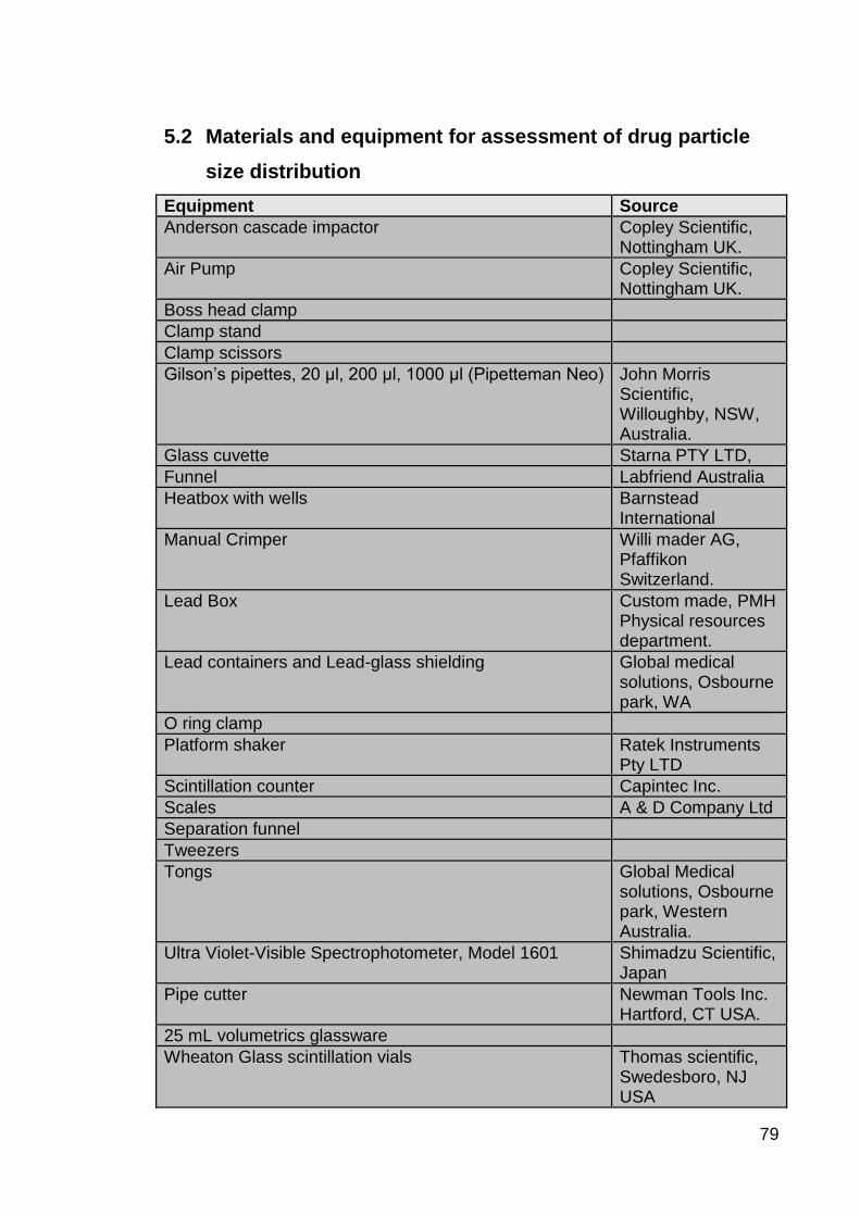
79
5.2 Materials and equipment for assessment of drug particle
size distribution
Equipment Source
Anderson cascade impactor Copley Scientific, Nottingham UK.
Air Pump Copley Scientific, Nottingham UK.
Boss head clamp
Clamp stand
Clamp scissors
Gilson’s pipettes, 20 μl, 200 μl, 1000 μl (Pipetteman Neo) John Morris Scientific, Willoughby, NSW, Australia.
Glass cuvette Starna PTY LTD,
Funnel Labfriend Australia
Heatbox with wells Barnstead International
Manual Crimper Willi mader AG, Pfaffikon Switzerland.
Lead Box Custom made, PMH Physical resources department.
Lead containers and Lead-glass shielding Global medical solutions, Osbourne park, WA
O ring clamp
Platform shaker Ratek Instruments Pty LTD
Scintillation counter Capintec Inc.
Scales A & D Company Ltd
Separation funnel
Tweezers
Tongs Global Medical solutions, Osbourne park, Western Australia.
Ultra Violet-Visible Spectrophotometer, Model 1601 Shimadzu Scientific, Japan
Pipe cutter Newman Tools Inc. Hartford, CT USA.
25 mL volumetrics glassware
Wheaton Glass scintillation vials Thomas scientific, Swedesboro, NJ USA

80
5.3 Consumables for radiolabelling and gamma scintigraphy
on study day
Consumables Source
Compact Space Chamber Plus® spacer Allersearch, VIC, Australia. Owned by EBOS Group Pty Ltd.
Exhalation Filters
QVAR™, 100 μg beclomethasone di-propionate per actuation iNova Pharmaceuticals (Aust) Pty Ltd
Flixotide®, 125 μg fluticasone propionate per actuation Allen & Hanburys respiratory care. Division of GlaxoSmithKline

81
5.4 Parent information and consent form

82

83

84
85
86

87
5.5 Physical examination

88
5.6 Vital sign measurements

89
5.7 Screening questionnaire

90
5.8 Look up table


![Thyroid pathophysiology scintigraphy[1]](https://img.pdfslide.net/doc/110x75/588a7dc81a28abad628b4ebd/thyroid-pathophysiology-scintigraphy1.jpg)

