Embed Size (px)
Citation preview

Topics in Fluorescence Spectroscopy Volume 8 Radiative Decay Engineering
Topics in Fluorescence Spectroscopy Edited by JOSEPH R. LAKOWICZ and CHRIS D. GEDDES
Volume 1: Techniques
Volume 2: Principles
Volume 3: Biochemical Applications
Volume 4: Probe Design and Chemical Sensing
Volume 5: Nonlinear and Two-Photon-Induced Fluorescence
Volume 6: Protein Fluorescence
Volume 7: DNA Technology
Volume 8: Radiative Decay Engineering

Topics in Fluorescence Spectroscopy Volume 8 Radiative Decay Engineering
Edited by
CHRIS D. GEDDES The Institute of Fluorescence Medical Biotechnology Center University of Maryland Biotechnology Institute Baltimore, Maryland
and
JOSEPH R. LAKOWICZ Center for Fluorescence Spectroscopy and Department of Biochemistry and Molecular Biology University of Maryland School of Medicine Baltimore, Maryland
Springer
The Library of Congress cataloged the first volume of this title as follows:
Topics in fluorescence spectroscopy/edited by Joseph R. Lakowicz. p. cm. Includes bibliographical references and index. Contents: v. 1. Techniques 1. Fluorescence spectroscopy. I. Lakowicz, Joseph R.
QD96.F56T66 1991 91-32671 543'.0858—dc20 CIP
Front cover—Surface Plasmon Coupled Emission (SPCE). See Journal of Fluorescence 14(1), 119-123, 2004, and chapter within
ISSN: 1574-1036
ISBN 0-387-22662-1 (HB)
Printed on acid-free paper
©2005 Springer Science-f Business Media, Inc. All rights reserved. This work may not be translated or copied in whole or in part without the written permission of the publisher (Springer Science+Business Media, Inc., 233 Spring Street, New York, NY 10013, USA), except for brief excerpts in connection with reviews or scholarly analysis. Use in connection with any form of information storage and retrieval, electronic adaptation, computer software, or by similar or dissimilar methodology now known or hereafter developed is forbidden. The use in this publication of trade names, trademarks, service marks and similar terms, even if they are not identified as such, is not to be taken as an expression of opinion as to whether or not they are subject to proprietary rights.
Printed in the United States of America
9 8 7 6 5 4 3 2 1 SPIN 11306818
springeronline.com
Contributors
Ricardo F. Aroca • Materials & Surface Science Group, Department of Chemistry and Biochemistry, University of Windsor, N9B 3P4, Windsor, Ontario, Canada
Kadir Asian • Institute of Fluorescence, University of Maryland Biotechnology Institute 725 West Lombard Street, Baltimore, Maryland 21201
Donna Chen • Department of Chemistry and Biochemistry, University of South Carolina, 631 Sumter Street, Columbia, South Carolina 29208
Ashutosh Chilkoti • Department of Biomedical Engineering, Duke University, Durham, North Carolina 27708
Paula E. Colavita • Department of Chemistry and Biochemistry, University of South Carolina, 631 Sumter Street, Columbia, South Carolina 29208
Michael Doescher • Department of Chemistry and Biochemistry, University of South Carolina, 631 Sumter Street, Columbia, South Carolina 29208
Eric Dulkeith • Photonics and Optoelectronics Group, Physics Department and Center for NanoScience, Ludwig-Maximilians-Universitat Miinchen, AmalienstraBe 54, 80799 Miinchen, Germany
Robert C. Dunn • Department of Chemistry, University of Kansas, Lawrence, Kansas, 66045
Jochen Feldmann • Photonics and Optoelectronics Group, Physics Department and Center for NanoScience, Ludwig-Maximilians-Universitat Miinchen, AmalienstraBe 54, 80799 Miinchen, Germany
Chris D. Geddes • Institute of Fluorescence, University of Maryland Biotechnology Institute, and Center for Fluorescence Spectroscopy, 725 West Lombard Street, Baltimore, Maryland 21201
Joel I. Gersten • Department of Physics, City College of the City University of New York, New York, New York 10031
Paul J.G. Goulet • Materials & Surface Science Group, Department of Chemistry and Biochemistry, University of Windsor, N9B 3P4, Windsor, Ontario, Canada
Ignacy Gryczynski • Center for Fluorescence Spectroscopy, University of Maryland, School of Medicine, 725 West Lombard Street, Baltimore, Maryland 21201

vi Contributorrs
Zygmunt Gryczynski • Center for Fluorescence Spectroscopy, University of Maryland, School of Medicine, 725 West Lombard Street, Baltimore, Maryland 21201
Amanda J. Haes • Northwestern University, Department of Chemistry, 2145 Sheridan Road, Evanston, Illinois 60208-3113
Christy L. Haynes • Northwestern University, Department of Chemistry, 2145 Sheridan Road, Evanston, Illinois 60208-3113
Amim Henglein • Hahn-Meitner Institut, 14109 Berlin, Germany
Thomas A. Klar • Photonics and Optoelectronics Group, Physics Department and Center for NanoScience, Ludwig-Maximilians-Universitat MUnchen, AmalienstraBe 54, 80799 Munchen, Germany
Wolfgang Knoll • Max-Planck-Institute for Polymer Research, Ackermannweg 10, 55128 Mainz, Germany
Joseph R. Lakowicz • Center for Fluorescence Spectroscopy, University of Maryland, School of Medicine, 725 West Lombard Street, Baltimore, Maryland 21201
Luis M. LiZ'Marzdn • Departamento de Quimica Fisica, Universidade de Vigo, 36200, Vigo, Spain
Joanna Malicka • Center for Fluorescence Spectroscopy, University of Maryland, School of Medicine, 725 West Lombard Street, Baltimore, Maryland 21201
C. Mayer • Analytical Biotechnology, Technical University of Delft, Julianalaan 67, 2628 BC Delft, The Netherlands
Adam D. McFarland • Northwestern University, Department of Chemistry, 2145 Sheridan Road, Evanston, Illinois 60208-3113
Paul Miney • Department of Chemistry and Biochemistry, University of South Carolina, 631 Sumter Street, Columbia, South Carolina 29208
Annabelle Molliet • Department of Chemistry and Biochemistry, University of South Carolina, 631 Sumter Street, Columbia, South Carolina 29208
David S. Moore-Nichols • Department of Chemistry, University of Kansas, Lawrence, Kansas, 66045
Michael L. Myrick • Department of Chemistry and Biochemistry, University of South Carolina, 631 Sumter Street, Columbia, South Carolina 29208
Nidhi Nath • Department of Biomedical Engineering, Duke University, Durham, North Carolina 27708
Thomas Neumann • Max-Planck-Institute for Polymer Research, Ackermannweg 10, 55128 Mainz, Germany
Contributors vii
Lifang Niu • Departments of Chemistry and of Materials Science, National University of Singapore, 10 Science Drive 4, Singapore 11754
Steven J. Oldenburg • Seashell Technology, La JoUa, California 92037
Isabel Pastoriza-Santos • Departamento de Quimica Fisica, Universidade de Vigo, 36200, Vigo, Spain
Darren Pearson • Department of Chemistry and Biochemistry, University of South Carolina, 631 Sumter Street, Columbia, South Carolina 29208
Jorge PereZ'Juste • Departamento de Quimica Fisica, Universidade de Vigo, 36200, Vigo, Spain
John Reddic • Department of Chemistry and Biochemistry, University of South Carolina, 631 Sumter Street, Columbia, South Carolina 29208
Th. Schalkhammer • Nanobioengineering, Vienna Biocenter, Universitat Wien Dr. Bohrgasse 9, 1030 Wien, Austria. Current address: Schalkhammer KG, Klausenstrasse 129, 2534 Alland, Austria
David A. Schultz • David A. Schultz, University of California, San Diego, La Jolla, California 92093-0319
Evelyne L. Schmid • Departments of Chemistry and of Materials Science, National University of Singapore, 10 Science Drive 4, Singapore 11754
Lindsay Taylor • Department of Chemistry and Biochemistry, University of South Carolina, 631 Sumter Street, Columbia, South Carolina 29208
Richard P. Van Duyne • Northwestern University, Department of Chemistry, 2145 Sheridan Road, Evanston, Illinois 60208-3113
Fang Yu • Max-Planck-Institute for Polymer Research, Ackermannweg 10, 55128 Mainz, Germany
Jing Zhou • Department of Chemistry and Biochemistry, University of South Carolina, 631 Sumter Street, Columbia, South Carolina 29208

Preface
Spatial control of photonic mode density is changing the practice of fluorescence spectroscopy. This laboratory has been active in fluorescence spectroscopy for nearly 30 years. During that time we have investigated many phenomena in fluorescence, including quenching, energy transfer and anisotropy, to name a few. Until recently we relied completely on the free-space emission properties of fluorophores observed in transparent media. The free-space quantities in fluorescence are determined by the values of the radiative and non-radiative properties of excited fluorophores. The observed changes in fluorescence intensities, lifetimes, etc. are due almost completely to changes in the non-radiative decay rates such as quenching. The rate of radiative decay is determined by the extinction coefficient or oscillator strength of the transition. This rate is essentially constant in most media.
In about 2000 we began to examine the effects of silver metallic particles on fluorescence. Examination of the literature revealed that proximity to silver particles could have dramatic effects on fluorescence quantum yields and lifetimes. Such changes are typically due to changes in the non-radiative decay rates. In contrast, the metal particles changed the radiative decay rate (F). These changes occur due to modifications of the photonic mode density (PMD) near the particle in T. This was the first time in 30 years that we saw an opportunity to modify this fundamental rate. Numerous opportunities became apparent as we considered the effects of PMD, including increased quantum yields, increased photostability and changes in resonance energy transfer. Additionally, we saw the opportunity to obtain directional rather than isotropic emission based on local changes in the PMD. We described these phenomena as radiative decay engineering (RDE) because we could engineer changes in the emission based on the fluorophore-metal particle geometries.
During these three years our enthusiasm for RDE has continually increased. Many of the early predictions have been confirmed experimentally. As one example we recently observed directional emission based on fluorophores located near a thin metal film, a phenomenon we call surface plasmon coupled emission (SPCE). We see numerous applications for RDE in biotechnology, clinical assays and analytical chemistry. The technology needed to implement RDE is straightforward and easily adapted by most laboratories. The procedures for making noble metal particles and surfaces are simple and inexpensive. The surface chemistry is well developed, and the noble metals are easily tolerated by biochemistry systems.

X Preface
While implementation of RDE is relatively simple, understanding the principles of RDE is difficult. The concepts are widely distributed in the optics and chemical physics literature, often described in terms difficult to understand by biophysical scientists. In this volume we have presented chapters from the experts who have studied metal particle optics and fluorophore-metal interactions. We believe this collection describes the fiindamental principles for the widespread use of radiative decay engineering in the biological sciences and nanotechnology.
Joseph R. Lakowicz and Chris D. Geddes Center for Fluorescence Spectroscopy
Bahimore, Maryland August 13, 2003
Contents
1. Preparation of Noble Metal Colloids and Selected Structures Isabel Pastoriza-Santos, Jorge Perez-Juste and Luis M. Liz-Marzan
1. Introduction 1 2. Preparation of Noble Metal Colloids 2
2.1. Spherical Nanoparticles in Water 2 2.1.1. Citrate Reduction 2 2.1.2. Borohydride Reduction 3 2.1.3. y-Radiolysis 4 2.1.4. Growth on Preformed Nanoparticles 5 2.1.5. Growth of Silica Shells on Metal Nanoparticles 5
2.2. Spherical Nanoparticles in Organic Solvents 6 2.2.1. Two-Phase Reduction 6 2.2.2. Reduction by the Solvent 6 2.2.3. Reduction within Microemulsions 7
2.3. Nanorods and Nanoprisms in Water 8 2.3.1. Synthesis of Nanorods within Porous Membranes 8 2.3.2. Nanorods from Wet Synthesis in Solution 8 2.3.3. Synthesis of Nanoprisms in Water 10
2.4. Nanorods and Nanoprisms in Organic Solvents 11 2.4.1. Reduction within Microemulsions 11 2.4.2. Reduction by the Solvent 11 2.4.3. Shape Control Using DMF 11
3. Metal Colloid Structures through Layer-by-Layer Assembly 13 3.1. Layer-by-Layer Assembly 13 3.2. Assembly of Au@Si02 13 3.3. Assembly of Au Nanoprisms 15
4. Conclusions 17 5. Acknowledgements 17 6. References 17
2. Near-Field Scanning Optical Microscopy: Alternative Modes of Use for NSOM Probes David S. Moore-Nichols and Robert C. Dunn
1. Introduction 25 2. Scanning Near-Field Fret Microscopy 27 3. Nanometric Biosensors andBioprobes 31 4. Applied Voltage Combined with NSOM for Structure/Dynamic Measurements.. 34

xii Contents
5. Interferometric NSOM Measurements 36 6. Fluorescence, Topography and Compliance Measurements Using
Tapping-Mode NSOM 40 7. Conclusions 43 8. Acknowledgments 44 9. References 44
Nanoparticles with Tunable Localized Surface Plasmon Resonances: Topics in Fluorescence Spectroscopy Christy L. Haynes, Amanda J. Haes, Adam D. McFarland, and Richard P. Van Duyne
Introduction 47 1.1. General Overview 47 1.2. Fabrication of Nanostructures with Tunable Optical Properties 48 1.3. Fundamental Studies of Tunable Optical Properties 52
1.3.1. Defining the Fundamental Characteristics of the Localized Surface Plasmon Resonance 52
1.3.2. Controlling the Localized Surface Plasmon Resonance 53 1.3.3. Implications for Related Phenomena 55
1.4. Applications of Tunable Optical Properties 57 1.5. Goals and Organizations 59 Tunable Localized Surface Plasmon Resonance 59 2.1. Introduction to Colloidal Nanoparticles 59 2.2. Colloidal Nanoparticle Experimental Section 60
2.2.1. Fabrication of Surfactant-Modified Silver Nanoparticles 60 2.2.2. Fabrication of Core-Shell Nanoparticles 60 2.2.3. Transmission Electron Microscopy Characterization 61
2.3. Structural and Optical Properties of Colloidal Nanoparticles 61 2.4. Study of Electromagnetic Coupling Using Electron Beam
Lithography Substrates 64 2.5. Experimental Methods 66
2.5.1. Sample Fabrication 66 2.5.2. Optical Characterization of Nanoparticle Arrays 67 2.5.3. Structural Characterization of Nanoparticle Arrays 68
2.6. Optical Properties of Electron Beam Lithography-Fabricated Nanoparticle Arrays 69
2.7. Tunable Localized Surface Plasmon Resonance Using Nanosphere Lithography 70 2.7.1. Effect of Nanoparticle Material on the LSPR 70 2.7.2. Effect of Nanoparticle size on the Ag LSPR 70 2.7.3. Effect of Nanoparticle Shape on the Ag LSPR 71 2.7.4. Effect of the External Dielectric Medium on the Ag LSPR 73 2.7.5. Effect of Thin Film Dielectric Overlayers on the LSPR 74 2.7.6. Effect of the Substrate Dielectric Constant on the LSPR 75

Contents xiii
3. Recent Applications of the Tunable Localized Surface Plasmon Resonance 75 3.1. Sensing with Nanoparticle Arrays 75
3.1.1. Experimental Procedure 76 3.1.2. Effect of the Alkanethiol Chain Length on the LSPR 16 3.1.3. Streptavidin Sensing Using LSPR Spectroscopy 78 3.1.4. Anti-Biotin Sensing Using LSPR Spectroscopy 80 3.1.5. Monitoring the Specific Binding of Streptavidin to Biotin and
Anti-Biotin to Biotin and the LSPR Response as a Function of Analyte Concentration 80
3.2. Sensing with Single Nanoparticles 82 3.2.1. Experimental Procedure 84 3.2.2. Single Nanoparticle Refractive Index Sensitivity 84 3.2.3. Single Nanoparticle Response to Adsorbates 85
3.3. Plasmon-Sampled Surface-Enhanced Raman Excitation Spectroscopy 86 3.3.1. Experimental Procedure 87 3.3.2. Varying the Excitation Wavelength in PS-SERES 89 3.3.3. Varying the Molecular Adsorbate in PS-SERES 89
4. Conclusions 92 5. Acknowledgements 93 6. References 93
4. Colloid Surface Chemistry Arnim Henglein
1. Introduction 101 2. Radiolytic Methods 101 3. Silver Colloid Preparation 103 4. Pulsed Particle Formation 105 5. Redox Potential and Particle Size 106 6. Polymer Stabilized Clusters 109 7. Electron Donation and Positive Hole Injection 109 8. PhotoelectronEmission I l l 9. Nano-Electrochemistry 114 10. Bimetallic Particles 115 11. Fermi Level Equilibration in Mixed Colloids 122 12. Adsorption of Electrophiles 124 13. Adsorption of Nucleophiles 126 14. Competitive Adsorption and Displacement Processes 130 15. Final Remarks 131 16. References 131

xiv Contents
5. Bioanalytical Sensing Using Noble Meal Colloids C. Mayer and Th. Schalkhammer
1. Bio-Nanotechnology 135 1.1. Metal Colloids 136 1.2. Metal Colloid Devices 140
2. Nano-Cluster Based Technology 141 2.1. Properties 141 2.2. Metal Colloids and Quantum Dots 144
2.2.1. Techniques to Prepare Noble Metal Colloids 145 2.3. Nano-Switches 151 2.4. Cluster-Cluster Aggregates 154 2.5. Coating Clusters with Biomolecules 156 2.6. AFM 158 2.7. Immune Colloidal Techniques 161 2.8. Binding and Assembly of Functionalized Colloids 161 2.9. Bio-Templating 163 2.10. Colloidal Particles and Electrodes 165 2.11. SPR-Transduction 165 2.12. Electroluminescence 166
3. Nano-Cluster and Field Effects 166 3.1. Surface Enhanced Optical Absorption (SEA) 166
3.1.1. Physical Principles 166 3.1.2. Applications 168 3.1.3. Distance Layer and Colloid Layers 169 3.1.4. SEA-Biochips 171
3.1.4.a. The SEA Chip 171 3.1.4.b. Applications and General Requirements 171 3.I.4.C. Setup 172 3.1.4.d. Example and Results 175
3.1.5. Nano-Distance Transduction via SEA Biochips 176 3.1.5.a. How It Works 176 3.1.5.b. Polyvinylpyrrolidone as Distance Layer 177 3.I.5.C. Proteins as Distance Layer 177 3.1.5.d. Spin-Coating of DNA 178 3.1.5.e. SetupofaMICORISChip 179
3.2. Resonance Enhanced Fluorescence (REF) 180 3.2.1. Physical Principles 180 3.2.2. Applications 182 3.2.3. REF in Microtiter-Plates 184 3.2.4. Cluster-Layer Enhanced Fluorescence DNA Chip Setup 184 3.2.5. Clusters Layer Fabrication Methods 185
3.3. Surface-Enhanced Infrared Absorption (SEIRA) 186 3.4. Scattered Evanescent Waves (SEW) 187 3.5. Surface-Enhanced Raman Scattering (SERS) 188 3.6. Cluster-Quenched Fluorescence 190 3.7. Cluster-Emission Devices (CED) 192
4. Acknowledgements 193 5. References 193

Contents xv
6. Theory of Fluorophore-Metallic Surface Interactions Joel I. Gersten
1. Introduction 197 2. Theory 199 3. Appendix A 216 4. References 220
7. Surface-Enhancement of Fluorescence Near Noble Metal Nanostructures Paul J.G. Goulet and Ricardo F. Aroca
1. Introduction 223 2. Electromagnetic Enhancement 225 3. Enhanced Absorption 229 4. Radiationless Energy Transfer and Distance Dependence 230 5. Coverage Dependence 233 6. Temperature Dependence 234 7. Quantum Efficiency and Enhancement 235 8. Enhancing Substrates 236 9. SEF of Langmuir-Blodgett Films 239 10. Summary and Outlook 243 11. Acknowledgements 244 12. References 244
8. Time-Resolved Fluorescence Measurements of Fluorophores Close to Metal Nanoparticles Thomas A. Klar, Eric Dulkeith, and Jochen Feldman
1. Introduction 249 2. Nanoparticle Plasmons 250 3. Fluorescence Decay Rates of Fluorophores in the Vicinity of Metal Structures ... 252
3.1. Theory 252 3.2. Time Resolved Spectroscopy 255
4. Time Resolved Spectroscopy of Fluorophores Bound to Metal Nanoparticles 257 5. Biophysical Applications 266 6. Acknowledgements 269 7. References 269
xvi Contents
9. Copper Coated Self-Assembled Monolayers: Alkanethiols and Prospective Molecular Wires Paula E. Colavita, Paul Miney, Lindsay Taylor, Michael Doescher, Annabelle Molliet, John Reddic, Jing Zhou, Darren Pearson, Donna Chen, and Michael L. Myrick
1. Introduction 275 2. Copper Overlayers on Alkanethiol Self-Assembled Monolayers 278
2.1. General Factors Affecting the Behavior of Metals Deposited onto Self-Assembled Monolayers 278
2.2. RAIRS Spectra of Alkanethiol SAMs in the C-H Stretching Region 280 2.3. Copper on Dodecanethiol and Octadecanethiol [30] 283
3. Copper on Conjugated Oligomers 293 4. Acknowledgments 300 5. References 300
10. Principles and Applications of Surface-Plasmon Field-Enhanced Fluorescence Techniques Wolfgang Knoll, Fang Yu, Thomas Neumann, Lifang Niu, and Evelyne L. Schmid
1. Introduction 305 2. Surface Plasmons as Interfacial Light 307 3. Chromophores Near Metal Surfaces 313 4. Recording Fluorescence from Chromophores Excited by Surface
Plasmon Waves 316 5. Surface Hybridization Studies 321 6. Protein Binding Studies — the Limit of Detection in SPFS 326 7. Surface Plasmon Fluorescence Microscopy 329 8. Conclusions 329 9. Acknowledgements 331 10. References 331
11. Optically Detectable Colloidal Metal Labels: Properties, Methods, and Biomedical Applications Steven J. Oldenburg and David A. Schultz
1. Introduction 333 2. Plasmon Resonance 334
2.1. Experimental and Theoretical Considerations 334 2.2. Particle Fabrication 337
2.2.1. Spherical Particles 337 2.2.2. Anisotropic, Elliptical or Rod Shaped Particles 338 2.2.3. Core: Shell Particles 339
2.3. Particle Characterization 339
Contents xvii
3. Particle Surface Modification to Produce Biological Labels 340 3.1. Particle Surface Modification 340
4. Dark Field Optical Microscope Designs for Plasmon Resonant Particle (PRP) Detection 341 4.1. Microscope Configuration 341 4.2. Illumination Light Sources 343 4.3. Apparatus for Individual Plasmon Resonant Particle
Spectral Determination 344 4.4. Single Particle Counting 344
5. Biological Applications 345 5.1. Individual PRP Detection and Counting in a Protein or DNA
Microarray Format 346 5.2. Individual PRP Detection of Immuno-Labeled Tissue 348 5.3. Plasmon Resonant Particles and Other Nanoparticles as Labels
for Biomedical Applications 349 6. References 349
12. Noble Metal Nanoparticle Biosensors Nidhi Nath and Ashutosh Chilkoti
1. Introduction 353 2. Noble Metal Nanoparticle 355
2.1. Optical Properties of Noble Metal Nanoparticles 355 2.2. Noble Metal Nanoparticles: Historical Perspective 357
3. Nanoparticle SPR Biosensor 360 3.1. Noble Metal Nanoparticles: Synthesis 361 3.2. Self-Assembly of Noble Metal Nanoparticles on Substrate 362 3.3. Optical Properties of self-Assembled Gold Nanoparticle on Glass 365
3.3.1. Refractive Index Response of Metal Nanoparticles 365 3.3.2. Spatial Sensitivity of Immobilized Gold Nanoparticles on Glass 368
3.4. Biosensing Using Noble Metal Nanoparticles 370 3.4.1. Receptor Presentation 371 3.4.2. Biomolecular Binding 373
4. Future Directions 376 5. Acknowledgements 376 6. References 377
13. Surface Plasmon-Coupled Emission: A New Method for Sensitive Fluorescence Detection Ignacy Gryczynski, Joanna Malicka, Zygmunt Gryczynski and Joseph R. Lalowicz
1. Introduction 381 2. Surface Plasmon Resonance Analysis 381

xviii Contents
3. Surface Plasmon-Coupled Emission 386 3.1. Properties of SPCE with Reverse Kretschmann Excitation 386 3.2. Properties of SPCE with Kretschmann Excitation 394 3.3. Background Rejection with SPCE 396 3.4. DNA Hybridization Using SPCE 398
4. Discussion 401 5. Acknowledgment 401 6. References 401
14.' Radiative Decay Engineering (RDE) Chris D. Geddes, Kadir Asian, Ignacy Gryczynski, Joanna Malicka and Joseph R. Lakowicz
1. Introduction 405 2. Enhanced Emission from Low and High Quantum Yield Species
Using Silver Island Films (SiFs) 411 3. Enhanced Intrinsic Fluorescence Using SiFs 415 4. Distance Dependence of Enhanced Fluorescence Using SiFs 417 5. Release of Self-Quenching Using SiFs 419 6. Other Metal-Nanostructures for MEF Using Indocyanine Green (ICG) 422
6.1. Silver Island Films (SiFs) 424 6.2. Immobilized Silver Colloids 425 6.3. Photo-Deposition of Silver onto Glass 426 6.4. Electoplating of Silver on Substrates 434 6.5. Roughened Silver Electrodes 435 6.6. Silver Fractal-like Structures on Glass 439 6.7. Silver Nanorods 443
7. Closing Remarks 445 8. Acknowledgments 446 9. References 446
PREPARATION OF NOBLE METAL COLLOIDS AND SELECTED STRUCTURES
Isabel Pastoriza-Santos, Jorge Perez-Juste, and Luis M. Liz-Marzan*
1. INTRODUCTION
Metal colloids have been around for centuries because of their striking optical properties. A typical example of ancient colloidal metal nanoparticles is the famous Lycurgus cup, which dates back to the fourth century AD. The cup is still at the British Museum^ and possesses the unique feature of changing color, since it is green when viewed in reflected light, but it appears red when a light is shone from inside and is transmitted through the glass. Analysis of the glass reveals that it contains a very small amount of tiny (ca. 70 nm) crystals of metal containing silver and gold in an approximate molar ratio or 14:1. It is the presence of these nanocrystals which imparts Lycurgus cup with its special color display. After such early origins, metal colloids, and particularly colloidal gold, were known and used in the Middle Ages by the alchemists for their health restorative properties, which are still popular nowadays.^ Although the discovery that glass could be colored red by adding a small amount of gold powder is often credited to Johann Kunckel, a German glassmaker in the late seventeenth century, it was not until 1857 that Michael Faraday performed a systematic study on the synthesis and color of colloidal gold. Since that pioneering work, thousands of scientific papers have been published on the synthesis, modification, properties, and assembly of metal nanoparticles, using a large variety of solvents and other substrates. All this has led not only to reliable procedures for the preparation of metal nanoparticles of basically any size and shape, but also to a deep understanding of many of the physico-chemical features that determine the special behavior of these systems.
One of the very interesting aspects of metal colloids is that their optical properties strongly depend on particle size and shape. Bulk gol4 for instance has a high absorbance along the whole visible range, except for a slight dip around 400-500 nm, which makes Au thin films look blue in transmission. As particle size is reduced fi om say 500 nm down to ca. 3 nm, the color changes gradually to orange, through several tones of purple and red. These effects are due to changes in the so-called surface plasmon resonance,"^
Isabel Pastoriza-Santos, Jorge Perez-Juste, and Luis M. Liz-Marzan, Departamento de Quimica Fisica, Universidade de Vigo, 36200, Vigo, Spain.
Topics in Fluorescence Spectroscopy, Volume 8: Radiative Decay Engineering Edited by Geddes and Lakowicz, Springer Science+Business Media, Inc., New York, 2005
2 I. PASTORIZA-SANTOS et al
which is the frequency at which conduction electrons oscillate as a response to the alternating electric field of an incoming electromagnetic wave. Below 3 nm, the color changes to brownish and eventually colorless, which is mainly due to quantum effects. Shape effects are even stronger, and slight deviations from the spherical geometry can lead to quite impressive color changes.
The plasmon resonance is one of the most characteristic properties of some noble metal colloids, and can be predicted using classical calculations of the scattering by small particles, which is usually known as Mie theory.^ Although UV-visible spectra for all metals can be calculated from such theory,^ only metals with free electrons (basically Au, Ag, Cu, and the alkali metals) possess plasmon resonances in the visible that provide them with intense colors. Elongated nanoparticles (ellipsoids and nanorods) display two distinct plasmon bands related to transversal and longitudinal electron oscillations, being the longitudinal one very sensitive to the aspect ratio of the particles.^ Apart from single particle properties, also the environment in which the metal particles are dispersed is of relevance to the optical properties.^ The refractive index of the dispersing medium^^ as well as the average distance between neighboring metal nanoparticles^* have been shown to influence the spectral features, which has been exploited for applications such as DNA detection.*^
Metal nanoparticles also have important applications in catalysis, '*" mainly because of their extremely large surface to volume ratio, which allows a more effective utilization of expensive transition metals, and additionally, since the metal nanoparticles can be either supported onto solid substrates or dispersed in a solvent, they can be used for both heterogeneous and homogeneous catalysis. We are not going to describe in detail the catalytic properties of metal colloids here.
In this chapter, we intend to introduce to the reader some of the most popular preparation methods of noble metal colloids, but do not intend by any means to thoroughly review the scientific literature on metal colloid synthesis. We have chosen examples of synthesis in aqueous and organic solvents, and differentiate between spherical and anisotropic nanoparticles (nanorods and nanoprisms). Additionally, in section 3 we shall describe one of the most popular recent procedures to assemble metal colloids into nanostructured materials (layer-by-layer assembly), as well as the properties of the resulting structures.
2. PREPARATION OF NOBLE METAL COLLOIDS
2.L Spherical Nanoparticles in Water
2.1.1. Citrate Reduction
Probably the most popular method to prepare Au nanospheres dispersed in water is the reduction with sodium citrate at high temperature. This method was first introduced by Turkevich and co-workers back in the fifties,*^ and frirther studied and modified in a number of later papers. ^"' This preparation is basically performed by heating a dilute (typically 1-5 x lO"" M) aqueous solution of HAuCU to its boiling point and then rapid addition of sodium citrate solution. The formation of the gold colloid is then rather slow, with a spectacular color change from light yellow (due to HAuCU) to colorless, then to gray, blue, purple, and finally (after some 10 minutes) deep wine red, revealing the
NOBLE METAL COLLOIDS. 3
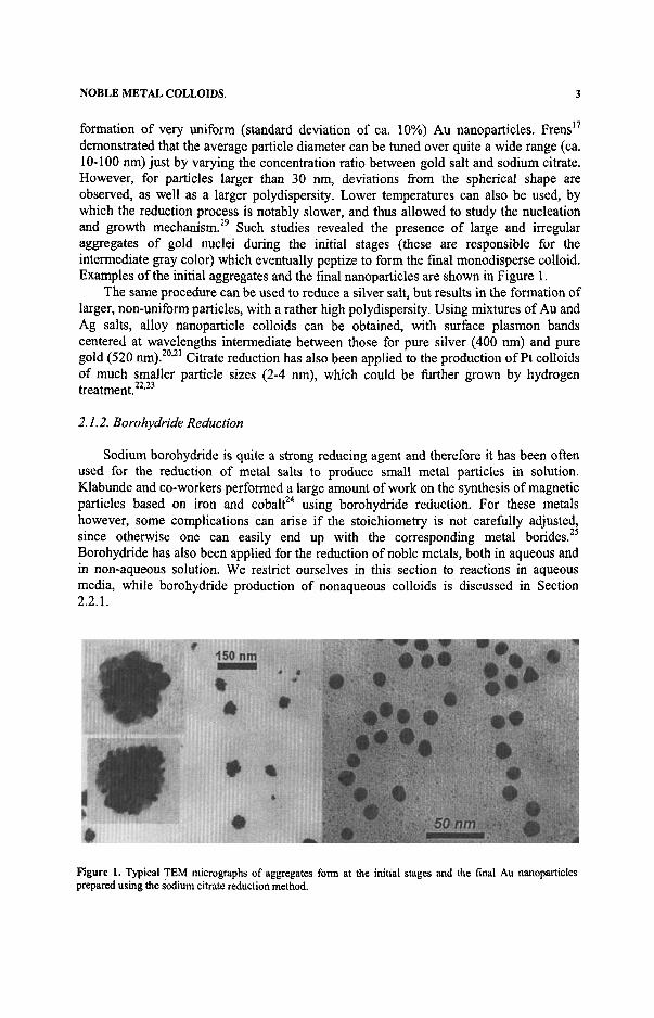
formation of very uniform (standard deviation of ca. 10%) Au nanoparticles. Frens^^ demonstrated that the average particle diameter can be tuned over quite a wide range (ca. 10-100 nm) just by varying the concentration ratio between gold salt and sodium citrate. However, for particles larger than 30 nm, deviations from the spherical shape are observed, as well as a larger polydispersity. Lower temperatures can also be used, by which the reduction process is notably slower, and thus allowed to study the nucleation and growth mechanism. ^ Such studies revealed the presence of large and irregular aggregates of gold nuclei during the initial stages (these are responsible for the intermediate gray color) which eventually peptize to form the final monodisperse colloid. Examples of the initial aggregates and the final nanoparticles are shown in Figure 1.
The same procedure can be used to reduce a silver salt, but results in the formation of larger, non-uniform particles, with a rather high polydispersity. Using mixtures of Au and Ag salts, alloy nanoparticle colloids can be obtained, with surface plasmon bands centered at wavelengths intermediate between those for pure silver (400 nm) and pure gold (520 nm). ' ^ Citrate reduction has also been applied to the production of Pt colloids of much smaller particle sizes (2-4 nm), which could be fiuther grown by hydrogen treatment. ' ^
2.1.2. Borohydride Reduction
Sodium borohydride is quite a strong reducing agent and therefore it has been often used for the reduction of metal salts to produce small metal particles in solution. Klabunde and co-workers performed a large amount of work on the synthesis of magnetic particles based on iron and cobalt " using borohydride reduction. For these metals however, some complications can arise if the stoichiometry is not carefiilly adjusted, since otherwise one can easily end up with the corresponding metal borides. ^ Borohydride has also been applied for the reduction of noble metals, both in aqueous and in non-aqueous solution. We restrict ourselves in this section to reactions in aqueous media, while borohydride production of nonaqueous colloids is discussed in Section 2.2.1.
Figure L Typical TEM micrographs of aggregates form at the initial stages and the fmal Au nanoparticles prepared using the sodium citrate reduction method.
4 I. PASTORIZA-SANTOS et al
The formation of metallic platinum by borohydride was first reported by Brown and Brown^^ related to the study of hydrogenation reactions. These authors did not use any stabilizing agent, and therefore obtained what they termed "finely-divided black precipitates". A more detailed and systematic study using polyvinylpyrrolidone (PVP) as a protective agent (this was the traditional term for polymers, surfactants or other molecules used to stabilize colloids, and which has nowadays been replaced by other terms such as stabilizer or capping agent) and comparing borohydride with other reducing agents, was performed by Van Rheenen et al. ^
Gold and silver, as well as bimetallic nanoparticles can also be easily formed using borohydride as a reductant, and suitable stabilizers. Liz-Marzan and Philipse^^ studied all possible combinations of Ag, Au and Ft, using clay fibers as supporting colloids. Optical analysis of the obtained colloids suggested that core-shell morphologies were obtained in some cases. Rather monodisperse silver nanoparticles can also be prepared by borohydride reduction in the presence of poly(acrylic acid), ^ and stabilization with citrate ions leads to small (ca. 3 nm) and rather uniform Au nanoparticles, which have been used as seeds for growth of larger spheres, ^ nanoshells, ^ and nanorods,^^ as detailed below.
2.1.3. y-Radiolysis
When speaking about metal nanoparticles and their properties. Prof Arnim Henglein's name should always be borne in mind. Henglein's pioneering contribution to understanding the special properties, not only of metal nanoparticles and clusters, ^ but also of semiconductor quantum dots " has inspired much of the work subsequently performed, as revealed in a recent festschrift issue in The Journal of Physical Chemistry. ^ One of these outstanding contributions has been the development of a very usefiil technique for the production of metal nanoparticles of controlled size, based on the formation of hydrated electrons and organic radicals in solution through 7-radiolysis, which then acted as reducing agents for metal ions, yielding metal atoms which subsequently coalesce to form larger particles.^^
Henglein and co-workers were pioneers in the application of radiation chemistry to synthesize very small metal nanoparticles, ' ^ as well as to study their photochemistry^^ and reactivity." " ^ They also synthesized larger metal particles of Au"" '"^ and Ag ^ with narrow size distributions by enlarging Au and Ag seeds, respectively, which in the case of Au constituted a usefiil material to study fast electronic processes." ^ Additionally, the same group applied radiation chemistry techniques to produce core-shell bimetallic particles. In the presence of suitable stabilizers, shells of different metals and various thickness could be grown around chemically performed seeds by varying the type and concentration of the metal ions in solution. Thus, high quality metal particles such as, Au@Pb,''''^ Au@Pb@Cd,'' Ag@Au,'^ Pt@Ag,'^ Au@Hg, ^ Pt@Au'2' ^ and Pd@Au'^'^' were produced to study their optical and chemical properties.
Other groups like those of Belloni ' ^ or Doudna ' ^ also applied 7-radiolysis to reduce metals. Most of the systems reported by these groups were metallic alloys with catalytic applications. Recently, Doudna et al. ^ reported a method based on radiolytic reduction to generate filament-like nanostructures of Ag@Pt.
NOBLE METAL COLLOIDS. 5
2.1.4. Growth on Preformed Nanoparticles
There are many synthetic methods to produce metal nanoparticles with sizes typically below 20 nm, but only a few methods produce particles of uniform size. To obtain larger particles, the difficulty in controlling the nucleation and growth steps occurring at intermediate stages results in a broad particle size distribution. ^ A common approach for controlling the size of nanoparticles is seeding growth. ' " ^ In seeding growth methods, small metal particles are prepared first and later used as seeds (nucleation centers) for the preparation of particles with larger size. Providing a controlled number of preformed seeds and growth conditions that inhibit any secondary nucleation, the particle size can be controlled simply by varying the ratio between seed and metal salt concentrations. The main difficulty is to fmd suitable growth conditions to avoid that a new nucleation process takes place during the growth stages, which strongly limits the application of these methods. ^ Henglein employed radiolytic reduction for seeded growth of gold and silver particles, since the flux of 7-rays can be conveniently controlled to vary the amount of metal atoms produced.'* ' ^ Natan and co-workers used a reducing agent (hydroxylamine) that is too weak to reduce the gold metal salt without the presence of seeds, since these act as catalysts." ^ Similarly, Murphy and co-workers used ascorbic acid as the reducing agent and added cetyltrimethyl ammonium bromide in the growth solution to complex the gold salt and prevent its reduction in the absence of seeds. ^ The growth mechanism consists of an electron transfer from the reducing agent to the metal particles, followed by the reduction of the gold salt on the surface of the seed, so that the growth process follows electrochemical mixed current theory. ' ^ Schematically,
Mn ' -' - + AuCU" -^ Au(„-,i)''' + 4Cr (1)
where Mn ' '* ' is a metal particle that has already accumulated x electrons. To avoid additional nucleation, a step-by-step particle enlargement method is more
useful, allowing a large metal to salt ratio to be maintained throughout successive growth steps. ^ A limitation of the seeded growth method is the lack of availability of smaller seeds with a narrow size distribution. The seeding growth method has been also used for the preparation of different metallic core-shell particles; Au@Ag, ® Ag@Au, ^ Pt@Au and Au@Pt. ^
2.1.5. Growth of Silica Shells on Metal Nanoparticles
Among the various surface modification processes developed for metal (and other) colloids, the growth of silica shells with tailored thickness has recently gathered great attention. One of the major reasons for silica coating is the anomalously high stability of silica colloids, especially in aqueous media, but other reasons are the easy control on the deposition process, its processibility, chemical inertness, controllable porosity, and optical transparency. All these properties make of silica an ideal material to modulate surface properties, while maintaining the physical properties of the underlying cores.
Silica coating of metal colloids presents the difficulty of a chemical mismatch between the core and the shell materials. Liz-Marzan and Philipse ^ first approached the synthesis of Au@Si02 particles through borohydride reduction of AuCU" in the presence
6 I. PASTORIZA-SANTOS et al.
of small silica spheres, followed by extensive silica growth in ethanol of the resulting heteroaggregates. Although coated gold nanoparticles were obtained, these were mixed with pure silica spheres, which were difficult to separate.
A method that has provided substantially better results was later designed by Liz-Marzan et dXP'^^ using silane coupling agents as primers to deposit a first monolayer of silanol groups on the surface, which would then be receptive toward deposition of thin silica layers from sodium silicate solution. Subsequent shell growth could be carried out in ethanol using the so-called Stober method. ^ Monodisperse colloids can be obtained using this procedure with nearly perfect core-shell structures. Examples of such particles are shown in Figure 5 below. This system has proved extremely useful for instance for systematic studies of optical properties, both in solution " ' ' ^ and within nanostructures. ' ' ^ A similar strategy can be applied to the coating of silver coUoids, ^ with the additional difficulty that dissolution of the Ag cores was observed when concentrated ammonia was added to increase the thickness of the silica shell, leading to formation of hollow silica shells. ^
Other methods have been devised for the preparation of Ag@Si02, such as consecutive reactions within microemulsion droplets,*^ or the reduction of Ag^ by A ,A -dimethylformamide (DMF) in the presence of an amino-silane.^^ However, none of these methods have been used for systematic studies until now.
2.2. Spherical Nanoparticles in Organic Solvents
2.2.1. Two-Phase Reduction
One of the currently most used methods for the preparation of gold nanoparticles has been developed by Brust et al. ' "* in 1994. This method combines borohydride reduction with the use of thio- or amino-derivatives as steric stabilizers, by means of a two-phase reaction. Basically, HAuCU is dissolved in water and subsequently transported into toluene by means of tetraoctylammonium bromide, which acts as a phase transfer agent. The toluene solution is then mixed and thoroughly stirred together with an aqueous solution of sodium borohydride, in the presence of thio-alkanes or amino-alkanes, which readily bind to the formed Au nanoparticles. Depending on the ratio between Au salt and capping agent (thiol/amine), the particle size can be tuned between ca. 1 and 10 nm. Several refinements of the preparative procedure including the development of analogous methods for the preparation of silver particles have been reported. ' ^ Murray and coworkers also contributed to enhance the method's popularity by offering an interesting and elegant alternative to the two-phase reduction method which has opened a new field of preparative chemistry. They explored routes to functionalized monolayer protected clusters by ligand place exchange reactions. ' ^ Simple alkane thiol ligands can be completely or partially exchanged by a variety of functional groups, which can be for instance electrochemically active ' ' ^ or photoluminiscent,^ allowing the chemical modification of their ligand shell.
2.2.2. Reduction by the Solvent
Several examples exist on the reduction of metallic salts by organic solvents. Probably the most popular one has been ethanol, which was long used by Toshima and co-workers for the preparation of metal nanoparticles such as Pt, Pd, Au or Rh (suitable

NOBLE METAL COLLOIDS. 7
for catalytic applications) in the presence of a protecting polymer. ' " Another interesting example is found in Figlarz's polyol method, which was initially developed for the formation of larger colloidal particles, ' ^ though later was also adapted and improved for the production of silver nanoparticles.^^ Propanol was also used by Mills and coworkers for the preparation of silver colloids in basic conditions.^^ Related to these processes we can also mention the reduction of noble metal salts by non-ionic surfactants, and more specifically by those with a large number of ethoxy groups, to which the reducing ability has been assigned. ' ^^
N,N-dimethylformamide (DMF) is also one of the usual organic solvents for numerous processes, and it has been shown to behave as an active reducing agent under suitable conditions. Pastoriza-Santos et al. reported the ability of DMF to reduce Ag" ions to the zero-valent metal, even at room temperature and in the absence of any external reducing agent. *' ^ Stable spherical silver nanoparticles were synthesized in DMF, using poly(vinyl pyrrolidone) as a stabilizer, ^^ but additionally silica-^^ and titania- " coated nanoparticles were produced by the same method, in the presence of aminopropyl-trimethoxysilane and titanium tetrabutoxide, respectively. It was also shown that formamide^^^ can act as a powerful reductant for silver or gold salts in the absence of oxygen. More recently, Diaz and coworkers reported the preparation of silver nanoparticles by the spontaneous reduction of silver 2-ethylhexanoate in dimethyl sulfoxide (DMSO) at room temperature. ^
2.2.3. Reduction within Microemulsions
Microemulsions have been used as confined reaction media during the past two decades, since, due to the very small size of the droplets, they can act as microreactors capable to control the size of the particles and at the same time to inhibit the aggregation by adsorption of the surfactants on the particle surface when the particle size approaches that of the microreactor droplet. The synthesis of nanoparticles using reactions in microemulsions was first described by Boutonnet and coworkers^^^ They synthesized monodispersed metal particles of Pt, Pd, Rh and Ir by reduction of metal salts with hydrogen or hydrazine in water in oil (w/o) microemulsions. Since then, many different types of materials have been prepared using microemulsions, including metal carbonates, ^^ metal oxides,' ' ^^ metal chalcogenides, ^ " ^^ polymers, " etc.
In the case of noble metals, several protocols have been described to produce metal nanosized particles by chemical reduction in microemulsions. Pileni and co-workers synthesized metallic particles of Ag, ' Cu, ^ Co ^ (and other materials) by reduction of the corresponding salts in w/o microemulsions using borohydride or hydrazine as reducing agents. They also showed a correlation between the structure of the mesophase in the surfactant system with the size and shape of the formation of metallic particle.
The wide range of applications (in catalysis, as biological stains, as condensers for electron storage in artificial photosynthesis or as ferrofluids) has also motivated studies by other groups on the formation of colloidal Ag, ^ Au, ^ Ni, ^ Cu, ^ Pd ^ and Co ^ particles. However, there is still a strong controversy on whether the microemulsion droplet size actually controls the final size of the obtained nanoparticles.
In recent years, the use of supercritical solvents has offered a new alternative in the synthesis of nanoparticles in microemulsions which presents several advantages over conventional organic solvents, because their solvation properties can be easily controlled
8 I. PASTORIZA-SANTOS et al
by varying pressure. Colloidal silver ^ ' ^ and copper ^^ have been synthesized using microemulsions of water in supercritical CO2 with fluorinated surfactants.
2.3. Nanorods and Nanoprisms in Water
23.1. Synthesis of Nanorods within Porous Membranes
The first procedure reported for the production of metal nanorods is called template synthesis. ^ ' ^ This method entails the preparation or deposition of the desired material within the cylindrical and monodisperse pores of a nanopore membrane. Martin and coworkers used polycarbonate filters, prepared by the track-etch method, ^^ and nanopore aluminas prepared electrochemically from Al foil/^^ as template materials. This method allows the preparation of cylindrical nanostructures with monodisperse diameters and lengths, and depending on the nature of the membrane and the synthetic method used, these may be solid nanowires or hollow nanotubes. ^^
The earliest applications of the template method consisted of the preparation of microscopic and macroscopic electrodes prepared by depositing Au within the pores of policarbonate membranes using electrochemical^^^ or electroless plating^^^ methods. In the electrochemical method, the Au deposition is more uniform and starts at the pore walls creating, at short deposition times, hollow Au nanotubes within the pores. Further deposition results in the formation of Au nanowires. The electroless method comprises the application of a catalyst to all the surfaces of the templating membrane. The membrane is then immersed into the electroless plating bath which contains Au and a chemical reducing agent. Considering that the reduction of Au to metallic Au only occurs in the presence of the catalyst, Au nanotubes are obtained along the pore walls. The thickness of the nanotube walls increases and the inner diameter of the tubes decreases with electroless plating time. At longer plating times, membranes containing nanowires are obtained.
A sequential electrodeposition within a porous template can be used to prepare striped nanowires with tailorable dimensions and composition. ^^ Variation in composition along the length of the wire can be used to incorporate electrical functionalities, ^^ optical contrast, ^^ and the desired surface chemistry. ^
Although very uniform thickness and controlled length were achieved, the amounts that could be made following this procedure were always very small, but still considerable basic work on the optical properties and alignment of these rods could be achieved. ^ - ^
2.3.2. Nanorods from Wet Synthesis in Solution
Wang and co-workers have developed an electrochemical solution phase synthesis of gold nanorods. " ' "^ Their synthetic approach is to control the growth by introducing into the electrochemical system appropriate "shape-inducing" cationic surfactants and other additives that were found empirically to favor rod formation and act as both the supporting electrolyte and the stabilizer for the resulting cylindrical Au nanoparticles. In the electrochemical method for Au nanorod formation the micellar system consists of two cationic surfactants: cetyltrimethyl ammonium bromide (CTAB) and the much more hydrophobic surfactant tetradecyl ammonium bromide (TDAB) or tetraoctyl ammonium bromide (TOAB). The ratio between the surfactants controls the average aspect ratio of
NOBLE METAL COLLOIDS. 9
the gold nanorods. The synthesis works in a small scale and is very difficult to carry out on a large scale, but represents a landmark in terms of shape control. The groups of El-Sayed and Hartland have shown that such rods possess quite unique and interesting optical properties. The particles may be selectively melted using laser pulses and show exciting coupling of the electron gas to bending modes. " ' " ^ Additionally, very strong optical polarization effects have been observed in nanorod polymer films. "^
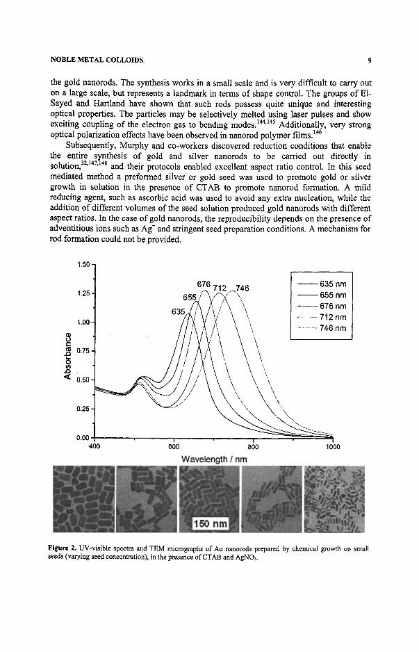
Subsequently, Murphy and co-workers discovered reduction conditions that enable the entire synthesis of gold and silver nanorods to be carried out directly in solution, ' " ' " ^ and their protocols enabled excellent aspect ratio control. In this seed mediated method a preformed silver or gold seed was used to promote gold or silver growth in solution in the presence of CTAB to promote nanorod formation. A mild reducing agent, such as ascorbic acid was used to avoid any extra nucleation, while the addition of different volumes of the seed solution produced gold nanorods with different aspect ratios. In the case of gold nanorods, the reproducibility depends on the presence of adventitious ions such as Ag" and stringent seed preparation conditions. A mechanism for rod formation could not be provided.
1.60
635 nm 655 nm 676 nm 712 nm 746 nm
Figure 2. UV-visible spectra and TEM micrographs of Au nanorods prepared by chemical growth on small seeds (varying seed concentration), in the presence of CTAB and AgNOa.
10 I. PASTORIZA-SANTOS et al
The seed mediated method has been recently improved by El-Sayed and coworkers/ ^ resulting in a spectacular increase in the yield of rod formation, similar to the photochemical method but easier to produce. Two modifications were applied to the seed-mediated growth method: (a) replacement of citrate with CTAB molecules in the seed formation step and (b) adjustment of the silver content of the growth solution to grow nanorods with controlled aspect ratios, resulting in the formation of only 0-1% of spheres. Examples of rods formed using this procedure are shown in Figure 2. In the same figure, the corresponding UV-visible spectra were plotted, to show how an increase in aspect ratio leads to a red-shift of the longitudinal plasmon band, while the transversal resonance only slightly varies due to differences in particle thickness.
Other methods that were recently reported include a seedless and surfactantless wet chemical method by Murphy and co-workers, to obtain silver nanowires in the presence of sodium citrate and sodium hydroxide, ^® as well as a photochemical method developed by Yang et al. for the synthesis of gold nanorods. ^ In contrast to the seed mediated mechanism proposed by Murphy, in the photochemical method the addition of a preformed gold seed is not needed. However, different amoimts of silver nitrate are added to the growth solution containing CTAB and the gold salt before irradiating the solution with UV light. Again, the role of the Ag^ in controlling the aspect ratio of the rods could not be determined.
Some work has also been recently carried out on the solution synthesis of bimetallic nanorods, employing gold nanorods as preformed cores. Jang and co-workers synthesized Au@Ag core-shell nanorods using the electrochemical method to obtain the gold nanorods and the seed-mediated mechanism to get the desired core-shell structure. ^ The combination of the different optical resonances from the core and the shell metals leads to a quite spectacular optical spectrum from these composite, anisotropic nanoparticles.
2.3J. Synthesis ofNanoprisms in Water
Although some triangular and polygonal flat nanoprisms are often obtained during every metal colloid preparation, the synthesis of metal flat nanoprisms with relatively high yield is quite recent. Only in November 2001, ^ Jin et al. reported the conversion of citrate-stabilized silver nanospheres into (truncated) triangular nanoprisms driven by irradiation with a 40-W fluorescent lamp, in the presence of bis(p-sulfonatophenyl) phenylphosphine. Apparently, the initial silver nanoparticles are first fragmented into smaller culsters, and subsequently there is a growth of particles with nanoprism shape, at the cost of the smaller clusters, which is induced by the phosphine present in solution. The optical spectra of the nanoprisms displayed bands for in-plane dipole resonance, as well as for in-plane and out-of-plane quadrupole resonances, while the out-of-plane dipole was only reflected as a small shoulder. Truncated triangles were also obtained by Chen and Carrol ^ using a procedure quite similar to the seeded growth method described for Au nanorod formation. In this case, the reduction was performed using ascorbic acid to reduce silver ions on silver seeds in a basic solution of concentrated CTAB.
Malikova et al. ^ synthesized Au nanoprisms in aqueous solution by reduction of neutralized HAuCU with salicylic acid at 80 °C. Although the yield of nanoprisms was not outstanding, the formation of thin films showed that strong optical coupling occurs when the nanoprisms are close enough to each other (see Section 3.3 below).
NOBLE METAL COLLOIDS. 11
2.4. Nanorods and Nanoprisms in Organic Solvents
2.4.1. Reduction within Microemulsions
While the microemulsion method has been widely applied to the production and stabilization of spherical metal particles with various sizes and compositions, shape control of noble metallic particles using this procedure has only been demonstrated in a handful of studies to date. Pileni and co-workers demonstrated that it is possible to control nanocrystal shape to some extent within microemulsions. ^ Although the shape of the templates plays a role during the growth of the nanocrystals, these authors showed that the particle shape can be controlled even if the microscopic structure of the self-assembled surfactant system used as a template remains unchanged and that addition of salt to the templates can induce drastic changes in the particle shape. Recently, the same group also reported the synthesis of silver nanodisks in reverse micellar solution by reduction of Ag(AOT) with hydrazine, with various sizes that depended on the relative amount of hydrazine, but with constant aspect ratio. ^
Ni nanorods have been synthesized by reduction of nickel chloride with hydrazine hydrate in w/o microemulsion. ^ Another use of interfaces to drive the synthesis of nanomaterials of variable morphology was introduced very recently by Sanyal and coworkers, ^ who demonstrated that the reduction of aqueous chloroaurate ions by anthracene anions bound to a liquid-liquid interface produced thin nanosheets of gold.
2.4.2. Reduction by the Solvent
Xia and coworkers recently demonstrated that the polyol method^^ can be applied for the production of silver nanowires by reducing silver nitrate with ethylene glycol in the presence of poly(vinyl pyrrolidone)(PVP).^^^ ^ Silver nanoparticles with high aspect ratios were only formed in the presence of (Ft) seeds formed in situ prior to the addition of the silver salt. In the course of refluxing, These authors claim that Ostwald ripening takes place at the high temperatures used, forming nanoparticles with larger sizes at the expense of smaller ones. It is likely that PVP controls the growth rates of various faces of silver by coordinating to the surface. As a result, silver nanowires with diameters in the range of 30-60nm and lengths up to -50|j.m were formed. They could control the dimensions of the silver nanowires by varying the experimental conditions (temperature, seed concentration, ratio between silver salt and PVP, etc.). In a later report, ^^ the same group showed that, again through control of the concentrations of silver salt and PVP, silver nanocubes can be produced, which are single crystals, but with slightly truncated comers and edges. Additionally, all these silver particles with different shapes can be converted into hollow nanostructures (nanotubes, nanocubes, etc.) by reacting with salts of Au, Pt or Pd in water at reflux,^^ which opens a new way to exciting morphologies with interesting properties.
2.4.3. Shape Control Using DMF
As it is previously shown, N,N-dimethylformamide (DMF) can reduce Ag* ions to the zero-valent metal, even at room temperature and in the absence of any external reducing agent, ^ which takes place through the following reaction:

12 I. PASTORIZA-SANTOS etal.
HC0NMe2 + 2Ag" + H2O -> 2Ag^ + MezNCOOH + 2¥t (2)
A basic difference between this reaction and the reduction by other organic compounds is that it proceeds at a meaningful rate even when performed at room temperature and in the dark. This is readily observed through yellow coloration of the solution, which deepens with time. This reducing ability under mild conditions points toward a larger tendency of this solvent for the reduction of Ag^ as compared to ethanol or other organic solvents.
Interestingly, the shape (and size) of the obtained nanoparticles depends on several parameters, such as silver salt and stabilizer concentrations, temperature and reaction time. Specifically, when poly(vinyl pyrrolidone) (PVP) is used as a protecting agent, spherical nanoparticles form at low AgNOs concentration (0.76 mM), while increasing silver concentration (up to 0.02 M) the formation of anisotropic particles is largely favored, though again the concentration of PVP and the reaction temperature strongly influence the shape of the final particles.
Figure 3 shows examples of nanospheres, nanorods and nanoprisms obtained by reduction of AgNOi in DMF, in the presence of PVP. It can be clearly observed that for high Ag concentration, at low PVP content nanorods are mainly formed, but when higher PVP concentrations are used, the formation of nanorods is suppressed, and mainly trianglular and other polygonal nanoplates predominate, which indicates that at low PVP:Ag ratios some crystallographic facets can become more active with respect to nanoparticle growth. In fact, TEM observation shows that initially, small spheres are formed which then assemble into certain shapes, which are determined by the crystallographic structure of the initial nanocrystals, and ultimately a restructuration takes place, which leads to fee single crystals with well-defined shapes. ^ Additionally, in the case of nanoprisms, it was also observed that they become larger with time, and a wider variety of shapes are found for longer boiling times.
Atomic force microscopy (AFM) measurements demonstrated the flat geometry of the nanoprisms, as shown in Figure 3, indicating an average thickness of ca. 35 nm for lateral dimensions of the order of 200 nm. It can also be observed in this image that most of the nanoprisms are truncated triangles, which sometimes leads to other polygonal shapes.
iOCO»n 3000mi»
Figure 3. TEM micrographs showing Ag nanoparticles obtained by reduction in DMF in the presence of PVP: (a) [PVP]=0.76 niM, [Ag"]=0.76 mM; (b) [PVP]=0.06 mM, [Ag"]=0.02 M. (c) AFM image of Ag nanosprisms obtained by reduction in DMF ([PVP]=0.4 mM, [Ag^]=0.02 M).

NOBLE METAL COLLOIDS. 13
3. METAL COLLOID STRUCTURES THROUGH LAYER-BY-LAYER ASSEMBLY
3.L Layer-by-Layer Assembly
The use of polyelectrolytes for the assembly of nanoparticles was developed only recently, ^^ but has experienced a tremendous advance during the last decade. The basis of this, so-called layer-by-layer (LBL) technique is the electrostatic attraction between oppositely charged species. ^ Briefly, a charged substrate is immersed within a solution of an oppositely charged polyelectrolyte, so that the surface charge is reversed, which is known as overcompensation effect. A subsequent immersion in a solution of nanoparticles with the appropriate surface charge leads to the deposition of a homogeneous, compact nanoparticle monolayer on the surface. The process can be repeated in successive cycles to deposit more monolayers and build up a hybrid film. If we compare this process to traditional deposition techniques, such as sputtering and vacuum evaporation, layer-by-layer assembly possesses features that make it specially valuable for the formation of complex nanostructures. Importantly, LBL can be carried out at room temperature and does not require subsequent annealing of the deposited film, so that a wide variety of substrates can be used. Furthermore, the shape and size of the constituting nanoparticles can be selected prior to assembly, which makes possible a careful design of the properties of the film by means of the proper choice of the colloid. With respect to other standard film formation techniques, such as Langmuir-Blodgett deposition, LBL assembly is much simpler to carry out and does not require special equipment such as Langmuir troughs. However, it is also true that Langmuir-Blodgett films show a higher in-plane order.
All these features make LBL an ideal technique for the assembly of core-shell and anisotropic nanoparticles (among others), since the nanoparticles can be completely synthesized prior to the assembly process, and the desired properties can be designed in the first synthetic stage. We only describe here two examples of the versatility of this technique for the assembly of metal nanoparticles, which allows to obtain structures with special optical features.
3.2. Assembly of Au@Si02
The interest of the assembly of silica-coated gold nanoparticles relies on the possibility of controlling the particle volume fraction by means of the variation of the silica shell thickness, provided that the assembled films are close-packed. In close-packing conditions, the separation between metal cores is just twice the thickness of the coating shell, which can be controlled during the synthesis of the colloids. This system has been recently studied in detail by Ung et al., ' ^ and described here are the main results on the influence of metal nanoparticles volume fraction (through interparticle interactions) on the optical properties of the films.
In refs. 11 and 168, experimental and calculated absorbance and reflectance spectra are compared for thin films with different metal contents (through variation of silica shell thickness). This comparison shows that, as the gold volume fraction increases (the separation between nanoparticles decreases), there is a red-shift of the plasmon resonance, as well as a broadening of the band. This effect originates in the dipole-dipole interactions between neighboring nanoparticles, and a separation of just 15 nm is