Embed Size (px)
Citation preview

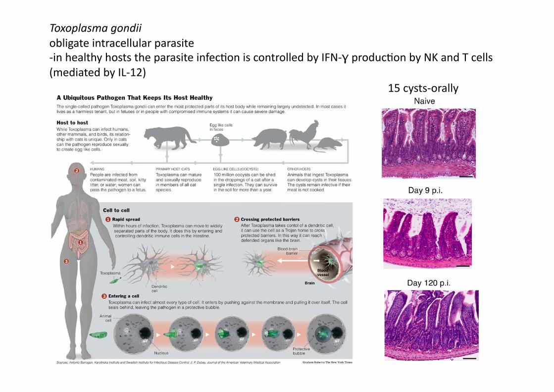
Toxoplasma gondii obligate intracellular parasite -‐in healthy hosts the parasite infec4on is controlled by IFN-‐γ produc4on by NK and T cells (mediated by IL-‐12)
15 cysts-‐orally
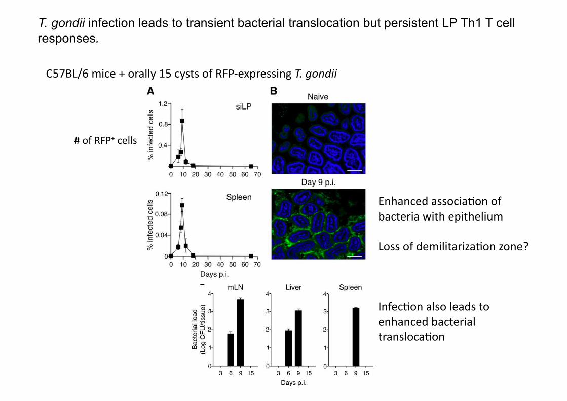
T. gondii infection leads to transient bacterial translocation but persistent LP Th1 T cell responses.
C57BL/6 mice + orally 15 cysts of RFP-‐expressing T. gondii
# of RFP+ cells
Enhanced associa4on of bacteria with epithelium
Loss of demilitariza4on zone?
Infec4on also leads to enhanced bacterial transloca4on
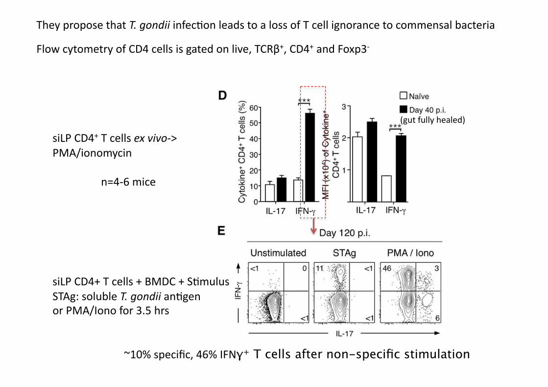
They propose that T. gondii infec4on leads to a loss of T cell ignorance to commensal bacteria
(gut fully healed)
siLP CD4+ T cells + BMDC + S4mulus STAg: soluble T. gondii an4gen or PMA/Iono for 3.5 hrs
siLP CD4+ T cells ex vivo-‐> PMA/ionomycin
n=4-‐6 mice
Flow cytometry of CD4 cells is gated on live, TCRβ+, CD4+ and Foxp3-‐
~10% specific, 46% IFNγ+ T cells after non-specific stimulation
Mouse models used:
CBir1 Tg –CD45.1 –backcrossed for 3 genera4ons -‐CBir1 Tg T cells also express CD45.1 on their cell surface -‐permits cells to be tracked in a CD45.2 host (C57BL/6 mice)
CBir1 Tg –Rag-‐/-‐ –backcrossed for 2 genera4ons -‐the only T cells produced are CBir1 Tg T cells
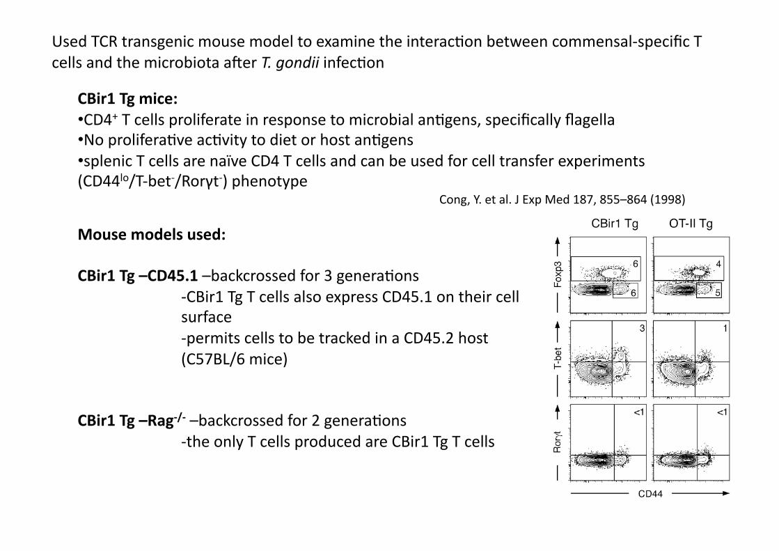
CBir1 Tg mice: • CD4+ T cells proliferate in response to microbial an4gens, specifically flagella • No prolifera4ve ac4vity to diet or host an4gens • splenic T cells are naïve CD4 T cells and can be used for cell transfer experiments (CD44lo/T-‐bet-‐/Rorγt-‐) phenotype
Cong, Y. et al. J Exp Med 187, 855–864 (1998)
Used TCR transgenic mouse model to examine the interac4on between commensal-‐specific T cells and the microbiota aker T. gondii infec4on
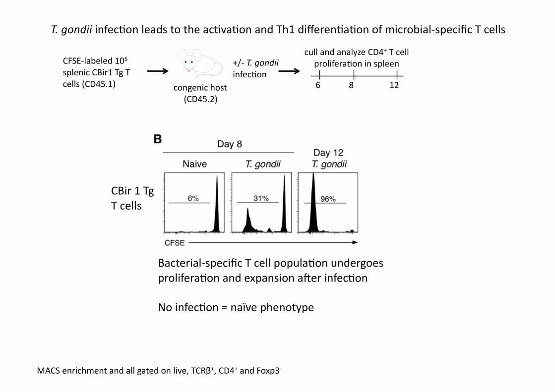
CFSE-‐labeled 105 splenic CBir1 Tg T cells (CD45.1) congenic host
(CD45.2)
+/-‐ T. gondii infec4on
6 8 12
cull and analyze CD4+ T cell prolifera4on in spleen
Bacterial-‐specific T cell popula4on undergoes prolifera4on and expansion aker infec4on
No infec4on = naïve phenotype
MACS enrichment and all gated on live, TCRβ+, CD4+ and Foxp3-‐
T. gondii infec4on leads to the ac4va4on and Th1 differen4a4on of microbial-‐specific T cells
CBir 1 Tg T cells
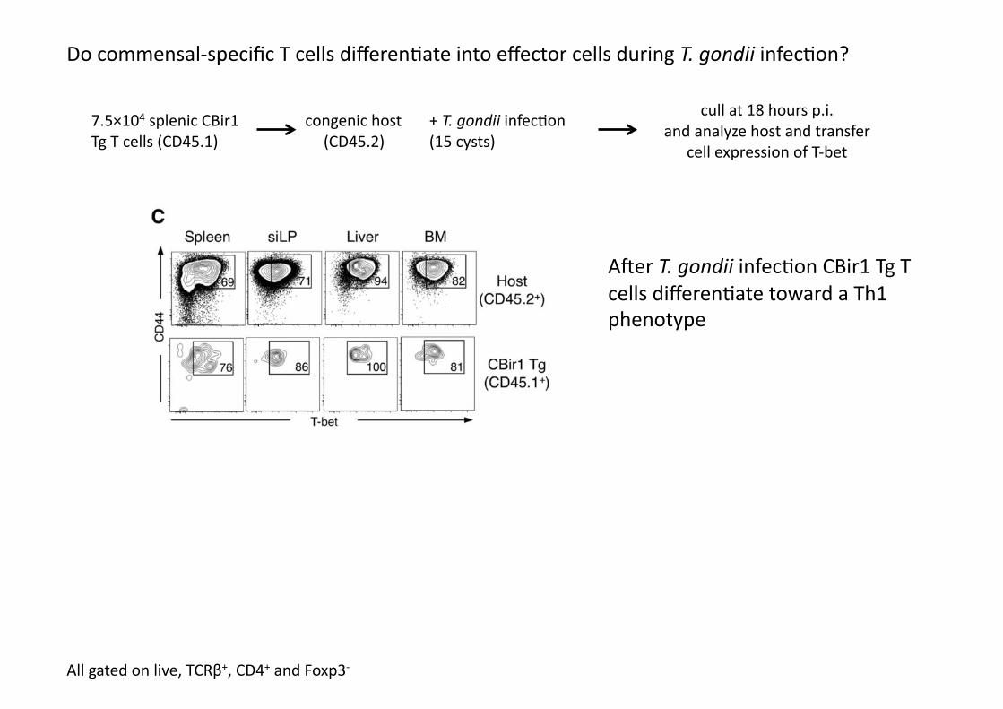
7.5×10⁴ splenic CBir1 Tg T cells (CD45.1)
congenic host (CD45.2)
+ T. gondii infec4on (15 cysts)
cull at 18 hours p.i. and analyze host and transfer
cell expression of T-‐bet
Do commensal-‐specific T cells differen4ate into effector cells during T. gondii infec4on?
Aker T. gondii infec4on CBir1 Tg T cells differen4ate toward a Th1 phenotype
All gated on live, TCRβ+, CD4+ and Foxp3-‐

7.5×10⁴ splenic CBir1 Tg or OTII T cells (CD45.1)
congenic host (CD45.2)
+ T. gondii infec4on (15 cysts)
splenocytes 8 days aker infec4on
CBir1 Tg cells proliferate & differen4ate to Th1
OTII Tg cells remain naïve
Analyzed for intracellular staining for IFN-‐γ & IL-‐17
All gated on live, TCRβ+, CD4+ and Foxp3-‐
Specific expansion of commensal-‐specific T cells aker T. gondii infec4on
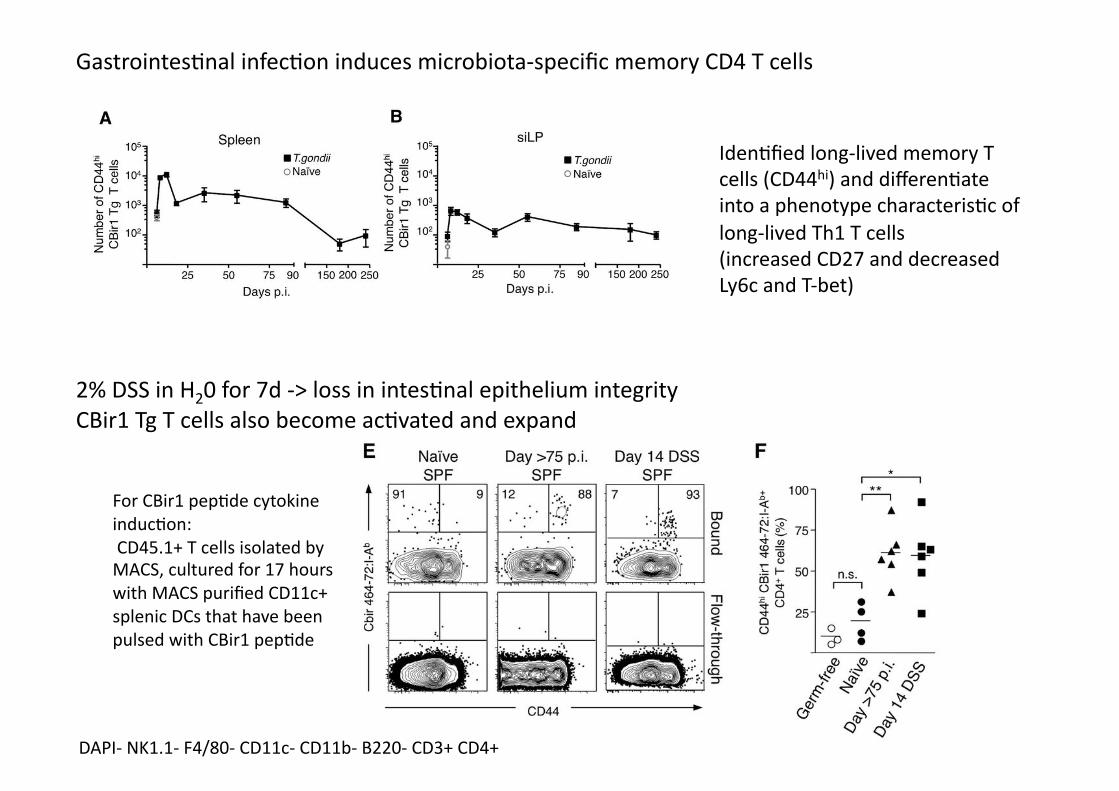
Gastrointes4nal infec4on induces microbiota-‐specific memory CD4 T cells
Iden4fied long-‐lived memory T cells (CD44hi) and differen4ate into a phenotype characteris4c of long-‐lived Th1 T cells (increased CD27 and decreased Ly6c and T-‐bet)
2% DSS in H20 for 7d -‐> loss in intes4nal epithelium integrity CBir1 Tg T cells also become ac4vated and expand
DAPI-‐ NK1.1-‐ F4/80-‐ CD11c-‐ CD11b-‐ B220-‐ CD3+ CD4+
For CBir1 pep4de cytokine induc4on: CD45.1+ T cells isolated by MACS, cultured for 17 hours with MACS purified CD11c+ splenic DCs that have been pulsed with CBir1 pep4de
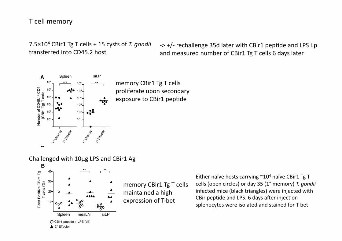
T cell memory
7.5×10⁴ CBir1 Tg T cells + 15 cysts of T. gondii transferred into CD45.2 host
-‐> +/-‐ rechallenge 35d later with CBir1 pep4de and LPS i.p and measured number of CBir1 Tg T cells 6 days later
memory CBir1 Tg T cells proliferate upon secondary exposure to CBir1 pep4de
Challenged with 10μg LPS and CBir1 Ag
memory CBir1 Tg T cells maintained a high expression of T-‐bet
Either naïve hosts carrying ~104 naïve CBir1 Tg T cells (open circles) or day 35 (1° memory) T. gondii infected mice (black triangles) were injected with CBir pep4de and LPS. 6 days aker injec4on splenocytes were isolated and stained for T-‐bet
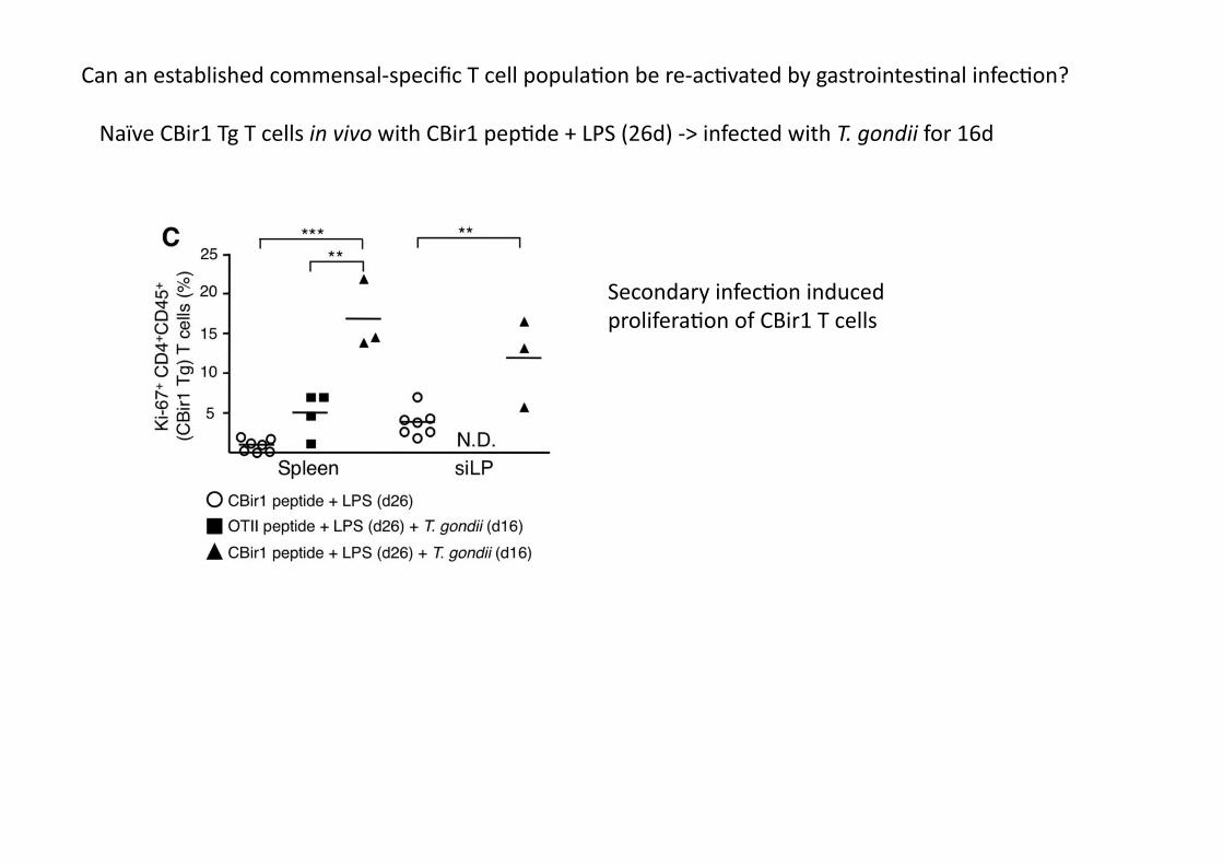
Can an established commensal-‐specific T cell popula4on be re-‐ac4vated by gastrointes4nal infec4on?
Naïve CBir1 Tg T cells in vivo with CBir1 pep4de + LPS (26d) -‐> infected with T. gondii for 16d
Secondary infec4on induced prolifera4on of CBir1 T cells
• Under homeosta4c condi4ons, the commensal microbiota can drive the differen4a4on of Tregs and Th17 cells
• But CBir1 Tg T cells did not induce the expression of IL-‐17A or the transcrip4on factors Rorγt or Foxp3 during T. gondii infec4on indica4ng that microbiota-‐specific T cells are differen4a4ng according to signals provided by the inflammatory milieu
• Novel finding: during gastrointes4nal infec4on bacteria-‐specific T cells become ac4vated and differen4ate into long-‐lived memory cells
Discussion
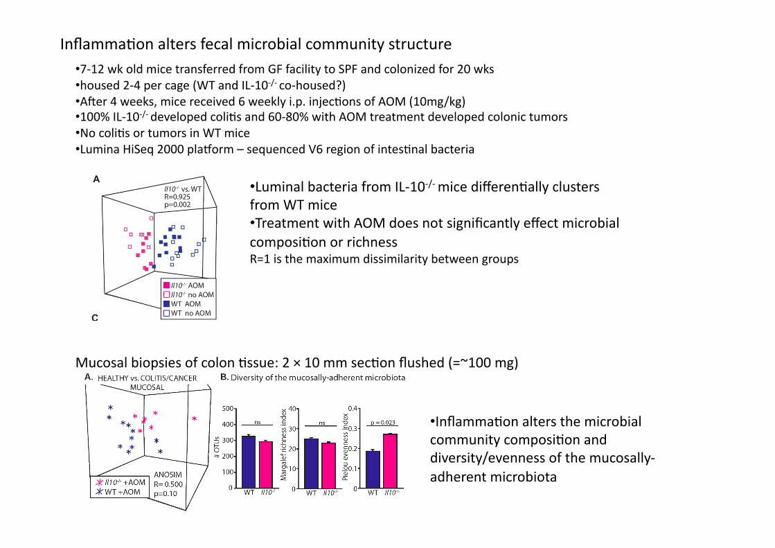
• Luminal bacteria from IL-‐10-‐/-‐ mice differen4ally clusters from WT mice • Treatment with AOM does not significantly effect microbial composi4on or richness R=1 is the maximum dissimilarity between groups
• Inflamma4on alters the microbial community composi4on and diversity/evenness of the mucosally-‐adherent microbiota
Inflamma4on alters fecal microbial community structure • 7-‐12 wk old mice transferred from GF facility to SPF and colonized for 20 wks • housed 2-‐4 per cage (WT and IL-‐10-‐/-‐ co-‐housed?) • Aker 4 weeks, mice received 6 weekly i.p. injec4ons of AOM (10mg/kg) • 100% IL-‐10-‐/-‐ developed coli4s and 60-‐80% with AOM treatment developed colonic tumors • No coli4s or tumors in WT mice • Lumina HiSeq 2000 plaworm – sequenced V6 region of intes4nal bacteria
Mucosal biopsies of colon 4ssue: 2 × 10 mm sec4on flushed (=~100 mg)
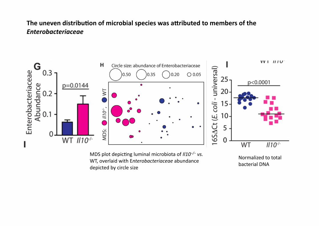
The uneven distribuAon of microbial species was aEributed to members of the Enterobacteriaceae
MDS plot depic4ng luminal microbiota of Il10−/− vs. WT, overlaid with Enterobacteriaceae abundance depicted by circle size
Normalized to total bacterial DNA

Monocoloniza4on of GF IL-‐10-‐/-‐ mice with E. faecalis or E. coli NC101 differen4ally impacts tumorigenesis without affec4ng inflamma4on
Colonic cytokines involved in inflamma4on and carcinogenesis were not significantly different between groups mRNA expression rela4ve to GF IL-‐10-‐/-‐ mean + SEM, n=4
E. faecalis human commensal E. coli NC101 murine adherent-‐invasive strain n=7-‐12 per group
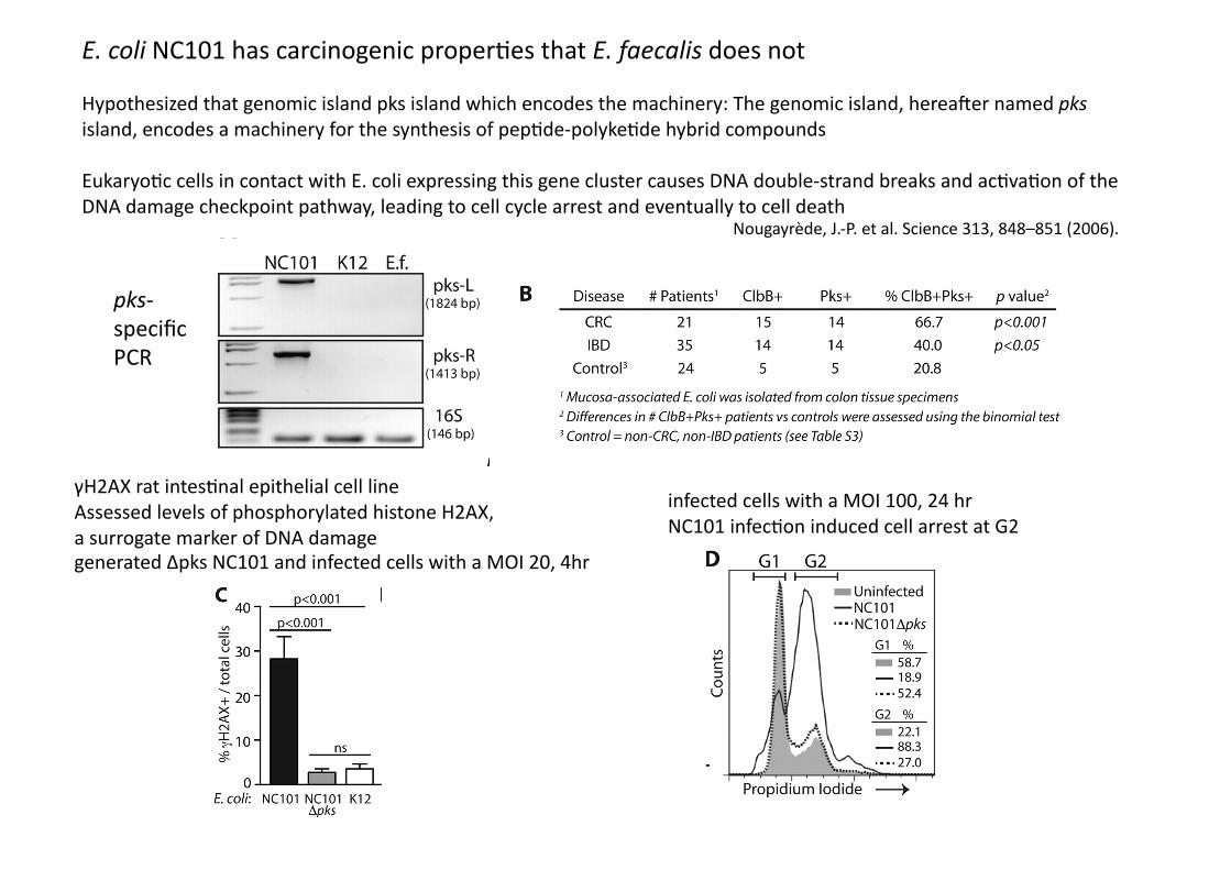
E. coli NC101 has carcinogenic proper4es that E. faecalis does not
Hypothesized that genomic island pks island which encodes the machinery: The genomic island, hereaker named pks island, encodes a machinery for the synthesis of pep4de-‐polyke4de hybrid compounds
Eukaryo4c cells in contact with E. coli expressing this gene cluster causes DNA double-‐strand breaks and ac4va4on of the DNA damage checkpoint pathway, leading to cell cycle arrest and eventually to cell death
Nougayrède, J.-‐P. et al. Science 313, 848–851 (2006).
pks-‐specific PCR
γH2AX rat intes4nal epithelial cell line Assessed levels of phosphorylated histone H2AX, a surrogate marker of DNA damage generated Δpks NC101 and infected cells with a MOI 20, 4hr
infected cells with a MOI 100, 24 hr NC101 infec4on induced cell arrest at G2
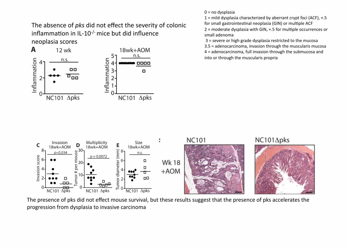
The absence of pks did not effect the severity of colonic inflamma4on in IL-‐10-‐/-‐ mice but did influence neoplasia scores
0 = no dysplasia 1 = mild dysplasia characterized by aberrant crypt foci (ACF), +.5 for small gastrointes4nal neoplasia (GIN) or mul4ple ACF 2 = moderate dysplasia with GIN, +.5 for mul4ple occurrences or small adenoma 3 = severe or high grade dysplasia restricted to the mucosa 3.5 = adenocarcinoma, invasion through the muscularis mucosa 4 = adenocarcinoma, full invasion through the submucosa and into or through the muscularis propria
The presence of pks did not effect mouse survival, but these results suggest that the presence of pks accelerates the progression from dysplasia to invasive carcinoma

Impact of pks on host DNA damage in vivo:
Colonocyte γH2AX+ nuclear foci in AOM/IL-‐10-‐/-‐ mice associated with bacteria for 14 wks
Cells with ≥4 γ-‐foci (nuclear foci staining posi4ve for γH2AX) were enumerated and expressed as γ-‐foci+ cells per crypt
γ-‐foci+ cells were counted in crypts throughout the colon by a blinded inves4gator in at least 40 fields of view
• this two-‐hit model, inflamma4on targets the microbiota to foster the expansion of bacteria with genotoxic poten4al, such as pks+ bacteria
• In parallel, inflamma4on creates an opportunity for pks+ bacteria to adhere to the colonic mucosa by decreasing protec4ve mucins and an4microbial pep4de produc4on, a process prevented by natural barrier func4on present in non-‐inflamed WT mice
• Novel finding: that inflamma4on and not cancer is associated with shiks in microbial concentra4on
Discussion