Embed Size (px)
Citation preview
Treating The Symptom By Knowing The Disease
Presenter
David E McDermott RCP
Vitas Innovative Hospice Services
Dallas Program
Respiratory distress is the most common symptom at the end of life but is consistently mismanaged
The standard of care for respiratory distress There is not a simple bandaid Mismanagement will make things worse Medications have side effects Masking will only live up to hospice stereotype,
costing our companies resources that are better utilized elsewhere
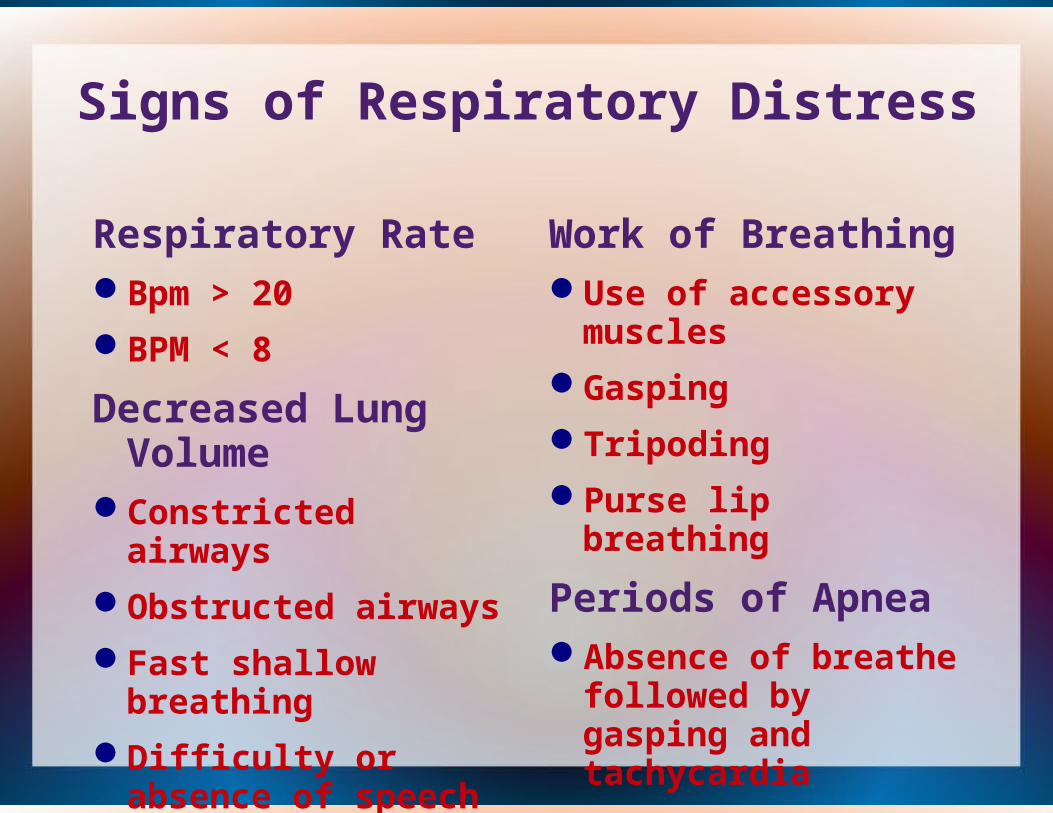
Signs of Respiratory Distress
Respiratory RateBpm > 20
BPM < 8
Decreased Lung Volume
Constricted airways
Obstructed airways
Fast shallow breathing
Difficulty or absence of speech
Work of BreathingUse of accessory
muscles
Gasping
Tripoding
Purse lip breathing
Periods of ApneaAbsence of breathe
followed by gasping and tachycardia
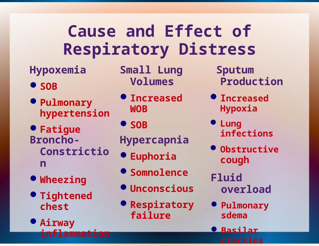
Cause and Effect of Respiratory Distress
Hypoxemia
SOB
Pulmonary hypertension
Fatigue
Small Lung Volumes
Increased WOB
SOB
Sputum Production
Increased Hypoxia
Lung infections
Obstructive cough
Fluid overloadPulmonary
sdema
Basilar crackles
Hypercapnia
Euphoria
Somnolence
Unconscious
Respiratory failure
Broncho-Constriction
Wheezing
Tightened chest
Airway inflammation
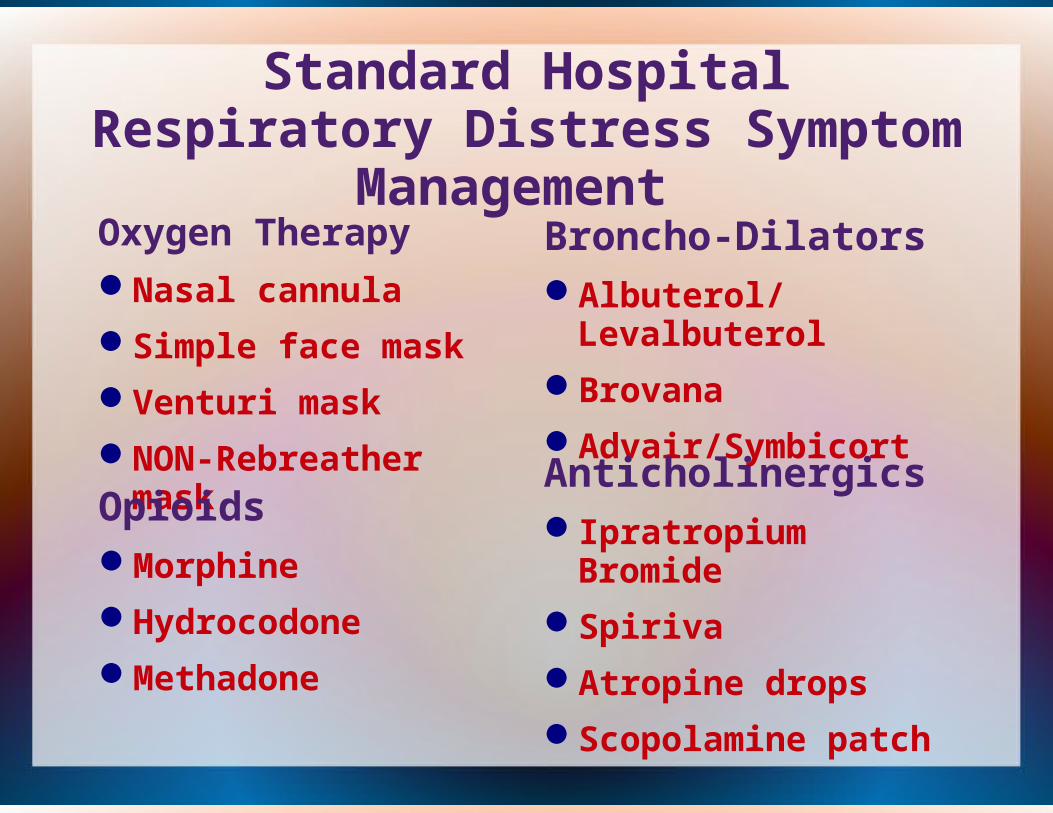
Standard Hospital Respiratory Distress Symptom Management
Oxygen Therapy
Nasal cannula
Simple face mask
Venturi mask
NON-Rebreather mask
Broncho-DilatorsAlbuterol/Levalbuterol
Brovana
Advair/Symbicort
AnticholinergicsIpratropium Bromide
Spiriva
Atropine drops
Scopolamine patch
OpioidsMorphine
Hydrocodone
Methadone

Additional Treatments
Anxiety Medications
Benzodiazepines
Xanax
Lorazepam
DiureticsFurosemide
Torsemide
Bumetanide
SteroidsPrednisone
DexamethasoneAntibiotics
Pulmonary Rehab
Surgery
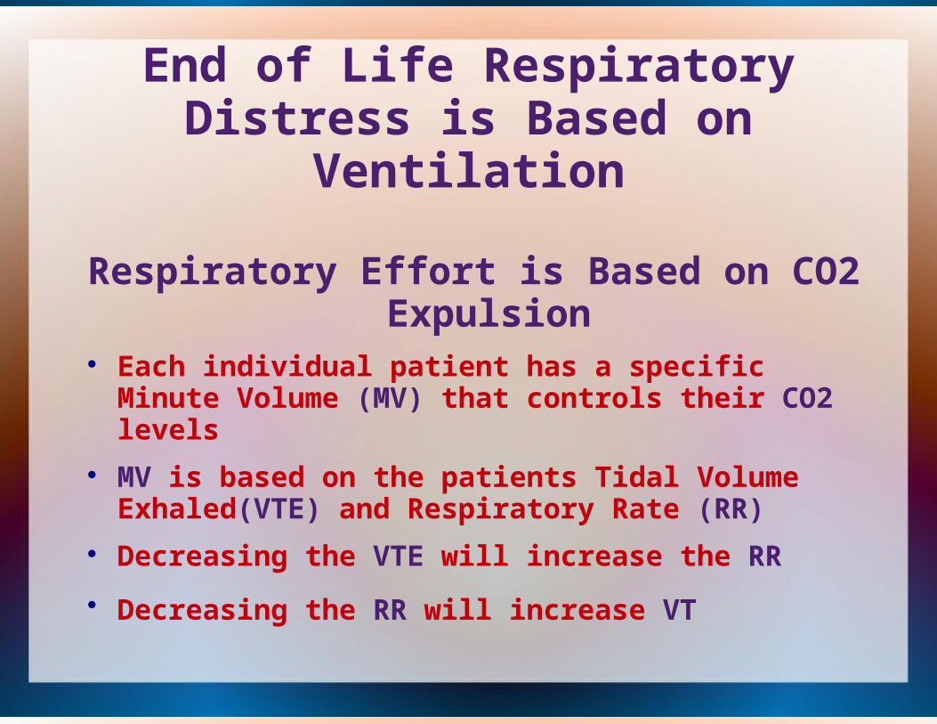
End of Life Respiratory Distress is Based on Ventilation
Respiratory Effort is Based on CO2 Expulsion Each individual patient has a specific Minute Volume
(MV) that controls their CO2 levels MV is based on the patients Tidal Volume
Exhaled(VTE) and Respiratory Rate (RR) Decreasing the VTE will increase the RR
Decreasing the RR will increase VT
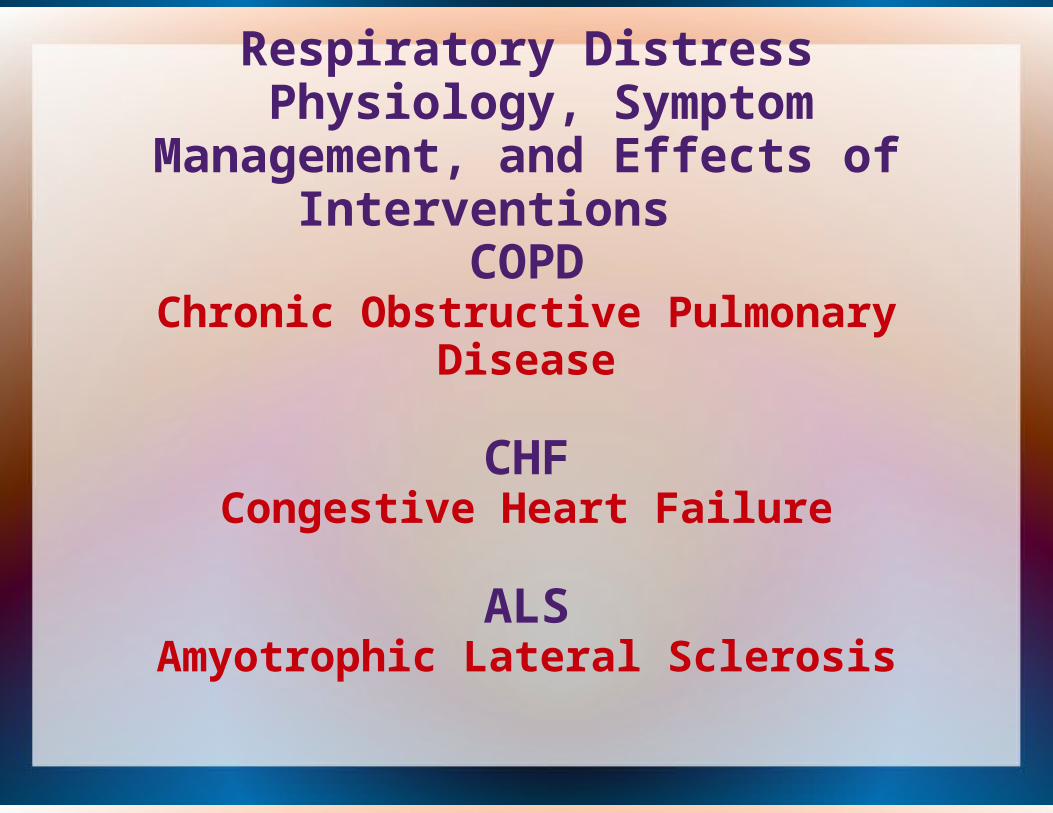
Respiratory Distress Physiology, Symptom Management,
and Effects of Interventions
COPDChronic Obstructive Pulmonary Disease
CHFCongestive Heart Failure
ALS
Amyotrophic Lateral Sclerosis
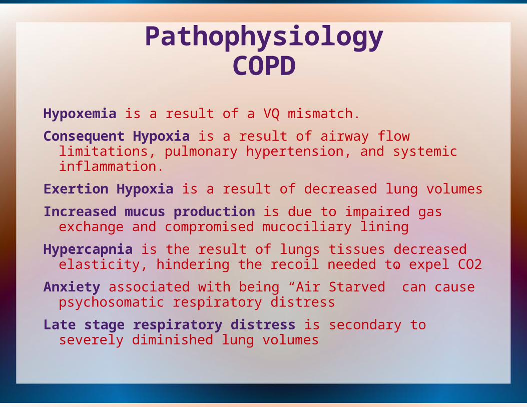
PathophysiologyCOPD
Hypoxemia is a result of a VQ mismatch.
Consequent Hypoxia is a result of airway flow limitations, pulmonary hypertension, and systemic inflammation.
Exertion Hypoxia is a result of decreased lung volumes
Increased mucus production is due to impaired gas exchange and compromised mucociliary lining
Hypercapnia is the result of lungs tissues decreased elasticity, hindering the recoil needed to expel CO2
Anxiety associated with being “Air Starved” can cause psychosomatic respiratory distress
Late stage respiratory distress is secondary to severely diminished lung volumes
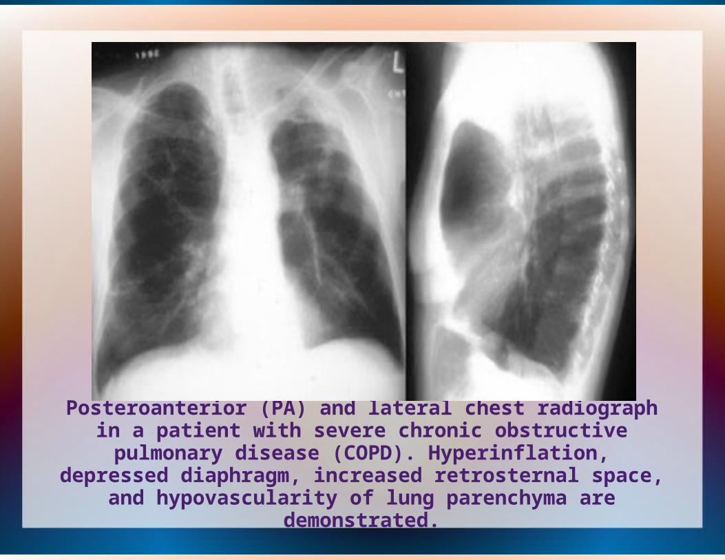
Posteroanterior (PA) and lateral chest radiograph in a patient with severe chronic obstructive pulmonary disease (COPD). Hyperinflation, depressed diaphragm, increased retrosternal
space, and hypovascularity of lung parenchyma are demonstrated.

Physiologic Based Treatments of COPD Related Respiratory Distress
Physiology Broncho-Constriction
Chronic Hypoxia
Excessive Mucus Production
Airway Swelling
Hypercapnia(>CO2)
Decreased Lung volumes
Anxiety
Exertion Induced Hypoxia
TreatmentBroncho-Dilators
Oxygen
Anticholinergics
Corticosteroids
BIPAP
Opioids
Benzodiazepines
Discontinuing Exertion
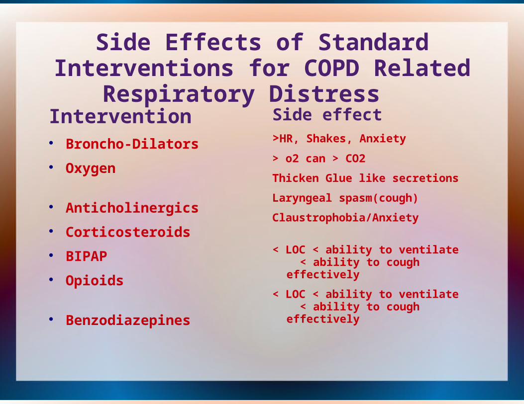
Side Effects of Standard Interventions
for COPD Related Respiratory Distress Intervention
Broncho-Dilators
Oxygen
Anticholinergics
Corticosteroids
BIPAP
Opioids
Benzodiazepines
Side effect>HR, Shakes, Anxiety
> o2 can > CO2
Thicken Glue like secretions
Laryngeal spasm(cough)
Claustrophobia/Anxiety
< LOC < ability to ventilate < ability to cough effectively
< LOC < ability to ventilate < ability to cough effectively
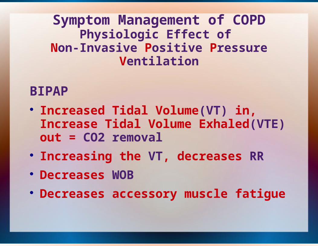
Symptom Management of COPD Physiologic Effect of
Non-Invasive Positive Pressure Ventilation
BIPAP Increased Tidal Volume(VT) in, Increase Tidal
Volume Exhaled(VTE) out = CO2 removal Increasing the VT, decreases RR Decreases WOB Decreases accessory muscle fatigue

Pathophysiology of CHFPressure and volume overloadLoss of cardiac muscle High output failure Reduction in cardiac output
Effects of left ventricular (LV) insufficiency/failure 1) There is an intrinsic decrease in muscle contractility2) Increased reload backup, resulting in pulmonary congestion and dyspnea.3) Systemic blood pressure is often reduced, but there is an increased after load, which can further reduce cardiac output4) Heart rate is generally increased as part of a compensatory mechanism. An increase in heart size, increasing wall tension and increasing myocardial oxygen consumption
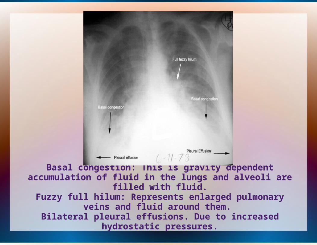
Basal congestion: This is gravity dependent accumulation of fluid in the lungs and alveoli are filled with fluid.
Fuzzy full hilum: Represents enlarged pulmonary veins and fluid around them.
Bilateral pleural effusions. Due to increased hydrostatic pressures.
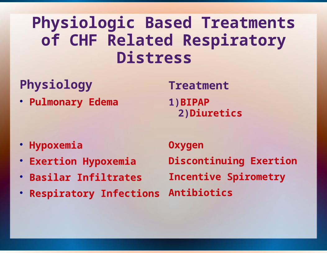
Physiologic Based Treatments of CHF Related Respiratory Distress
Physiology Pulmonary Edema
Hypoxemia Exertion Hypoxemia Basilar Infiltrates Respiratory Infections
Treatment
1)BIPAP 2)Diuretics
Oxygen
Discontinuing Exertion
Incentive Spirometry
Antibiotics
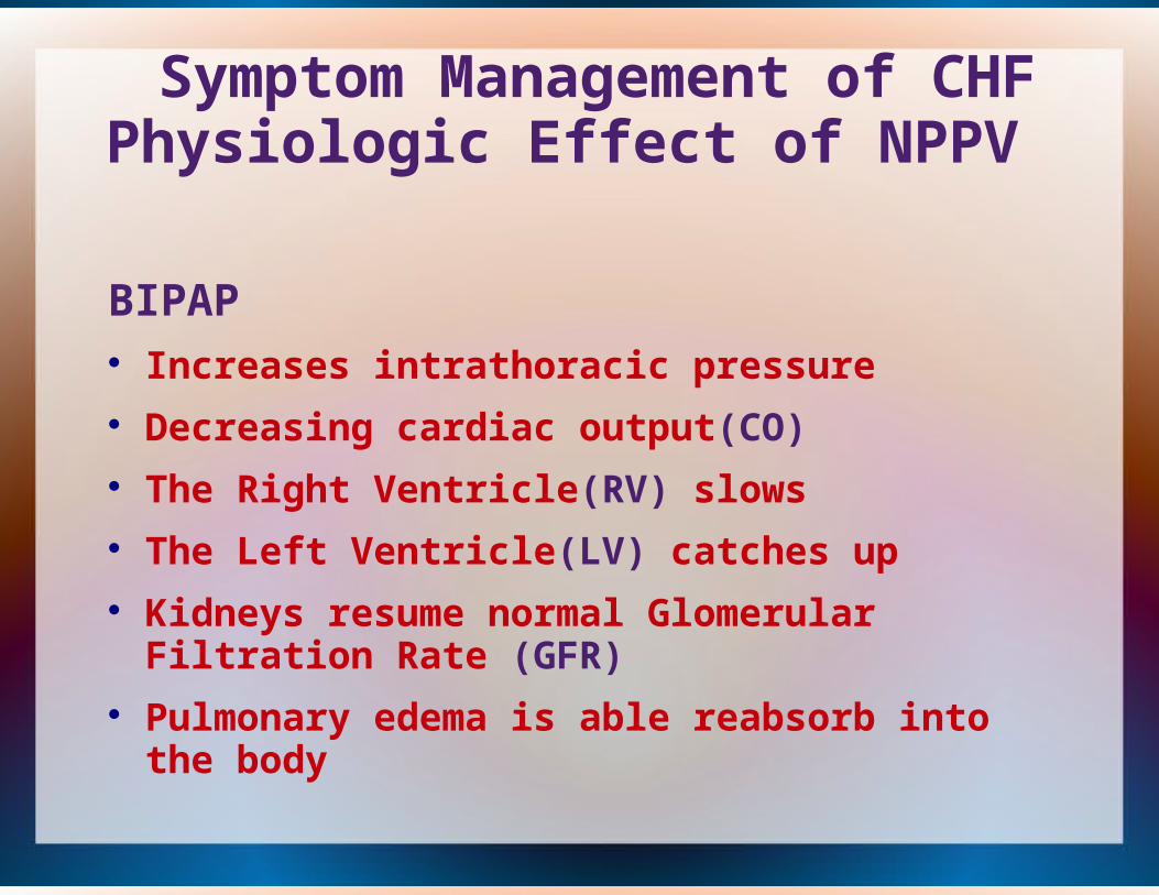
Symptom Management of CHF Physiologic Effect of NPPV
BIPAP Increases intrathoracic pressure Decreasing cardiac output(CO) The Right Ventricle(RV) slows The Left Ventricle(LV) catches up Kidneys resume normal Glomerular Filtration Rate
(GFR) Pulmonary edema is able reabsorb into the body
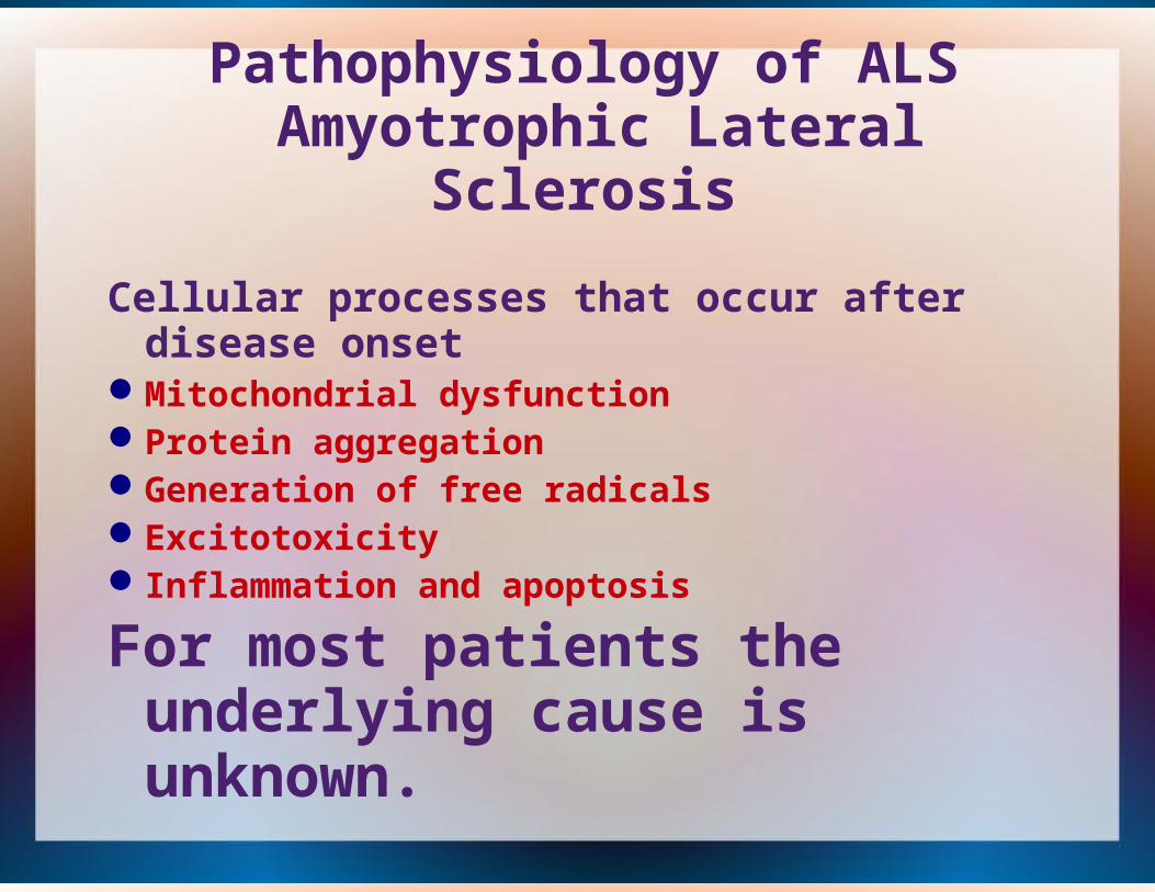
Cellular processes that occur after disease onset
Mitochondrial dysfunctionProtein aggregation Generation of free radicals ExcitotoxicityInflammation and apoptosis
For most patients the underlying cause is unknown.
Pathophysiology of ALS Amyotrophic Lateral Sclerosis
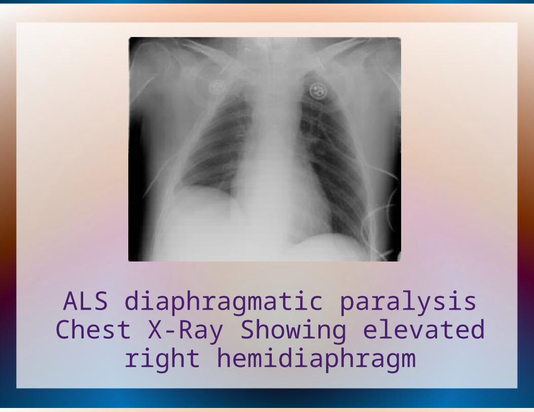
ALS diaphragmatic paralysis Chest X-Ray Showing elevated right
hemidiaphragm
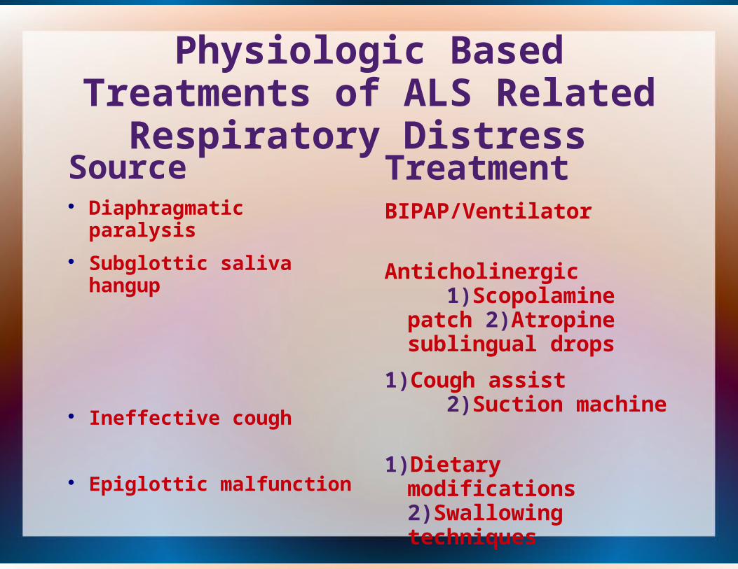
Physiologic Based Treatments of ALS Related Respiratory Distress
Source Diaphragmatic paralysis Subglottic saliva hangup
Ineffective cough
Epiglottic malfunction
TreatmentBIPAP/Ventilator
Anticholinergic 1)Scopolamine patch 2)Atropine sublingual drops
1)Cough assist 2)Suction machine
1)Dietary modifications 2)Swallowing techniques

Side Effects of Standard, Appropriate and Inappropriate, Interventions for ALS Related Respiratory Distress
Intervention BIPAP/Ventilator
Anticholinergic's
Oxygen Bronchodilators Cough Assist
Adverse EffectsPS set to high or to low
1)Barotrauma 2)Atelectatic basilar lung lobes
Over drying of mouth and airways
No physiologic benefit
>HR, Shakes, Anxiety
1)Respiratory distress 2)Barotrauma
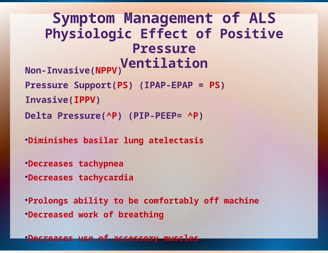
Symptom Management of ALSPhysiologic Effect of Positive Pressure
VentilationNon-Invasive(NPPV)
Pressure Support(PS) (IPAP-EPAP = PS)
Invasive(IPPV)
Delta Pressure(^P) (PIP-PEEP= ^P) Diminishes basilar lung atelectasis Decreases tachypneaDecreases tachycardia Prolongs ability to be comfortably off machine Decreased work of breathing
Decreases use of accessory muscles
References
http://www.ncbi.nlm.nih.gov/pubmed/21128691http://www.ncbi.nlm.nih.gov/pubmed/4014051
http://radiologymasterclass.co.uk/tutorials/chest/chest_pathology/chest_pathology_page8.htmlhttp://circheartfailure.ahajournals.org/content/4/6/677.fullhttp://www.webmd.com/lung/picture-of-the-lungs