Embed Size (px)
Citation preview
TrkB and TrkC Signaling Are Required for Maturation andSynaptogenesis of Hippocampal Connections
Albert Martınez,1 Soledad Alcantara,1 Vıctor Borrell,1 Jose A. Del Rıo,1 Joan Blasi,2 Raquel Otal,1Narciso Campos,3 Albert Boronat,3 Mariano Barbacid,4,5 Inmaculada Silos-Santiago,5,6 and Eduardo Soriano1
1Department of Animal and Plant Cell Biology, University of Barcelona, Barcelona 08028, Spain, 2Department of CellBiology and Pathology, University of Barcelona, L’Hospitalet de Llobregat, Barcelona 08907, Spain, 3Department ofBiochemistry and Molecular Biology, University of Barcelona, Barcelona 08028, Spain, 4Centro Nacional deInvestigaciones Oncologicas Carlos III, Instituto de Salud Carlos III, 28220 Majalahonda, Madrid, Spain, 5Department ofMolecular Oncology, Bristol-Myers Squibb, Pharmaceutical Research Institute, Princeton, New Jersey 08543-4000, and6Department of Neurobiology, Millennium Pharmaceuticals Inc., Cambridge, Massachusetts 02139-4815
Recent studies have suggested a role for neurotrophins in thegrowth and refinement of neural connections, in dendriticgrowth, and in activity-dependent adult plasticity. To unravelthe role of endogenous neurotrophins in the development ofneural connections in the CNS, we studied the ontogeny ofhippocampal afferents in trkB (2/2) and trkC (2/2) mice. In-jections of lipophilic tracers in the entorhinal cortex and hip-pocampus of newborn mutant mice showed that the ingrowthof entorhinal and commissural/associational afferents to thehippocampus was not affected by these mutations. Similarly,injections of biocytin in postnatal mutant mice (P10–P16) didnot reveal major differences in the topographic patterns ofhippocampal connections.
In contrast, quantification of biocytin-filled axons showedthat commissural and entorhinal afferents have a reduced num-ber of axon collaterals (21–49%) and decreased densities ofaxonal varicosities (8–17%) in both trkB (2/2) and trkC (2/2)mice. In addition, electron microscopic analyses showed that
trkB (2/2) and trkC (2/2) mice have lower densities of synapticcontacts and important structural alterations of presynapticboutons, such as decreased density of synaptic vesicles. Fi-nally, immunocytochemical studies revealed a reduced expres-sion of the synaptic-associated proteins responsible for synap-tic vesicle exocytosis and neurotransmitter release (v-SNAREsand t-SNAREs), especially in trkB (2/2) mice. We conclude thatneither trkB nor trkC genes are essential for the ingrowth orlayer-specific targeting of hippocampal connections, althoughthe lack of these receptors results in reduced axonal arboriza-tion and synaptic density, which indicates a role for TrkB andTrkC receptors in the developmental regulation of synapticinputs in the CNS in vivo. The data also suggest that the genesencoding for synaptic proteins may be targets of TrkB and TrkCsignaling pathways.
Key words: TrkB receptors; TrkC receptors; neurotrophic fac-tors; mutant mice; neuronal connections; synaptogenesis;synaptic-associated proteins; hippocampus
The neurotrophins, including nerve growth factor (NGF), brain-derived neurotrophic factor (BDNF), neurotrophin 3 (NT3), andneurotrophin 4/5 (NT4/5), are essential for the survival of popu-lations of neurons in the PNS and CNS [Jones et al. (1994);Minichiello and Klein (1996); Alcantara et al. (1997); for review,see Snider (1994); Farinas and Reichardt (1996); Snider andSilos-Santiago (1996)]. Recent studies indicate that neurotrophinsmay regulate dendritic and axonal growth (Cabelli et al., 1995,1997; Cohen-Cory and Fraser, 1995; McAllister et al., 1995, 1996,1997; Bolz et al., 1997; Inoue and Sanes, 1997; Paves and Saarma,1997) and the efficacy of synaptic transmission (Lohof et al., 1993;Kang and Schuman, 1995; Korte et al., 1995; Thoenen, 1995;Figurov et al., 1996; Prakash et al., 1996; Wang and Poo, 1997).
Moreover, target-derived neurotrophins in the PNS may regulatethe maturation of synaptic contacts and the density of synapticinnervation (Miller et al., 1994; Wang et al., 1995; Causing et al.,1997). In addition, a role for BDNF and NT4/5 has been shownfor the activity-dependent development of ocular dominancecolumns in the visual cortex (Cabelli et al., 1995, 1997; Galuske etal., 1996).
Most of the data on the effects of neurotrophins on axonalgrowth were obtained in vitro or after application of exogenousneurotrophins. To our knowledge, the only study that has ana-lyzed the role of endogenous neurotrophins in the developmentof neural connections in the CNS was performed by infusion ofneurotrophin receptors antagonists (TrkB-IgG) in the visual cor-tex (Cabelli et al., 1997). To determine the contribution of en-dogenous neurotrophins to the developmental pattern of neuro-nal connections and synaptogenesis in the CNS, we studied theontogeny of the main hippocampal afferents in mice lacking trkBand trkC genes, which encode for the receptors of BDNF andNT4/5, and NT3, respectively (Klein et al., 1991, 1992; Lamballeet al., 1991; Soppet et al., 1991). We chose the hippocampal areanot only because neurotrophic factors and their receptors areabundantly expressed in this region (Gall and Isackson, 1989;Ernfors et al., 1990, 1991; Hofer et al., 1990; Gall et al., 1991;
Received April 8, 1998; revised July 1, 1998; accepted July 6, 1998.This work was supported by grants from Comision Interministerial de Ciencia y
Tecnologıa, Spain (SAF98-0106), and Direccion General de Investigaciones Cien-tıficas y Tecnicas (P.M.95-102), and by the Ramon Areces Foundation (Spain), theInternational Institute for Research in Paraplegia (Switzerland), and the Marato ofTV3 to E.S. We thank Robin Rycroft for editorial assistance, and G. Barnstable andR. Jahn for generously providing antibodies to synaptic proteins.
A.M. and S.A. contributed equally to this work.Correspondence should be addressed to Dr. Eduardo Soriano, Department of
Animal and Plant Cell Biology, Faculty of Biology, University of Barcelona, Diag-onal 645, Barcelona 08028, Spain.Copyright © 1998 Society for Neuroscience 0270-6474/98/187336-15$05.00/0
The Journal of Neuroscience, September 15, 1998, 18(18):7336–7350
Isackson et al., 1991; Rocamora et al., 1996), but also becausemost studies on modulation of synaptic activity by neurotrophinshave been performed in this brain area (Kang and Schuman,1995; Korte et al., 1995; Figurov et al., 1996). In addition, theanalysis of the phenotype of hippocampal connections in trkB(2/2) and trkC (2/2) mice allows us to assess the developmentalfunctions of neurotrophins in a region where the main afferentconnections are organized both in a layer-specific manner and ina precise topographic order (Amaral and Witter, 1995).
MATERIALS AND METHODSMutant mice. Single-mutant trkB (2/2) and trkC (2/2) mice and double-mutant homozygous mice were generated by mating single and doubleheterozygous animals (Klein et al., 1993, 1994; Fritzsch et al., 1997).Because no significant differences were observed in the phenotype ofwild-type and heterozygous littermates, both groups of mice were used ascontrols for the quantitative analyses.
In situ hybridization. Embryonic day (E14, E16, E18), postnatal day(P0, P5, P10, P15, P21), and adult mice (NMRI strain; Iffa Credo, Lyon,France) were perfused with 4% paraformaldehyde in 0.1 M phosphatebuffer, cryoprotected in 30% sucrose, and frozen. Coronal sections(30–50 mm thick) were obtained and hybridized as described elsewhere(Lecea et al., 1995, 1997). Briefly, sections were deproteinized with 0.2 NHCl for 10 min, acetylated with 0.25% acetic anhydride in 0.1 M trieth-anolamine buffer, pH 8.0, and post-fixed for 10 min in 4% paraformal-dehyde. Sections were then hybridized overnight at 60°C withdigoxigenin-labeled antisense RNA probes to mouse BDNF and NT3and to the tyrosine kinase domains of mouse TrkB and TrkC in a solutioncontaining 50% formamide, 20 mM PIPES, 53 Denhardt’s solution, 10%dextran sulfate, 250 mg/ml yeast tRNA, 250 mg/ml salmon sperm DNA,50 mM dithiothreitol, 0.62 M NaCl, and 10 mM EDTA solution. Sectionswere then digested with RNase A (37°C for 1 hr) and washed in 0.53 SSCplus 50% formamide (55°C) and in 0.13 SSC plus 0.5% sarcosyl (60°C).After hybridization, sections were blocked with 10% sheep normal serum(2 hr), incubated overnight with an alkaline phosphatase-labeled anti-digoxigenin antibody (1:2000), and developed with a 5-bromo-4-chloro-3-idolyl phosphate (BCIP) and nitroblue tetrazolium (NBT) substrate.After several washes, sections were mounted onto slides and coverslippedwith Mowiol. Sections hybridized with sense riboprobes did not givesignals above background levels.
Anterograde tracing. Newborn and P5 mice were perfused with 4%paraformaldehyde in phosphate buffer, and their brains were dissectedout. Crystals of 1–19-dioctadecyl-3,3,39,39-tetramethylindocarbocyanineperchlorate (DiI) were injected into the entorhinal area or hippocampusas described (Super and Soriano, 1994). Brains were stored in phosphate-buffered 4% paraformaldehyde for 5–8 weeks in the dark at roomtemperature. Coronal sections were obtained with a vibratome, counter-stained with the DNA-specific dye bisbenzimide, and mounted with anantifading mounting medium. The sections were analyzed and pho-todocumented in a Polyvar (Reichert) microscope equipped with fluo-rescent filters. P10–P16 mice were injected by iontophoresis of biocytin(20 min; positive current; 7 sec on/off cycle) in the entorhinal cortex orthe hippocampus. After 24 hr of survival, animals were perfused with 4%paraformaldehyde, and brains were post-fixed in the same fixative andsectioned at 50 mm using a vibratome. Free-floating sections were thenincubated with the avidin–biotin–peroxidase complex (ABC) (VectorLabs, Burlingame, CA), developed with 0.05% diaminobenzidine (DAB)and 0.01% hydrogen peroxide in the presence of 0.02% nickel sulfate(Del Rıo et al., 1997), and counterstained with cresyl violet. A total of 97postnatal mice were used (n 5 11 trkB (1/1), 24 trkB (6), 23 trkB (2/2),11 trkC (1/1), 16 trkC (6), 12 trkC (2/2)). Five newborn double-mutanttrkB (2/2)/trkC (2/2) mice were also injected with DiI. Selected ento-rhinal and commissural fibers from trkB (2/2) and trkC (2/2) mice(P13–P14) and from control littermates were drawn using a 403 oil-immersion objective lens (n 5 2–4 animals per group, 32–58 fibers).Length of fibers was measured using a planimeter, and the branchingindex (Del Rıo et al., 1997) and density of axonal varicosities (number ofvaricosities per 100 mm) were calculated. Statistical analysis was per-formed using the Student’s t test.
Electron microscopy. trkB (2/2) (P13, n 5 2), trkC (2/2) (P12–13, n 53), and control littermates [trkB (1/1), n 5 1; trkB (1/2), n 5 1; trkC(1/1), n 5 1; trkC (1/2), n 5 2] were perfused with 2% glutaralde-hyde–1% paraformaldehyde in 0.12 M phosphate buffer. Brains were
removed from the skull and fixed in the same solution overnight. Tissueslices were post-fixed with 2% osmium tetroxide, stained with 2% uranylacetate, and embedded in Araldite. Ultrathin sections were collectedonto formvar-coated slot grids and stained with lead citrate. Electronmicrographs covering 100 mm 2 (final magnification 19,0003) were ran-domly taken from each hippocampal layer, and the number of synapticcontacts was counted (n 5 30–31 micrographs for each layer and group).For the morphometric analysis of presynaptic boutons and synapses,randomly selected synaptic contacts were micrographed at 58,0003 finalmagnification (n 5 81–170 axon terminals and synapses per group andlayer). The total number of synaptic vesicles and vesicles clustered nearthe active zone [whose membrane was closer than 50 nm (Rosahl et al.,1995)] was counted. The area of axon terminals and the length andthickness of synaptic specializations were calculated using the IMATimage analysis program (Scientific-Technical Services, University of Bar-celona). Statistical analysis was performed using the Student’s t test.
Organotypic slice cultures. Entorhinohippocampal cocultures and hip-pocampus/hippocampus cocultures were prepared from newborn trkB(2/2), trkC (2/2), and double-mutant trkB (2/2)/trkC (2/2) mice, aswell as from control littermates as described (Del Rıo et al., 1997).Animals were anesthetized by hypothermia, their brains were removed,and the hippocampus and entorhinal cortex were dissected out under amicroscope. Horizontal sections (350 mm thick) were obtained using aMcIlwain tissue chopper. Selected slices were cocultured using the in-terphase membrane method (Stoppini et al., 1991). A total of 52 ento-rhinohippocampal and 50 hippocampus/hippocampus mixed slice cocul-tures of mutant and control mice were prepared. In addition, culturesfrom single-mutant mice were also prepared (n 5 38). A crystal ofbiocytin was placed in the entorhinal cortex or the hippocampus 24 hrbefore fixation. Cocultures were fixed with 4% paraformaldehyde after9–15 d in vitro (DIV). Horizontal sections (40 mm thick) were obtained,incubated with the ABC complex, and developed with diaminobenzidine/nickel and hydrogen peroxide as described above. Cultures were thencounterstained with cresyl violet and coverslipped.
Immunocytochemistry and immunoblot. Mutant and control littermates(P13; trkB (2/2), n 5 2, trkB (1/1), n 5 1; trkB (1/2), n 5 1; trkC(2/2), n 5 2; trkC (1/1), n 5 1; trkC (1/2), n 5 1) were perfused with4% paraformaldehyde in PBS, cryoprotected in 30% sucrose, and sec-tioned at 25 mm. Sections from mutant and control mice were processedin bulk. Free-floating sections were blocked with 15% fetal bovine serumsolution and incubated overnight at 4°C with one of the followingprimary antibodies (diluted 1:1000–1500): anti-a-tubulin (Sigma, PooleDorset, UK), anti-synapsin I [MAB 355, Chemicon, Temecula, CA(recognizing the C-terminal fragment of synapsin I)], anti-synaptophysin(MAB SVP-38, Sigma), anti-synaptotagmin I [MAB 41.1, gift from R.Jahn (Gottingen, Germany) (Brose et al., 1992)], anti-SNAP-25 (MABSMI 81, Sternberger-Meyer, Jarrettsville, MD), anti-synaptobrevin 2[MAB 69.1, gift from R. Jahn (Edelmann et al., 1995)], anti-syntaxin 1[MAB HPC-1 recognizing isoforms 1A and 1B; gift from G. Barnstable(New Haven, CT) (Barnstable et al., 1985)], and anti-Rab3a [MAB 42.2,gift from R. Jahn (Matteoli et al., 1991)]. Thereafter, sections wereincubated for 3 hr at room temperature in the dark with FITC-conjugated secondary antibodies, mounted onto slides, and coverslippedwith an antifading mounting medium. Sections were viewed in a LeicaTCS 4D confocal scanning laser microscope. Two serial confocal images,2 mm apart, from each example were used to quantify the fluorescenceintensity of synaptic-associated protein immunolabeling. The intensitiesof fluorescence (expressed as gray levels) were measured along verticalstrips extending from the stratum oriens to the hilar region, with the aidof the IMAT image analysis program. Continuous linear profiles fromcontrol and null-mutant mice were then averaged, and the fluorescenceintensities were calculated for each hippocampal layer. Statistical analysiswas performed using the Student’s t test.
The forebrains of two trkB (2/2) and trkC (2/2) mice and littermatecontrols were frozen and homogenized in HEPES/NaOH 10 mM, pH 7.4,containing 0.32 M sucrose, 1 mg/ml leupeptin, 5 mg/ml aprotinin, 1 mMPMSF, and 1 mM EGTA. To remove nuclei and cellular debris, thehomogenates were centrifuged at 2000 rpm in a Beckman JA21 rotor for2 min followed by 12,000 rpm for 11 min in the same rotor. Supernatantscontaining cytosolic and light membrane fractions were subjected toSDS-PAGE and immunoblot analysis using 10 mg of protein per lane.Gels were then electrotransferred to nitrocellulose membranes using asemi-dry blotting system. Nitrocellulose membranes were blocked with5% nonfat milk in TBS (140 mM NaCl, 10 mM Tris/HCl, pH 7.4, with0.1% Tween-20) for 30 min at room temperature and incubated over-
Martınez et al. • Hippocampal connections in trkB (2/2) and trkC (2/2) mice J. Neurosci., September 15, 1998, 18(18):7336–735 7337
night with the monoclonal antibodies described above. After severalwashes, membranes were incubated with peroxidase-conjugated anti-mouse antibodies for 1 hr at room temperature and developed using theECL method (Amersham, Bucks, UK), placed in contact with x-ray filmsfor 10–60 sec, and quantified (Phoretix 1D Gel Analysis system). Thevalues were normalized to a-tubulin to correct for possible inequalities inprotein content.
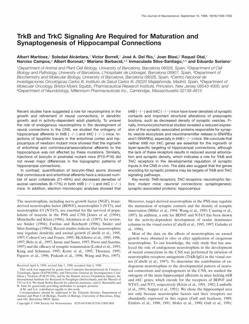
RESULTSExpression of neurotrophic factors and their receptorsin the developing hippocampal regionIn the mouse, developing entorhinal afferents reach the hip-pocampus at embryonic day 15 (E15) and invade the appropriatetarget layer (stratum lacunosum-moleculare) by E16, whereascommissural /associational afferents reach the target layers (stra-tum oriens and stratum radiatum) 2–3 d later (Super and Soriano,1994; Super et al., 1998). To investigate the role of neurotrophinsand their receptors in the development of these principal hip-pocampal connections, we first analyzed the developmental ex-pression of trkB, trkC, bdnf, and nt3 genes. At embryonic stages(E14–E18), both TrkB and TrkC mRNAs were detected in post-mitotic neurons of the entorhinal cortex, in the pyramidal neuronsof the hippocampus proper (CA1–CA4 subfields), and in thegranule cells of the dentate gyrus. At postnatal stages (P0–P15),when hippocampal connections mature, levels of expression forboth transcripts increased steadily and were maximal (Fig. 1A,B).mRNA hybridization signals decreased to adult-like levels by P21.BDNF mRNA was also widely expressed in the entorhinohip-pocampal system at all ages (Fig. 1C), with levels of expressionincreasing from P5 on to reach maximal expression levels in theadult. NT3 transcripts, which in the adult hippocampus are re-stricted to the dentate gyrus and the CA2 region (Ernfors et al.,1990; Lindvall et al., 1992; Isackson, 1995), were upregulated atprenatal and early postnatal stages and were expressed in mostneurons within the hippocampus and entorhinal areas (data notshown). No remarkable differences in the pattern of expressionfor either transcript were observed between the medial and lateralfields of the entorhinal cortex. These results show that TrkB andTrkC receptors and their ligands, BDNF and NT3, are expressedin the hippocampal region at the time of ingrowth and develop-ment of the main hippocampal afferents (Super and Soriano,1994; Del Rıo et al., 1997; Super et al., 1998). Furthermore, TrkBand TrkC receptors are expressed in entorhinohippocampal pro-jection cells and in the neurons of the CA3 subfield that give riseto the commissural /associational pathway (Amaral and Witter,1995).
Pattern of hippocampal connectivity in trkB (2/2) andtrkC (2/2) miceRecently, it has been suggested that neurotrophins may be in-volved in the attraction of the axonal growth cone (Paves andSaarma, 1997; Song et al., 1997). To test whether TrkB or TrkCsignaling is required for the ingrowth and targeting of afferentfibers in the hippocampal region we next examined the develop-ment of hippocampal connections in trkB (2/2) and trkC (2/2)mice. Injections of the anterograde tracer DiI in the entorhinalarea at P0–P5 showed that in trkB (2/2) and trkC (2/2) mice
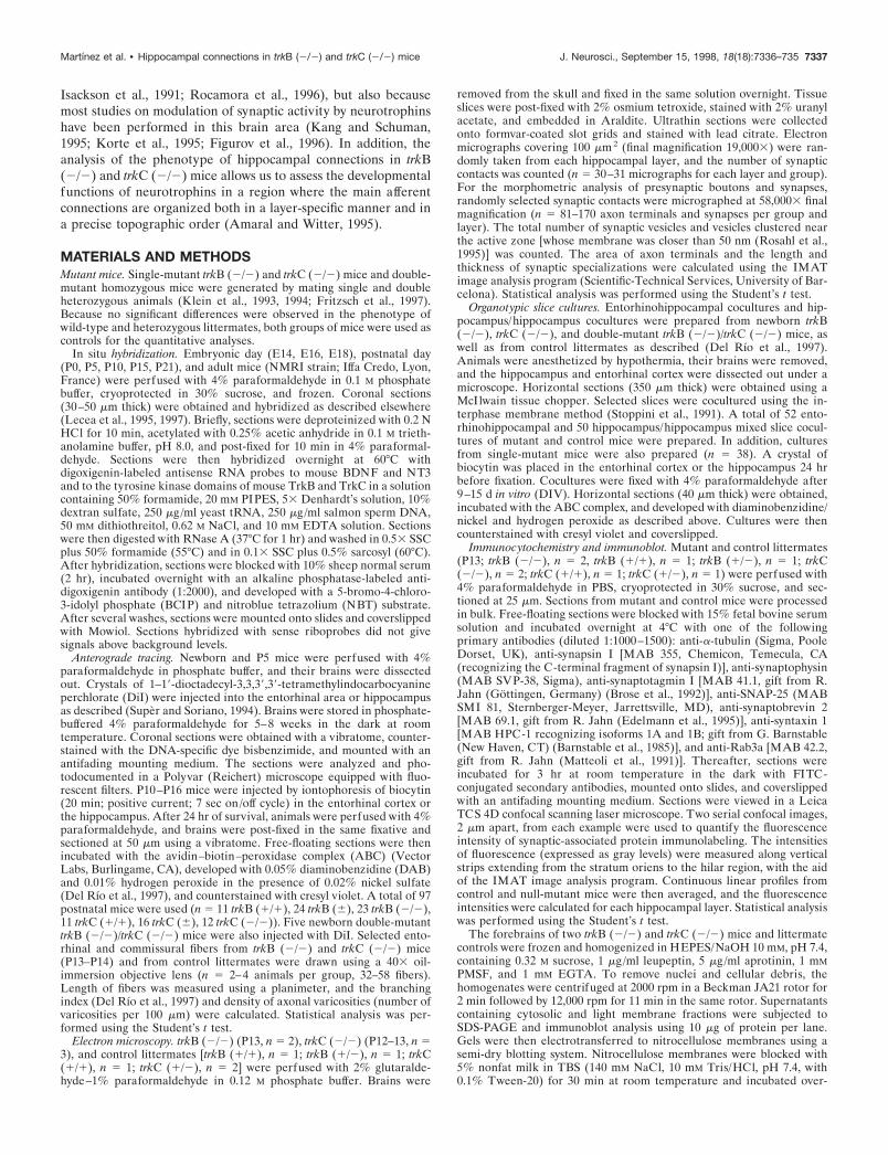
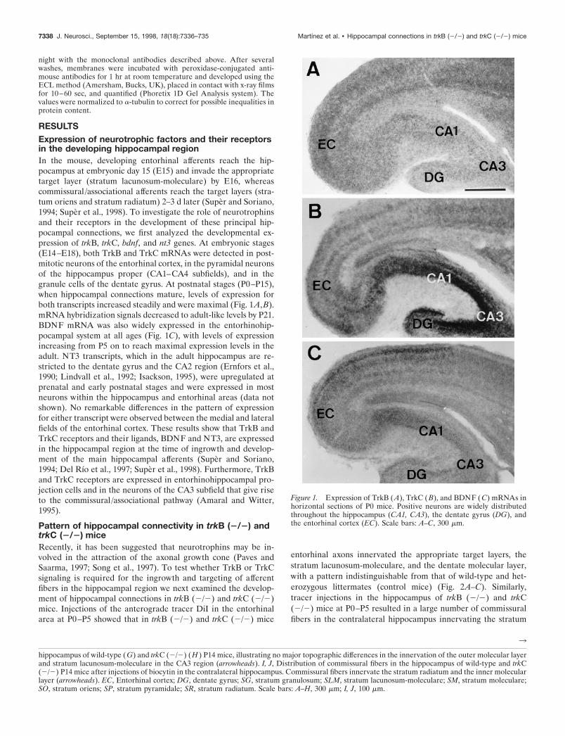
entorhinal axons innervated the appropriate target layers, thestratum lacunosum-moleculare, and the dentate molecular layer,with a pattern indistinguishable from that of wild-type and het-erozygous littermates (control mice) (Fig. 2A–C). Similarly,tracer injections in the hippocampus of trkB (2/2) and trkC(2/2) mice at P0–P5 resulted in a large number of commissuralfibers in the contralateral hippocampus innervating the stratum
3
hippocampus of wild-type (G) and trkC (2/2) (H ) P14 mice, illustrating no major topographic differences in the innervation of the outer molecular layerand stratum lacunosum-moleculare in the CA3 region (arrowheads). I, J, Distribution of commissural fibers in the hippocampus of wild-type and trkC(2/2) P14 mice after injections of biocytin in the contralateral hippocampus. Commissural fibers innervate the stratum radiatum and the inner molecularlayer (arrowheads). EC, Entorhinal cortex; DG, dentate gyrus; SG, stratum granulosum; SLM, stratum lacunosum-moleculare; SM, stratum moleculare;SO, stratum oriens; SP, stratum pyramidale; SR, stratum radiatum. Scale bars: A–H, 300 mm; I, J, 100 mm.
Figure 1. Expression of TrkB (A), TrkC (B), and BDNF (C) mRNAs inhorizontal sections of P0 mice. Positive neurons are widely distributedthroughout the hippocampus (CA1, CA3), the dentate gyrus (DG), andthe entorhinal cortex (EC). Scale bars: A–C, 300 mm.
7338 J. Neurosci., September 15, 1998, 18(18):7336–735 Martınez et al. • Hippocampal connections in trkB (2/2) and trkC (2/2) mice
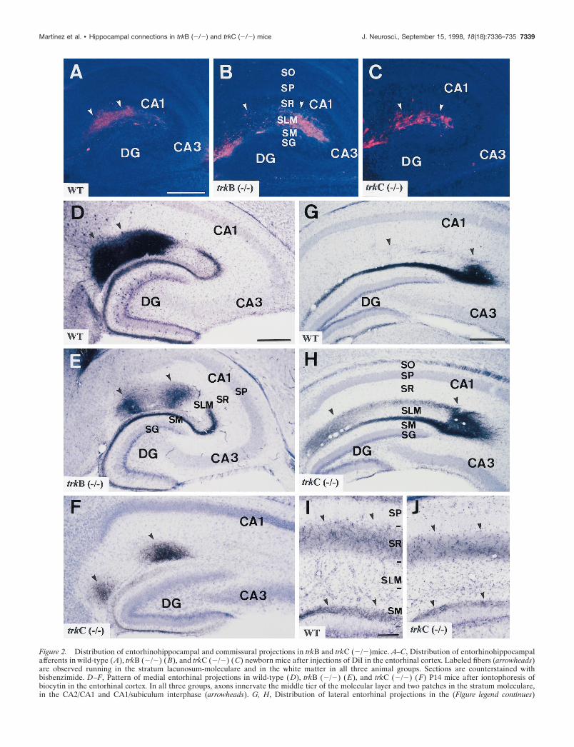
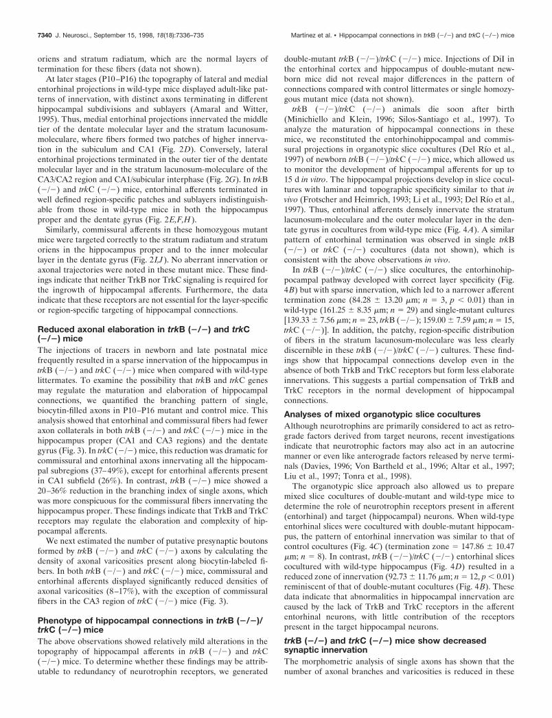
Figure 2. Distribution of entorhinohippocampal and commissural projections in trkB and trkC (2/2)mice. A–C, Distribution of entorhinohippocampalafferents in wild-type (A), trkB (2/2) (B), and trkC (2/2) (C) newborn mice after injections of DiI in the entorhinal cortex. Labeled fibers (arrowheads)are observed running in the stratum lacunosum-moleculare and in the white matter in all three animal groups. Sections are counterstained withbisbenzimide. D–F, Pattern of medial entorhinal projections in wild-type (D), trkB (2/2) (E), and trkC (2/2) (F) P14 mice after iontophoresis ofbiocytin in the entorhinal cortex. In all three groups, axons innervate the middle tier of the molecular layer and two patches in the stratum moleculare,in the CA2/CA1 and CA1/subiculum interphase (arrowheads). G, H, Distribution of lateral entorhinal projections in the (Figure legend continues)
Martınez et al. • Hippocampal connections in trkB (2/2) and trkC (2/2) mice J. Neurosci., September 15, 1998, 18(18):7336–735 7339
oriens and stratum radiatum, which are the normal layers oftermination for these fibers (data not shown).
At later stages (P10–P16) the topography of lateral and medialentorhinal projections in wild-type mice displayed adult-like pat-terns of innervation, with distinct axons terminating in differenthippocampal subdivisions and sublayers (Amaral and Witter,1995). Thus, medial entorhinal projections innervated the middletier of the dentate molecular layer and the stratum lacunosum-moleculare, where fibers formed two patches of higher innerva-tion in the subiculum and CA1 (Fig. 2D). Conversely, lateralentorhinal projections terminated in the outer tier of the dentatemolecular layer and in the stratum lacunosum-moleculare of theCA3/CA2 region and CA1/subicular interphase (Fig. 2G). In trkB(2/2) and trkC (2/2) mice, entorhinal afferents terminated inwell defined region-specific patches and sublayers indistinguish-able from those in wild-type mice in both the hippocampusproper and the dentate gyrus (Fig. 2E,F,H).
Similarly, commissural afferents in these homozygous mutantmice were targeted correctly to the stratum radiatum and stratumoriens in the hippocampus proper and to the inner molecularlayer in the dentate gyrus (Fig. 2 I,J). No aberrant innervation oraxonal trajectories were noted in these mutant mice. These find-ings indicate that neither TrkB nor TrkC signaling is required forthe ingrowth of hippocampal afferents. Furthermore, the dataindicate that these receptors are not essential for the layer-specificor region-specific targeting of hippocampal connections.
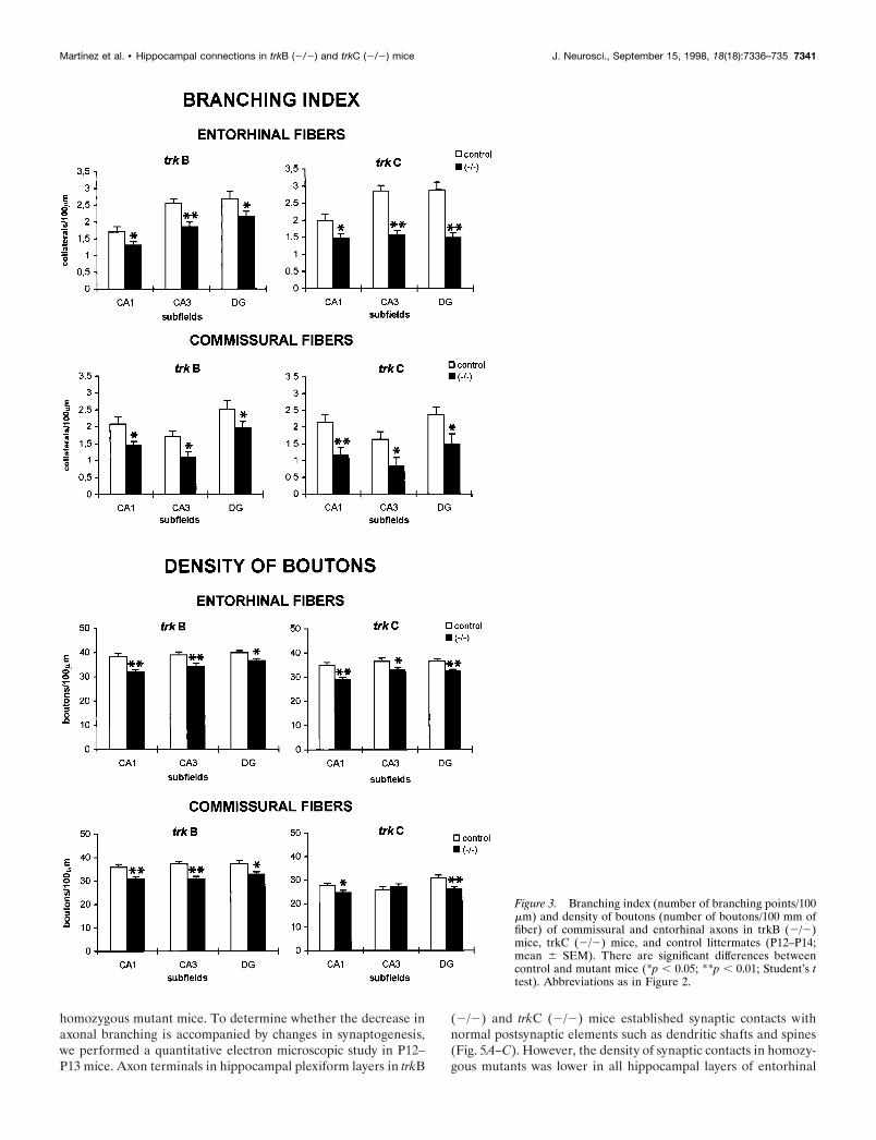
Reduced axonal elaboration in trkB (2/2) and trkC(2/2) miceThe injections of tracers in newborn and late postnatal micefrequently resulted in a sparse innervation of the hippocampus intrkB (2/2) and trkC (2/2) mice when compared with wild-typelittermates. To examine the possibility that trkB and trkC genesmay regulate the maturation and elaboration of hippocampalconnections, we quantified the branching pattern of single,biocytin-filled axons in P10–P16 mutant and control mice. Thisanalysis showed that entorhinal and commissural fibers had feweraxon collaterals in both trkB (2/2) and trkC (2/2) mice in thehippocampus proper (CA1 and CA3 regions) and the dentategyrus (Fig. 3). In trkC (2/2) mice, this reduction was dramatic forcommissural and entorhinal axons innervating all the hippocam-pal subregions (37–49%), except for entorhinal afferents presentin CA1 subfield (26%). In contrast, trkB (2/2) mice showed a20–36% reduction in the branching index of single axons, whichwas more conspicuous for the commissural fibers innervating thehippocampus proper. These findings indicate that TrkB and TrkCreceptors may regulate the elaboration and complexity of hip-pocampal afferents.
We next estimated the number of putative presynaptic boutonsformed by trkB (2/2) and trkC (2/2) axons by calculating thedensity of axonal varicosities present along biocytin-labeled fi-bers. In both trkB (2/2) and trkC (2/2) mice, commissural andentorhinal afferents displayed significantly reduced densities ofaxonal varicosities (8–17%), with the exception of commissuralfibers in the CA3 region of trkC (2/2) mice (Fig. 3).
Phenotype of hippocampal connections in trkB (2/2)/trkC (2/2) miceThe above observations showed relatively mild alterations in thetopography of hippocampal afferents in trkB (2/2) and trkC(2/2) mice. To determine whether these findings may be attrib-utable to redundancy of neurotrophin receptors, we generated
double-mutant trkB (2/2)/trkC (2/2) mice. Injections of DiI inthe entorhinal cortex and hippocampus of double-mutant new-born mice did not reveal major differences in the pattern ofconnections compared with control littermates or single homozy-gous mutant mice (data not shown).
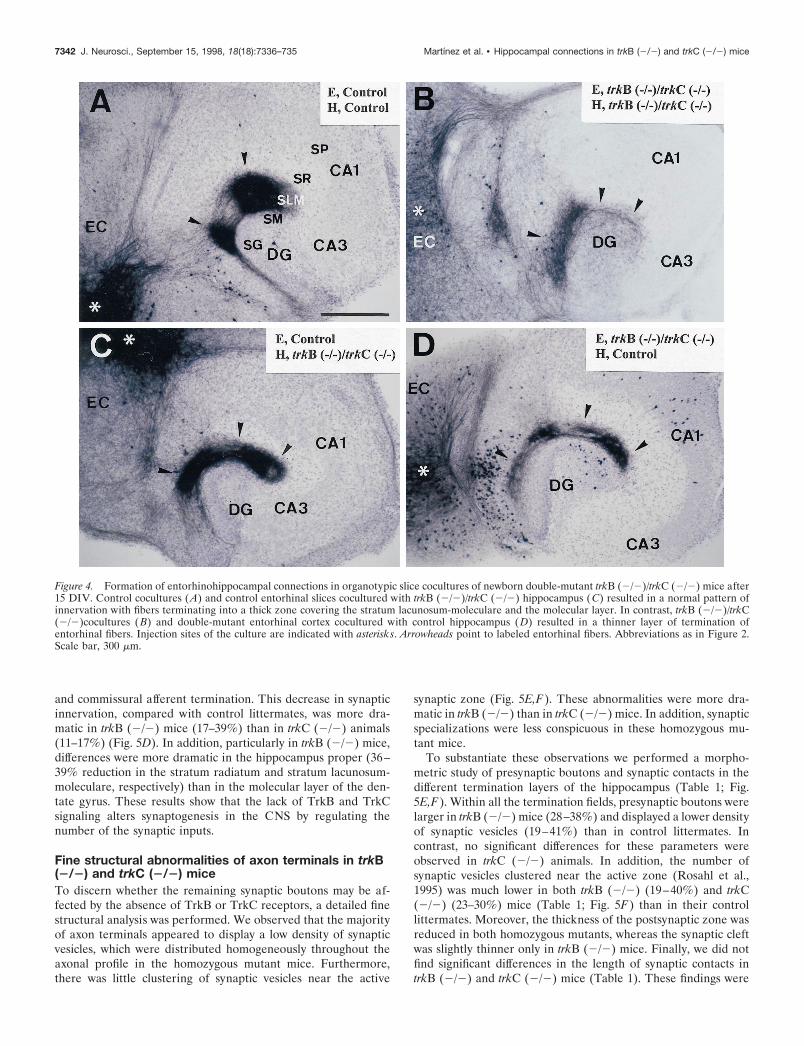
trkB (2/2)/trkC (2/2) animals die soon after birth(Minichiello and Klein, 1996; Silos-Santiago et al., 1997). Toanalyze the maturation of hippocampal connections in thesemice, we reconstituted the entorhinohippocampal and commis-sural projections in organotypic slice cocultures (Del Rıo et al.,1997) of newborn trkB (2/2)/trkC (2/2) mice, which allowed usto monitor the development of hippocampal afferents for up to15 d in vitro. The hippocampal projections develop in slice cocul-tures with laminar and topographic specificity similar to that invivo (Frotscher and Heimrich, 1993; Li et al., 1993; Del Rıo et al.,1997). Thus, entorhinal afferents densely innervate the stratumlacunosum-moleculare and the outer molecular layer in the den-tate gyrus in cocultures from wild-type mice (Fig. 4A). A similarpattern of entorhinal termination was observed in single trkB(2/2) or trkC (2/2) cocultures (data not shown), which isconsistent with the above observations in vivo.
In trkB (2/2)/trkC (2/2) slice cocultures, the entorhinohip-pocampal pathway developed with correct layer specificity (Fig.4B) but with sparse innervation, which led to a narrower afferenttermination zone (84.28 6 13.20 mm; n 5 3, p , 0.01) than inwild-type (161.25 6 8.35 mm; n 5 29) and single-mutant cultures[139.33 6 7.56 mm; n 5 23, trkB (2/2); 159.00 6 7.59 mm; n 5 15,trkC (2/2)]. In addition, the patchy, region-specific distributionof fibers in the stratum lacunosum-moleculare was less clearlydiscernible in these trkB (2/2)/trkC (2/2) cultures. These find-ings show that hippocampal connections develop even in theabsence of both TrkB and TrkC receptors but form less elaborateinnervations. This suggests a partial compensation of TrkB andTrkC receptors in the normal development of hippocampalconnections.
Analyses of mixed organotypic slice coculturesAlthough neurotrophins are primarily considered to act as retro-grade factors derived from target neurons, recent investigationsindicate that neurotrophic factors may also act in an autocrinemanner or even like anterograde factors released by nerve termi-nals (Davies, 1996; Von Bartheld et al., 1996; Altar et al., 1997;Liu et al., 1997; Tonra et al., 1998).
The organotypic slice approach also allowed us to preparemixed slice cocultures of double-mutant and wild-type mice todetermine the role of neurotrophin receptors present in afferent(entorhinal) and target (hippocampal) neurons. When wild-typeentorhinal slices were cocultured with double-mutant hippocam-pus, the pattern of entorhinal innervation was similar to that ofcontrol cocultures (Fig. 4C) (termination zone 5 147.86 6 10.47mm; n 5 8). In contrast, trkB (2/2)/trkC (2/2) entorhinal slicescocultured with wild-type hippocampus (Fig. 4D) resulted in areduced zone of innervation (92.73 6 11.76 mm; n 5 12, p , 0.01)reminiscent of that of double-mutant cocultures (Fig. 4B). Thesedata indicate that abnormalities in hippocampal innervation arecaused by the lack of TrkB and TrkC receptors in the afferententorhinal neurons, with little contribution of the receptorspresent in the target hippocampal neurons.
trkB (2/2) and trkC (2/2) mice show decreasedsynaptic innervationThe morphometric analysis of single axons has shown that thenumber of axonal branches and varicosities is reduced in these
7340 J. Neurosci., September 15, 1998, 18(18):7336–735 Martınez et al. • Hippocampal connections in trkB (2/2) and trkC (2/2) mice
homozygous mutant mice. To determine whether the decrease inaxonal branching is accompanied by changes in synaptogenesis,we performed a quantitative electron microscopic study in P12–P13 mice. Axon terminals in hippocampal plexiform layers in trkB
(2/2) and trkC (2/2) mice established synaptic contacts withnormal postsynaptic elements such as dendritic shafts and spines(Fig. 5A–C). However, the density of synaptic contacts in homozy-gous mutants was lower in all hippocampal layers of entorhinal
Figure 3. Branching index (number of branching points/100mm) and density of boutons (number of boutons/100 mm offiber) of commissural and entorhinal axons in trkB (2/2)mice, trkC (2/2) mice, and control littermates (P12–P14;mean 6 SEM). There are significant differences betweencontrol and mutant mice (*p , 0.05; **p , 0.01; Student’s ttest). Abbreviations as in Figure 2.
Martınez et al. • Hippocampal connections in trkB (2/2) and trkC (2/2) mice J. Neurosci., September 15, 1998, 18(18):7336–735 7341
and commissural afferent termination. This decrease in synapticinnervation, compared with control littermates, was more dra-matic in trkB (2/2) mice (17–39%) than in trkC (2/2) animals(11–17%) (Fig. 5D). In addition, particularly in trkB (2/2) mice,differences were more dramatic in the hippocampus proper (36–39% reduction in the stratum radiatum and stratum lacunosum-moleculare, respectively) than in the molecular layer of the den-tate gyrus. These results show that the lack of TrkB and TrkCsignaling alters synaptogenesis in the CNS by regulating thenumber of the synaptic inputs.
Fine structural abnormalities of axon terminals in trkB(2/2) and trkC (2/2) miceTo discern whether the remaining synaptic boutons may be af-fected by the absence of TrkB or TrkC receptors, a detailed finestructural analysis was performed. We observed that the majorityof axon terminals appeared to display a low density of synapticvesicles, which were distributed homogeneously throughout theaxonal profile in the homozygous mutant mice. Furthermore,there was little clustering of synaptic vesicles near the active
synaptic zone (Fig. 5E,F). These abnormalities were more dra-matic in trkB (2/2) than in trkC (2/2) mice. In addition, synapticspecializations were less conspicuous in these homozygous mu-tant mice.
To substantiate these observations we performed a morpho-metric study of presynaptic boutons and synaptic contacts in thedifferent termination layers of the hippocampus (Table 1; Fig.5E,F). Within all the termination fields, presynaptic boutons werelarger in trkB (2/2) mice (28–38%) and displayed a lower densityof synaptic vesicles (19–41%) than in control littermates. Incontrast, no significant differences for these parameters wereobserved in trkC (2/2) animals. In addition, the number ofsynaptic vesicles clustered near the active zone (Rosahl et al.,1995) was much lower in both trkB (2/2) (19–40%) and trkC(2/2) (23–30%) mice (Table 1; Fig. 5F) than in their controllittermates. Moreover, the thickness of the postsynaptic zone wasreduced in both homozygous mutants, whereas the synaptic cleftwas slightly thinner only in trkB (2/2) mice. Finally, we did notfind significant differences in the length of synaptic contacts intrkB (2/2) and trkC (2/2) mice (Table 1). These findings were
Figure 4. Formation of entorhinohippocampal connections in organotypic slice cocultures of newborn double-mutant trkB (2/2)/trkC (2/2) mice after15 DIV. Control cocultures (A) and control entorhinal slices cocultured with trkB (2/2)/trkC (2/2) hippocampus (C) resulted in a normal pattern ofinnervation with fibers terminating into a thick zone covering the stratum lacunosum-moleculare and the molecular layer. In contrast, trkB (2/2)/trkC(2/2)cocultures (B) and double-mutant entorhinal cortex cocultured with control hippocampus (D) resulted in a thinner layer of termination ofentorhinal fibers. Injection sites of the culture are indicated with asterisks. Arrowheads point to labeled entorhinal fibers. Abbreviations as in Figure 2.Scale bar, 300 mm.
7342 J. Neurosci., September 15, 1998, 18(18):7336–735 Martınez et al. • Hippocampal connections in trkB (2/2) and trkC (2/2) mice
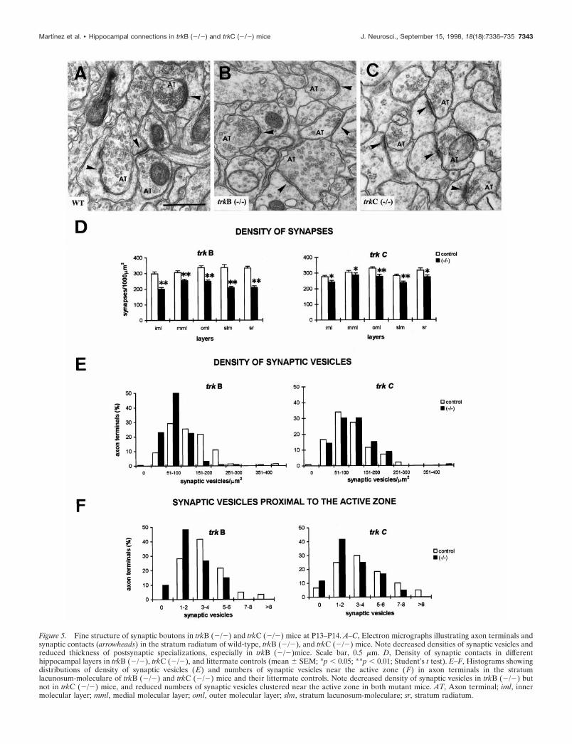
Figure 5. Fine structure of synaptic boutons in trkB (2/2) and trkC (2/2) mice at P13–P14. A–C, Electron micrographs illustrating axon terminals andsynaptic contacts (arrowheads) in the stratum radiatum of wild-type, trkB (2/2), and trkC (2/2) mice. Note decreased densities of synaptic vesicles andreduced thickness of postsynaptic specializations, especially in trkB (2/2)mice. Scale bar, 0.5 mm. D, Density of synaptic contacts in differenthippocampal layers in trkB (2/2), trkC (2/2), and littermate controls (mean 6 SEM; *p , 0.05; **p , 0.01; Student’s t test). E–F, Histograms showingdistributions of density of synaptic vesicles (E) and numbers of synaptic vesicles near the active zone (F) in axon terminals in the stratumlacunosum-moleculare of trkB (2/2) and trkC (2/2) mice and their littermate controls. Note decreased density of synaptic vesicles in trkB (2/2) butnot in trkC (2/2) mice, and reduced numbers of synaptic vesicles clustered near the active zone in both mutant mice. AT, Axon terminal; iml, innermolecular layer; mml, medial molecular layer; oml, outer molecular layer; slm, stratum lacunosum-moleculare; sr, stratum radiatum.
Martınez et al. • Hippocampal connections in trkB (2/2) and trkC (2/2) mice J. Neurosci., September 15, 1998, 18(18):7336–735 7343
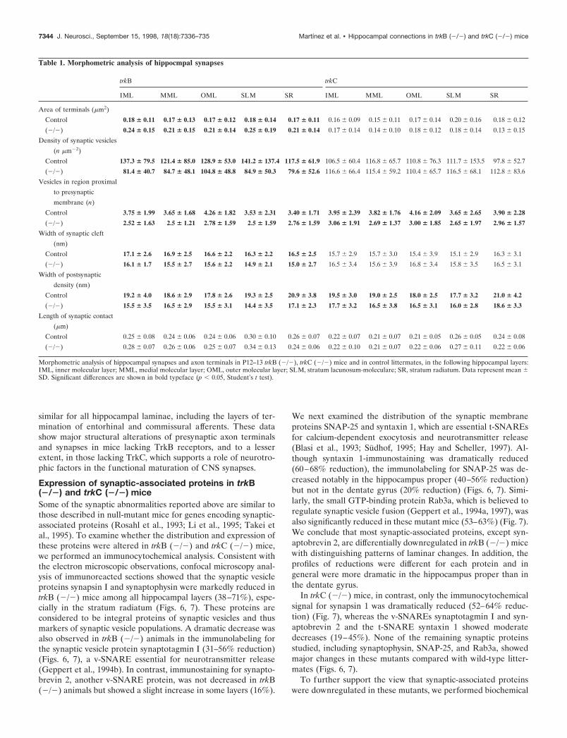
similar for all hippocampal laminae, including the layers of ter-mination of entorhinal and commissural afferents. These datashow major structural alterations of presynaptic axon terminalsand synapses in mice lacking TrkB receptors, and to a lesserextent, in those lacking TrkC, which supports a role of neurotro-phic factors in the functional maturation of CNS synapses.
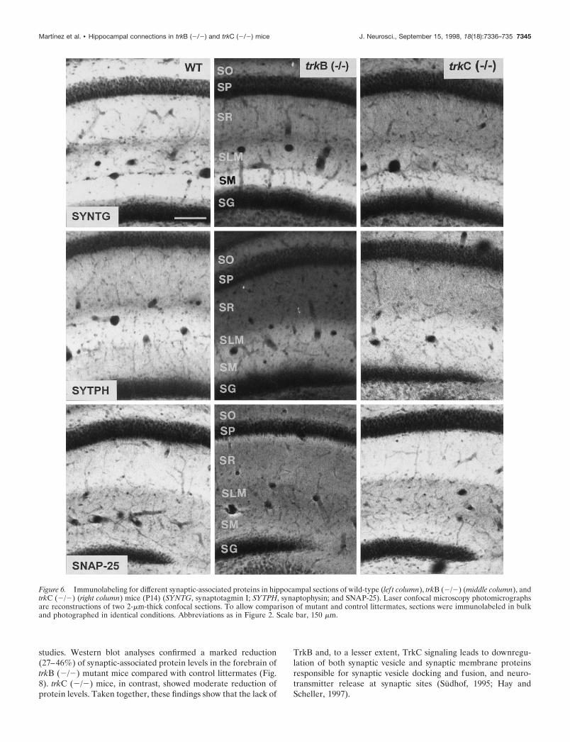
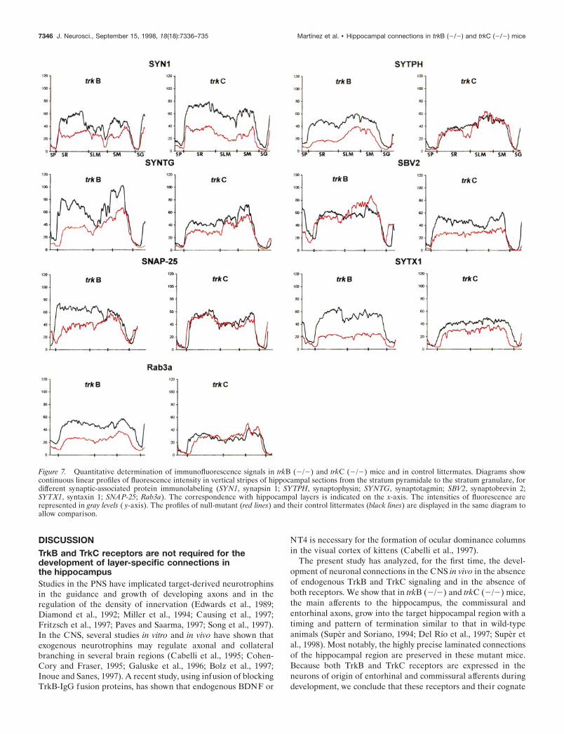
Expression of synaptic-associated proteins in trkB(2/2) and trkC (2/2) miceSome of the synaptic abnormalities reported above are similar tothose described in null-mutant mice for genes encoding synaptic-associated proteins (Rosahl et al., 1993; Li et al., 1995; Takei etal., 1995). To examine whether the distribution and expression ofthese proteins were altered in trkB (2/2) and trkC (2/2) mice,we performed an immunocytochemical analysis. Consistent withthe electron microscopic observations, confocal microscopy anal-ysis of immunoreacted sections showed that the synaptic vesicleproteins synapsin I and synaptophysin were markedly reduced intrkB (2/2) mice among all hippocampal layers (38–71%), espe-cially in the stratum radiatum (Figs. 6, 7). These proteins areconsidered to be integral proteins of synaptic vesicles and thusmarkers of synaptic vesicle populations. A dramatic decrease wasalso observed in trkB (2/2) animals in the immunolabeling forthe synaptic vesicle protein synaptotagmin I (31–56% reduction)(Figs. 6, 7), a v-SNARE essential for neurotransmitter release(Geppert et al., 1994b). In contrast, immunostaining for synapto-brevin 2, another v-SNARE protein, was not decreased in trkB(2/2) animals but showed a slight increase in some layers (16%).
We next examined the distribution of the synaptic membraneproteins SNAP-25 and syntaxin 1, which are essential t-SNAREsfor calcium-dependent exocytosis and neurotransmitter release(Blasi et al., 1993; Sudhof, 1995; Hay and Scheller, 1997). Al-though syntaxin 1-immunostaining was dramatically reduced(60–68% reduction), the immunolabeling for SNAP-25 was de-creased notably in the hippocampus proper (40–56% reduction)but not in the dentate gyrus (20% reduction) (Figs. 6, 7). Simi-larly, the small GTP-binding protein Rab3a, which is believed toregulate synaptic vesicle fusion (Geppert et al., 1994a, 1997), wasalso significantly reduced in these mutant mice (53–63%) (Fig. 7).We conclude that most synaptic-associated proteins, except syn-aptobrevin 2, are differentially downregulated in trkB (2/2) micewith distinguishing patterns of laminar changes. In addition, theprofiles of reductions were different for each protein and ingeneral were more dramatic in the hippocampus proper than inthe dentate gyrus.
In trkC (2/2) mice, in contrast, only the immunocytochemicalsignal for synapsin 1 was dramatically reduced (52–64% reduc-tion) (Fig. 7), whereas the v-SNAREs synaptotagmin I and syn-aptobrevin 2 and the t-SNARE syntaxin 1 showed moderatedecreases (19–45%). None of the remaining synaptic proteinsstudied, including synaptophysin, SNAP-25, and Rab3a, showedmajor changes in these mutants compared with wild-type litter-mates (Figs. 6, 7).
To further support the view that synaptic-associated proteinswere downregulated in these mutants, we performed biochemical
Table 1. Morphometric analysis of hippocmpal synapses
trkB trkC
IML MML OML SLM SR IML MML OML SLM SR
Area of terminals (mm2)
Control 0.18 6 0.11 0.17 6 0.13 0.17 6 0.12 0.18 6 0.14 0.17 6 0.11 0.16 6 0.09 0.15 6 0.11 0.17 6 0.14 0.20 6 0.16 0.18 6 0.12
(2/2) 0.24 6 0.15 0.21 6 0.15 0.21 6 0.14 0.25 6 0.19 0.21 6 0.14 0.17 6 0.14 0.14 6 0.10 0.18 6 0.12 0.18 6 0.14 0.13 6 0.15
Density of synaptic vesicles
(n mm22)
Control 137.3 6 79.5 121.4 6 85.0 128.9 6 53.0 141.2 6 137.4 117.5 6 61.9 106.5 6 60.4 116.8 6 65.7 110.8 6 76.3 111.7 6 153.5 97.8 6 52.7
(2/2) 81.4 6 40.7 84.7 6 48.1 104.8 6 48.8 84.9 6 50.3 79.6 6 52.6 116.6 6 66.4 115.4 6 59.2 110.4 6 65.7 116.5 6 68.1 112.8 6 83.6
Vesicles in region proximal
to presynaptic
membrane (n)
Control 3.75 6 1.99 3.65 6 1.68 4.26 6 1.82 3.53 6 2.31 3.40 6 1.71 3.95 6 2.39 3.82 6 1.76 4.16 6 2.09 3.65 6 2.65 3.90 6 2.28
(2/2) 2.52 6 1.63 2.5 6 1.21 2.78 6 1.59 2.5 6 1.59 2.76 6 1.59 3.06 6 1.91 2.69 6 1.37 3.00 6 1.85 2.65 6 1.97 2.96 6 1.57
Width of synaptic cleft
(nm)
Control 17.1 6 2.6 16.9 6 2.5 16.6 6 2.2 16.3 6 2.2 16.5 6 2.5 15.7 6 2.9 15.7 6 3.0 15.4 6 3.9 15.1 6 2.9 16.3 6 3.1
(2/2) 16.1 6 1.7 15.5 6 2.7 15.6 6 2.2 14.9 6 2.1 15.0 6 2.7 16.5 6 3.4 15.6 6 3.9 16.8 6 3.4 15.8 6 3.5 16.5 6 3.1
Width of postsynaptic
density (nm)
Control 19.2 6 4.0 18.6 6 2.9 17.8 6 2.6 19.3 6 2.5 20.9 6 3.8 19.5 6 3.0 19.0 6 2.5 18.0 6 2.5 17.7 6 3.2 21.0 6 4.2
(2/2) 15.5 6 3.5 16.5 6 2.9 15.5 6 3.1 14.4 6 3.5 17.1 6 2.3 17.7 6 3.2 16.5 6 3.8 16.5 6 3.1 16.0 6 2.8 18.6 6 3.3
Length of synaptic contact
(mm)
Control 0.25 6 0.08 0.24 6 0.06 0.24 6 0.06 0.30 6 0.10 0.26 6 0.07 0.22 6 0.07 0.21 6 0.07 0.21 6 0.05 0.26 6 0.05 0.24 6 0.08
(2/2) 0.28 6 0.07 0.26 6 0.06 0.25 6 0.07 0.34 6 0.13 0.24 6 0.06 0.22 6 0.10 0.21 6 0.07 0.22 6 0.06 0.27 6 0.11 0.22 6 0.06
Morphometric analysis of hippocampal synapses and axon terminals in P12–13 trkB (2/2), trkC (2/2) mice and in control littermates, in the following hippocampal layers:IML, inner molecular layer; MML, medial molecular layer; OML, outer molecular layer; SLM, stratum lacunosum-moleculare; SR, stratum radiatum. Data represent mean 6SD. Significant differences are shown in bold typeface (p , 0.05, Student’s t test).
7344 J. Neurosci., September 15, 1998, 18(18):7336–735 Martınez et al. • Hippocampal connections in trkB (2/2) and trkC (2/2) mice
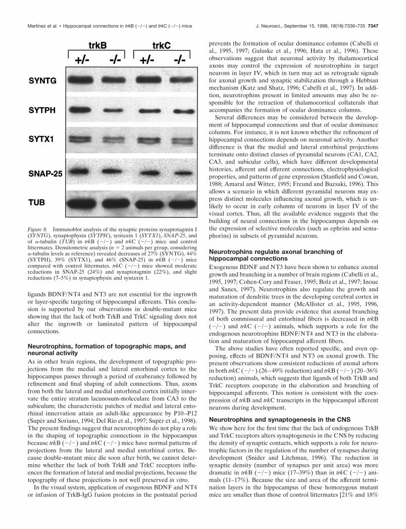
studies. Western blot analyses confirmed a marked reduction(27–46%) of synaptic-associated protein levels in the forebrain oftrkB (2/2) mutant mice compared with control littermates (Fig.8). trkC (2/2) mice, in contrast, showed moderate reduction ofprotein levels. Taken together, these findings show that the lack of
TrkB and, to a lesser extent, TrkC signaling leads to downregu-lation of both synaptic vesicle and synaptic membrane proteinsresponsible for synaptic vesicle docking and fusion, and neuro-transmitter release at synaptic sites (Sudhof, 1995; Hay andScheller, 1997).
Figure 6. Immunolabeling for different synaptic-associated proteins in hippocampal sections of wild-type (lef t column), trkB (2/2) (middle column), andtrkC (2/2) (right column) mice (P14) (SYNTG, synaptotagmin I; SYTPH, synaptophysin; and SNAP-25). Laser confocal microscopy photomicrographsare reconstructions of two 2-mm-thick confocal sections. To allow comparison of mutant and control littermates, sections were immunolabeled in bulkand photographed in identical conditions. Abbreviations as in Figure 2. Scale bar, 150 mm.
Martınez et al. • Hippocampal connections in trkB (2/2) and trkC (2/2) mice J. Neurosci., September 15, 1998, 18(18):7336–735 7345
DISCUSSIONTrkB and TrkC receptors are not required for thedevelopment of layer-specific connections inthe hippocampusStudies in the PNS have implicated target-derived neurotrophinsin the guidance and growth of developing axons and in theregulation of the density of innervation (Edwards et al., 1989;Diamond et al., 1992; Miller et al., 1994; Causing et al., 1997;Fritzsch et al., 1997; Paves and Saarma, 1997; Song et al., 1997).In the CNS, several studies in vitro and in vivo have shown thatexogenous neurotrophins may regulate axonal and collateralbranching in several brain regions (Cabelli et al., 1995; Cohen-Cory and Fraser, 1995; Galuske et al., 1996; Bolz et al., 1997;Inoue and Sanes, 1997). A recent study, using infusion of blockingTrkB-IgG fusion proteins, has shown that endogenous BDNF or
NT4 is necessary for the formation of ocular dominance columnsin the visual cortex of kittens (Cabelli et al., 1997).
The present study has analyzed, for the first time, the devel-opment of neuronal connections in the CNS in vivo in the absenceof endogenous TrkB and TrkC signaling and in the absence ofboth receptors. We show that in trkB (2/2) and trkC (2/2) mice,the main afferents to the hippocampus, the commissural andentorhinal axons, grow into the target hippocampal region with atiming and pattern of termination similar to that in wild-typeanimals (Super and Soriano, 1994; Del Rıo et al., 1997; Super etal., 1998). Most notably, the highly precise laminated connectionsof the hippocampal region are preserved in these mutant mice.Because both TrkB and TrkC receptors are expressed in theneurons of origin of entorhinal and commissural afferents duringdevelopment, we conclude that these receptors and their cognate
Figure 7. Quantitative determination of immunofluorescence signals in trkB (2/2) and trkC (2/2) mice and in control littermates. Diagrams showcontinuous linear profiles of fluorescence intensity in vertical stripes of hippocampal sections from the stratum pyramidale to the stratum granulare, fordifferent synaptic-associated protein immunolabeling (SYN1, synapsin 1; SYTPH, synaptophysin; SYNTG, synaptotagmin; SBV2, synaptobrevin 2;SYTX1, syntaxin 1; SNAP-25; Rab3a). The correspondence with hippocampal layers is indicated on the x-axis. The intensities of fluorescence arerepresented in gray levels ( y-axis). The profiles of null-mutant (red lines) and their control littermates (black lines) are displayed in the same diagram toallow comparison.
7346 J. Neurosci., September 15, 1998, 18(18):7336–735 Martınez et al. • Hippocampal connections in trkB (2/2) and trkC (2/2) mice
ligands BDNF/NT4 and NT3 are not essential for the ingrowthor layer-specific targeting of hippocampal afferents. This conclu-sion is supported by our observations in double-mutant miceshowing that the lack of both TrkB and TrkC signaling does notalter the ingrowth or laminated pattern of hippocampalconnections.
Neurotrophins, formation of topographic maps, andneuronal activityAs in other brain regions, the development of topographic pro-jections from the medial and lateral entorhinal cortex to thehippocampus passes through a period of exuberancy followed byrefinement and final shaping of adult connections. Thus, axonsfrom both the lateral and medial entorhinal cortex initially inner-vate the entire stratum lacunosum-moleculare from CA3 to thesubiculum; the characteristic patches of medial and lateral ento-rhinal innervation attain an adult-like appearance by P10–P12(Super and Soriano, 1994; Del Rıo et al., 1997; Super et al., 1998).The present findings suggest that neurotrophins do not play a rolein the shaping of topographic connections in the hippocampusbecause trkB (2/2) and trkC (2/2) mice have normal patterns ofprojections from the lateral and medial entorhinal cortex. Be-cause double-mutant mice die soon after birth, we cannot deter-mine whether the lack of both TrkB and TrkC receptors influ-ences the formation of lateral and medial projections, because thetopography of these projections is not well preserved in vitro.
In the visual system, application of exogenous BDNF and NT4or infusion of TrkB-IgG fusion proteins in the postnatal period
prevents the formation of ocular dominance columns (Cabelli etal., 1995, 1997; Galuske et al., 1996; Hata et al., 1996). Theseobservations suggest that neuronal activity by thalamocorticalaxons may control the expression of neurotrophins in targetneurons in layer IV, which in turn may act as retrograde signalsfor axonal growth and synaptic stabilization through a Hebbianmechanism (Katz and Shatz, 1996; Cabelli et al., 1997). In addi-tion, neurotrophins present in limited amounts may also be re-sponsible for the retraction of thalamocortical collaterals thataccompanies the formation of ocular dominance columns.
Several differences may be considered between the develop-ment of hippocampal connections and that of ocular dominancecolumns. For instance, it is not known whether the refinement ofhippocampal connections depends on neuronal activity. Anotherdifference is that the medial and lateral entorhinal projectionsterminate onto distinct classes of pyramidal neurons (CA1, CA2,CA3, and subicular cells), which have different developmentalhistories, afferent and efferent connections, electrophysiologicalproperties, and patterns of gene expression (Stanfield and Cowan,1988; Amaral and Witter, 1995; Freund and Buzsaki, 1996). Thisallows a scenario in which different pyramidal neurons may ex-press distinct molecules influencing axonal growth, which is un-likely to occur in early columns of neurons in layer IV of thevisual cortex. Thus, all the available evidence suggests that thebuilding of neural connections in the hippocampus depends onthe expression of selective molecules (such as ephrins and sema-phorins) in subsets of pyramidal neurons.
Neurotrophins regulate axonal branching ofhippocampal connectionsExogenous BDNF and NT3 have been shown to enhance axonalgrowth and branching in a number of brain regions (Cabelli et al.,1995, 1997; Cohen-Cory and Fraser, 1995; Bolz et al., 1997; Inoueand Sanes, 1997). Neurotrophins also regulate the growth andmaturation of dendritic trees in the developing cerebral cortex inan activity-dependent manner (McAllister et al., 1995, 1996,1997). The present data provide evidence that axonal branchingof both commissural and entorhinal fibers is decreased in trkB(2/2) and trkC (2/2) animals, which supports a role for theendogenous neurotrophins BDNF/NT4 and NT3 in the elabora-tion and maturation of hippocampal afferent fibers.
The above studies have often reported specific, and even op-posing, effects of BDNF/NT4 and NT3 on axonal growth. Thepresent observations show consistent reductions of axonal arborsin both trkC (2/2) (26–49% reduction) and trkB (2/2) (20–36%reduction) animals, which suggests that ligands of both TrkB andTrkC receptors cooperate in the elaboration and branching ofhippocampal afferents. This notion is consistent with the coex-pression of trkB and trkC transcripts in the hippocampal afferentneurons during development.
Neurotrophins and synaptogenesis in the CNSWe show here for the first time that the lack of endogenous TrkBand TrkC receptors alters synaptogenesis in the CNS by reducingthe density of synaptic contacts, which supports a role for neuro-trophic factors in the regulation of the number of synapses duringdevelopment (Snider and Litchman, 1996). The reduction insynaptic density (number of synapses per unit area) was moredramatic in trkB (2/2) mice (17–39%) than in trkC (2/2) ani-mals (11–17%). Because the size and area of the afferent termi-nation layers in the hippocampus of these homozygous mutantmice are smaller than those of control littermates [21% and 18%
Figure 8. Immunoblot analysis of the synaptic proteins synaptotagmin I(SYNTG), synaptophysin (SYTPH ), syntaxin 1 (SYTX1), SNAP-25, andof a-tubulin (TUB) in trkB (2/2) and trkC (2/2) mice and controllittermates. Densitometric analysis (n 5 2 animals per group, consideringa-tubulin levels as reference) revealed decreases of 27% (SYNTG), 44%(SYTPH), 39% (SYTX1), and 46% (SNAP-25) in trkB (2/2) micecompared with control littermates. trkC (2/2) mice showed moderatereductions in SNAP-25 (24%) and synaptotagmin (22%), and slightreductions (7–5%) in synaptophysin and syntaxin 1.
Martınez et al. • Hippocampal connections in trkB (2/2) and trkC (2/2) mice J. Neurosci., September 15, 1998, 18(18):7336–735 7347
reduction of layers in trkB (2/2) and trkC (2/2) mice, respec-tively, as estimated in Nissl-stained sections], the reduction in thetotal number of synaptic inputs is higher. This reduction in thetotal number of hippocampal synapses can be estimated as 34–52% and 25–30% for trkB (2/2) and trkC (2/2) mice,respectively.
Postnatal trkB (2/2) mice show increased neuronal cell deathin some of the populations of hippocampal afferent neurons, suchas the CA3 pyramidal cells (Alcantara et al., 1997). The de-creased synaptic innervation in part may be the result of areduction in the number of neurons. Although neuronal celldeath may account for some of the reduction in synaptic inner-vation, our findings show that homozygous mutant mice displaydecreased branching and elaboration of single axonal arbors andreduced densities of axonal varicosities along axon collaterals(Fig. 3). These results indicate that the reduced synaptic inner-vation is also attributable to an effect of the mutations on devel-oping afferent axons.
Neurotrophins and the functional maturation ofsynapses: regulation of v-SNAREs and t-SNAREsThis study reports for the first time that the axon terminals of trkBand trkC kinase-deficient mice display dramatic fine structuralabnormalities, such as decreased density of synaptic vesicles andless prominent clustering of synaptic vesicles near the active zone.These findings suggest that the absence of TrkB and TrkC sig-naling interferes with the functional maturation of the presynap-tic machinery, in particular by altering the number of synapticvesicles or their exocytotic /endocytotic cycle or both. In agree-ment with this notion, we report here a dramatic and specificdownregulation of presynaptic proteins, including t-SNARE andv-SNARE proteins, responsible for synaptic vesicle fusion (Blasiet al., 1993; Sudhof, 1995; Geppert et al., 1997; Hay and Scheller,1997; Martin, 1997). Such a downregulation is unlikely to besolely the consequence of a decreased synaptic density becausethe reductions of protein levels are very heterogeneous (Figs. 6,7). For instance, decreased immunocytochemical signals in trkB(2/2) mice range from the virtual disappearance of syntaxin1-immunostaining to no detectable changes in synaptobrevin 2.This is more evident in trkC (2/2) animals, which show a selec-tive reduction in the immunological signals of only three synapticproteins. Taken together these observations support a role ofneurotrophins in the developmental maturation of synaptic struc-ture and function by regulating the levels of some, but not all,presynaptic proteins (Wang et al., 1995; Takei et al., 1997).
Because similar structural and biochemical changes in micelacking synaptic-associated proteins are linked to altered synapticfunction and neurotransmitter release (Rosahl et al., 1993; Li etal., 1995; Takei et al., 1995; Castillo et al., 1997), it is tempting tospeculate that Ca21-dependent synaptic vesicle dynamics andneural transmission may be altered in trkB and trkC-deficientmice. Neurotrophins and presynaptic Trk receptors have beenshown to potentiate neurotransmitter release (Lohof et al., 1993;Kang and Schuman, 1995; Korte et al., 1995; Wang and Poo,1997), and BDNF is necessary for induction of LTP (Korte et al.,1995). These actions are thought to be mediated by presynapticTrk activation and phosphorylation of some synaptic proteins,such as synapsin I (Jovanovic et al., 1996). We propose that TrkBand TrkC receptors not only modify the presynaptic machinerylocally, but they also regulate synaptic protein levels during de-velopment. This may suggest that transcriptional regulation ofv-SNAREs and t-SNAREs is one of the mechanisms by which
neurotrophins contribute to the activity-dependent plasticity ofthe developing and adult CNS.
REFERENCESAlcantara S, Frisen J, Del Rıo JA, Soriano E, Barbacid M, Silos-Santiago
I (1997) TrkB signaling is required for postnatal survival of CNSneurons and protects hippocampal and motor neurons from axotomy-induced cell death. J Neurosci 17:3623–3633.
Altar CA, Cai N, Bliven T, Juhasz M, Conner JM, Acheson AL, LindsayRM, Wiegand SJ (1997) Anterograde transport of brain-derived neu-rotrophic factor and its role in the brain. Nature 386:856–860.
Amaral DG, Witter MP (1995) Hippocampal formation. In: The ratnervous system (Paxinos G, ed), pp 433–493. San Diego: Academic.
Barnstable CJ, Hofstein R, Akagawa K (1985) A marker for amacrinecell development in rat retina. Dev Brain Res 20:286–290.
Blasi J, Chapman ER, Link E, Binz T, Yamasaki S, DeCamilli P, SudhofTC, Niemann H, Jahn R (1993) Botulinum neurotoxin A selectivelycleaves the synaptic protein SNAP-25. Nature 365:160–163.
Bolz J, Castellani V, Batardiere A (1997) Neurotrophic factors play arole in the elaboration of local cortical circuits. Soc Neurosci Abstr23:1433.
Brose N, Petrenko AG, Sudhof TC, Jahn R (1992) Synaptotagmin: acalcium sensor on the synaptic vesicle surface. Science 256:1021–1025.
Cabelli RJ, Hohn A, Shatz T (1995) Inhibition of ocular dominancecolumn formation by infusion of NT-4/5 or BDNF. Science267:1662–1666.
Cabelli RJ, Shelton DL, Segal RA, Shatz CJ (1997) Blockade of endog-enous ligands of TrkB inhibits formation of ocular dominance columns.Neuron 19:63–76.
Castillo PE, Janz R, Sudohf TC, Tzounopoulos T, Malenka RC, NicollRA (1997) Rab3a is essential for mossy fibre long-term potentiation inthe hippocampus. Nature 388:590–593.
Causing CG, Gloster A, Aloyz R, Bamji SX, Chang E, Fawcett J, KuchelG, Miller FD (1997) Synaptic innervation density is regulated byneuron-derived BDNF. Neuron 18:257–267.
Cohen-Cory S, Fraser SE (1995) Effects of brain-derived neurotrophicfactor on optic axon branching and remodelling in vitro. Nature378:192–196.
Davies AM (1996) Paracrine and autocrine actions of neurotrophic fac-tors. Neurochem Res 21:749–753.
Del Rıo JA, Heimrich B, Borrell V, Forster E, Drakew A, Alcantara S,Nakajima K, Miyata T, Ogawa M, Mikoshiba K, Derer P, Frotscher M,Soriano E (1997) A role of Cajal-Retzius cells and reelin in thedevelopment of hippocampal connections. Nature 385:70–74.
Diamond J, Holmes M, Coughlin M (1992) Endogenous NGF and nerveimpulses regulate the collateral sprouting of sensory axons in the skinof the adult rat. J Neurosci 12:1454–1466.
Edelmann L, Hanson PI, Chapman ER, Jahn R (1995) Synaptobrevinbinding to Synaptophysin: a potential mechanism for controlling theexocytotic fusion machine. EMBO J 14:224–231.
Edwards RM, Rutter WJ, Hanahan D (1989) Directed expression ofNGF to pancreatic b cells in transgenic mice leads to selective hyper-innervation of the islets. Cell 58:161–170.
Ernfors P, Wetmore C, Olson L, Persson H (1990) Identification of cellsin rat brain and peripheral tissues expressing mRNA for members ofthe nerve growth factor family. Neuron 5:511–526.
Ernfors P, Bengzon J, Kokaia Z, Persson H, Lindvall A (1991) Increasedlevels of messenger RNAs for neurotrophic factors in the brain duringkindling epileptogenesis. Neuron 7:165–176.
Farinas I, Reichardt LF (1996) Neurotrophic factors and their receptors:implications of genetic studies. Semin Neurosci 8:133–143.
Figurov A, Pozzo-Miller LD, Olaffson P, Wang T, Lu B (1996) Regula-tion of synaptic responses to high-frequency stimulation and LTP byneurotrophins in the hippocampus. Nature 381:706–709.
Freund TF, Buzsaki G (1996) Interneurons of the hippocampus. Hip-pocampus 6:345–470.
Fritzsch B, Silos-Santiago I, Bianchi LM, Farinas I (1997) The role ofneurotrophic factors in regulating the development of inner ear inner-vation. Trends Neurosci 20:159–164.
Frotscher M, Heimrich B (1993) Formation of layer-specific fiber pro-jections to the hippocampus in vitro. Proc Natl Acad Sci USA90:10400–10403.
7348 J. Neurosci., September 15, 1998, 18(18):7336–735 Martınez et al. • Hippocampal connections in trkB (2/2) and trkC (2/2) mice
Gall CM, Isackson PJ (1989) Limbic seizures increase neuronal pro-duction of messenger RNA for nerve growth factor. Science245:758 –761.
Gall CM, Murray K, Isackson PJ (1991) Kainic acid-induced seizuresstimulate increased expression of nerve growth factor mRNA in rathippocampus. Mol Brain Res 9:113–123.
Galuske RAW, Kim D-S, Castren E, Thoenen H, Singer W (1996)Brain-derived neurotrophic factor reverses experience-dependent syn-aptic modifications in kitten visual cortex. Eur J Neurosci 8:1554–1559.
Geppert M, Bolshakov VY, Siegelbaum SA, Takel K, De Camilli P,Ha-mmer RE, Sudhof TC (1994a) The role of Rab3A in neurotransmitterrelease. Nature 369:493–497.
Geppert M, Goda Y, Hammer RE, Li C, Roshal TW, Stevens CF, SudhofTC (1994b) Synaptotagmin I: a major Ca 21 sensor for transmitterrelease at a central synapse. Cell 79:717–727.
Geppert M, Goda Y, Stevens CF, Sudhof TC (1997) The small GTP-binding protein Rab3a regulates a late step in synaptic vesicle fusion.Nature 387:810–814.
Hata Y, Katsuyama N, Fukuda M, Ohshima M, Tsumoto T, Hatanaka H(1996) Brain-derived neurotrophic factor disrupts effects of monoculardeprivation in kitten visual cortex. Soc Neurosci Abstr 22:1728.
Hay JC, Scheller RH (1997) SNAREs and NSF in targeted membranefusion. Curr Opin Cell Biol 9:505–512.
Hofer M, Pagliusi SR, Hohn A, Leibrok J, Barde Y-A (1990) Regionaldistribution of brain-derived neurotrophic factor RNA in the adultmouse brain. EMBO J 9:2459–2464.
Inoue A, Sanes JR (1997) Lamina-specific connectivity in the brain:regulation by N-cadherin, neurotrophins, and glycoconjugates. Science276:1428–1431.
Isackson PJ (1995) Trophic factor response to neuronal stimuli or injury.Curr Opin Neurobiol 5:350–357.
Isackson PJ, Huntsman MM, Murray KD, Gall CM (1991) BDNFmRNA expression is increased in adult rat forebrain after limbicseizures: temporal patterns of induction distinct from NGF. Neuron6:937–948.
Jones KR, Farinas I, Backus C, Reichardt LF (1994) Targeted disrup-tion of the BDNF gene perturbs brain and sensory neuron developmentbut not motor neuron development. Cell 76:989–999.
Jovanovic JN, Benfenati F, Siow YL, Sihra TS, Sanghera JS, Pelech SL,Greengard P, Czernik J (1996) Neurotrophins stimulate phosphoryla-tion of synapsin I by MAP kinase and regulate synapsin I-actin inter-actions. Proc Natl Acad Sci USA 93:3679–3683.
Kang H, Schuman EM (1995) Long-lasting neurotrophin induced en-hancement of synaptic transmission in the adult hippocampus. Science267:1658–1662.
Katz LC, Shatz CJ (1996) Synaptic activity and the construction ofcortical circuits. Science 274:1133–1138.
Klein R, Nanduri V, Jing S, Lamballe F, Tapley P, Bryant S, Cordon-Cardo C, Jones KR, Reichardt LF, Barbacid M (1991) The trkBtyrosine protein kinase is a receptor for brain-derived neurotrophicfactor and neurotrophin-3. Cell 66:395–403.
Klein R, Lamballe F, Bryant S, Barbacid M (1992) The trkB tyrosineprotein kinase is a receptor for neurotrophin-4. Neuron 8:947–956.
Klein R, Smeyne RJ, Wurst W, Long LK, Auerbach BA, Joyner AL,Barbacid M (1993) Targeted disruption of the trkB neurotrophin re-ceptor gene results in nervous system lesions and neonatal death. Cell75:113–122.
Klein R, Silos-Santiago I, Smeyne RJ, Lira S, Brambilla S, Bryant S,Zhang L, Snider WD, Barbacid M (1994) Disruption of the neurotro-phin receptor gene trkC eliminates muscle afferents and results inabnormal movements. Nature 368:249–251.
Korte M, Carroll P, Wolf E, Brem G, Thoenen H, Bonhoeffer T (1995)Hippocampal long-term potentiation is impaired in mice lackingBDNF. Proc Natl Acad Sci USA 92:8856–8880.
Lamballe F, Klein R, Barbacid M (1991) trkC, a new member of the trkfamily of tyrosine protein kinases, is a receptor for neurotrophin-3.Cell 66:967–979.
Lecea L, Del Rıo JA, Soriano E (1995) Developmental expression ofparvalbumin mRNA in the cerebral cortex and hippocampus of the rat.Mol Brain Res 32:1–13.
Lecea L, Del Rıo JA, Alcantara S, Criado JR, Morales M, Henriksen S,Soriano E, Sutcliffe G (1997) Cortistatin is expressed in a distinctsubset of cortical interneurons. J Neurosci 17:5868–5880.
Li D, Field PM, Starega U, Li Y, Raisman G (1993) Entorhinal axon
project to dentate gyrus in organotypic slice co-culture. Neuroscience52:799–813.
Li L, Chin LS, Shupliakov O, Brodin L, Shira TS, Hvalby O, Jensen V,Zheng D, McNamara JO, Greengard P, Andersen P (1995) Impair-ment of synaptic vesicle clustering and of synaptic transmission, andincreased seizure propensity, in synapsin I-deficient mice. Proc NatlAcad Sci USA 92:9235–9239.
Lindvall O, Ernfors P, Bengzon J, Kokaia Z, Smith ML, Siesjo BK,Persson H (1992) Differential regulation of mRNAs for nerve growthfactor, brain-derived neurotrophic factor, and neurotrophin-3 in theadult rat brain following cerebral ischemia and hypoglycemic coma.Proc Natl Acad Sci USA 89:648–652.
Liu ZZ, Zhu LQ, Eide FF (1997) Critical role of TrkB and brain-derived neurotrophic factor in the differentiation and survival of retinalpigment epithelium. J Neurosci 17:8749–8755.
Lohof AM, Ip NY, Poo M (1993) Potentiation of developing neuromus-cular synapses by the neurotrophins NT-3 and BDNF. Nature363:350–353.
Martin TFJ (1997) Stages of regulated exocytosis. Trends Cell Biol7:271–276.
Matteoli M, Takei K, Cameron R, Hurlbut P, Johnston PA, Sudhof TC,Jahn R, De Camilli P (1991) Association of Rab3a with synapticvesicles at the late stages of the secretory pathway. J Cell Biol115:625–633.
McAllister AK, Lo DC, Katz LC (1995) Neurotrophins regulate den-dritic growth in developing visual cortex. Neuron 15:791–803.
McAllister AK, Katz LC, Lo DC (1996) Neurotrophin regulation ofcortical dendritic growth requires activity. Neuron 17:1057–1064.
McAllister AK, Katz LC, Lo DC (1997) Opposing roles for endogenousBDNF and NT-3 in regulating cortical dendritic growth. Neuron18:767–778.
Miller FD, Speelma A, Mathew TC, Fabian J, Chang E, Pozniak C, TomaJG (1994) Nerve growth factor derived from terminals selectively in-creases the ratio of p75 to trkA NGF receptors on mature sympatheticneurons. Dev Biol 161:206–217.
Minichiello L, Klein R (1996) TrkB and TrkC neurotrophin receptorscooperate in promoting survival of hippocampal and cerebellar granuleneurons. Genes Dev 19:2849–2858.
Paves H, Saarma M (1997) Neurotrophins as in vitro growth cone guid-ance molecules for embryonic sensory neurons. Cell Tissue Res290:285–298.
Prakash N, Cohen-Cory S, Frostig RD (1996) Rapid and opposite effectsof BDNF and NGF on the functional organization of the adult cortex“in vivo”. Nature 381:702–706.
Rocamora N, Pascual M, Ascady L, de Lecea L, Freund T (1996) Ex-pression of NGF and NT3 mRNAs in hippocampal interneurons in-nervated by the GABAergic septohippocampal pathway. Neuron16:3991–4004.
Rosahl TW, Geppert M, Spillane D, Herz J, Hammer RE, Malenka RC,Sudhof TC (1993) Short-time synaptic plasticity is altered in micelacking synapsin I. Cell 75:661–670.
Rosahl TW, Spillane D, Missler M, Herz J, Selig DK, Wolff JR, HammerRE, Malenka RC, Sudhof TC (1995) Essential function of synapsins Iand II in synaptic vesicle regulation. Nature 375:488–493.
Silos-Santiago I, Fagan AM, Garber M, Fritzsch B, Barbacid M (1997)Severe sensory deficits but normal CNS development in newborn micelacking TrkB and TrkC tyrosine protein kinase receptors. Eur J Neu-rosci 9:2045–2056.
Snider WD (1994) Functions of the neurotrophins during nervous sys-tem development: what are knockouts teaching us. Cell 77:627–638.
Snider WD, Litchman J (1996) Are neurotrophins synaptotrophins?.Mol Cell Neurosci 7:433–442.
Snider WD, Silos-Santiago I (1996) Dorsal root ganglion neurons re-quire functional neurotrophin receptors for survival during develop-ment. Philos Trans R Soc Lond B Biol Sci 351:395–403.
Song H-J, Ming G-L, Poo M-M (1997) cAMP-induced switching inturning direction of nerve growth cones. Nature 388:275–279.
Soppet D, Escandon E, Maragos J, Middlemas DS, Reid SW, Blair J,Burton LE, Stanton BR, Kaplan DR, Hunter T, Nickolics K, ParadaLF (1991) The neurotrophic factors brain-derived neurotrophic factorand neurotrophin-3 are ligands for the trkB tyrosine kinase receptor.Cell 65:895–903.
Stanfield BB, Cowan WM (1988) The development of the hippocam-
Martınez et al. • Hippocampal connections in trkB (2/2) and trkC (2/2) mice J. Neurosci., September 15, 1998, 18(18):7336–735 7349
pal region. In: Development and maturation of cerebral cortex.Cerebral Cortex. Vol VII (Peters A, Jones EG), pp 91–122. NewYork: Plenum.
Stoppini L, Buchs PA, Muller D (1991) A simple method for organotypiccultures of nervous tissue. J Neurosci Methods 37:173–182.
Sudhof TC (1995) The synaptic vesicle cycle: a cascade of protein-protein interactions. Nature 375:645–653.
Super H, Soriano E (1994). The organization of the embryonic and earlypostnatal murine hippocampus. II. Development of entorhinal, com-missural, and septal connections studied with the lipophilic tracer DiI.J Comp Neurol 344:101–120.
Super H, Martınez A, Del Rıo JA, Soriano E (1998) Involvement ofdistinct pioneer neurons in the formation of layer-specific connectionsin the hippocampus. J Neurosci 18:4616–4626.
Takei Y, Harada A, Takeda S, Kobayashi K, Terada S, Noda T, Taka-hashi T, Hirokawa N (1995) Synapsin I deficiency results in the struc-tural change in the presynaptic terminals in the murine nervous system.J Cell Biol 131:1789–1800.
Takei N, Sasaoka K, Inoue K, Takahashi M, Endo Y, Hatanaka H (1997)Brain-derived neurotrophic factor increases the stimulation-evoked re-lease of glutamate and the levels of exocytosis-associated proteins incultured cortical neurons from embryonic rats. J Neurochem68:370–375.
Thoenen H (1995) Neurotrophins and neuronal plasticity. Science270:593–598.
Tonra JR, Cutis R, Wong V, Cliffer KD, Park JS, Timmes A, Nguyen T,Lindsay RM, Acheson A, DiStefano PS (1998) Axotomy upregulatesthe anterograde transport and expression of BDNF by sensory neurons.J Neurosci 18:4374–4383.
Von Bartheld CS, Byers MR, Williams R, Bothwell M (1996) Antero-grade transport of neurotrophins and axodendritic transfer in thedeveloping visual system. Nature 379:830–833.
Wang T, Xie K, Lu B (1995) Neurotrophins promote maturation ofdeveloping neuromuscular synapses. J Neurosci 15:4796–4805.
Wang X-H, Poo M-M (1997) Potentiation of developing synapses bypostsynaptic release of neurotrophin-4. Neuron 19:825–835.
7350 J. Neurosci., September 15, 1998, 18(18):7336–735 Martınez et al. • Hippocampal connections in trkB (2/2) and trkC (2/2) mice