Embed Size (px)
Citation preview
INTRODUCTION
The Trk gene family encodes receptor tyrosine kinases(RTKs) that include trk, trkB and trkC (for review see:Chao, 1992). The best characterized of these genes ishuman trk, which was originally described as a dominantlyacting oncogene (Martin-Zanca et al., 1986) and recognizedto encode a RTK (Martin-Zanca et al., 1989). However, thenormal biological role of gp145prototrk remained enigmaticuntil expression studies provided the first clues regardingpossible function (Martin-Zanca et al., 1990). Biochemicalanalysis demonstrated that the gp140prototrk RTK interactsdirectly with Nerve Growth Factor (NGF; Kaplan et al.,1991a,b; Klein et al., 1991), is required for NGF function(Loeb et al., 1991) and forms part of the high affinity NGFreceptor (Klein et al., 1991; Hempstead et al., 1991). Fur-thermore, the NGF-related neurotrophins have been identi-fied as ligands for the Trk-related RTK-encoding genes trkBand trkC (Berkemeier et al., 1991; Ip et al 1992; Soppet etal., 1991; Squinto et al., 1991; Lamballe et al., 1991; Tsoul-fas et al., 1993). The interaction between neurotrophins andtheir respective receptors stimulates receptor tyrosinekinase activity and elicits different biological responsesdepending on the cellular environment of receptorexpression. For example, in the PC12 rat pheochromocy-
toma cell line, neurotrophic factors promote neuronal sur-vival and differentiation upon binding to the different mem-bers of the Trk receptor family (Kaplan et al., 1991a; Loebet al., 1991; Squinto et al., 1991; Tsoulfas et al., 1993; D.Soppet and L. F. P., unpublished data). In contrast, stimu-lation of Trk receptors by neurotrophins induces prolifera-tion in transfected NIH-3T3 fibroblasts (Glass et al., 1991;Barbacid et al., 1991). Other examples of RTKs exhibitingpleiotropic effects depending on the particular site ofexpression are the let-23 and c-kit genes. In Caenorhabdi -tis elegans, genetic evidence indicates that the let-23 gene,which encodes a putative RTK of the epidermal growthfactor (EGF) receptor subfamily, can control two opposingpathways in vulval differentiation and functions in at leastfive others tissues (Aroian and Sternberg, 1991). In themouse, the c-kit RTK acts differently in cells originatingfrom diverse embryonic lineages such as primordial germcells, melanocytes and hematopoietic stem cells (Geissleret al., 1981, 1988). Similarly, findings that NGF has specificeffects in the rat seminiferous epithelium (Parvinen et al.,1992) and B-lymphocytes (Otten et al., 1989) suggest that,in addition to its neurotrophic actions, this factor and pre-sumably its related neurotrophins, brain-derived neu-rotrophic factor (BDNF; Leibrock et al., 1989), neu-rotrophin-3 (NT-3; Enfors et al., 1990; Hohn et al., 1990;
463Development 118, 463-475 (1993)Printed in Great Britain © The Company of Biologists Limited 1993
The Trk family of tyrosine kinases encodes receptors fornerve growth factor-related neurotrophins. Here wepresent a developmental expression study of trkC, whichencodes a receptor for neurotrophin-3 (NT-3). Like therelated genes, trk and trkB, trkC is expressed primarilyin neural lineages although the pattern is complex andincludes non-neuronal cells. Direct comparison with trkand trkB developmental expression patterns permits thefollowing observations. (1) trkC is expressed in novelneural tissues where other Trk genes are silent. (2) Sometissues appear to coexpress trkB and trkC receptors inthe embryo and in the adult. (3) trkC expression can bedetected in the gastrulating embryo. These data provide
insights into the role of Trk-family receptors and nervegrowth factor-related neurotrophins during develop-ment and suggest that, in addition to regulating neu-ronal survival and differentiation, the neurotrophin/Trkreceptor system may have broader physiological effects.Finally, interspecific mouse backcrosses have been usedto map the location of each of the Trk genes on mousechromosomes. Alignment with available chromosomalmaps identify possible linkage between the Trk genesand known neurological mutations.
Key words: trkC; tyrosine kinase; neural development,neurotrophin; NT-3; mouse nervous system
SUMMARY
trkC, a receptor for neurotrophin-3, is widely expressed in the developing
nervous system and in non-neuronal tissues
Lino Tessarollo, Pantelis Tsoulfas, Dionisio Martin-Zanca*, Debra J. Gilbert, Nancy A. Jenkins,Neal G. Copeland and Luis F. Parada†
Molecular Embryology Section and Mammalian Genetics Laboratory, ABL-Basic Research Program, NCI-Frederick CancerResearch and Development Center, Frederick, Maryland 21702-1201, USA
*Present address: Instituto de Microbiologia Bioquimica CSIC, Departamento de Microbiologia, Facultad de Ciencias, 37008 Salamanca, Spain†Author for correspondence

464 L. Tessarollo and others
Fig. 1. RNAse protection analysis of trkC expression inmouse embryos from 7.5 to 17.5 days of gestation.Approximately 10 µg of total RNA from embryonic trunks(T) and heads (H) were analyzed using a 32P-labeled trkC-specific probe (see Materials and methods). A β-actin probewas used in the same hybridization mixture with trkCas aninternal control for RNA quantitation. Co-electrophoresedDNA size markers are indicated at the left.
Fig. 2. trkC expression in 9.5, 11.5 and 13.5 day embryos. (A,C) Dark-field and (B,D) light-field views of a sagittal (A,B) and a frontal(C,D) section of a 9.5 day embryo hybridized with a trkC-specific probe. (E) Dark-field and (F) light-field optics of an 11.5 day mouseembryo sagittal section. (G,H,I) Dark-field views of serial adjacent sections from a 13.5 day embryo. Arrows in A-D indicate the formingDRG and migratory neural crest cells whereas in E-I they indicate the DRG (single arrow) and the sympathetic (double arrows). n, neuraltube; a, dorsal aorta; cerebellum (Cb), trigeminal (V) and vestibulo-cochlear (VIII) ganglia, stomach (g).
465Developmental expression of the trkC receptor tyrosine kinase
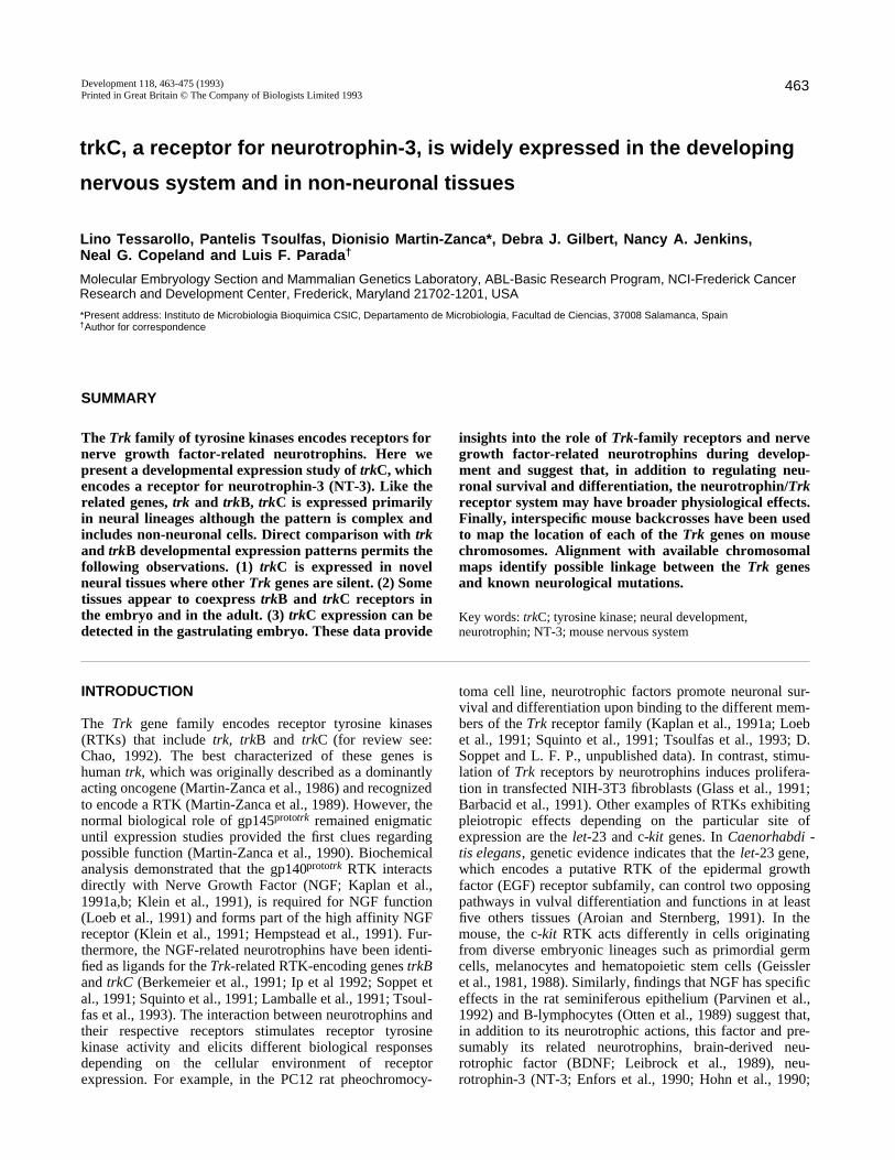
Fig. 3. Comparative expression of Trk-family genes. Dark-field views of serial adjacent sagittal sections of a 17.5 day mouse embryohybridized with a trk (A), trkB (B) or trkC (C)-specific probe. White triangles indicate the tooth papilla; arrows, DRG; Sr, striatum; t,thalamus; T, tongue; V, fifth cranial ganglion; C, superior cervical ganglion; S, sympathetic ganglia; W, whisker pad; i, intestine. Theblack triangles in panel A point to an artifact.
Fig. 4. trkC expression in embryonic CNS. (A) Dark-field and (B) bright-field of a coronal section from 17.5 day embryonic brain.(C) Dark-field and (D) bright-field magnification of 17.5 day embryonic cortex. (E) Dark-field and (F) bright-field view of a sectionthrough the mid-lumbar region of a 15.5 day embryo spinal cord. cx, cortex; c, cortical layer; i, intermediate layer; v, ventricular layer; cp,caudate putamen; h, hippocampus; t, thalamus; m, ventral horn (motor neuron region of the spinal cord); f, floor plate.
466
Jones and Reichardt, 1990; Kaisho et al., 1990; Maison-pierre et al., 1990a; Rosenthal et al., 1990), Xenopus NT-4 (xNT-4, Hallböök et al., 1991; Ip et al., 1992) and humanNT-5 (hNT-5; Berkemeier et al., 1991), may have broaderphysiological effects.
The rat trkC locus is complex, encoding multiple distinctreceptors (Tsoulfas et al., 1993). These trkC isoforms bindNT-3, a widely expressed neurotrophin (Maisonpierre et al.,1990b), in chemical cross-linking and equilibrium bindinganalyses (Lamballe et al., 1991, Tsoulfas et al., 1993).
In this study, we have investigated the expression of trkCin the mouse embryo and adult CNS. We find trkC tran-scripts throughout the nervous system but also in non-neuraltissues. Furthermore, we contrast the expression of the trkCgene with that of trk and trkB. Finally, we have mappedthe chromosomal location of the three Trk loci and showthat they are unlinked and map in the vicinity of previouslyexisting neurological mutations. This information may leadto a direct association of Trk genes with the phenotypeselicited by known spontaneous mutations in the mouse.
MATERIALS AND METHODS
RNA preparation and RNAse protection analysisRNA was extracted using RNAzol (Cinna/Biotecx) following themanufacturer’s recommendations. RNAse protection experimentswere performed as previously described (Tessarollo et al.,1992)using the RPA kit (Ambion). A genomic trkC-specific probe that
spans 196 nucleotides of the extracellular domain (aa 336-401 ofthe rat sequence; Tsoulfas et al., 1993) and including 64 down-stream intronic nucleotides was used to generate an antisense RNAprobe employed in RNAse protection analysis. A 250 base β-actinc-RNA probe (Alonso et al., 1986) was included in the same reac-tion as a means of assessing relative levels of RNA present ineach hybridization.
In situ hybridizationIn situ hybridization protocols were as described (Martin-Zancaet al. 1990) with the following modifications. Dissected embryoswere fixed overnight in 4% paraformaldehyde, dehydrated withalcohols and xylenes, and embedded in paraffin. Embryos weresectioned at 5 µm thickness and mounted on gelatin-coated slides.Slides were deparaffinized in xylene and rehydrated in graded(100-30%) ethanol solutions. After fixing in 4% paraformalde-hyde, the tissues were pretreated with proteinase K (20 µg/ml)(Boehringer Mannheim), refixed and immersed in triethanolaminebuffer containing acetic anydrate and dehydrated. Sections werehybridized with antisense cRNA probes (5×10⁵ cts/minute) in abuffer containing 50% formamide, 0.3 M NaCl, 20 mM Tris-Cl(pH 7.4), 1× Denhardt’s solution, 0.5 mg/ml yeast tRNA and 10mM DTT at 50°C for 20 hours. After hybridization, washes wereperformed in 4× SSC and 10 mM DTT at 50°C. The slides werethen incubated for 30 minutes at 37°C with RNase A (20 µg/ml)and RNase T1 (2 µg/ml) followed by a 30 minute incubation at55°C in 50% formamide, 0.2× SSC, 10 mM DTT, washed twicefor 30 minutes in 0.2× SSC, 1% sodium pyrophosphate (w/v), 10mM DTT and dehydrated. The slides were dipped in Kodak emul-sion NTB-2 and exposed for up to 10 days at 4°C. The slideswere then developed in Kodak D-19, fixed as recommended bymanufacturer and stained in 0.2% toluidine blue. All photomi-
L. Tessarollo and others
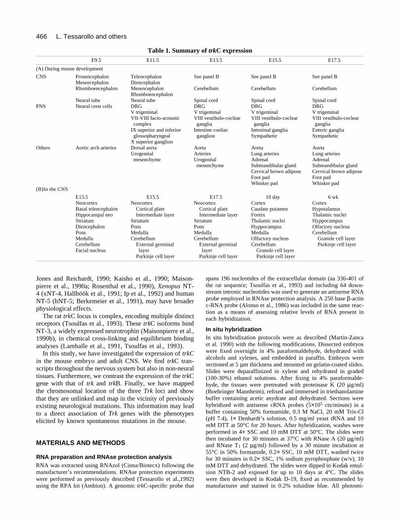
Table 1. Summary of trkC expressionE9.5 E11.5 E13.5 E15.5 E17.5
(A) During mouse development
CNS Prosencephalon Telencephalon See panel B See panel B See panel BMesencephalon DiencephalonRhomboencephalon Mesencephalon Cerebellum Cerebellum Cerebellum
RhomboencephalonNeural tube Neural tube Spinal cord Spinal cord Spinal cord
PNS Neural crest cells DRG DRG DRG DRGV trigeminal V trigeminal V trigeminal V trigeminalVII-VIII facio-acoustic VIII vestibulo-coclear VIII vestibulo-coclear VIII vestibulo-coclearcomplex ganglia ganglia ganglia
IX superior and inferior Intestine coeliac Intestinal ganglia Enteric gangliaglossopharyngeal ganglion Sympathetic Sympathetic
X superior ganglionOthers Aortic arch arteries Dorsal aorta Aorta Aorta Aorta
Urogenital Arteries Lung arteries Lung arteriesmesenchyme Urogenital Adrenal Adrenal
mesenchyme Submandibular gland Submandibular glandCervical brown adipose Cervical brown adiposeFoot pad Foot padWhisker pad Whisker pad
(B)In the CNS
E13.5 E15.5 E17.5 10 day 6 wkNeocortex Neocortex Neocortex Cortex CortexBasal telencephalon Cortical plate Cortical plate Caudate putamen HypotalamusHippocampal neo Intermediate layer Intermediate layer Fornix Thalamic nucleiStriatum Striatum Striatum Thalamic nuclei HyppocampusDiencephalon Pons Pons Hyppocampus Olfactory nucleusPons Medulla Medulla Medulla CerebellumMedulla Cerebellum Cerebellum Olfactory nucleus Granule cell layerCerebellum External germinal External germinal Cerebellum Purkinje cell layerFacial nucleus layer layer Granule cell layer
Purkinje cell layer Purkinje cell layer Purkinje cell layer

467Developmental expression of the trkC receptor tyrosine kinase
croscopy was done on a Zeiss Axiophot microscope. Sense andanti-sense probes labeled with 35S were prepared by standard pro-cedures (Krieg and Melton, 1987) by using UTP as the labelednucleotide. The trkC antisense probe was synthesized from a ~2kb rat trkC cDNA (NRT-8) as described by Tsoulfas et al. (1993).The trk and trkB probes are described in detail by Martin-Zancaet al. (1990) and Klein et al. (1989), respectively.
Interspecific backcross mappingInterspecific backcross progeny were generated by mating(C57BL/6J × Mus spretus)F1 females and C57BL/6J males asdescribed (Copeland and Jenkins, 1991). A total of 205 N2 prog-eny were obtained; a random subset of these N2 mice were usedto map the Trk loci (see text for details). DNA isolation, restric-tion enzyme digestion, agarose gel electrophoresis, Southern blottransfer and hybridization were performed as described (Jenkinset al., 1982). All blots were prepared with Zetabind nylon mem-brane (AMF-Cuno). Hybridization probes were labeled with[α32P] dCTP using a nick translation labeling kit (IL-9)(Boehringer Mannheim) or random priming (trk, trkB, trkC)(Amersham Corporation). Washing was done to a final stringencyof 0.2-0.8× SSCP, 0.1% SDS, 65°C.
The three mouse Trk loci, trk, trkB and trkC, have been desig-nated Ntrk1, Ntrk2 and Ntrk3, respectively, to conform with thecurrent human locus designations for these genes. The trk probewas a 1.5 kb EcoRI genomic fragment from the mouse trk extra-cellular domain (Martin-Zanca et al., 1990). The trk probe hybrid-ization to a 6.4 kb fragment in BglII-digested C57BL/6J (B6) DNAand 5.0 and 1.3 kb fragments in M. spretus (S) DNA. The twoM. spretus -specific fragments cosegregated and were followed inbackcross mice. The trkB probe was a 938 bp corresponding toamino acid 1-308 of mouse trkB sequences (Klein et al., 1989).The trkB probe hybridized to 3.8, 2.5, 1.1, 0.9 and 0.35 kb frag-ments in TaqI-digested B6 DNA and 3.5, 3.1, 2.5, 1.4, 1.1 and0.35 kb fragments in S DNA. The 3.5, 3.1 and 1.4 kb M. spre -tus-specific fragments cosegregated and were followed in back-cross mice. The trkC probe was a 288 bp cDNA correspondingto amino acid 282-378 of the mouse trkC gene (Tsoulfas et al.,1993). The TrkC probe hybridized to a 1.3 kb fragment in EcoRI-digested B6 DNA and a 3.6 kb fragment in S DNA. The 1.3 kbM. spretus -specific fragment was followed in backcross mice.
A description of most of the probes and RFLPs for loci usedto position the Trk loci on the interspecific map have beenreported. The loci in include: fibrinogen, gamma polypeptide(Fgg), connexin-40 (Cnx-40) and nerve growth factor beta (Ngfb)on chromosome 3 (Mucenski et al., 1988; Cox et al., 1991; Hae-fliger et al., 1992); neuroendocrine convertase-1 (Nec-1) on chro-mosome 13 (Copeland et al., 1992); and insulin-like growth factor1 receptor (Igflr), feline sarcoma oncogene (Fes) and tyrosinase(Tyr) on chromosome 7 (Lunsford et al., 1990; Copeland et al.,1992). One locus, interleukin-9 (IL-9) on chromosome 13 has notbeen previously reported for our panel. The IL-9 probe (pP40.2B4)was a 550 bp mouse cDNA (Van Snick et al., 1989). The IL-9probe hybridized to an 18.0 kb fragment in SphI-digested B6 DNAand a 5.4 kb fragment in S DNA. The 5.4 kb M. spretus-specificfragment was followed in backcross mice.
Recombination distances were calculated as described (Green,1981) using the computer program SPRETUS MADNESS. Geneorder was determined by minimizing the number of recombina-tion events required to explain the allele distribution patterns.
RESULTS
To determine the temporal expression pattern for trkCduring murine development, a trkC-specific probe was used
in RNAse protection assays employing a β-actin internalcontrol as described in Materials and methods. 10 µg oftotal mouse embryo RNA were assayed and, although afaint protected band is first observed on day 7.5, trkC geneexpression is clearly detected at embryonic day 8.5 coin-ciding with the timing of neural tube formation (Fig. 1).Relative to the internal β-actin control, a substantialincrease in trkC mRNA expression occurs around embry-onic day 10.5 (25-35 somites).
The trk and trkB genes are expressed predominantly inthe nervous system (Martin-Zanca et al., 1990; Klein et al.,1990a). Having determined that trkC transcripts are presentin the embryo from the earliest stages of neural induction,we next performed RNA in situ hybridization of postim-plantation embryo sections from day 7.5 through day 17.5and in neo-nate and adult central nervous system (CNS; seeMaterials and methods). For comparison, in the course ofthis study, we hybridized adjacent embryo sections with trk,trkB and trkC probes.
The results obtained from our in situ experiments on trkCexpression are summarized in Table 1. The expression ofthis gene is highly complex. We first observed trkC hybrid-ization as a weak signal in 7.5 day egg cylinders with apattern indicating expression in the early neuroectoderm(not shown). Whole-mount in situ experiments are inprogress to extend these observations. trkC gene expressionis evident throughout the neuroepithelium of 9.5 (12-30somites) day embryos (Fig. 2A-D). At these early stages,we also observe trkC transcripts in regions where neuralcrest cells are known to localize. These include formingdorsal root ganglia (DRG) and regions adjacent to theneural tube and the dorsal aorta (Fig. 2C,D; arrows), loca-tions identified as migratory routes used by cells of neuralcrest origin (see below).
During organogenesis, additional sites of active trkC arefound. Fig. 2E-F shows a sagittal section from an 11.5 dayembryo (35 somites). trkC expression is exhibited through-out the CNS (telencephalon, diencephalon, mesencephalon,rhombencephalon and neural tube) and comparison with anadjacent trkB hybridized section (not shown) indicatesoverlapping expression in the mantle (postmitotic) layer ofthe spinal cord, whereas the ependymal (mitotic) layerexpresses only trkB. The trkB and trkC genes are coex-pressed in the branchial arch arteries; however, more caudaltrkB transcripts are present in the mesonephric region whiletrkC is expressed in the midgut and genital ridge associ-ated mesenchyme (see Fig. 2E-F).
At 13.5 days of development, the trkC gene is expressedthroughout the neural network (Fig. 2G-I). At this stage,trkC mRNA is present in neural tissues where trk and trkBgene activity has not been previously detected (Martin-Zanca et al., 1990; Klein et al., 1989, 1990a). One strikingexample is the expression of trkC in neural-crest-derivedcells of the developing enteric nervous system (Figs 2H, 7).In contrast, trkC and trkB transcripts are present in thetrigeminal (Vth cranial) ganglion (Fig. 2H,I) where punc-tate hybridization reflects expression in a small subset ofcells (Fig. 2I). The three afferent branches of the trigemi-nal ganglion (ophthalmic, maxillary and mandibular) canbe readily traced by abundant trkC hybridization, presum-ably reflecting expression in the neural crest-derived
468
Schwann cells that migrate and differentiate along thetrigeminal processes (Fig. 2G).
Several insights can be drawn from a direct comparisonof trk, trkB and trkC expression at later stages in develop-ment. Fig. 3 shows in situ hybridization of day 17.5 sec-tions, from a single fetus, which were hybridized withprobes to the three related genes. trk expression is distinc-tive in its tight regulation primarily limited to trigeminal(V), superior cervical (C), sympathetic (S) and dorsal rootganglia (arrows; Fig. 3A; Martin-Zanca et al., 1990). Incomparison, trkB is expressed in these ganglia and at highlevels in the CNS and additional structures including thewhisker pad (W), tongue (T) and tooth buds (white arrow-head; Fig. 3B). This complex expression pattern is consis-tent throughout mid-gestation (days 14-18; L. F. P., unpub-lished observations). Expression of the trkC gene mirrorsthat of trkB in many regions of the embryo (Fig. 3B,C).Like the trkB gene (Klein et al 1990a; Parada et al., 1992),trkC is expressed throughout the nervous system includingmany of the same cranial and spinal ganglia (V, VII, VIII,X, DRG). The trkC and trkB genes are also apparently co-expressed in several non-neural structures including thetongue, the whisker pad (vibrissae) mesenchyme and form-ing teeth (Fig. 3B,C). In the tooth, trkB is expressed in theouter dermal papilla while trkC transcripts appear confinedto the core of the dermal papilla (compare Fig. 3B and C).
trkC transcripts are also found in several additional
L. Tessarollo and others
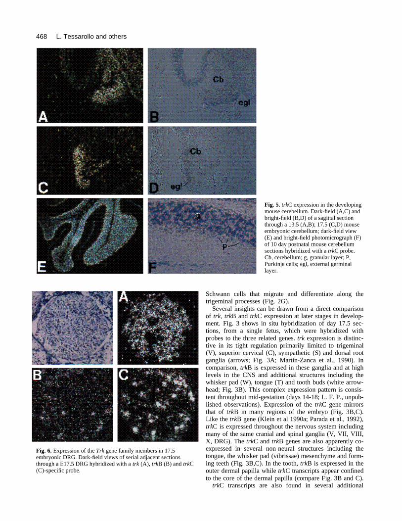
Fig. 5. trkC expression in the developingmouse cerebellum. Dark-field (A,C) andbright-field (B,D) of a sagittal sectionthrough a 13.5 (A,B); 17.5 (C,D) mouseembryonic cerebellum; dark-field view(E) and bright-field photomicrograph (F)of 10 day postnatal mouse cerebellumsections hybridized with a trkC probe.Cb, cerebellum; g, granular layer; P,Purkinje cells; egl, external germinallayer.
Fig. 6. Expression of the Trk gene family members in 17.5embryonic DRG. Dark-field views of serial adjacent sectionsthrough a E17.5 DRG hybridized with a trk (A), trkB (B) and trkC(C)-specific probe.
469Developmental expression of the trkC receptor tyrosine kinase
neural crest-derived structures including enteric ganglia(see Fig. 7) and in other non-neural cells (see below).
Embryonic CNSThe pattern of trkC gene expression is summarized in Table1 and examples of in situ hybridization in the embryonicCNS are shown in Figs 2, 3C and 4. Interestingly, this geneshows both overlapping and exclusive expression profilesin the cephalic CNS when compared to the trkB gene. Inthe telencephalic cortical plate, both trkC and trkB tran-scripts are highly represented whereas in the intermediatelayer only trkC mRNA was observed. In the ventricularlayer, trkB expression is high and trkC expression is low(Figs 3B,C, 4A-D). In the striatum (Sr), trkC mRNA is mostabundant centrally while trkB transcripts predominate alongthe ependymal layer. Finally, the thalamic nuclei (t) appearto express either trkB or trkC (Fig. 3).
A cross section through the caudal spinal cord of a 15.5day embryo reveals trkC transcripts throughout the dorsal-ventral axis of the mantle layer including the ventral hornwhere motor neurons are located (Fig. 4E,F). No evidenceof expression was seen in the floor plate at any develop-mental stages examined.
trkC transcripts are also abundant in the embryonicmetencephalon (cerebellum). Fig. 5 provides examples oftrkC expression in cerebellum at embryonic days 13.5, 17.5and postnatal day 10. Transcripts are seen throughoutembryonic stages with high levels in the immature but post-mitotic Purkinje cells (Fig. 5A-D; Altman and Bayer, 1985).No expression was observed in the external germinal layer(egl). In all postnatal stages examined, including adult, trkCmRNA levels are very high in the cerebellar granule layersand in Purkinje neurons (Fig. 5E,F and not shown).
Peripheral nervous systemThe neural crest gives rise to a large component of theperipheral nervous system (PNS). trkC transcripts are foundin many PNS structures and also in cells that appear to bemigratory, neural crest cells (Table 1, Fig. 2A,C,E, andunpublished results). As previously noted (Figs 2, 3), all
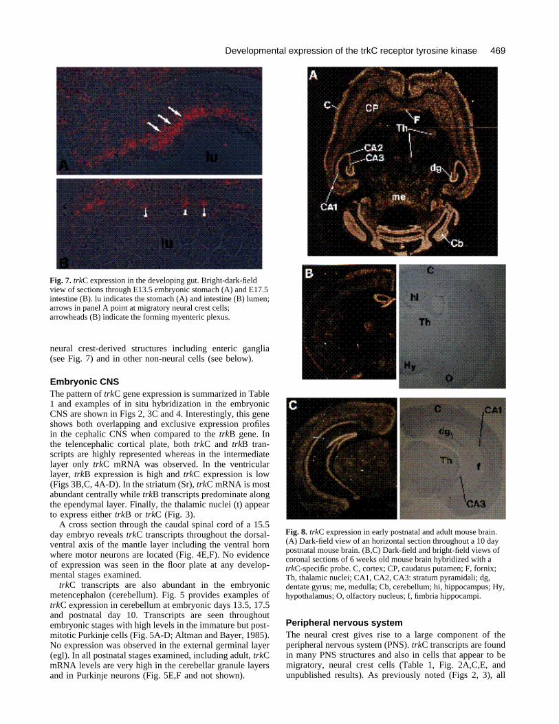
Fig. 7. trkC expression in the developing gut. Bright-dark-fieldview of sections through E13.5 embryonic stomach (A) and E17.5intestine (B). lu indicates the stomach (A) and intestine (B) lumen;arrows in panel A point at migratory neural crest cells;arrowheads (B) indicate the forming myenteric plexus.
Fig. 8. trkC expression in early postnatal and adult mouse brain.(A) Dark-field view of an horizontal section throughout a 10 daypostnatal mouse brain. (B,C) Dark-field and bright-field views ofcoronal sections of 6 weeks old mouse brain hybridized with atrkC-specific probe. C, cortex; CP, caudatus putamen; F, fornix;Th, thalamic nuclei; CA1, CA2, CA3: stratum pyramidali; dg,dentate gyrus; me, medulla; Cb, cerebellum; hi, hippocampus; Hy,hypothalamus; O, olfactory nucleus; f, fimbria hippocampi.

470
Trk genes are expressed to a greater or lesser extent inneural crest-derived embryonic cranial, spinal and sympa-thetic ganglia. Fig. 6 shows in situ hybridization of threeadjacent DRG sections from a 17.5 day embryo. These sec-tions (5 µm) provide direct comparison of Trk geneexpression and, in agreement with a report by Carroll andco-workers (1992), show that a majority of neurons expresstrk (Fig. 6A), while trkB (Fig. 6B) and trkC (Figs 6C, 2I)transcripts are present in a reduced subset of neurons. Verysimilar distribution of the three genes is seen in trigeminalganglion (Fig. 3; see also Martin-Zanca et al., 1990; andKlein et al., 1990a).
The ganglia of the enteric nervous system are formedfrom migratory neural crest cells originating in the vagalcrest region (Le Douarin and Teillet, 1973). These cellshybridize with trkC probes (Fig. 7A) as exemplified byhybridization of embryonic day 13.5 stomach where trkC-expressing migratory neural crest cells can be observedintercalated within the mesenchyme. Several days later,trkC transcripts can be localized to the forming ganglia thatappear throughout the muscle and submucosal layers of theintestine (Fig. 7B). We do not observe trkC expression inthe epithelium (Fig. 7). Thus, trkC expression is found indiverse PNS structures some of which do not appear toexpress other Trk-family receptors (Martin-Zanca et al.,1990; Klein et al., 1990a).
Adult CNS expressiontrkC gene expression remains abundant in the adult mouseCNS. The cerebral cortex exhibits high levels of transcriptsparticularly in the superficial layers and in the deep layerswith reduced expression in the intermediate layers (Fig. 8).TrkC transcripts are also abundant in the caudate putamenfrom embryonic stages (Fig. 4A,B), a pattern that remainsunchanged in the adult CNS (Fig. 8B). Similarly, thalamicnuclei maintain trkC expression throughout developmentand in the adult (Fig. 8A,B). Thalamic nuclei do not appearto express trkB transcripts in adult brain (Klein et al.,1990a).
In the hippocampus of adult brain, the pyramidal celllayer as well as the neurons of the dentate gyrus exhibithigh levels of trkC mRNA (Fig. 8A-C). In the cerebellum,both the Purkinje and the granule cell layer express highlevels of trkC transcripts. Thus, expression of the trkC geneobserved in the mature brain is consistent with that exhib-ited during embryonic development.
TrkC expression outside the nervous systemAs can be appreciated from Figs 2 and 3, trkC-encodingtranscripts are abundant throughout the embryo includingsites that are not necessarily consistent with expression inneural cells (see Table 1). Note the presence of trkC tran-scripts in vibrissae and dental papillae (Fig. 3). We alsofind trkC transcripts in cells of the submandibular gland(Fig. 9A,B) and in the mesenchyme surroundingmesonephric and urogenital ducts (Fig. 9C,D). High levelsof trkC transcripts can also be localized to the brown adi-pose tissue dorsal to the cervical spinal cord (Fig. 9D,E);the cortex of the metanephros; and adrenal gland (Fig.9F,G). Finally, abundant trkC expression is found along the
subendothelial mesenchyme of arteries throughout thedeveloping embryo (Figs 9H,I and 2, 3).
Chromosomal mappingWe next wished to identify the chromosomal location ofthe three Trk genes to determine whether they are linkedor map to separate chromosomal locations and to assess thepossibility that these three genes might be associated withpreviously reported neurological mutations in the mouse.The chromosomal location of trk, trkB and trkC was deter-mined by interspecific backcross analysis using progenyderived from matings of [(C57BL/6J × Mus spretus)F1 ×C57BL/6J] mice that have been typed for over 1100 locidistributed among all mouse chromosomes (Copeland andJenkins, 1991; see Materials and methods).
Each of the Trk genes mapped to a different mouse auto-some (Fig. 10), indicating that they have become well dis-persed during chromosome evolution. To determinewhether any of the Trk genes mapped near a known mousemutation with a phenotype that might be consistent with adefect in a Trk gene, we aligned our interspecific maps ofchromosomes 3, 13 and 7 with composite linkage maps thatreport the map location of many uncloned mouse mutations(compiled from GBASE, a computerized database main-tained at The Jackson Laboratory, Bar Harbor, ME). Inter-estingly, all three genes mapped near loci that exhibit neu-rological phenotypes.
trk mapped in the vicinity of the spontaneous neurolog-ical mutation spastic (spa) located on mouse chromosome3 (Chai, 1961; Van Heyningen et al., 1975; Wilson et al.,1986; reviewed in Green, 1989). trkB mapped in the vicin-ity of the spontaneous neurological mutation Purkinje celldegeneration (pcd) located on chromosome 13 (Mullen etal., 1976; O’Gorman, 1985; reviewed in Green, 1989).Finally, trkC mapped in the vicinity of a gene on mousechromosome 7 that has been shown to affect susceptibilityof inbred mice to audiogenic seizures. (Neumann andCollins, 1991).
DISCUSSION
Recent studies from a number of laboratories, showing thattrk and trkB are expressed mainly in the nervous systemand encode functional receptors for the neurotrophins, havebeen important for advancing our understanding of themechanisms that regulate neural development.
In the present study, we show that trkC, a third memberof the Trk gene family (Lamballe et al., 1991; Tsoulfas etal., 1993), is expressed in a broad component of the ner-vous system and in non-neural tissues, including cell typeswhere other Trk genes are not active.
trkC is expressed early in developmenttrkC expression was first observed in the gastrulatingembryo, coordinate with the temporal expression pattern ofits ligand NT-3, the more abundant neurotrophin detectedearly in development (Maisonpierre et al., 1990b). This isthe earliest example of an active Trk gene reported to date,raising the possibility that the trkC RTK may mediateimportant functions in early neuroepithelium formation.
L. Tessarollo and others
471Developmental expression of the trkC receptor tyrosine kinase
Neural crest Kalcheim and co-workers (1992) have reported a prolifer-ative activity of NT-3 in migratory neural crest cells. In thepresent study, we provide evidence for NT-3 receptorexpression in migratory neural crest cells, thus supportingthe notion that Trk receptors and their ligands may medi-ate alternative functions to trophic signals during develop-ment (for review, see Barbacid et al., 1991).
The trkC gene is expressed in many neural crest deriva-tives including cranial, dorsal root and sympathetic ganglia.In trigeminal and dorsal root ganglia, all Trk genes areactive. trk mRNA is perhaps most abundant and confinedto neurons in these ganglia. We note that in addition toexpression in sympathetic ganglia late in development, trkexpression can also be detected in isolated neurons of thebasal forebrain, hindbrain and in other locations of the lateembryonic and adult brain (Holtzman et al., 1992; L. F. P.,unpublished observations). Like trkB, trkC is expressed infewer neurons in sensory ganglia. However, in the trigem-inal ganglion, the afferent projections are outlined byexpression of these genes suggesting that trkB and trkC areexpressed in neural crest-derived Schwann cells thatmigrate along the axonal projections to their sites of differ-entiation.
Another notable site of trkC-specific expression is theenteric neural crest and ganglia. Through trkC hybridiz-ation, it is possible to trace the migration of vagal neuralcrest cells into the gut region, suggesting that neurotrophinsmay have important functions in the migratory and trophicpotential of enteric neurons.
trkC is also expressed in a group of non-neural tissuesthat, in quail-chick studies (Le Douarin, 1982) or inradioisotopic labeling experiments in the amphibian embryo(Chibon, 1964), have suggested neural crest origin. Amongthese are the artery walls derived from aortic arches, toothpapillae (Chibon, 1970) and salivary glands. Using RNAseprotection and northern analyses, we also observe trkC tran-scripts in the thymus (Tsoulfas et al., 1993), another tissuewhose connective tissue component is derived from theneural crest.
Brown adipose tissue, particularly in the cervical regiondorsal to the spinal cord, also expresses high levels of trkCtranscripts (Fig. 9E,F). Because of the high silver grain den-sity caused by strong expression, we are unable to identifyprecisely the expressing cell types within this tissue. Elec-tron microscopy studies indicate that brown adipose is par-ticularly well innervated and Schwann cells have beenobserved sheathing unmyelinated nerve fibers in the lob-ules, close to the vessels, and between adipocytes(Bargmann et al., 1968; Linck et al., 1973). Therefore, trkCexpression in this tissue may reflect the presence of tran-scripts in neural cells and not in adipocytes. However, inchick, the subcutaneous adipose of the face and the ventralneck region is derived from the neural crest (Le Douarin,1982), while the precise origin of the brown adipose tissuehas not been well established (Néchad, 1986). Thus, it isalso possible that this adipose tissue may be of neural crestorigin and retain trkC expression.
Trk genes are expressed in a broad spectrum of neuralcrest derivatives implying that neurotrophins may functionin cells where activity has not previously been associated.
The trkB and trkC expression profiles exhibited in migra-tory neural crest cells along the descending aorta suggestthat trkB and trkC products may represent good markersfor subsets of neural-crest-derived cells.
CNS In the CNS, the trkB and trkC genes are coexpressed anddifferentially expressed in intriguing patterns. We observedcoexpression of these genes in the hippocampus (pyrami-dal and granule neurons), in the cerebellum (Purkinje cells),in the ventral spinal cord and in regions of the cerebralcortex (L. T. and L. F. P., unpublished data). In contrast,thalamic nuclei appear to express trkC transcripts prefer-entially in the adult. Similarly in the cerebellum, trkCexpression is abundant in the granular layer where little orno trkB transcripts could be detected (see Klein et al.,1990b).
The neurotrophin theory has been well substantiated inthe sensory nervous system through the study of NGF-related neurotrophins and through the availability of well-defined primary culture systems (Barde, 1989). The searchfor trophic molecules acting on motor neurons has provenmore elusive. Culturing of motor neurons is technically dif-ficult and, although tissue extracts have been reported topromote motor neuron survival, only one molecule in par-ticular, CNTF (Sendtner et al., 1992a) has been identifiedto promote survival of these cells in vivo. Until recently,efforts to identify a motor neuron response to the NGFfamily of neurotrophins have not been successful. Veryrecently BDNF has been shown to rescue motor neurons invivo in rat and chick (Yan et al., 1992; Oppenheim et al.,1992; Sendtner et al., 1992b). The present results indicatethe existence of trkC and trkB (Klein et al., 1990a) tran-scripts during embryogenesis in the ventral horn of thespinal cord and in other regions where motor neurons arelocalized. These results therefore suggest the need for arenewed effort at understanding neurotrophin function inmotor neurons.
Chromosomal locationIn mouse, the three Trk family members map to differentmouse autosomes and are located in regions containingknown neurological mutations. For example, the mouse trkgene maps near the spa mutation on chromosome 13. spahomozygotes show spastic symptoms although no anatom-ical abnormalities have been described. Glycine receptordeficiencies have been reported in these mice (White andHeller, 1982; White, 1985), but it remains to be determinedwhether these deficiencies are the cause or a secondaryeffect of the spa mutation.
Likewise, trkB maps on chromosome 3, in a region wherethe pcd mutation has previously been mapped. pcd homozy-gotes show a moderate ataxia beginning at 3 to 4 weeks ofage. They are smaller than their normal littermates but livea fairly normal life span although adult males are infertile.In these mice, Purkinje cells begin to degenerate by 15 to18 days of age followed by a slower degeneration of thephotoreceptor cells of the retina and mitral cells of theolfactory bulb. Later (50-60 days of age) discrete popula-tions of thalamic neurons also degenerate. Studies of fusionchimeras between pcd/pcd and +/+ embryos suggest that

472 L. Tessarollo and others

473Developmental expression of the trkC receptor tyrosine kinase
pcd acts directly within the Purkinje cells themselves(Mullen, 1977). The trkB gene is expressed in Purkinjecells, in retinal ganglion cells, in motor neurons and in aspectrum of other neural tissues. Thus the pcd phenotypecould be accounted for by mutations in the trkB locus.
The trkC locus maps in a region of chromosome 7 thathas been associated with increased tendency to exhibit audi-tory defects in mice (Neumann and Collins, 1991). Theseauditory defect susceptibilities are ill-defined at this timebut it is possible that the trkC locus may contribute to suchabnormalities since the NT-3 gene and its receptor (data notshown) is expressed in the sensory components of thedeveloping ear during embryogenesis (Pirvola et al., 1992).As germline mutations are induced in the Trk genes viahomologous recombination in embryonic stem cells, itshould be possible to perform allelism studies to determinewhether any of these mutations do in fact result fromdefects in Trk genes.
The present mouse mapping studies can also be used topredict where the Trk genes will map in humans. For exam-ple, trkB maps between Il-9 and Nec-1 which have bothbeen mapped to the long arm of human chromosome 5 (Fig.10), suggesting that trkB will reside on the long arm ofhuman chromosome 5 as well. Likewise, trkC does notrecombine with Fes, which has been mapped to 15q25-qter.Finally, trk has been mapped by two groups to the long armof human chromosome 1 (Miozzo et al., 1990; Morris etal., 1991). This placement is consistent with the mousemapping studies shown in Fig. 10.
CONCLUSION
We have studied the expression of the trkC gene duringdevelopment and in the adult CNS, and compared these datawith that of the other identified Trk gene family members(Martin-Zanca et al., 1990; Klein et al., 1990a). These genesmap to unlinked chromosomal locations and are primarily,
though not exclusively, expressed in the nervous system.Understanding the expression patterns of Trk genes pro-vides important information regarding potential sites ofneurotrophin action and identification of cells that prefer-entially coexpress combinations of Trk receptors or whichuniquely express one specific receptor. These data suggestthat experimental strategies must be defined to explore thephysiological significance of Trk receptor coexpression andof their possible function in previously unidentified sites ofactivity including motor neurons and non-neural cells.Finally knowledge of the sites of Trk-gene expression willprovide important insights for the analysis of mice that lackTrk-gene function due to mutations generated by homolo-gous recombination in embryonic stem cells.
We thank Dan Soppet for the mouse trkB sequence and formany helpful discussions. We are grateful to the members of theParada lab for their support and, in particular, to James Pickel andDan Soppet for their advise and critical reading of the manuscript.We also thank B. Cho and M. Barnstead for technical assistanceand Richard Fredrickson for his skillful assistance with artworkand Cindy Fitzpatrick for manuscript preparation. This researchwas supported by the National Cancer Institute, DHHS, under con-tract NO1-CO-74101 with ABL.
REFERENCES
Alonso, S., Minty, A., Bourlet, Y. and Buckingham, M. (1986).Comparison of three actin-coding sequences in the mouse; evolutionaryrelationships between the actin genes of warm-blooded vertebrates. J.Molec. Evolution 23, 11-22.
Altman, J. and Bayer, S. A. (1985). Embryonic development of the ratcerebellum. I. Delineation of the cerebellar primordium and early cellmovements. J. Comp. Neurol. 231, 1-6.
Aroian, R. V. and Sternberg, P. W. (1991). Multiple functions of let-23, aCaenorhabditis elegans receptor tyrosine kinase gene required for vulvalinduction. Genetics 128, 251-267.
Barbacid, M., Lamballe, F., Pulido, D. and Klein, R. (1991). The trkfamily of tyrosine protein kinase receptors. BBA Rev. Cancer 1072, 115-127.
Fig. 10. Linkage maps showing the chromosomal locations of Ntrk loci in mouse. The Ntrk loci were mapped by interspecific backcrossanalysis. The number of recombinant N2 animals over the total number of animals typed plus the recombination frequencies, expressed asgenetic distance in centimorgans (± 1 s.e.), is shown for each pair of loci on the left of the chromosome maps. Where no recombinantswere found between loci, the upper 95% confidence limit of the recombination distance is given in parentheses. The position of loci inhuman chromosomes, determined in previously reported studies, are shown to the right of the chromosome maps. References for thehuman map positions of loci mapped in this study can be obtained from GDB (Genome Data Base), a computerized database of humanlinkage information maintained by The William H. Welch Medical Library of The Johns Hopkins University (Baltimore, MD).
474
Barde, Y.-A. (1989). Trophic factors and neuronal survival. Neuron 2,1525-1534.
Bargmann, W., von Hehn, G. and Lindner, E. (1968). Über die zellen desbraunen fettgeund ihre innervation. Z. Zellforsch. Mikroski. Anat. 85, 601.
Berkemeier, L. R., Winslow, J. W., Kaplan, D. R., Nikolics, K., Goeddel,D. V. and Rosenthal, A. (1991). Neurotrophin-5: A novel neurotrophicfactor that activates trk and trkB. Neuron 7, 857.
Carroll, S. L., Silos-Santiago, I., Frese S. E., Ruit, K. G., Milbrandt, J.and Snider, W. D. (1992). Dorsal root ganglion neurons expressing trkare selectively sensitive to NGF deprivation in utero. Neuron 9, 779-788.
Chai, C. K. (1961). Hereditary spasticity in mice. J. Hered. 52, 241-243.Chao, M. V. (1992). Neurotrophin receptors: a window into neuronal
differentiation. Neuron 9, 583-593.Chibon, P. (1964). Analyse par la méthode de marquage nucléaire a la
thymidine tritiée des dérivés de la crête neurale céphalique chez l’UrodèlePleurodeles waltlii Michah. C.R. Acad. Sci. Paris Ser. III 159, 3624-3627.
Chibon, P. (1970). L’origine de l’organe adamantin des dents. Etudeaumoyen du marquage nucleaire de l’ectoderme stomodeal. Ann.Embryol. Morphog. 3, 203-213.
Copeland, N. G. and Jenkins, N. A. (1991). Development and applicationsof a molecular genetic linkage map of the mouse genome. Trends Genet.7, 113-118.
Copeland, N. G., Gilbert, D. J., Chretien, M., Seidah, N. G. and Jenkins,N. A. (1992). Regional localization of three convertases, PC1 (Nec-1),PC2 (Nec-2), and Furin (Fur), on mouse chromosomes. Genomics 13,1356-1358.
Cox, R. D., Copeland, N. G., Jenkins, N. A. and Lehrach, H. (1991).Interspersed repetitive element polymerase chain reaction productmapping using a mouse interspecific backcross. Genomics 10, 375-384.
Ernfors, P., Ibáñez, C. F., Ebendal, T., Olson, L. and Persson, H. (1990).Molecular cloning and neurotrophic activities of a protein with structuralsimilarities to nerve growth factor: Developmental and topographicalexpression in the brain. Proc. Natl. Acad. Sci. USA 87, 5454.
Geissler, E. N., McFarland, E. C. and Russel, E. S. (1981). Analysis ofpleiotropism at the dominant white-spotting (W) locus of the housemouse: a description of ten new W alleles. Genetics 97, 337-361.
Geissler, E. N., Ryan, M. A. and Housman, D. E. (1988). The dominantwhite-spotting (W) locus of the mouse encodes the c-kit proto-oncogene.Cell 55, 185-192.
Glass, D. J., Nye, S. H., Hantzopoulos, P., Macchi, M. J., Squinto, S. P.,Goldfarb, M. and Yancopoulos, G. D. (1991). TrkB mediatesBDNF/NT-3-dependent survival and proliferation in fibroblasts lackingthe low affinity NGF receptor. Cell 66, 405-413.
Green, E. L. (1981). Linkage, recombination and mapping. In Genetics andProbability in Animal Breeding Experiments, pp. 77-113. New York:Oxford University Press.
Green, M. C. (1989). Catalog of mutant genes and polymorphic loci. InGenetic Variants and Strains of the Laboratory Mouse (ed. M. F. Lyonand A. G. Searle), pp. 12-403. New York: Oxford University Press.
Haefliger, J.-A., Bruzzone, R., Jenkins, N. A., Gilbert, D. J., Copeland,N. G. and Paul, D. L. (1992). Four novel members of the connexin familyof gap junction proteins. J. Biol. Chem. 267, 2057-2064.
Hallböök, F., Ibáñez, C. F. and Persson, H. (1991). Evolutionary studiesof the nerve growth factor family reveal a novel member abundantlyexpressed in xenopus ovary. Neuron 6, 845.
Hempstead, B., Kaplan, D. R., Martin-Zanca, D., Parada, L. F. andChao, M. V. (1991). High affinity NGF binding requires co-expression ofthe trk proto-oncogene product and the low affinity NGF receptor. Nature350, 678.
Hohn, A., Leibrock, J., Bailey, K. and Barde, Y.-A. (1990). Identificationand characterization of a novel member of the nerve growth factor brain-derived neurotrophic factor family. Nature 344, 339.
Holtzman, D. M., Li, Y., Parada, L. F., Kinsman, S., Chen, C.-K.,Valletta, J. S., Zhou, J., Long, J. B. and Mobley, W. C. (1992). p140trk
mRNA marks NGF responsive forebrain neurons: evidence that trk geneexpression is induced by NGF. Neuron 9, 465-478.
Ip, N. Y., Ibáñez, C. F., Nye, S. H., McClain, J., Jones, P. F., Gies, D. R.,Belluscio, L., LeBeau, M. M., Espinosa III, R., Squinto, S. P., Persson,H., Yancopoulos, G. D. (1992). Mammalian neurotrophin-4: Structure,chromosomal localization, tissue distribution, and receptor specificity.Proc. Natl. Acad. Sci. USA 89, 3060.
Jenkins, N. A., Copeland, N. G., Taylor, B. A. and Lee, B. K. (1982).Organization, distribution, and stability of endogenous ecotropic murine
leukemia virus DNA sequences in chromosomes of Mus musculus. J.Virol. 43, 26-36.
Jones, K. R. and Reichardt, L. F. (1990). Molecular cloning of the humangene that is a member of the nerve growth factor family. Proc. Natl. Acad.Sci. USA 87, 8060.
Kaisho, Y., Yashimura, K. and Nakahama, K. (1990). Cloning andexpression of a cDNA encoding a novel human neurotrophic factor. FEBSLett. 266, 187.
Kalcheim, C., Carmeli, C. and Rosenthal, A. (1992). Neurotrophin 3 is amitogen for cultured neural crest cells. Proc. Natl. Acad. Sci. USA 89,1661-1665.
Kaplan, D. R., Martin-Zanca, D. and Parada, L. F. (1991a). Tyrosinephosphorylation and tyrosine kinase activity of the trk protooncogeneproduct induced by NGF. Nature 350, 158.
Kaplan, D. R., Hempstead, B. L., Martin-Zanca, D., Chao, M. V. andParada, L. F. (1991b). The trk proto-oncogene product: A signaltransducing receptor for nerve growth factor. Science 252, 554.
Klein, R., Parada, L. F., Coulier, F. and Barbacid, M. (1989). TrkB, anovel tyrosine protein kinase receptor expressed during mouse neuraldevelopment. EMBO J. 8, 3701.
Klein, R., Martin-Zanca, D., Barbacid, M. and Parada, L. F. (1990a).Expression of the tyrosine kinase receptor gene trkB is confined to themurine embryonic and adult nervous system. Development 109, 845.
Klein, R., Conway, D., Parada, L. F. and Barbacid, M. (1990b). The trkBtyrosine kinase gene codes for a second neurogenic receptor that lacks thecatalytic domain. Cell 61, 647.
Klein, R., Jing, S., Nanduri, V., O’Rourke, E. and Barbacid, M. (1991).The Trk proto-oncogene encodes a receptor for nerve growth factor. Cell65, 189.
Krieg, P. A. and Melton, D. A. (1987). In vitro RNA synthesis with SP6RNA polymerase. Methods Enzymol. 155, 397-405.
Lamballe, F., Klein, R. and Barbacid, M. (1991). trkC: a new member ofthe trk family of tyrosine protein kinases, is a receptor for neurotrophin-3.Cell 66, 967.
Le Douarin, N. M. (1982). The Neural Crest, pp 54-107. Cambridge Univ.Press.
Le Douarin, N. M. and Teillet, M. A. (1973). The migration of neural crestcells to the wall of the digestive tract in avian embryo. J. Embryol. Exp.Morph. 30, 31-48.
Leibrock, J., Lottspeich, F., Hohn, A., Hofer, M., Hengerer, B.,Masiakowski, P., Thoenen, H. and Barde, Y.-A. (1989). Molecularcloning and expression of brain-derived neurotrophic factor. Nature 341,149.
Linck, G., Stoeckel, M. E., Porte, A. and Petrovic A. (1973). An electronmicroscope study of the specialized cell contacts and innervation ofadipocyte in the brown fat of the european hamster (Cricetus cricetus).Cytobiologie 7, 431.
Loeb, D. M., Maragos, J., Martin-Zanca, D., Chao, M. V., Parada, L. F.and Greene, L. A. (1991). The trk proto-oncogene rescues NGFresponsiveness in mutant NGF-nonresponsive PC12 cell lines. Cell 66,961.
Lunsford, R. D., Jenkins, N. A., Kozak, C. A., Liang, L-F., Silan, C. M.,Copeland, N. G. and Dean, J. (1990). Genomic mapping of murine Zp-2and Zp-3, two oocyte-specific loci encoding zona pellucida proteins.Genomics 6, 184-187.
Maisonpierre, P. C., Belluscio, L., Squinto, S., Ip, N. Y., Furth, M. E.,Lindsay, R. M. and Yancopoulos, G. D. (1990a). A neurotrophin-3: Aneurotrophic factor related to NGF and BDNF. Science 247, 1446.
Maisonpierre, P. C., Belluscio, L., Friedman, B., Alderson, R. F.,Wiegand, S. J., Furth, M. E., Lindsay, R. M. and Yancopoulos, G. D.(1990b). NT-3, BDNF, and NGF in the developing rat nervous system:parallel as well as reciprocal patterns of expressions. Neuron 5, 5101-5109.
Martin-Zanca, D., Hughes, S. H. and Barbacid, M. (1986). A humanoncogene formed by the fusion of truncated tropomyosin and proteintyrosine kinase sequences. Nature 319, 743-748.
Martin-Zanca, D., Oskam, R., Mitra, G., Copeland, T. and Barbacid,M. (1989). Molecular and biochemical characterization of the human trkproto-oncogene. Mol. Cell. Biol. 9, 24.
Martin-Zanca, D., Barbacid, M. and Parada, L. F. (1990). Expression ofthe trk proto-oncogene is restricted to the sensory cranial and spinalganglia of neural crest origin in mouse development. Genes Dev. 4, 683.
Miozzo, M., Pierotti, M. A., Sozzi, G., Radie, P., Bongarzone, I., Spurr,
L. Tessarollo and others

475Developmental expression of the trkC receptor tyrosine kinase
N. K. and Della Porta, G. (1990). Human TRK proto-oncogene maps tochromosome 1q32-q41. Oncogene 5, 1411-1414.
Morris, C. M., Hao, Q. L., Heisterkamp, N., Fitzgerald, P. H. andGroffen, J. (1991). Localization of the TRK proto-oncogene to humanchromosome bands 1q23-1q24. Oncogene 6, 1093-1095.
Mucenski, M. L., Taylor, B. A., Copeland, N. G. and Jenkins, N. A.(1988). Chromosomal location of Evi-1, a common site of ecotropic viralintegration in AKXD murine myeloid tumors. Oncogene Res. 2, 219-233.
Mullen, R. J. (1977). Site of pcd gene action and Purkinje cell mosaicism incerebella of chimeric mice. Nature 270, 245-247.
Mullen, R. J., Eicher, E. M. and Sidman, R. L. (1976). Purkinje celldegeneration, a new neurological mutation in the mouse. Proc. Natl.Acad. Sci. USA 73, 208-212.
Néchad, M. (1986). Structure and development of the brown adipose tissue.In Brown adipose tissue (ed. P. Trayhurn and D. G. Nicholls), pp. 1-30.London: Arnold.
Neumann, P. E. and Collins, R. L. (1991). Genetic dissection ofsusceptibility to audiogenic seizures in inbred mice. Proc. Natl. Acad. Sci.USA 88, 5408-5412.
O’Gorman, S. (1985). Degeneration of thalamic nuclei in ‘Purkinje celldegeneration’ mutant mice. II. Cytology of neuron loss. J. Comp. Neurol.234, 298-316.
Oppenheim, R. W., Qin-Wei, Y., Prevette, D. and Yan, Q. (1992). Brain-derived nerotrophic factor rescues developing avian motoneurons fromcell death. Nature 360, 755-757.
Otten U., Ehrhard P. and Peck, R. (1989). Nerve growth factor inducesgrowth and differentiation of human B lymphocytes. Proc. Natl. Acad.Sci. USA 86, 10059-10063.
Parada, L. F., Tsoulfas, P., Tessarollo, L., Blair, J., Reid, S. and Soppet,D. (1992). The Trk family of tyrosine kinases: receptors for NGF-relatedneurotrophins. In CSH Series. Cold Spring Harbor Symp. Quant. Biol. 57,43-51.
Parvinen, M., Pelto-Huikko, M., Soder, O., Schultz, R., Kaipia, A.,Mali, P., Toppari, J., Hakovirta, H., Lönnerberg, P., Ritzén, E.M.,Ebendal, T., Olson, L., Hökfelt, T. and Persson, H. (1992). Expressionof β-Nerve Growth Factor and its receptor in rat seminiferous epithelium:specific function at the onset of meiosis. J. Cell Biol. 117, 629-641.
Pirvola, U., Ylikoski, J., Palgi, J., Lehtonen, E., Arumäe, U. andSaarma, M. (1992). Brain-derived neurotrophic factor and neurotrophin3 mRNAs in the peripheral target fields of developing inner ear ganglia.Proc. Natl. Acad. Sci. USA 89, 9915-9919.
Rosenthal, A., Goeddel, D. V., Nyugen, T., Lewis, M., Shih, A.,Laramee, G. R., Nikolics, K. and Winslow, J. W. (1990). Primarystructure and biological activity of a novel human neurotrophic factor.Neuron 4, 767-773.
Sendtner, M., Schmalbruch, H. Stockli, K. A., Carrol, P., Kreutzberg,
G. W. and Thoenen, H. (1992a). Ciliary neurotrophic factor preventsdegeneration of motor neurons in mouse mutant progressive motorneuronopathy. Nature 358, 502-504.
Sendtner, M., Holtmann, B., Kolbeck, R., Thoenen, H. and Barde, Y-A.(1992b). Brain-derived neurotrophin factor prevents the death ofmotoneurons in newborn rats after nerve section. Nature 360, 757-759.
Soppet, D., Escandon, E., Maragos, J., Middlemas, D. S., Reid, S. W.,Blair, J., Burton, L. E., Stanton, B. R., Kaplan, D. R., Hunter, T.,Nikolics, K. and Parada, L. F. (1991). The neurotrophic factor andneurotrophin-3 are ligands for the trkB tyrosine kinase receptor. Cell 65,895.
Squinto, S. P., Stitt, T. N., Aldrich, T. H., Davis, S., Bianco, S. M.,Radziejewski, C., Glass, D. J., Masiakowski, P., Furth, M. E.,Valenzuela, D. M., DiStefano, P. S. and Yancopoulos, G. D. (1991).TrkB encodes a functional receptor for brain-derived neurotrophic factorand neurotrophin-3 but nerve growth factor. Cell 54, 885.
Tessarollo, L., Nagarajan, L., Parada, L. F. (1992) C-ros: the vertebratehomolog of the sevenless tyrosine kinase receptor is tightly regulatedduring organogenesis in mouse embryonic development. Development115, 11.
Tsoulfas, P., Soppet, D., Escandon E., Tessarollo, L., Mendoza-Ramirez, J.-L., Rosenthal, A., Nikolics K. and Parada, L. F. (1993)The rat trkC locus encodes multiple neurogenic receptors that exhibitdifferential response to NT-3 in PC12 cells. Neuron (In press).
Van Heyningen, V., Bobrow, M., Bodmer, W. F., Gardiner, S. E., Povey,S. and Hopkinson, D. A. (1975). Chromosome assignment of somehuman enzyme loci: mitochondrial malate dehydrogenase to 7, mannosephosphate isomerase and pyruvate kinase to 15 and probably, esterase Dto 13. Ann. Hum. Genet. 38, 295-303.
Van Snick, J., Goethals, A., Renauld, J.-C., Van Roost, E., Uyttenhove,C., Rubira, M. R., Moritz, R. L. and Simpson, R. J. (1989). Cloningand characterization of a cDNA for a new mouse T cell growth factor(P40). J. Exp. Med. 169, 363-368.
White, W. F. (1985). The glycine receptor in the mutant mouse spastic(spa): strychnine binding characteristics and pharmacology. Brain Res.329, 1-6.
White, W. F. and Heller, A. H. (1982). Glycine receptor alteration in themutant mouse spastic. Nature 298, 655-657.
Wilson, D. E., Woodard, D., Sandler, A., Erikson, J. and Gurney, A.(1986). The gene for uridine monophosphatase-2 is on mousechromosome 11. Am. J. Hum. Genet. 39, 173.
Yan, Q., Elliott, J. and Snider W. D. (1992). Brain-derived neurotrophicfactor rescues spinal motor neurons from axotomy-induced cell death.Nature 360, 753-755.
(Accepted 1 March 1993)