Embed Size (px)
Citation preview
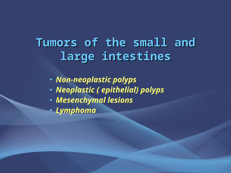
Tumors of the small and large Tumors of the small and large intestinesintestines
• Non-neoplastic polyps• Neoplastic ( epithelial) polyps• Mesenchymal lesions• Lymphoma
Tumors of the small and large intestineTumors of the small and large intestine
• Epithelial tumors are a major cause of morbidity and mortality worldwide
• The colorectal cancer is the GIT segment most commonly affected by tumors
• It is the host to more primary tumors than any other tumor of the body.
• Colonic carcinoma is second to bronchogenic carcinoma as a cause of death in USA.
• Adenocarcinoma in colorectum represent 70% of all malignancy of GIT
• Benign tumors, primarily epithelial, are present in 25 to 50% of older adults.
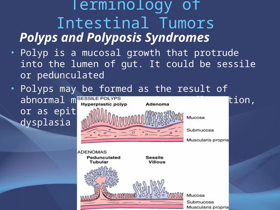
Terminology of Intestinal Tumors
Polyps and Polyposis Syndromes• Polyp is a mucosal growth that protrude into the lumen of
gut. It could be sessile or pedunculated• Polyps may be formed as the result of abnormal mucosal
maturation, inflammation, or as epithelial proliferation with dysplasia
Terminology of Intestinal Tumors
• Polypoid lesions is the inflammatory masses, hamartomas and tumors arising from the submucosa or muscle coat, but also protruding into the lumen.
• Polyposis is multiple polyps.
• Polyposis syndrome is hereditary, characterized by the presence of multiple pedunculated or sessile tumors of the mucosa
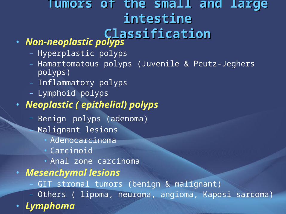
Tumors of the small and large intestineTumors of the small and large intestineClassificationClassification
• Non-neoplastic polyps– Hyperplastic polyps– Hamartomatous polyps (Juvenile & Peutz-Jeghers polyps)– Inflammatory polyps– Lymphoid polyps
• Neoplastic ( epithelial) polyps– Benign polyps (adenoma)– Malignant lesions
• Adenocarcinoma• Carcinoid• Anal zone carcinoma
• Mesenchymal lesions– GIT stromal tumors (benign & malignant)– Others ( lipoma, neuroma, angioma, Kaposi sarcoma)
• Lymphoma
Intestinal PolypsNon-Neoplastic Polyps
• Represent 90% of all epithelial polypi found in large intestine.
• Found in more than half of all persons age 60 years or older.
• Types: 1. Hyperplastic polyp.
2. Hamartomatous polyp (Juvenile & Peutz-Jeghers)
3. Inflammatory polyp
4. Lymphoid polyp
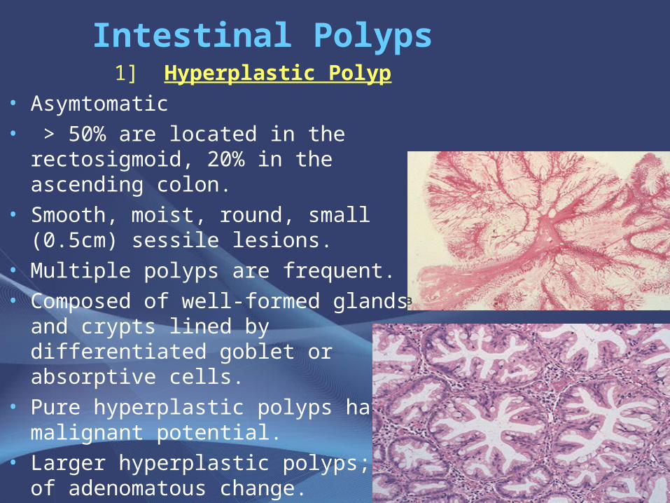
Intestinal Polyps 1] Hyperplastic Polyp
• Asymtomatic• > 50% are located in the rectosigmoid,
20% in the ascending colon.• Smooth, moist, round, small (0.5cm)
sessile lesions.• Multiple polyps are frequent.• Composed of well-formed glands and
crypts lined by differentiated goblet or absorptive cells.
• Pure hyperplastic polyps have no malignant potential.
• Larger hyperplastic polyps; foci of adenomatous change.
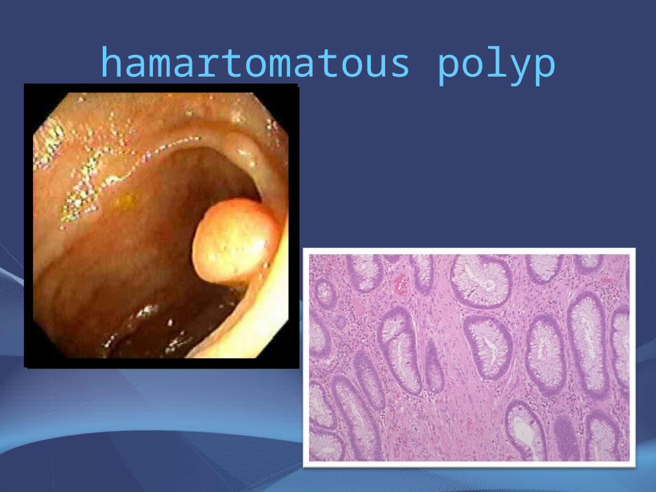
Non-Neoplastic Polyp2] Hamartomatous polyp
Juvenile Polyps (retention polyp)• Developmental malformations affecting the glands and
lamina propria, having no malignant potential.• commonly occur in children under 5 years old in the rectum.
In adult called retention polyp.• Painless rectal bleeding after defecation.• Large, rounded, smooth lesions with a stalk• Histology: mucus-filled, cystically dilated tubules lined by
normal or inflamed mucosa.
• Juvenile polyposis syndrome. Occurrence of multiple hamartomatous polyps throughout the GI tract.
Non-Neoplatic Polyps
2] Hamartomatous Polyps
Peutz-Jehgers polyps• Uncommon hamartomatous polyps accompanied by mucosal
and cutaneous pigmentation around the lips, oral mucosa, face and genitalia.
• Rare, autosomal dominant.• Caused by germ-line mutation in the LKB1 gene, which
encodes a serine threonine kinase.• Polyps tend to be large and pedunculated.• May occur anywhere in the GI tract.• Have an increased risk of developing carcinoma of the
pancreas, breast, lung, ovary and uterus.
hamartomatous polyp
Non-Neoplastic Polyps
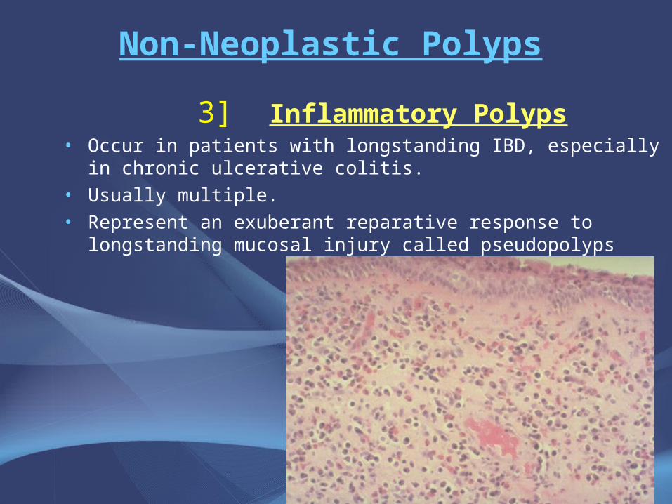
3] Inflammatory Polyps• Occur in patients with longstanding IBD, especially in chronic
ulcerative colitis.• Usually multiple.• Represent an exuberant reparative response to longstanding
mucosal injury called pseudopolyps
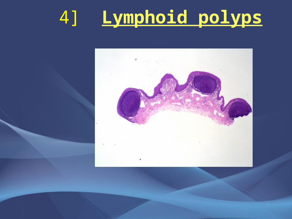
4] Lymphoid polyps
Neoplastic Polyps (Adenomas)
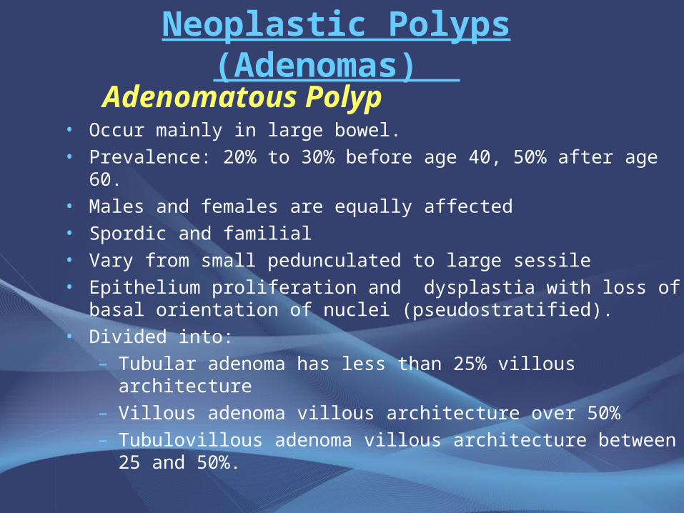
Adenomatous Polyp• Occur mainly in large bowel.• Prevalence: 20% to 30% before age 40, 50% after age 60.• Males and females are equally affected• Spordic and familial • Vary from small pedunculated to large sessile• Epithelium proliferation and dysplastia with loss of basal
orientation of nuclei (pseudostratified).• Divided into:
– Tubular adenoma has less than 25% villous architecture– Villous adenoma villous architecture over 50% – Tubulovillous adenoma villous architecture between 25
and 50%.
• All adenomatous lesions arise as the result of epithelial proliferation and dysplasia, which may range from mild to so severe as to represent transformation to carcinoma.
Neoplastic Polyps
1] Tubular adenoma• Represents 75% of all neoplastic polyps.• Occurs sporadicaly and in well defined hereditary syndromes.• Average age is 60 years• 75 % occur in the distal colon and rectum.• More than 50% occur singly.• Size: few millimeters (sessile) to many centimeters (have
stalk).• Stalk has a central core of fibrovascular tissue, covered with
dysplastic colonic mucosa.• Severe dysplasia and invasive carcinoma may supervene.
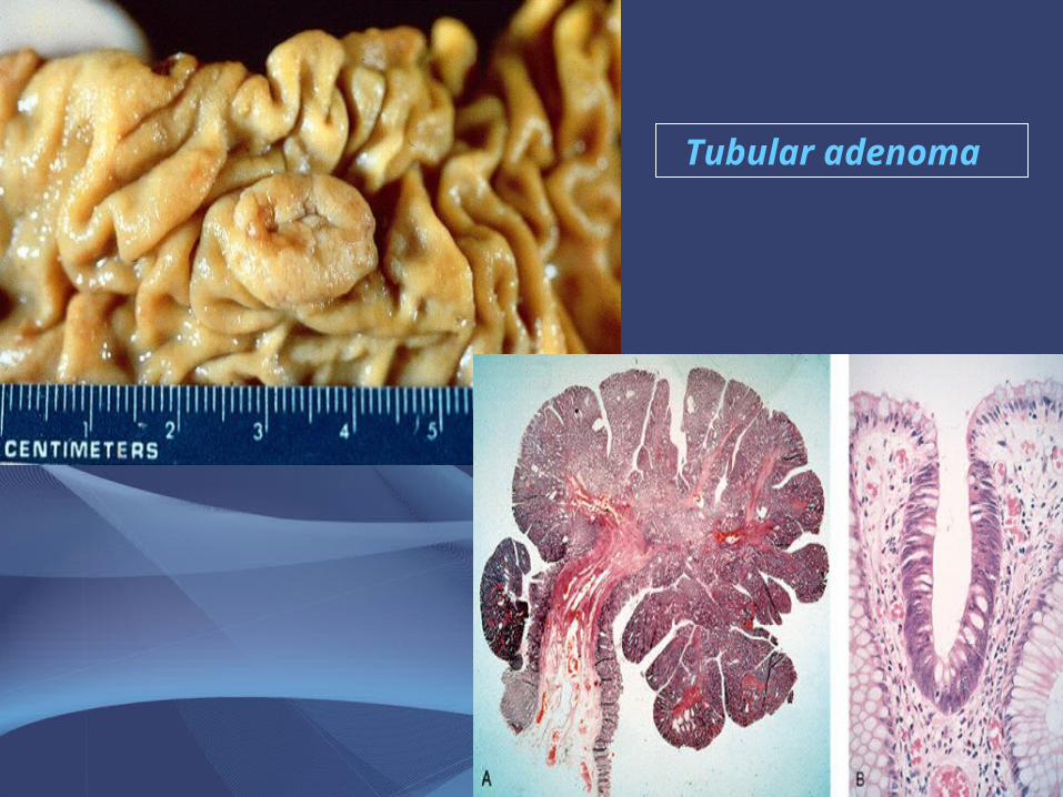
Tubular adenoma
Neoplastic Polyps
2] Villous Adenoma• The least common, largest and most ominous of epithelial
polyps.• Age: 60 to 65 years, M:F ratio roughly equal.• Present with rectal bleeding or anemia, large ones may
secrete copious amounts of mucoid material rich in protein.
• 75% located in rectosigmoid area.
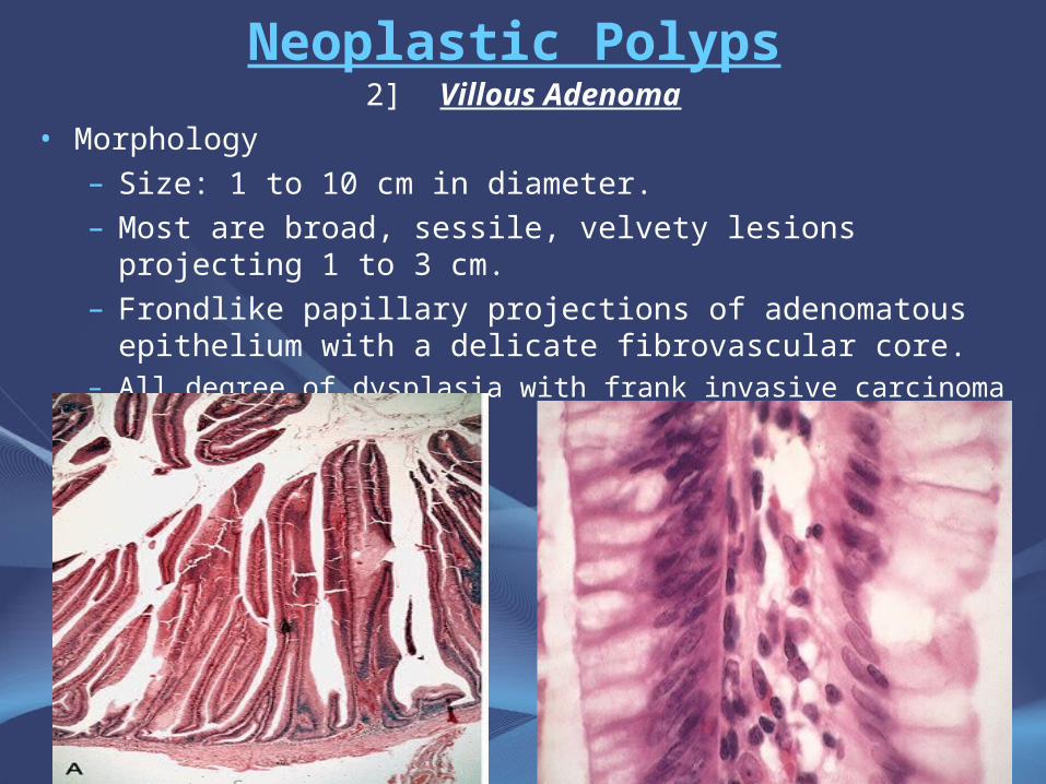
Neoplastic Polyps2] Villous Adenoma
• Morphology
– Size: 1 to 10 cm in diameter.
– Most are broad, sessile, velvety lesions projecting 1 to 3 cm.
– Frondlike papillary projections of adenomatous epithelium with a delicate fibrovascular core.
– All degree of dysplasia with frank invasive carcinoma in up to 40%.
Neoplastic Polyps
3] Tubulovillous adenoma
• Intermmediate in size, frequency of having a stalk, degree of dysplasia and malignant potential between tubular and villous adenomas.
Neoplastic Polyps
Clinical features• The smaller adenomas are usually asymptomatic,
occult bleeding.• Villous adenomas are much more frequently
symptomatic because of overt or occult rectal bleeding or mucoid material rich in protein and potassium to produce hypoproteinemia or hypokalemia.
• Adenomas in the immediate vicinity of the ampulla of Vater may produce biliary obstruction.
Relationship of Neoplastic Polyps to Carcinoma
• Adenoma to carcinoma sequence is documented by several observations and genetic alterations.
• The probability of carcinoma occuring in a neoplastic polyp is related to:
1. The size of the polyp.
2. The relative proportion of its villous features.
3. The presence of significant cytologic atypia
(dysplasia) in the neoplastic cells.
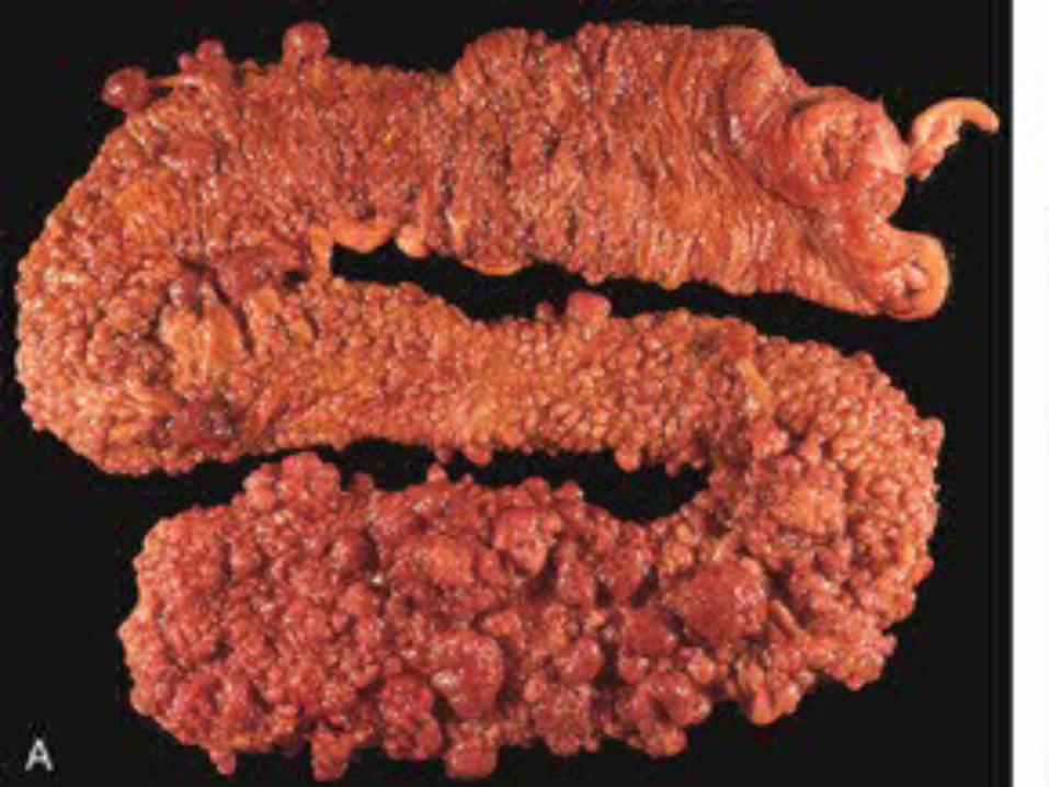
Familial Polyposis Syndrome• Patients have genetic tendencies to develop neoplastic
polyps, most often autosomal dominant.Familial polyposis coli (FPC)
• Genetic defect ch5 q21.• Innumerable neoplastic polyps in the colon (500 to 2500)• Polyps are also found elsewhere in alimentary tract• Most polyps are tubular adenomas• The risk of colorectal cancer is 100% by midlife.
Gardener’s syndrome• Polyposis coli, multiple osteomas, epidermal cysts, and
fibromatosis.Turcot syndrome
• Polyposis coli, glioma and fibromatosis
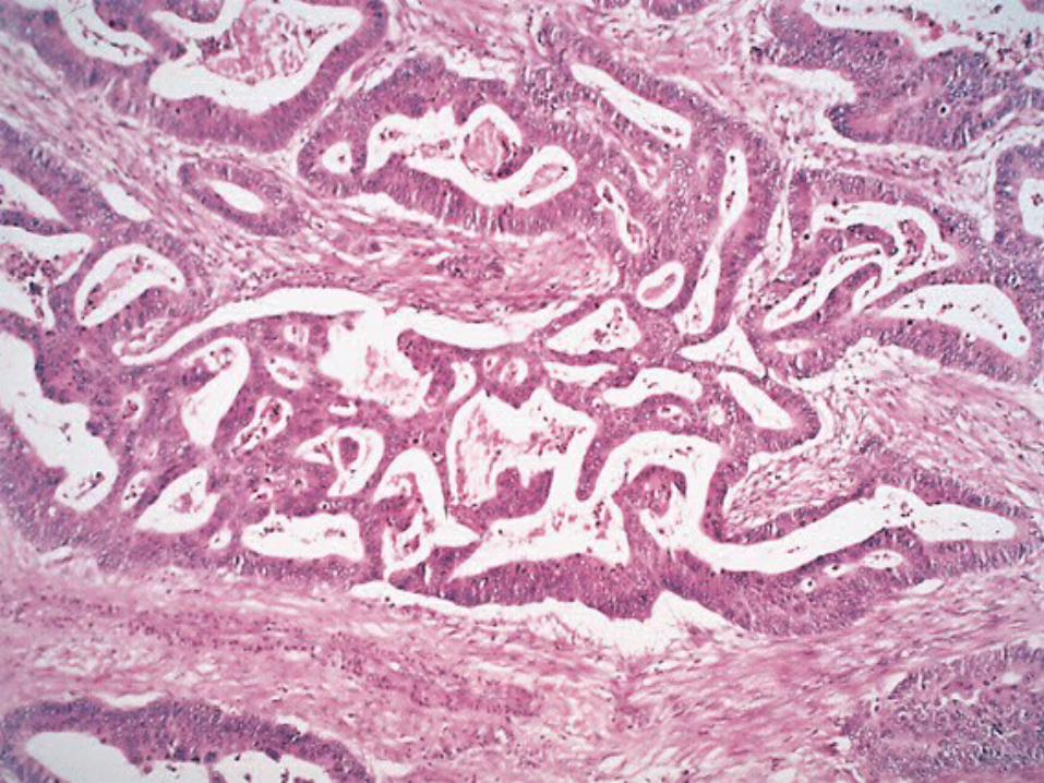
Malignant Tumors of Large IntestineAdenocarcinoma
• Constitutes 98% of all cancers in the large intestine.• Worldwide distribution, highest incidence in west.• Causes 15% of all cancer-related death in the USA.• The mortality rate and incidence is higher in blacks.• Peak incidence in the sixth to seventh decade.
Malignant Tumors of Large IntestineAdenocarcinoma
• Predisposing factors: IBD, polyposis syndrome.• Male:female ratio is 2:1 in rectal cancer, roughly equal in colon
cancer, generally males are affected about 20% more than females.• Diet appears to play an important role in the risk for colon cancer:
- Low content of unabsorpable vegetable fibre.
- High content of refined carbohydrates.
- High fat content.
- ? Increased intake of nitrites, nitrates (nitrosamines).
- Reduced intake of vit A, C & E.• The use of aspirin and NSAID (cyclooxygenase-2 inhibitors) exerts
a protective effect against colon cancer
• When colorectal cancer is found in a young person, preexisting ulcerative colitis or one of the polyposis syndromes must be suspected.
• Individuals with hereditary nonpolyposis colorectal cancer syndrome (HNPCC, also known as Lynch syndrome), caused by germ-line mutations of DNA mismatch repair genes, are at a high risk of developing colorectal cancers.– (HNPCC patients are also at risk of developing other tumors, such
as cholangiocarcinomas.)
Malignant Tumors of Large IntestineAdenocarcinoma
Colorectal carcinogenesis• Two pathogenetically distinct pathways for the
development of colon cancer, both seem to result from accumulation of multiple mutations– The APC/B-catenin pathway
• chromosomal instability that results in stepwise accumulation of mutations in a series of oncogenes and tumor suppressor genes.
• Localized colon epithelial proliferation followed by the formation of small adenomas, become more dysplastic, and ultimately develop into invasive cancers.
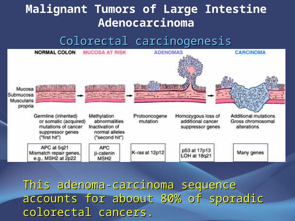
This adenoma-carcinoma sequence accounts This adenoma-carcinoma sequence accounts for aboout 80% of sporadic colorectal cancers.for aboout 80% of sporadic colorectal cancers.
Malignant Tumors of Large IntestineAdenocarcinoma
Colorectal carcinogenesisColorectal carcinogenesis
Malignant Tumors of Large IntestineAdenocarcinoma
The DNA mismatch repair genes pathway:
•10% to 15% of sporadic cases.•There is accumulation of mutations (as in the
APC/B-catenin schema) but the involved genes are different.
•Unlike in the adenoma-carcinoma sequence, there are no clearly identifiable morphologic correlates
Malignant Tumors of Large IntestineAdenocarcinoma
• In DNA mismatch repair genes pathway defective DNA repair caused by inactivation of DNA mismatch repair genes is the fundamental and the most likely initiating event in colorectal cancers
• Inherited mutations in one of five DNA mismatch repair genes (MSH2, MSH6, MLH1, PMS1, AND PMS2) give rise to the hereditary non polyposis colon carcinoma (HNPCC)
• MLH1 gene is the one most commonly involved in sporadic colon carcinomas
Malignant Tumors of Large IntestineAdenocarcinoma
• Microsatellite instability (MSI) is the molecular signature of defective DNA mismatch repair
• Most microsatellite sequences are in noncoding regions of the genes so mutations in these genes are probably harmless.
• However, some micorsatellite sequences are located in the coding or promoter region of genes involved in regulation of cell growth.
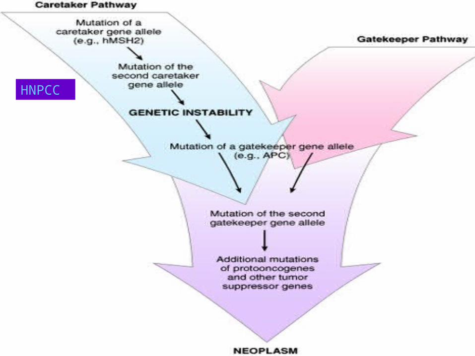
HNPCC
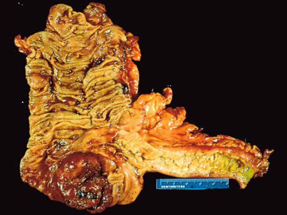
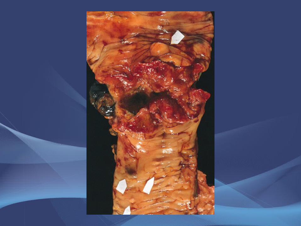
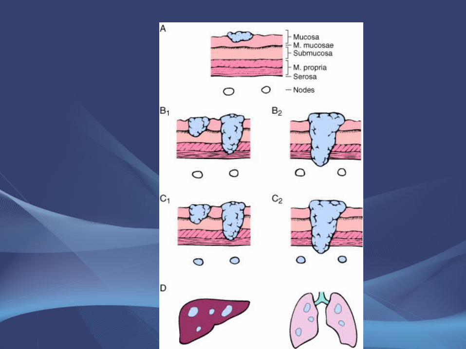
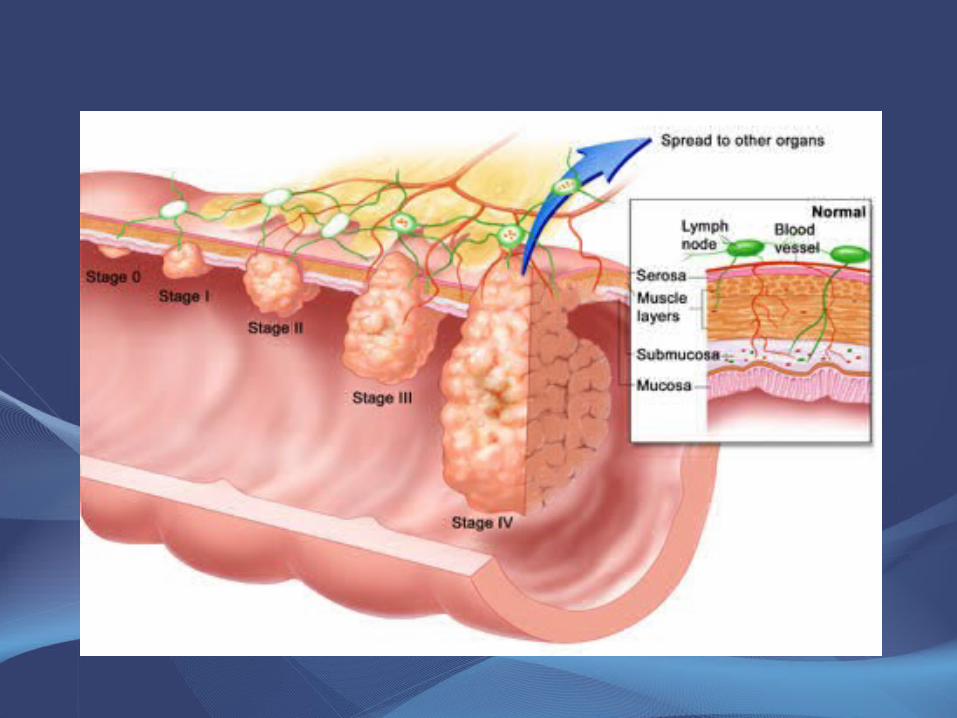
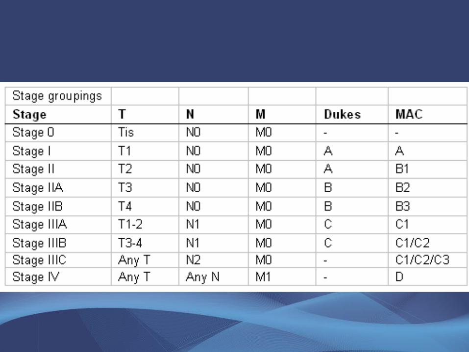
Colorectal Carcinoma
Morphology• Sixty to 70% of colorectal carcinomas are in the
rectum, rectosigmoid and sigmoid colon.• Left-sided carcinomas tend to be annular,
encircling lesions with early symptoms of obstruction.
• Neoplasms start superficially, slowly invading the deeper layers with ulceration and eventually metastasis.
Colorectal Carcinoma
Morphology• Right-sided carcinomas tend to grow as polypoid,
fungating masses, obstruction is uncommon.• Invasion of the wall and extend to the mesentery,
regional lymph nodes and more distal sites.• Mucinous adenocarcinoma secret abundant mucin
that may dissect through cleavage planes in the wall.
• Small cell undifferentiated carcinomas are rare (arising from neuroendocrine cells)
• In UC, poorly differentiated infiltrative adenocarcinoma without an exophytic growth.
Colorectal Carcinoma
Clinical features
• The condition tends to be present for a considerable time before producing symptoms.
• Left-sided lesions tend to present earlier but
also have a more infiltrative growth pattern and a poorer prognosis.
• Right-sided lesions tend to present with weakness, malaise, weight loss, unexplained anemia (secondary to early bleeding).
Colorectal Carcinoma
• Spread: - direct extension.
- metastasis through:
- lymphatic
- blood vessels
- favored sites are regional lymph node, liver, lungs, bones.
• Serum levels of carcinoembryonic antigen (CEA) are related to tumor size and extent of spread. They are helpful in monitoring for recurrence of tumor after resection.
• Overall 5-year survival is 35 to 49% in the United States.
Colorectal Neoplasm
Other Tumors• Malignant spindle cell (mesenchymal) tumors and
lymphomas.• Grossly and microscopically resemble those
arising elsewhere in the GI tract.• Carcinoid tumors may arise anywhere in the
colon, especially the rectum.• Squamous cell carcinomas are largely limited to
the rectal canal. Initially present as plaque-like lesions, later becoming ulcerated or fungating.
• Malignant melanoma at the anal verge.
Small Intestinal Neoplasms• 3-6% of GIT neoplasm, slight preponderance to benign tumors.
BENIGN
• Discovered incidentally, leiomyoma, adenoma and lipoma
• Large lesions may cause obstruction, bleeding, intussusception, volvulus.
ADENOMAS
• Single or multiple polyps, most often in the duodenum and ileum.
• There is a risk of malignancy with larger adenomatous polyps.
MALIGNANT
• In descending order of frequency: carcinoid, adenocarcinomas, lymphomas and leiomyosarcomas.
• Leiomyosarcomas have tyrosine kinase receptors, can be treated by STI-571
Small Intestinal NeoplasmsAdenocarcinoma of small intestine
• Tumors grow as polypoid fungating ulcerating mass or encircling pattern
• Site: duodenum ( ampulla of Vater)• Presentation: abdominal cramping pain,
vomiting and weight loss• Patients present late• 5 years survival is 70% after en bloc
resection
Small Intestinal Neoplasms
Carcinoid Tumors• Neoplasms arising from endocrine cells Kulchitsky or
enterochromaffin cells found along the length of GIT mucosa. Cells have an affinity for silver salts.
• 60 to 80% appendix and terminal ileum: 10 to 20% rectum, the remainder in the stomach, duodenum or esophagus.
• Other Location: Lungs, pancreas, biliary tract, ovaries and liver.• Peak age: 6th decade, comprise 2% of colorectal carcinoma and 50%
of small intestinal carcinoma.• Tumors in the appendix and rectum, although spreading locally,
seldom metastasize.• Ileal, gastric, and colonic carcinoids are frequently malignant.
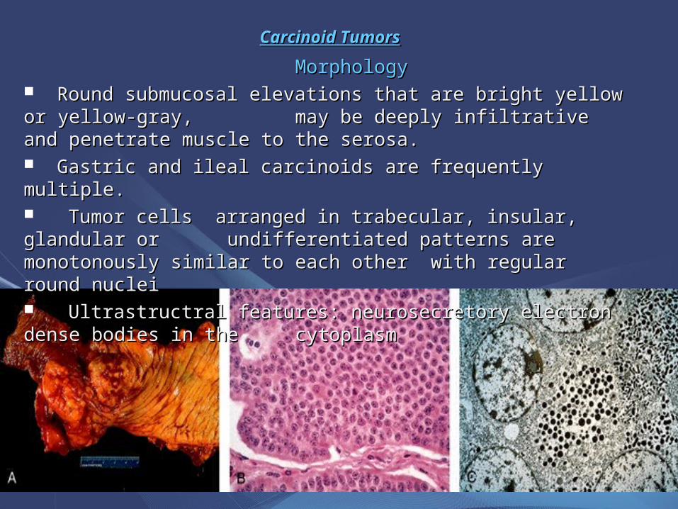
Carcinoid TumorsCarcinoid Tumors
MorphologyMorphology Round submucosal elevations that are bright yellow or yellow-gray, Round submucosal elevations that are bright yellow or yellow-gray, may be deeply infiltrative and penetrate muscle to the serosa.may be deeply infiltrative and penetrate muscle to the serosa. Gastric and ileal carcinoids are frequently multiple.Gastric and ileal carcinoids are frequently multiple. Tumor cells arranged in trabecular, insular, glandular or Tumor cells arranged in trabecular, insular, glandular or undifferentiated patterns are monotonously similar to each other undifferentiated patterns are monotonously similar to each other with with regular round nucleiregular round nuclei Ultrastructral features: neurosecretory electron dense bodies in the Ultrastructral features: neurosecretory electron dense bodies in the
cytoplasmcytoplasm
Small Intestinal Neoplasms
Carcinoid Tumor
Clinical features• Asymptomatic• May cause obstruction, intussusception or bleeding.• May elaborate hormones: Zollinger-Ellison, Cushing’s
carcinoid or other syndromes.• 5 years survival rate is 90%, small bowel Carcinoid with
liver metastasis the 5 years survival rate is better than 50%
Small Intestinal NeoplasmsCarcinoid tumor
Carcinoid syndrome
• Syndrome occur in 1% of all pt. with carcinoid & in 20% of those of widespread metastasis
• Paroxymal flushing, episodes of asthma-like wheezing, right-sided heart failure, attacks of watery diarrhea, abdominal pain, edema and pellagra-like lesions of the skin and oral mucosa.
• The principal chemical mediator is serotonin (5-hydroxy-tryptamine-5HT).
• 5-HT is decarboxylated in the liver and lungs to 5-hydroxy-indoleacetic acid (5HIAA)• The syndrome is classically associated with ileal carcinoids
with hepatic metastases.
Small Intestinal Neoplasms
Lymphoma• Up to 40% of lymphomas arise in sites
other than lymph nodes, gut is the most.• 1% to 4% of all gastrointestinal
malignancies are lymphomas. • Primary GIT lymphomas exhibit no
evidence of liver, spleen, or bone marrow involvement at the time of diagnosis.
Small Intestinal NeoplasmsLymphoma
• Sporadic lymphoma arise from the B cells of mucosa-associated lymphoid tissue (MALT).
• This usually affects adults, lacks a sex predilection, and may arise anywhere in the gut: stomach - 55% to 60% -
small intestine - 25% to 30% - proximal colon - 10% to 15% -
distal colon - up to 10% -
Small Intestinal Neoplasms
Lymphoma
• Gastric MALT lymphomas arise in the setting of mucosal lymphoid activation, as a result of Helicobacter associated chronic gastritis.
• Celiac disease is associated with a higher than normal risk of T-cell lymphomas.
Small Intestinal Neoplasms
Lymphoma
• Primary GIT lymphomas have a better prognosis than do those arising in other sites.
• Treatment: combined surgery, chemotherapy, and radiation therapy.


![Prolapsed Ano-Rectal Neoplastic Polyps in Elderly Patients ...A].pdf · over 65 years of age with prolapsed anorectal polyps which we treated by transanal excision. Patients who underwent](https://img.pdfslide.net/doc/110x75/608a7dd07ab5bf3eba4c0539/prolapsed-ano-rectal-neoplastic-polyps-in-elderly-patients-apdf-over-65.jpg)