Embed Size (px)
Citation preview

David Rapkin, MD, FACOGAssistant Professor, Division of General Obstetrics & Gynecology
Department of Obstetrics and GynecologyUniversity of South Florida
Tampa, FL
��Chief of Obstetrics and Gynecology at Tampa General Hospital
Shoulder DystociaManagement and Documentation

Disclosure
I have no relevant financial
conflicts of interest to disclose
Objectives Identify risk factors for shoulder dystocia
Review the management of shoulder dystocia
Explain how simulation can reduce the incidence of
poor outcomes in shoulder dystocia deliveries
Identify the key components to appropriately document
the events of a shoulder dystocia

Background
Incidence and Risk Factors
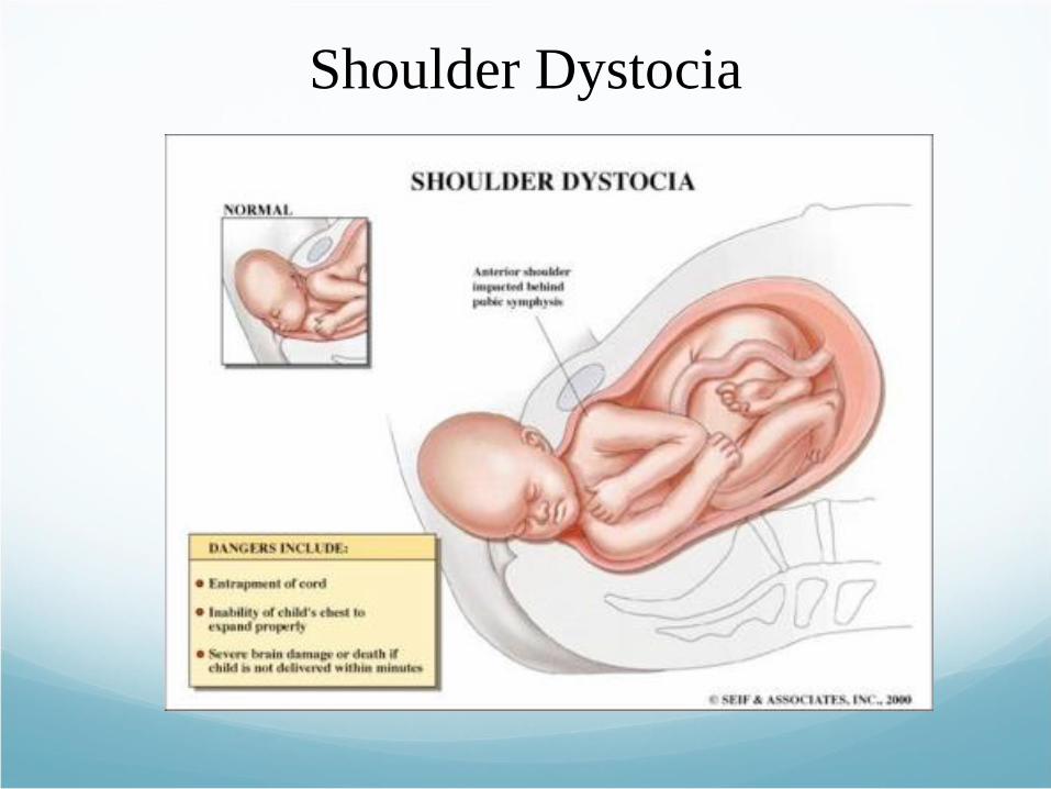
Shoulder Dystocia

ACOG Practice Bulletin
“Delivery that requires additional
obstetric maneuvers following the
failure of gentle downward traction on
the fetal head to effect the delivery of
the shoulders”
Shoulder dystocia. ACOG Practice Bulletin No. 40. American College of Obstetricians and Gynecologists. Obstet Gynecol 2002;100:1045–50.

Birth Weight ComparisonsAnthropometric >4000g <4000 g Measurements (n=202) (n=100) p
Birth weight (± 1 SD) 4247 g ± 471 3399 g ± 36
Gestational age 40.7 wks ± 1.3 39.6 wks ± 3.9 NS
Length 54.6 cm ± 2.8 51.7 cm ± 2.6 <.001
Head circumference 36.3 cm ± 1.2 34.6 cm ± 1.3 <.001
Chest circumference 36.2 cm ± 2.8 33.6 cm ± 1.8 <.001
Chest-head 0.06 cm ± 1.6 -1.1 cm ± 1.5 <.001

Shoulder Dystocia versus No
Shoulder Dystocia
Shoulder No ShoulderAnthropometric Dystocia DystociaMeasurements (n=10) (n=130) p
Birth weight 4416 g ± 294 4254 g ± 323 NS
Gestational age 41.2 wks ± 1.6 40.4 wks ± 3.6 NS
Length 54.5 cm ± 1.9 54.8 cm ± 2.3 NS
Head circum. 36.1 cm ± 1.6 36.2 cm ± 1.1 NS
Chest circum. 37.7 cm ± 1.5 36.0 cm ± 3.3 NS
Chest-head 1.6 cm ± 2.2 0.2 cm ± .8 <.025
Mean ± 1 SD

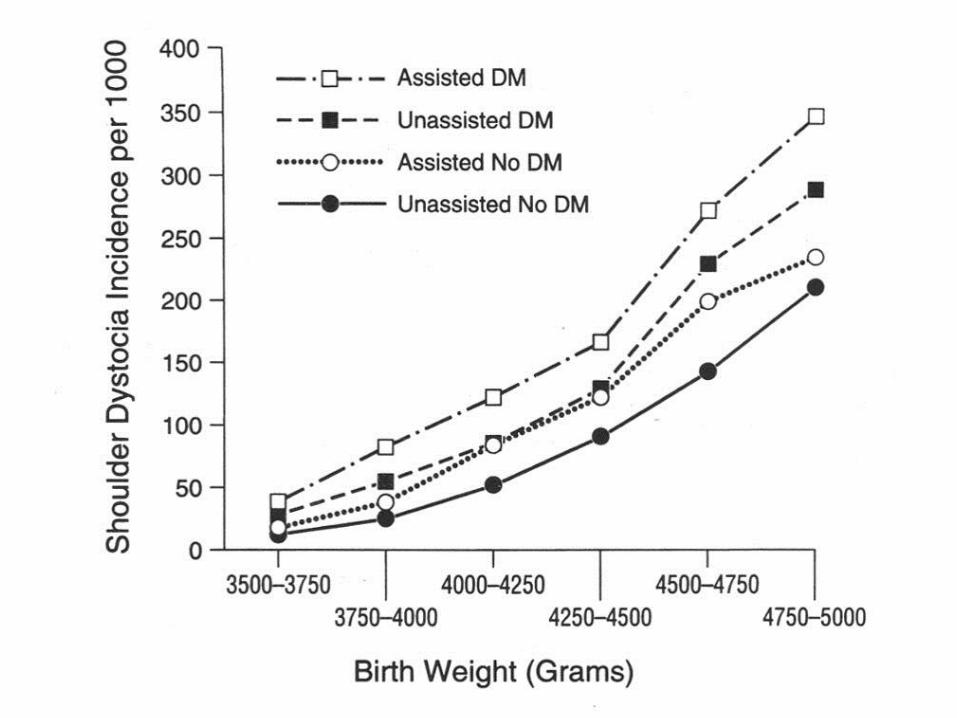
Proposed Risk Factors Shoulder Dystocia
Fetal weight (actual vs estimated)
Obesity
Diabetes
Prior shoulder dystocia
Excessive weight gain
Prior macrosomic infant
>42 weeks
Labor pattern
Operative vaginal delivery
Antepartum:
Intrapartum:

The Seattle Times, Friday, February 1, 2002.
11# baby

Shoulder Dystocia Lawsuit
Verdict of $56M Appealed by
Hospital August 12, 2010 Written by: Staff Writers
A New York hospital is attempting to overturn a $56
million verdict in a birth injury lawsuit won by a family
last year after their son’s shoulder became stuck on the
mother’s pelvic bone during delivery.
http://www.aboutlawsuits.com/shoulder-dystocia-lawsuit-verdict-appealed-
11885/


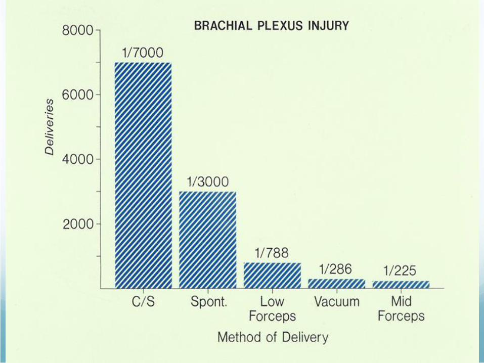
Obstetric Brachial Plexus PalsyNatural History
The 2 studies of 7 which come closest to
an ideal study show a tendency towards a 20–30%
residual deficit in contrast to the optimistic view of
over 90% complete or almost complete recovery
Physicians should exercise caution in predicting
excellent recovery shortly
after birth
Pondaag 2004. Develop Med and Child Neurology. Vol 46.


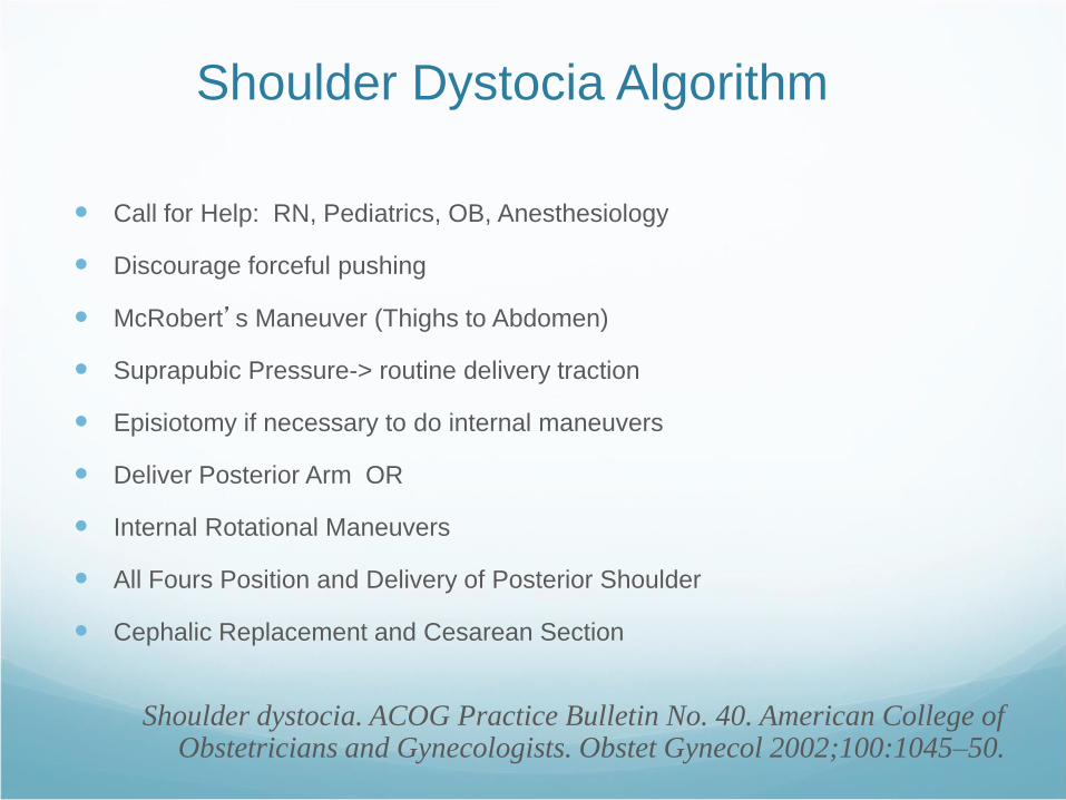
Shoulder Dystocia Algorithm
Call for Help: RN, Pediatrics, OB, Anesthesiology
Discourage forceful pushing
McRobert’s Maneuver (Thighs to Abdomen)
Suprapubic Pressure-> routine delivery traction
Episiotomy if necessary to do internal maneuvers
Deliver Posterior Arm OR
Internal Rotational Maneuvers
All Fours Position and Delivery of Posterior Shoulder
Cephalic Replacement and Cesarean Section
Shoulder dystocia. ACOG Practice Bulletin No. 40. American College of Obstetricians and Gynecologists. Obstet Gynecol 2002;100:1045–50.

McRoberts Maneuver

Suprapubic Pressure

Wood’s Maneuver

Rubin’s Maneuver

Delivery of Posterior Arm (1/3)

Delivery of Posterior Arm (2/3)
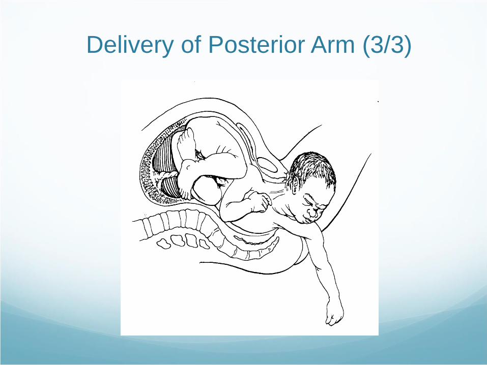
Delivery of Posterior Arm (3/3)

Effect of Barnum Maneuver(reducing obstructing part of fetal shoulder)

Zavanelli Maneuver (1/2)

Zavanelli Maneuver (2/2)

Gaskin Maneuver
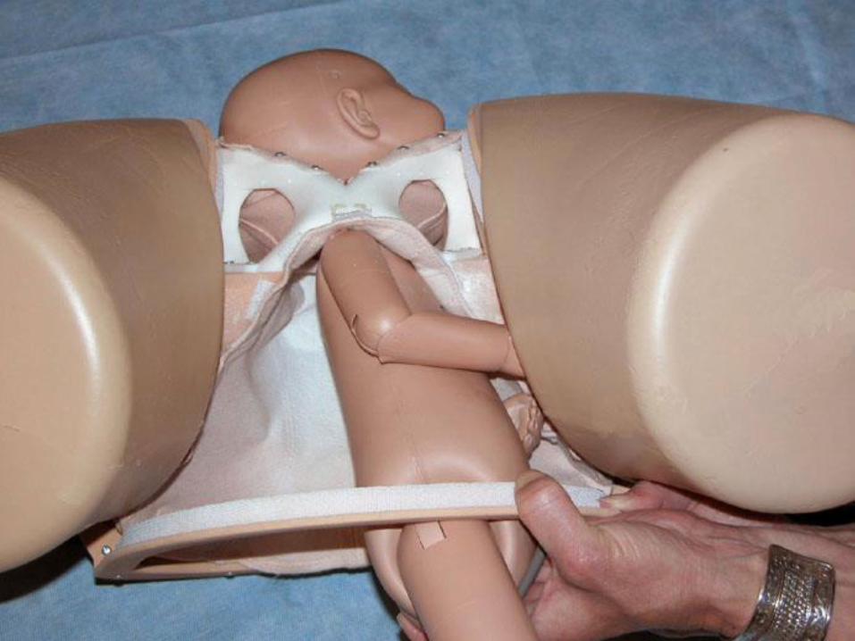


Fig. 1
Fig. 1. Difficulties gaining vaginal
access. A. Attempting to gain anterior
access. B. Attempting to gain lateral
access. C. Entering with two fingers as
if performing a routine vaginal
examination. D. Leaving the thumb
out.Crofts. Lessons for Shoulder
Dystocia Training. Obstet Gynecol
2008.
Copyright © 2015 Obstetrics & Gynecology. Published by Lippincott Williams & Wilkins. 37
Observations From 450 Shoulder
Dystocia Simulations: Lessons for
Skills Training
Crofts, Joanna F.; Fox, Robert; Ellis,
Denise; Winter, Catherine; Hinshaw,
Kim; Draycott, Timothy J.
Obstetrics & Gynecology. 112(4):906-
912, October 2008.
doi: 10.1097/AOG.0b013e3181865f55

Fig. 2
Fig. 2. Facilitation of vaginal access.
Posterior vaginal access with whole
hand.Crofts. Lessons for Shoulder
Dystocia Training. Obstet Gynecol
2008.
Copyright © 2015 Obstetrics & Gynecology. Published by Lippincott Williams & Wilkins. 38
Observations From 450 Shoulder
Dystocia Simulations: Lessons for
Skills Training
Crofts, Joanna F.; Fox, Robert; Ellis,
Denise; Winter, Catherine; Hinshaw,
Kim; Draycott, Timothy J.
Obstetrics & Gynecology. 112(4):906-
912, October 2008.
doi: 10.1097/AOG.0b013e3181865f55

Fig. 3
Fig. 3. Vaginal access training. A.
Incorrect. B. Incorrect. C. Correct. D.
Correct. E. Incorrect. F. Correct.Crofts.
Lessons for Shoulder Dystocia Training.
Obstet Gynecol 2008.
Copyright © 2015 Obstetrics & Gynecology. Published by Lippincott Williams & Wilkins. 39
Observations From 450 Shoulder
Dystocia Simulations: Lessons for
Skills Training
Crofts, Joanna F.; Fox, Robert; Ellis,
Denise; Winter, Catherine; Hinshaw,
Kim; Draycott, Timothy J.
Obstetrics & Gynecology. 112(4):906-
912, October 2008.
doi: 10.1097/AOG.0b013e3181865f55
1. Performing a manual removal of a
placenta
2. Putting on a tight bracelet
3. Removing the last potato chip in
the tube!

Fig. 1
Fig. 1. Birth training mannequins used
for shoulder dystocia training. A.
Prototype 2. B. Prototype 3 with skin on.
C. Prototype 3 with skin off.Draycott.
Improved Outcomes After Shoulder
Dystocia Training. Obstet Gynecol
2008.
Copyright © 2015 Obstetrics & Gynecology. Published by Lippincott Williams & Wilkins. 40
Improving Neonatal Outcome Through
Practical Shoulder Dystocia Training
Draycott, Timothy J.; Crofts, Joanna F.;
Ash, Jonathan P.; Wilson, Louise V.; Yard,
Elaine; Sibanda, Thabani; Whitelaw,
Andrew
Obstetrics & Gynecology. 112(1):14-20,
July 2008.
doi: 10.1097/AOG.0b013e31817bbc61

Fig. 1
Fig. 1. Birth training mannequins used
for shoulder dystocia training. A.
Prototype 2. B. Prototype 3 with skin on.
C. Prototype 3 with skin off.Draycott.
Improved Outcomes After Shoulder
Dystocia Training. Obstet Gynecol
2008.
Copyright © 2015 Obstetrics & Gynecology. Published by Lippincott Williams & Wilkins. 41
Improving Neonatal Outcome Through
Practical Shoulder Dystocia Training
Draycott, Timothy J.; Crofts, Joanna F.;
Ash, Jonathan P.; Wilson, Louise V.; Yard,
Elaine; Sibanda, Thabani; Whitelaw,
Andrew
Obstetrics & Gynecology. 112(1):14-20,
July 2008.
doi: 10.1097/AOG.0b013e31817bbc61
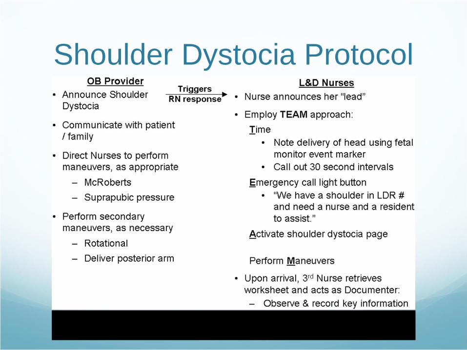
Shoulder Dystocia Protocol
Grobman WA,BurkeC,HornbogenA,CostelloR.Development and implementation of a team-centered shoulder dystocia protocol. Simul Healthc.
2010;5:199–203. 25.
GrobmanWA,MillerD,BurkeC,HornbogenA,TamK,CostelloR. Outcomes associated with introduction of a shoulder dystocia protocol. Am J Obstet
Gynecol. 2011;205:513–517.

Concerns Regarding Management of Shoulder
Dystocia and Solutions Embedded Within the Shoulder
Dystocia Protocol
Concern SolutionLack of common understanding that a Unambiguous statement by delivering
shoulder dystocia had occurred provider once a shoulder dystocia is diagnosed
Difficulty summoning desired staff Single page number created and implemented in the
hospital paging system that simultaneously notifies
desired individuals
Lack of role clarity Institution of protocol with delineation of roles
Reduced situational awareness for Duration of shoulder dystocia announced at standard
delivering provider intervals; implementation of protocol that does not
require him or her to direct all actions
Variability of documentation Standard worksheet completed by designated nurse

Shoulder Dystocia Documentation Worksheet
EVENTS COMMENTS
Time head delivered
Time infant delivered LOT ROT
Duration of shoulder dystocia
Position of fetal head at restitution LOT ROT
MANEUVERS CHECK IF PERFORMED COMMENTS
McRoberts’
Suprapubic pressure
Woods’ screw
Rubin’s maneuver
Other type of rotational maneuver
Delivery of posterior arm
Zavanelli
Other
SHOLDER TEAM PRESENT IN DELIVERY ROOM NAMES
OB Attending/CNM
OB Resident/Fellow
RN#1
RN#2
RN Documenter
Anesthesiologist
Pediatrician
Others

ACOG Documentation
WorksheetDate_____________ Patient_____________________________Date of birth_________
MR #____________Physician or certified nurse–midwife____________________________
Gravidity/Parity________________
Timing: Onset of active labor__________ Start of second stage______
Delivery of head__________ Time shoulder dystocia recognized and help called_________
Delivery of posterior shoulder__________Delivery of infant________
Antepartum documentation:
❏Assessment of pelvis
❏History of prior cesarean delivery: Indication for cesarean delivery:
❏History of prior shoulder dystocia
❏History of gestational diabetes
❏Largest prior newborn birth weight
❏Estimated fetal weight
❏Cesarean delivery offered if estimated fetal weight greater than 4,500 g (if the patient has
diabetes mellitus) or greater than 5,000 g (if patient does not have diabetes mellitus)
http://www.acog.org/-/media/Patient-Safety-
Checklists/psc006.pdf?dmc=1&ts=20150209T1606083475

Intrapartum documentation:
❏Mode of delivery of vertex:
❏Spontaneous
❏Operative delivery: Indication:
❏Vacuum❏Forceps
❏Anterior shoulder: ❏Right ❏Left
❏Traction on vertex: ❏None ❏Standard
❏No fundal pressure applied
❏Maneuvers utilized :
❏Hip flexion (McRoberts maneuver) ❏Suprapubic pressure
❏Delivery of posterior arm ❏All fours (Gaskin maneuver)
❏Posterior scapula (Woods maneuver) ❏Anterior scapula (Rubin)
❏Abdominal delivery ❏Zavanelli maneuver
❏Episiotomy: ❏None ❏Median ❏Mediolateral ❏Proctoepisiotomy
❏Extension of episiotomy: ❏None ❏Third degree ❏Fourth degree
❏Laceration: ❏Third degree ❏Fourth degree
❏Cord blood gases sent to the laboratory:
❏Yes: Results:_____________________________❏No
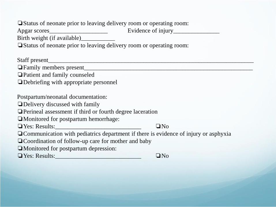
❏Status of neonate prior to leaving delivery room or operating room:
Apgar scores___________________ Evidence of injury_______________
Birth weight (if available)___________
❏Status of neonate prior to leaving delivery room or operating room:
Staff present___________________________________________________________________
❏Family members present_______________________________________________________
❏Patient and family counseled
❏Debriefing with appropriate personnel
Postpartum/neonatal documentation:
❏Delivery discussed with family
❏Perineal assessment if third or fourth degree laceration
❏Monitored for postpartum hemorrhage:
❏Yes: Results:____________________________ ❏No
❏Communication with pediatrics department if there is evidence of injury or asphyxia
❏Coordination of follow-up care for mother and baby
❏Monitored for postpartum depression:
❏Yes: Results:____________________________ ❏No

Summary The chest to head circumference ratio plays an important roll in
shoulder dystocia
Exercise caution in predicting excellent recovery of a brachial plexus
injury shortly after birth
Have a protocol to manage shoulder dystocia
Practice the maneuvers required to relieve a shoulder dystocia with
simulation
Preferably in a multi-team scenario
Have a designated person in the room to document the timeline and
events
Document, document, document

References1. Shoulder dystocia. ACOG Practice Bulletin No. 40. American College of Obstetricians and
Gynecologists. Obstet Gynecol 2002;100:1045–5
2. Pondaag 2004. Develop Med and Child Neurology. Vol 46.
3. Draycott TJ. Improving Neonatal Outcome Through Practical Shoulder Dystocia Training.
Obstet Gynecol. 2008 July;112(1):14-20.
4. Crofts JF et al. Observations From 450 Shoulder Dystocia Simulations: Lessons for Skills
Training. Obstet Gynecol. 2008 Oct;112(4):906-12.
5. http://www.acog.org/-/media/Patient-Safety-
Checklists/psc006.pdf?dmc=1&ts=20150209T1606083475


![Three patients presenting with severe macrosomia and ......predictive of fetal macrosomia [12, 13]. Sardesai et al. reported the case of a fetus with fatal HCM and macrosomia. The](https://img.pdfslide.net/doc/110x75/6141240183382e045471e526/three-patients-presenting-with-severe-macrosomia-and-predictive-of-fetal.jpg)