Embed Size (px)
Citation preview

Ultrasound, Positron Emission Tomography, and Single Photon Emission Tomography
Allen T. Newton, Ph.D.PAVE 2014
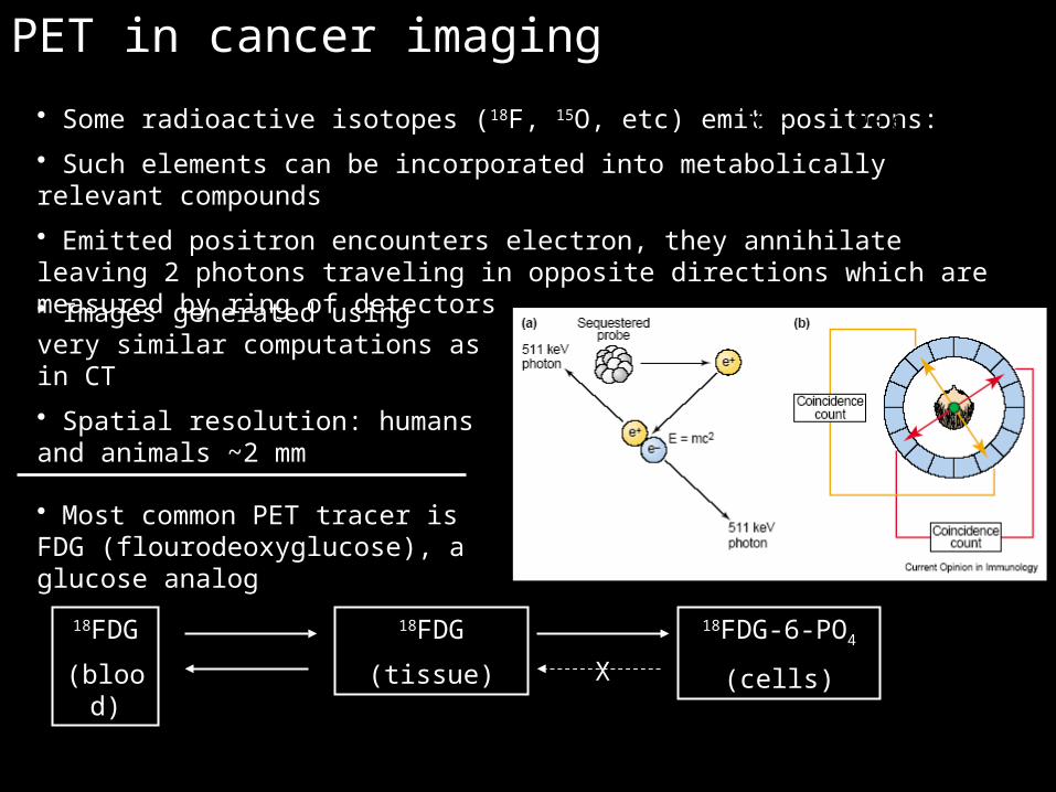
PET in cancer imaging
• Images generated using very similar computations as in CT
• Spatial resolution: humans and animals ~2 mm
• Some radioactive isotopes (18F, 15O, etc) emit positrons:
• Such elements can be incorporated into metabolically relevant compounds
• Emitted positron encounters electron, they annihilate leaving 2 photons traveling in opposite directions which are measured by ring of detectors
1
A AX Q e
Z Z
• Most common PET tracer is FDG (flourodeoxyglucose), a glucose analog
18FDG
(blood)
18FDG
(tissue)
18FDG-6-PO4
(cells)X

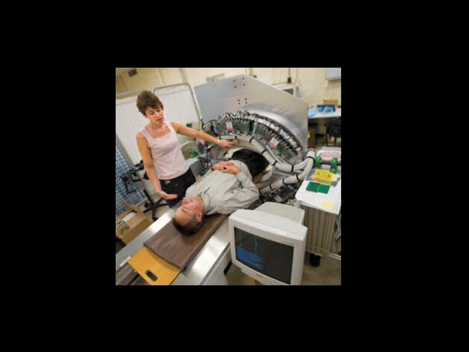
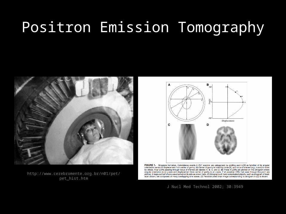
Positron Emission Tomography
J Nucl Med Technol 2002; 30:3949
http://www.cerebromente.org.br/n01/pet/pet_hist.htm
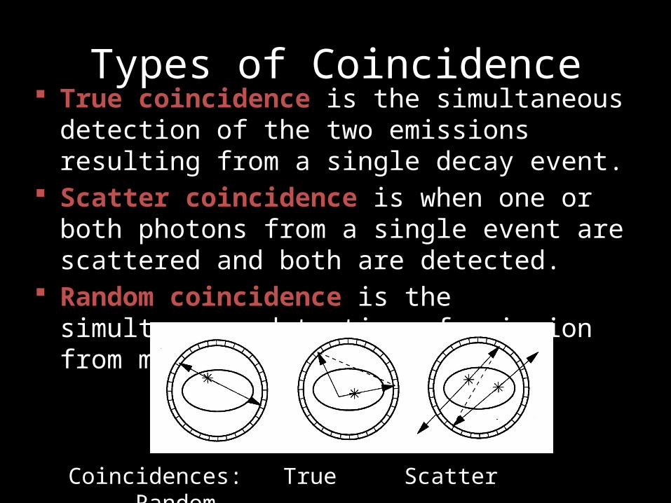
Types of Coincidence True coincidence is the simultaneous detection of the
two emissions resulting from a single decay event. Scatter coincidence is when one or both photons
from a single event are scattered and both are detected.
Random coincidence is the simultaneous detection of emission from more than one decay event.
Coincidences: True Scatter Random
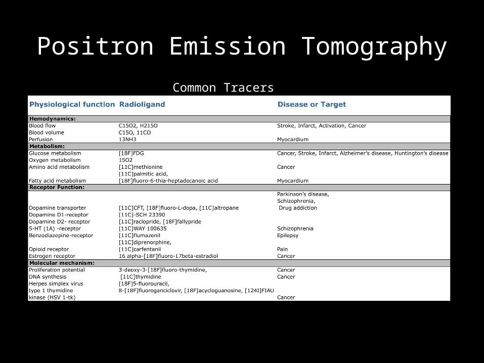
Positron Emission TomographyCommon Tracers

Positron Emission TomographyCommon Tracers
Half life = 110minHalf life = 20min
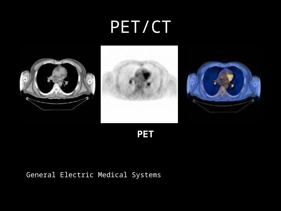
PET/CT
CT PET CT+PET
General Electric Medical Systems
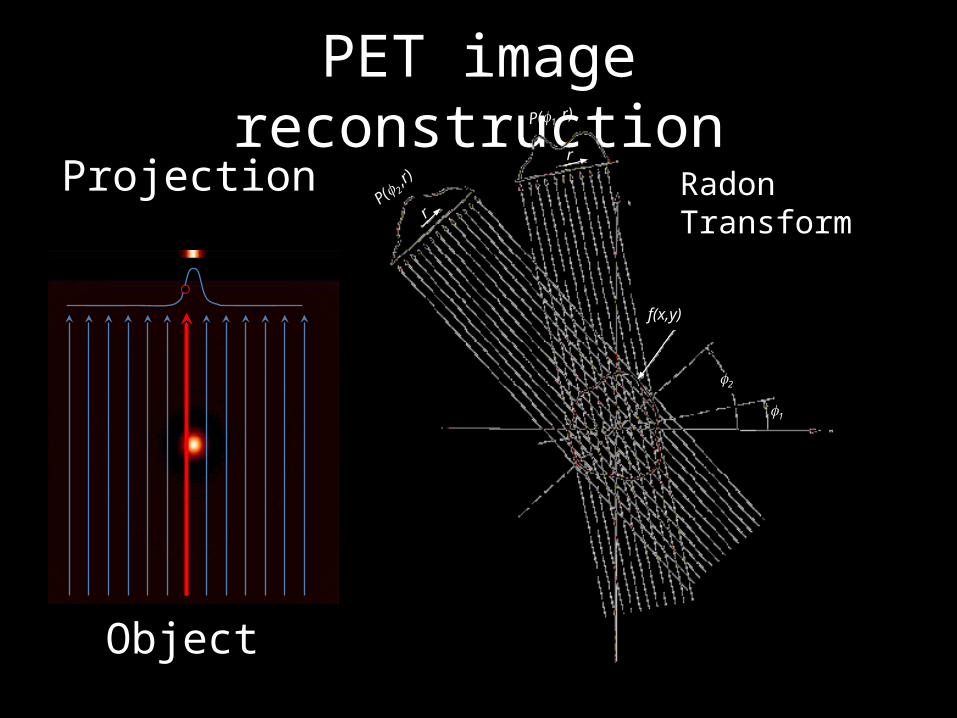
PET image reconstructionProjection
Object
P( 2 ,r) Radon Transform
1
2
P(1 ,r)
f(x,y)
r
r
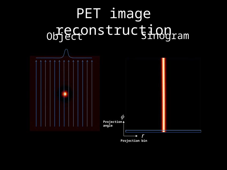
PET image reconstructionSinogram
r
Projectionangle
Projection bin
Object
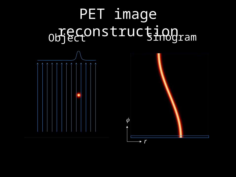
PET image reconstructionSinogramObject
r
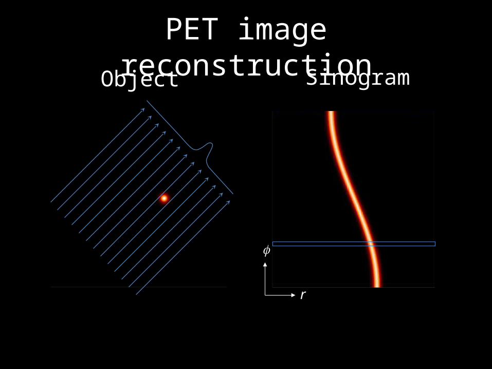
PET image reconstructionSinogramObject
r

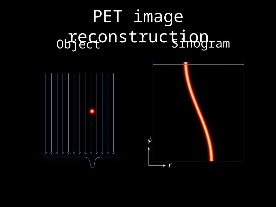
PET image reconstructionSinogramObject
r

PET image reconstructionSinogramObject
r
PET image reconstruction
r
SinogramObject
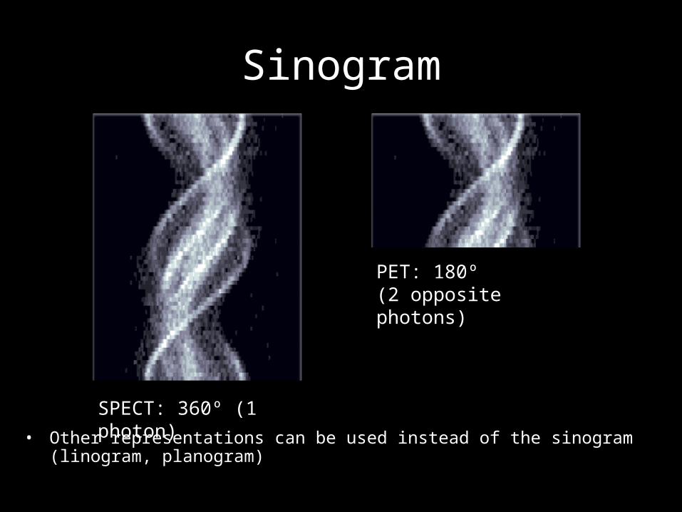
Sinogram
• Other representations can be used instead of the sinogram (linogram, planogram)
SPECT: 360º (1 photon)
PET: 180º (2 opposite photons)
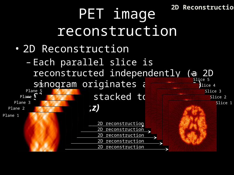
PET image reconstruction
• 2D Reconstruction– Each parallel slice is reconstructed independently
(a 2D sinogram originates a 2D slice)– Slices are stacked to form a 3D volume f(x,y,z)
2D reconstruction
Plane 5
Slice 5etc
etc
2D reconstruction
Plane 4
Slice 4
2D reconstruction
Plane 3
Slice 3
2D reconstruction
Plane 2
Slice 2
2D reconstruction
Plane 1
Slice 1
2D Reconstruction
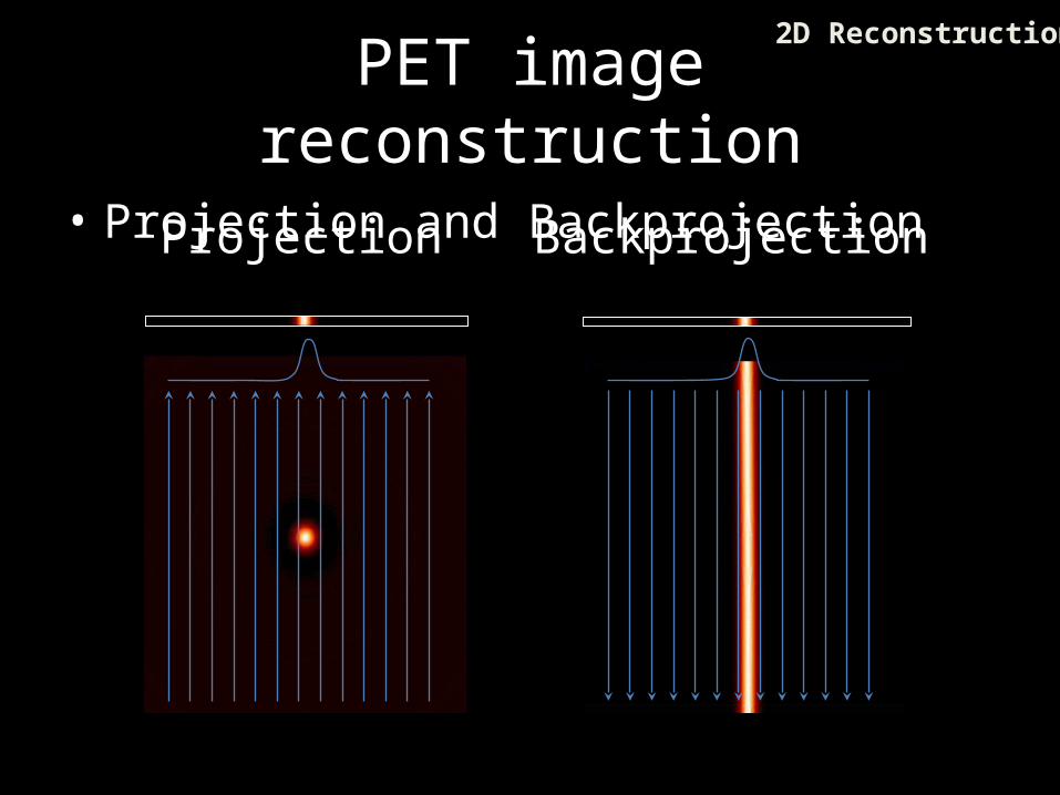
PET image reconstruction
• Projection and BackprojectionProjection Backprojection
2D Reconstruction
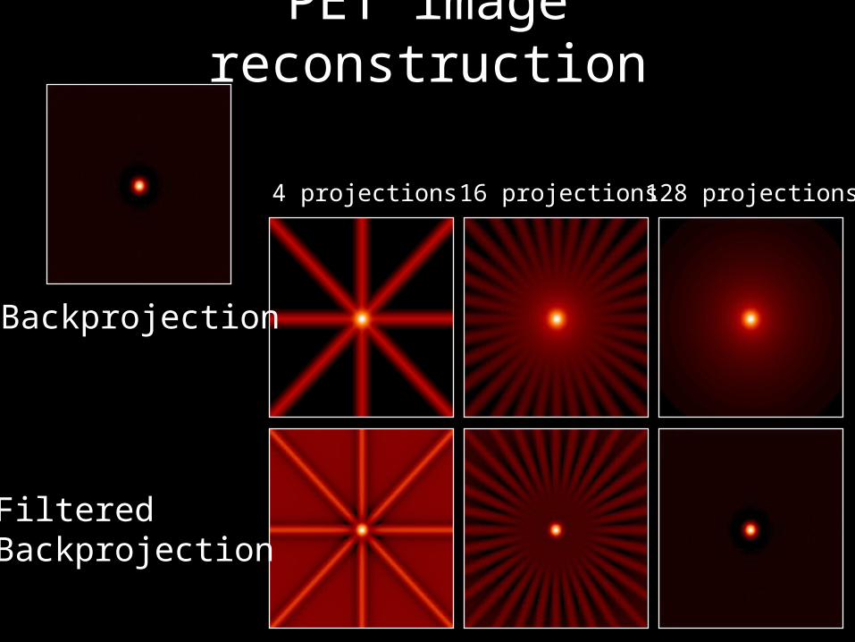
4 projections 16 projections 128 projections
Backprojection
FilteredBackprojection
PET image reconstruction
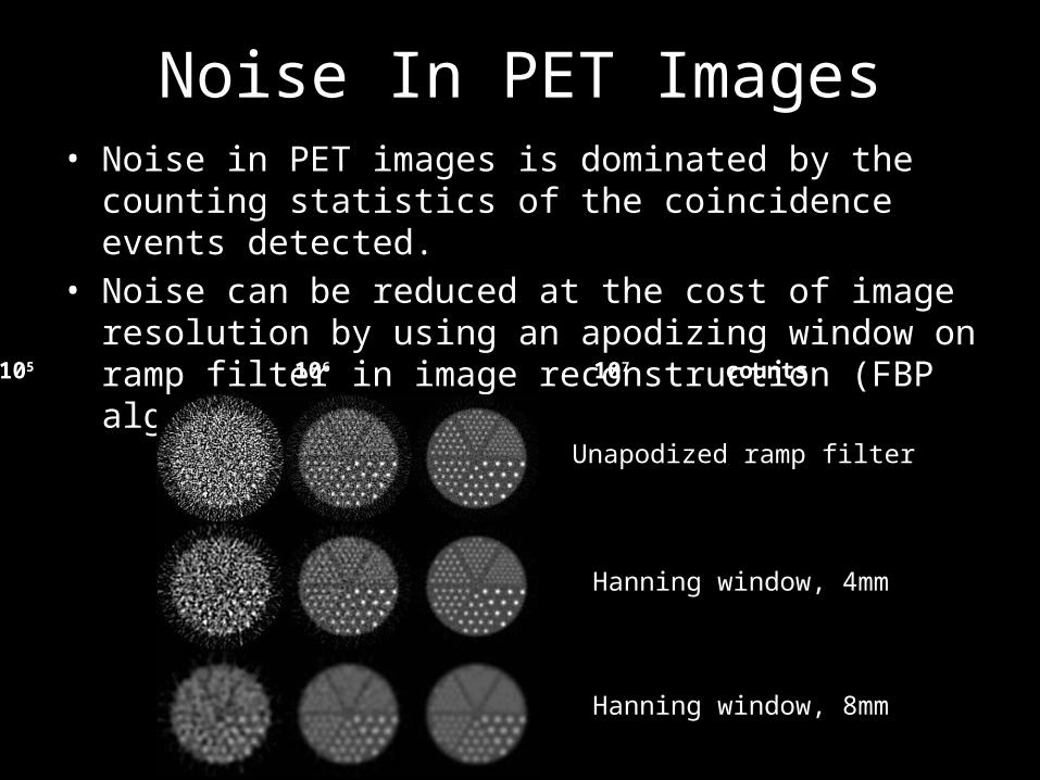
Noise In PET Images• Noise in PET images is dominated by the counting statistics of
the coincidence events detected.• Noise can be reduced at the cost of image resolution by using
an apodizing window on ramp filter in image reconstruction (FBP algorithm).
105 106 107 counts
Unapodized ramp filter
Hanning window, 4mm
Hanning window, 8mm
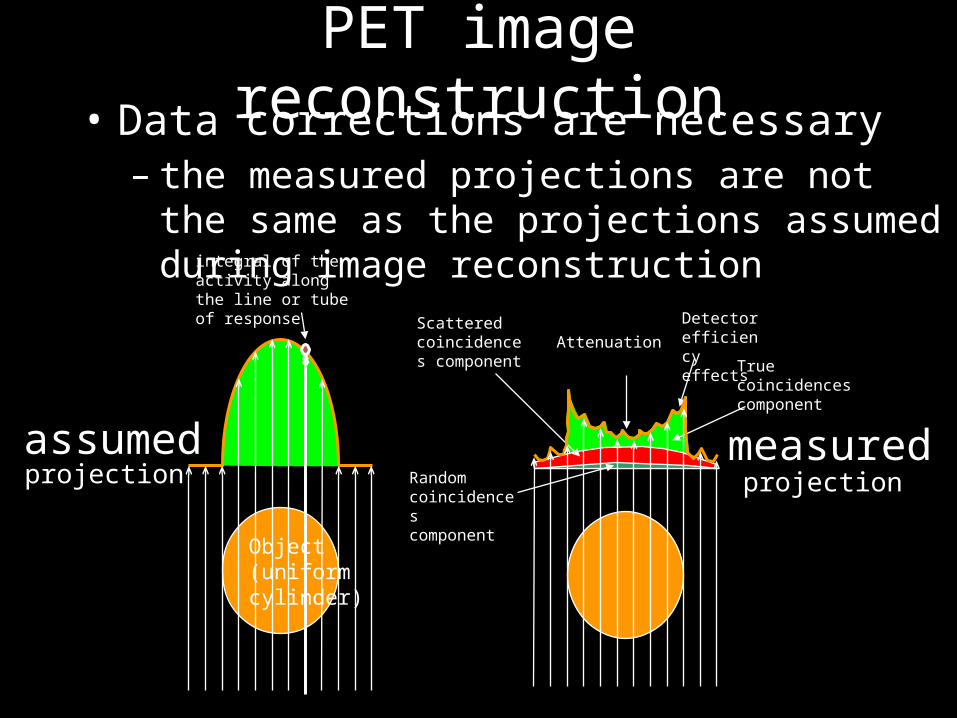
Scatteredcoincidences component
Attenuation
Random coincidencescomponent
Detector efficiencyeffects True
coincidencescomponent
PET image reconstruction• Data corrections are necessary
– the measured projections are not the same as the projections assumed during image reconstruction
Object(uniformcylinder)
projectionmeasured
projectionassumed
integral of the activity along the line or tube of response
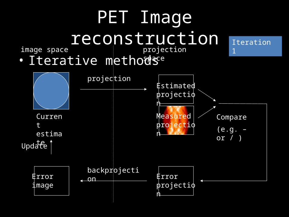
PET Image reconstruction
• Iterative methods
Current estimate
Measured projection
Compare
(e.g. – or / )
Error projection
projectionEstimated projection
image space projection space
backprojectionErrorimage
Update
Iteration 1

PET Image reconstruction
• Iterative methods
Current estimate
Measured projection
Compare
(e.g. - or / )
Error projection
projectionEstimated projection
image space projection space
backprojectionErrorimage
Update
Estimated projection
Estimated projection
Iteration 2
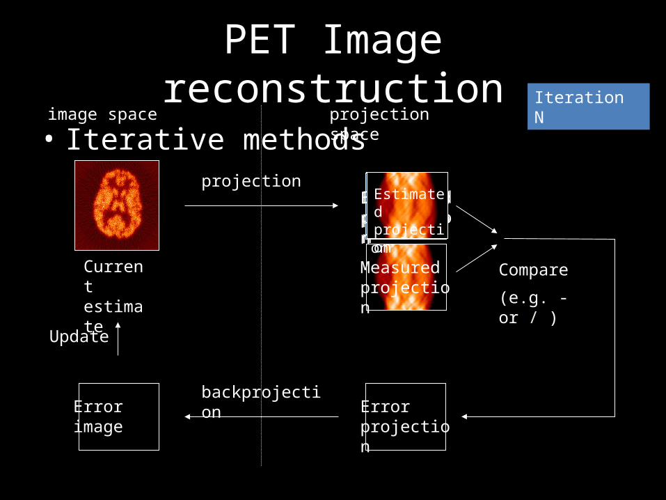
PET Image reconstruction
• Iterative methods
Current estimate
Measured projection
Compare
(e.g. - or / )
Error projection
projection
image space projection space
backprojectionErrorimage
Update
Estimated projection
Estimated projection
Estimated projection
Estimated projection
Iteration N
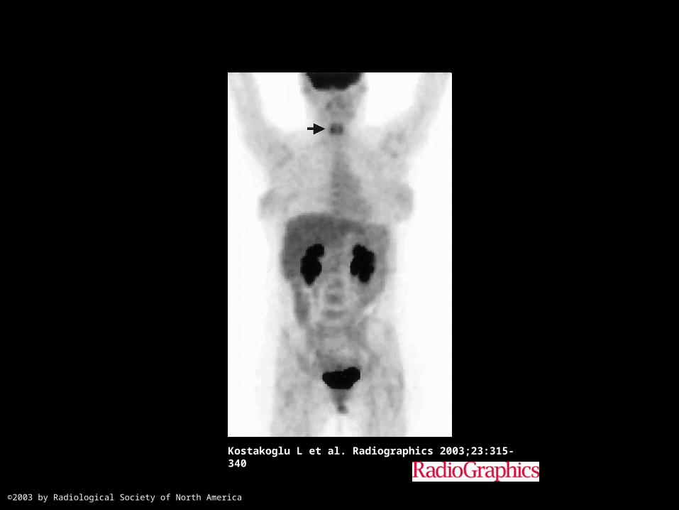
Kostakoglu L et al. Radiographics 2003;23:315-340
©2003 by Radiological Society of North America
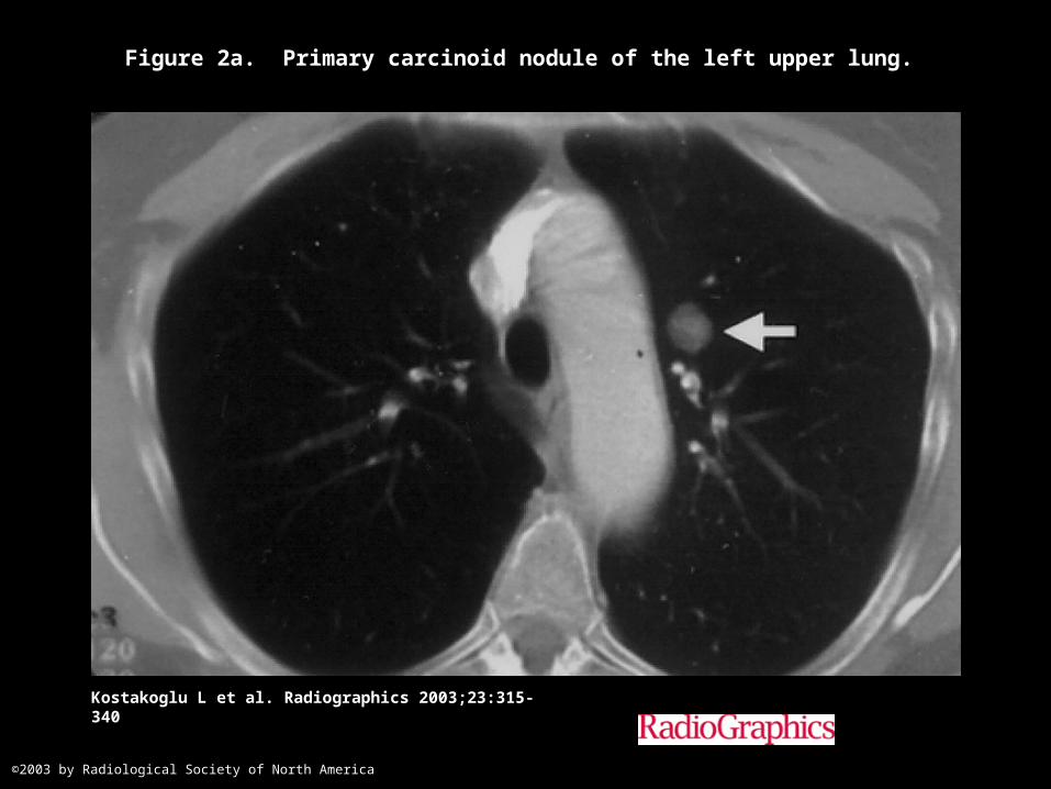
Figure 2a. Primary carcinoid nodule of the left upper lung.
Kostakoglu L et al. Radiographics 2003;23:315-340
©2003 by Radiological Society of North America
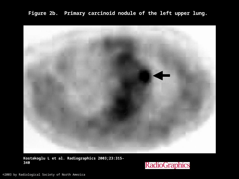
Figure 2b. Primary carcinoid nodule of the left upper lung.
Kostakoglu L et al. Radiographics 2003;23:315-340
©2003 by Radiological Society of North America
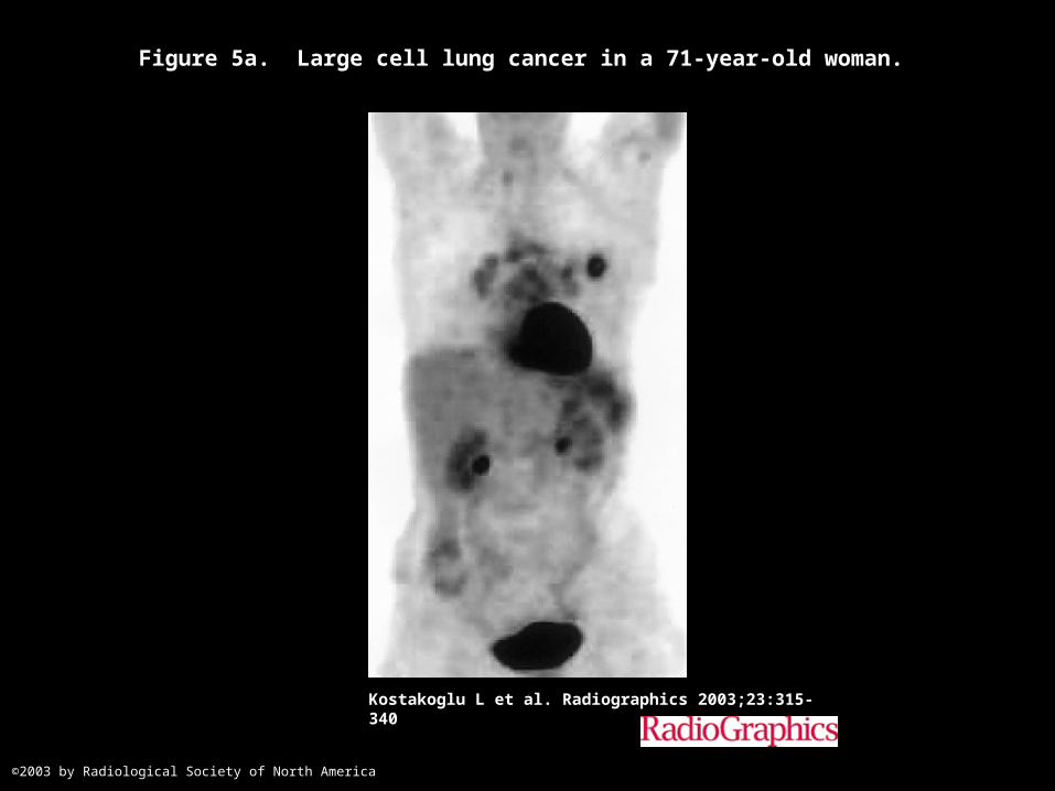
Figure 5a. Large cell lung cancer in a 71-year-old woman.
Kostakoglu L et al. Radiographics 2003;23:315-340
©2003 by Radiological Society of North America
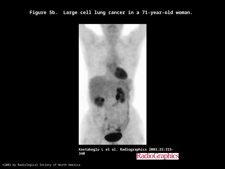
Figure 5b. Large cell lung cancer in a 71-year-old woman.
Kostakoglu L et al. Radiographics 2003;23:315-340
©2003 by Radiological Society of North America
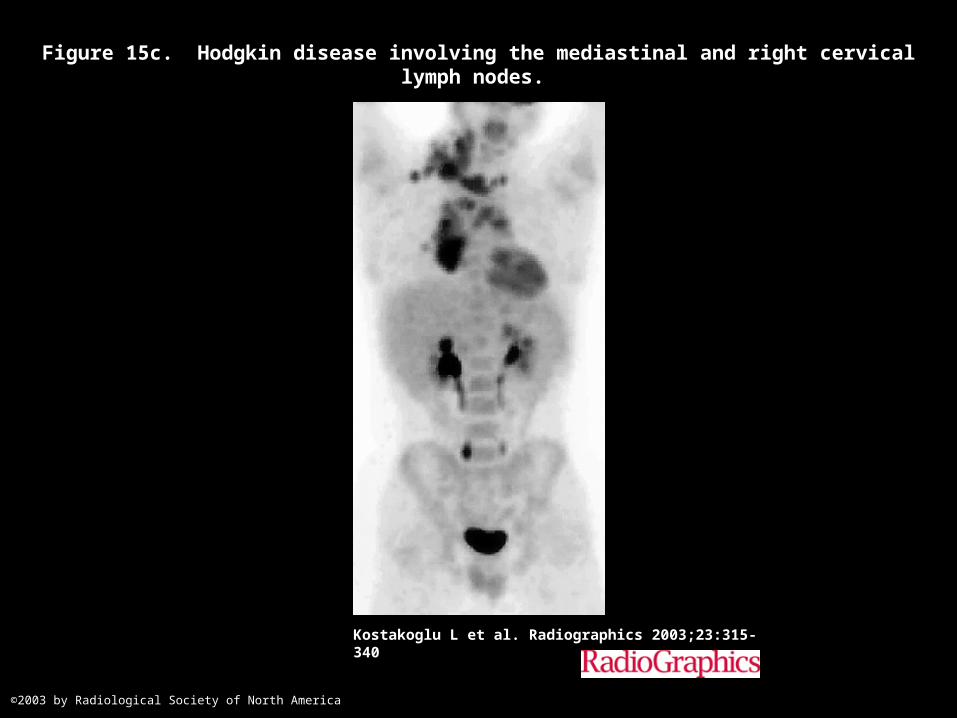
Figure 15c. Hodgkin disease involving the mediastinal and right cervical lymph nodes.
Kostakoglu L et al. Radiographics 2003;23:315-340
©2003 by Radiological Society of North America
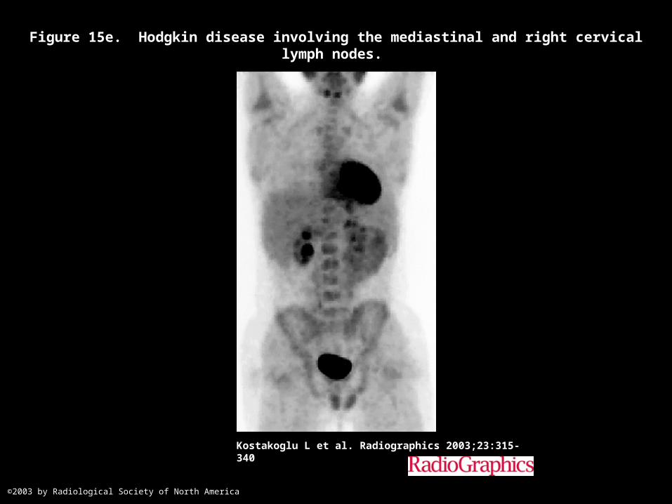
Figure 15e. Hodgkin disease involving the mediastinal and right cervical lymph nodes.
Kostakoglu L et al. Radiographics 2003;23:315-340
©2003 by Radiological Society of North America
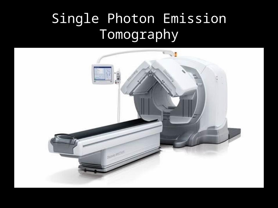
Single Photon Emission Tomography
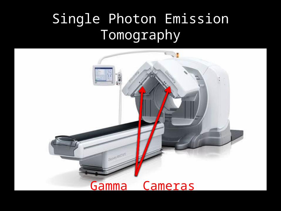
Single Photon Emission Tomography
Gamma Cameras
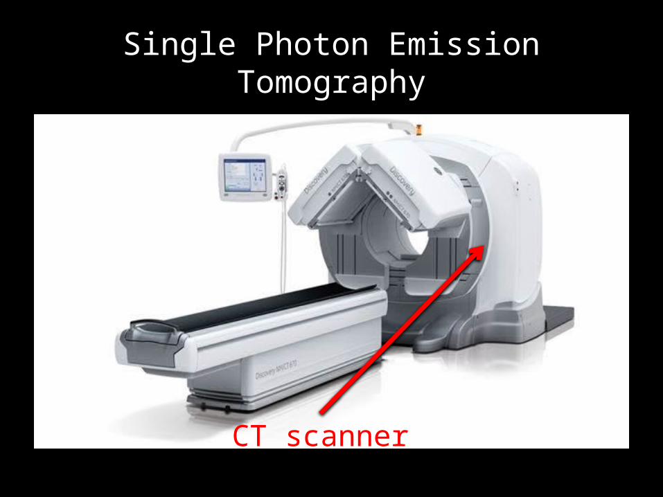
Single Photon Emission Tomography
CT scanner

Single Photon Emission Tomography
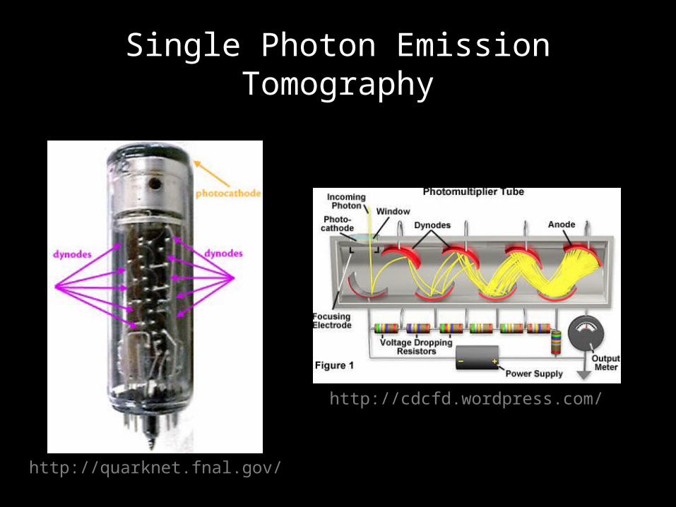
Single Photon Emission Tomography
http://cdcfd.wordpress.com/
http://quarknet.fnal.gov/
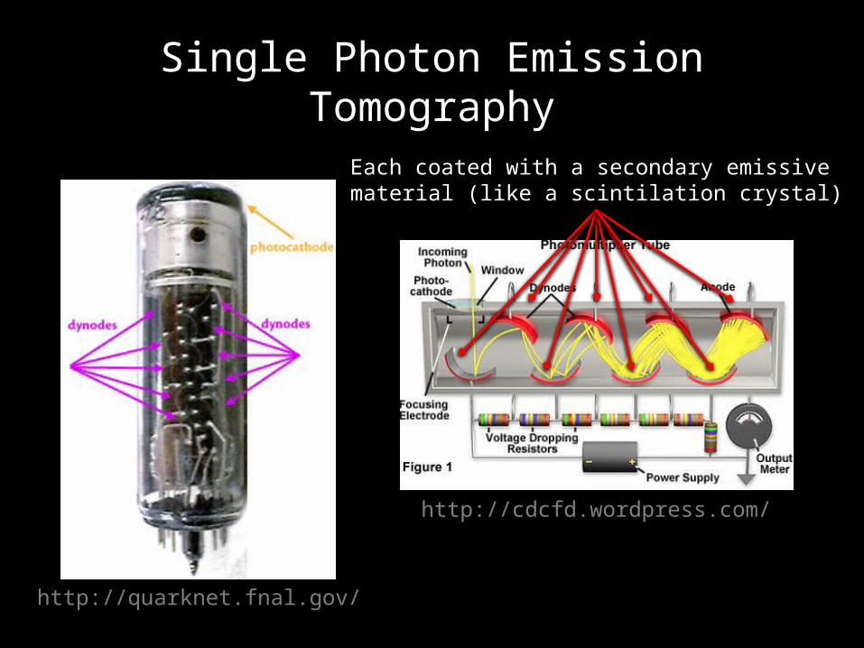
Single Photon Emission Tomography
http://cdcfd.wordpress.com/
http://quarknet.fnal.gov/
Each coated with a secondary emissive material (like a scintilation crystal)
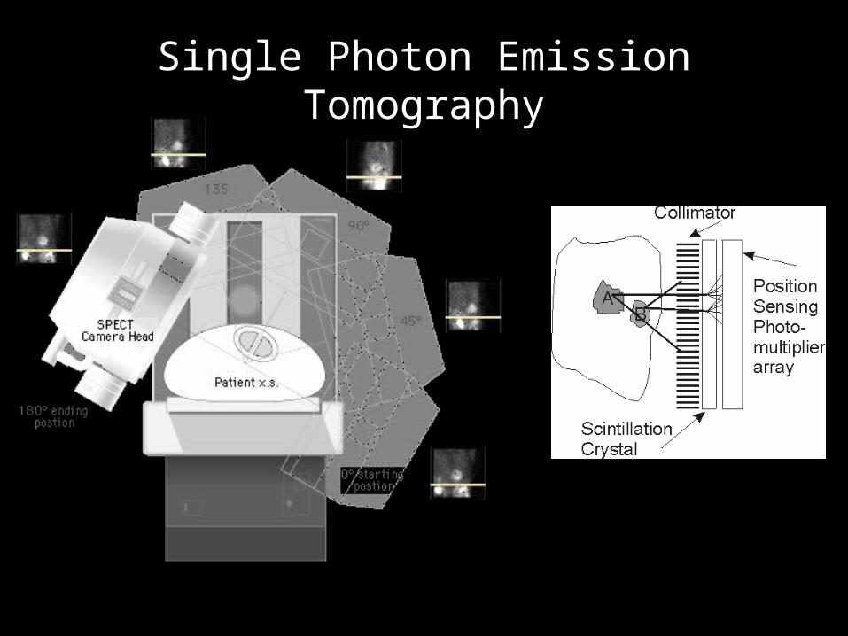
Single Photon Emission Tomography
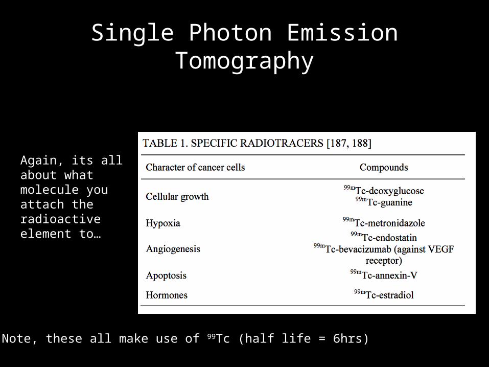
Single Photon Emission Tomography
Again, its all about what molecule you attach the radioactive element to…
Note, these all make use of 99Tc (half life = 6hrs)
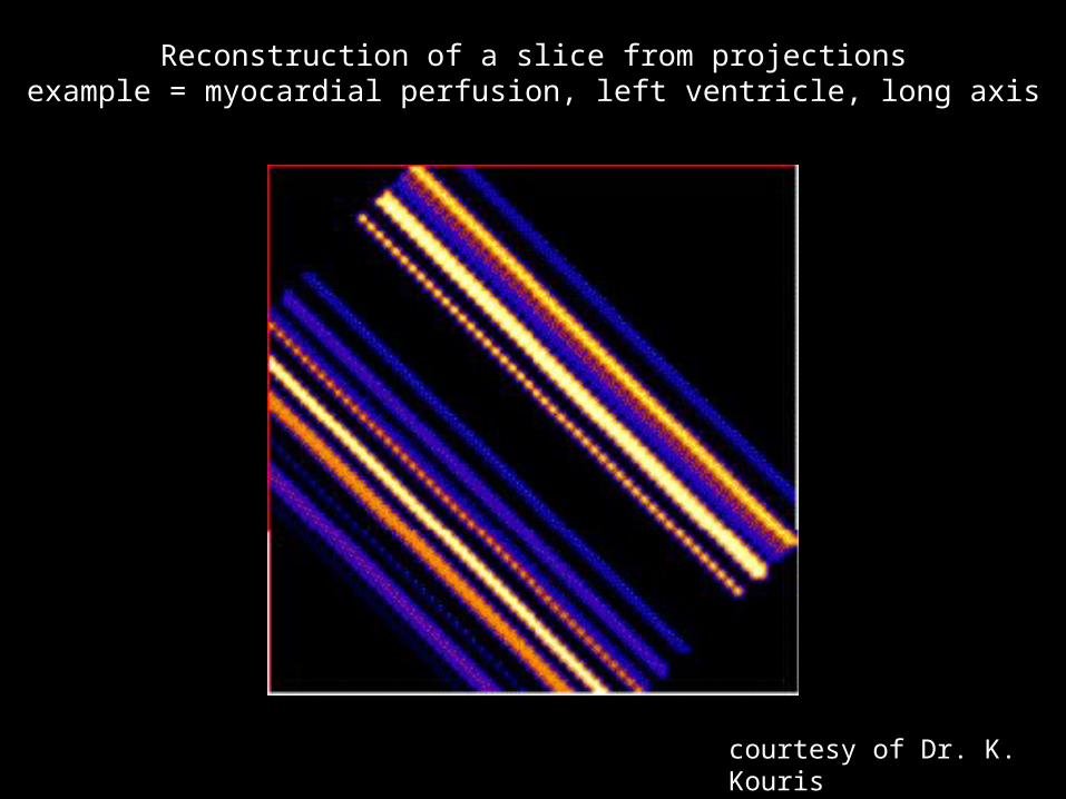
Reconstruction of a slice from projectionsexample = myocardial perfusion, left ventricle, long axis
courtesy of Dr. K. Kouris
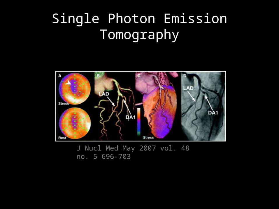
Single Photon Emission Tomography
J Nucl Med May 2007 vol. 48 no. 5 696-703
Single Photon Emission Tomography
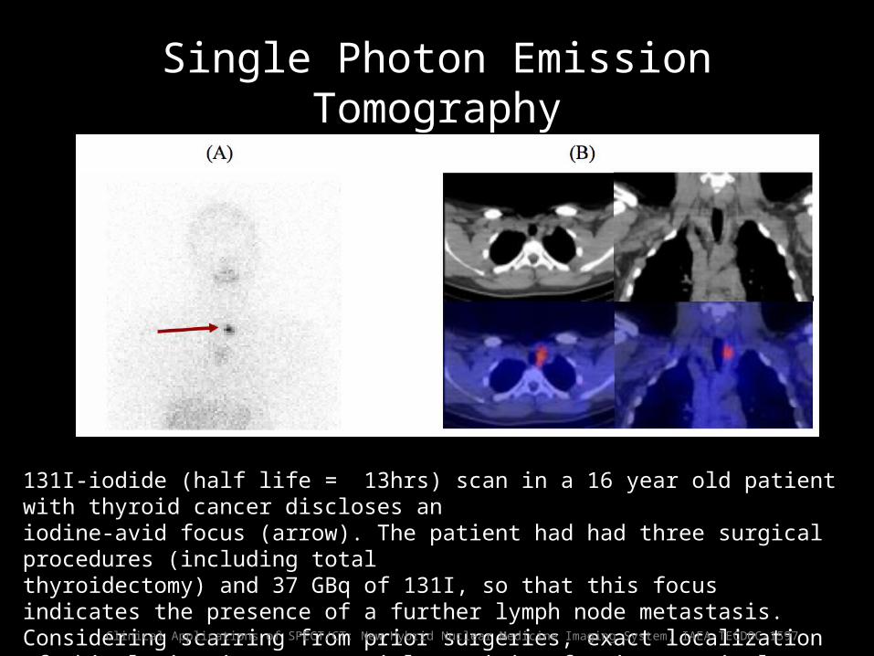
131I-iodide (half life = 13hrs) scan in a 16 year old patient with thyroid cancer discloses an iodine-avid focus (arrow). The patient had had three surgical procedures (including total thyroidectomy) and 37 GBq of 131I, so that this focus indicates the presence of a further lymph node metastasis. Considering scarring from prior surgeries, exact localization of this lesion is an essential requisite for its surgical resection
Clinical Applications of SPECT/CT: New Hybrid Nuclear Medicine Imaging System, IAEA-TECDOC-1597
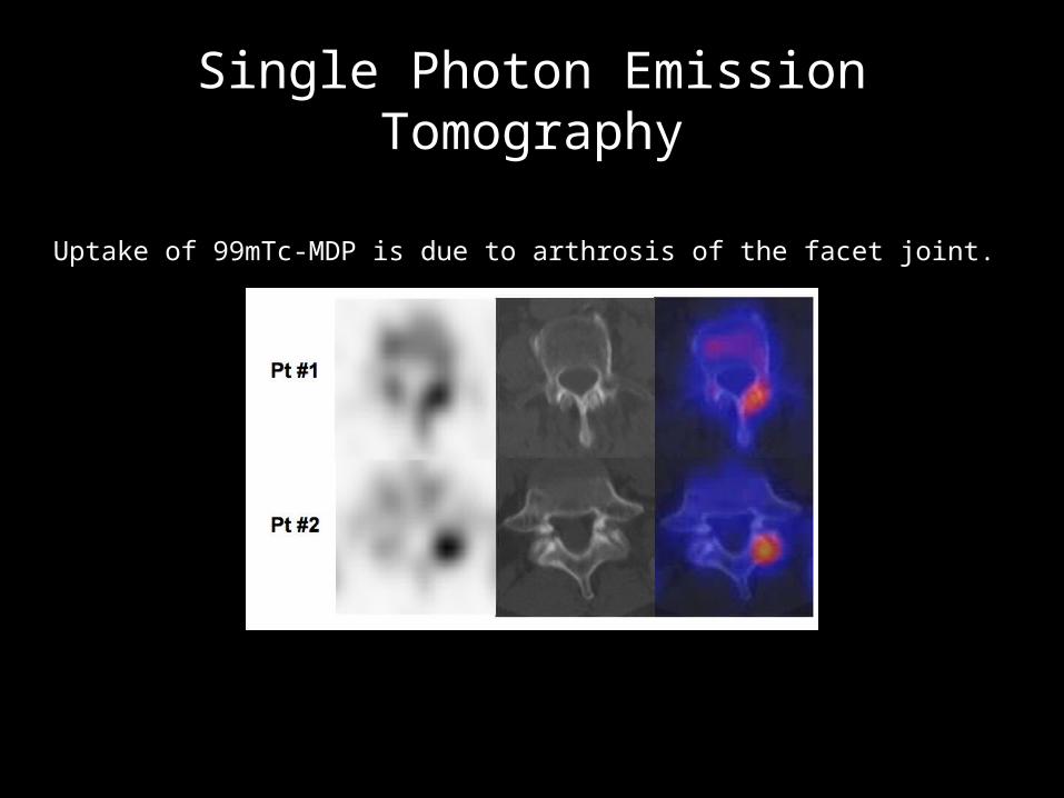
Single Photon Emission Tomography
Uptake of 99mTc-MDP is due to arthrosis of the facet joint.
Single Photon Tomography
“In a recent study comparing the diagnostic accuracy of 99mTc-phosphonate skeletal scintigraphy to that of [18F]FDG-PET in patients with thyroid carcinoma [64], sensitivity of the conventional procedure was not significantly different from that of [18F]FDG-PET. However, its specificity was significantly worse.”
“…there are several highly prevalent benign conditions leading to focally increased uptake of the radiolabelled phosphonates in the skeleton. Most of these conditions reflect degenerative processes of the joints increasing in frequency with age, such as spondylarthrosis or coxarthrosis. Additional benign causes of enhanced uptake are rheumatic disease or benign bone tumours.
Since most of these benign conditions are readily identifiable on CT, SPECT/CT is expected to improve specificity of skeletal scintigraphy without reducing its sensitivity. Besides single case reports illustrating this assumption, several prospective studies have investigated this issue.”
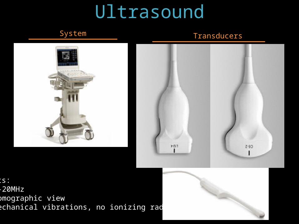
UltrasoundSystem Transducers
Facts:• 1-20MHz• Tomographic view• Mechanical vibrations, no ionizing radiation
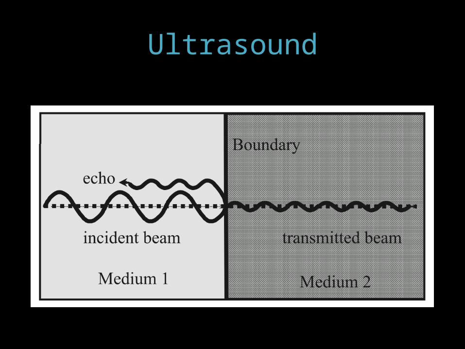
Ultrasound
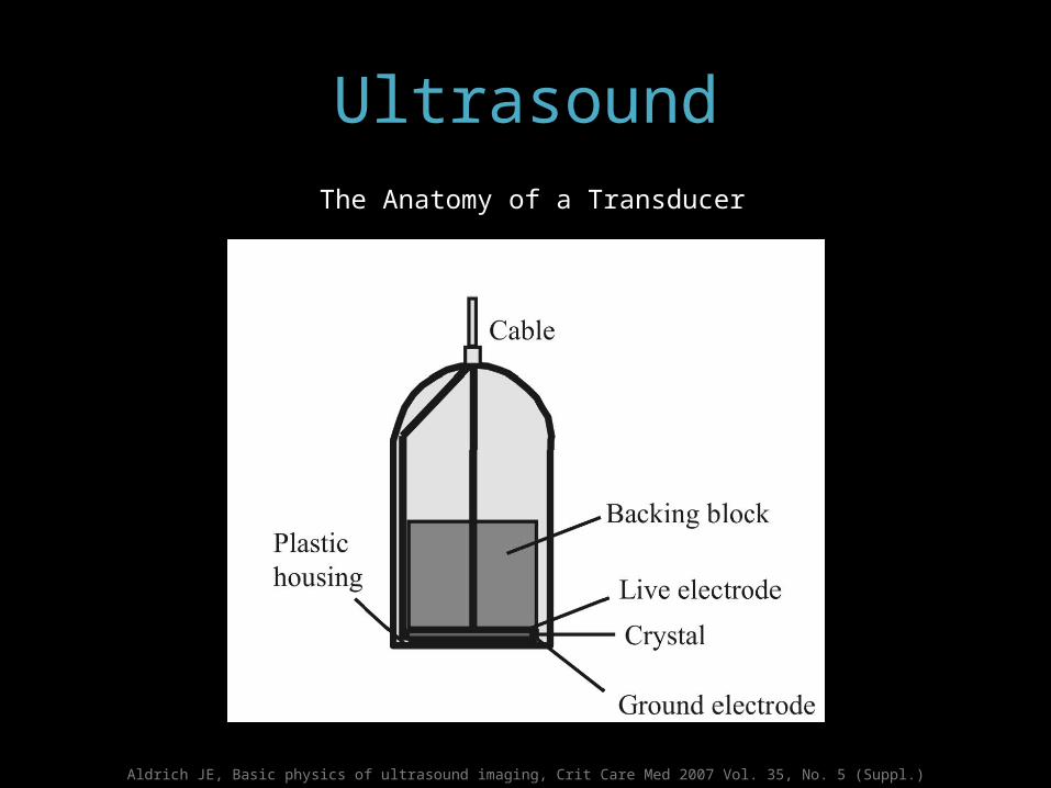
Aldrich JE, Basic physics of ultrasound imaging, Crit Care Med 2007 Vol. 35, No. 5 (Suppl.)
The Anatomy of a Transducer
Ultrasound
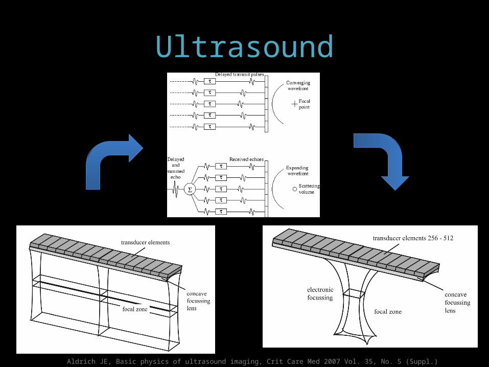
Ultrasound
Aldrich JE, Basic physics of ultrasound imaging, Crit Care Med 2007 Vol. 35, No. 5 (Suppl.)
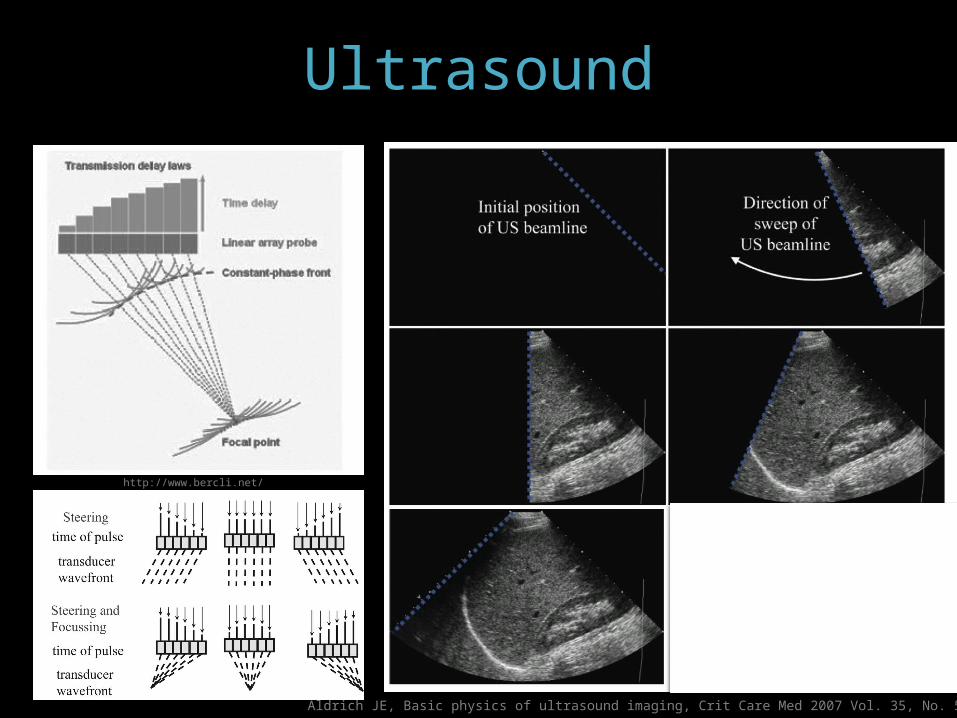
Ultrasound
Aldrich JE, Basic physics of ultrasound imaging, Crit Care Med 2007 Vol. 35, No. 5 (Suppl.)
http://www.bercli.net/
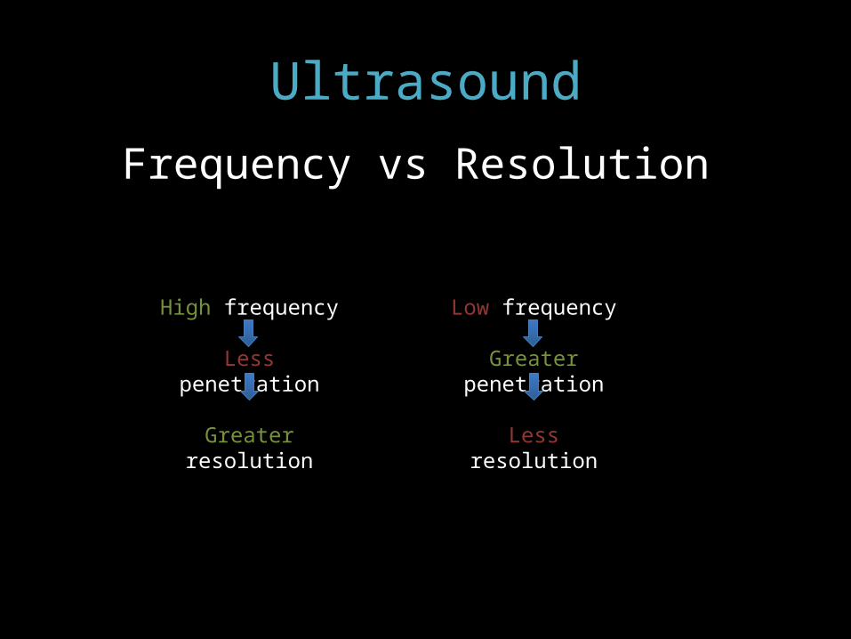
Ultrasound
High frequency
Less penetration
Greater resolution
Frequency vs Resolution
Low frequency
Greater penetration
Less resolution
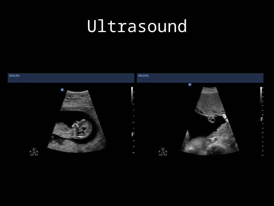
Ultrasound
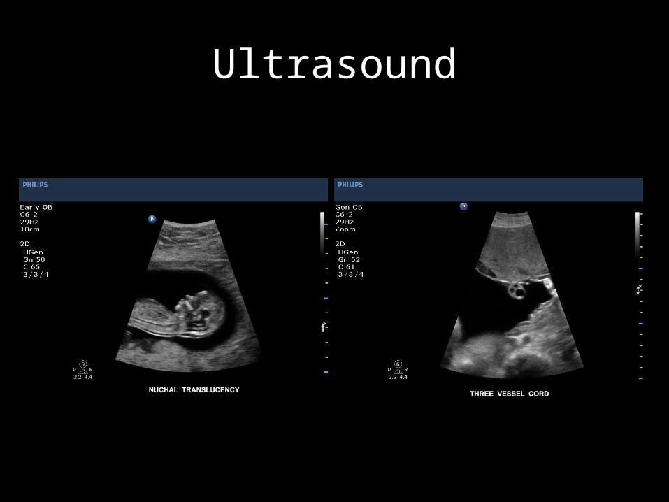
Ultrasound
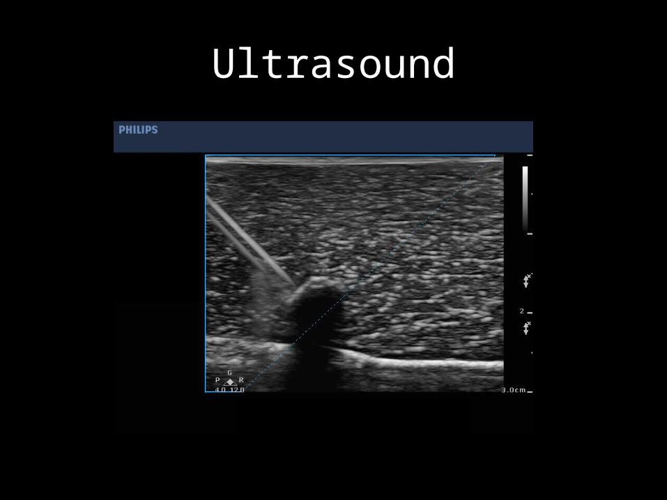
Ultrasound
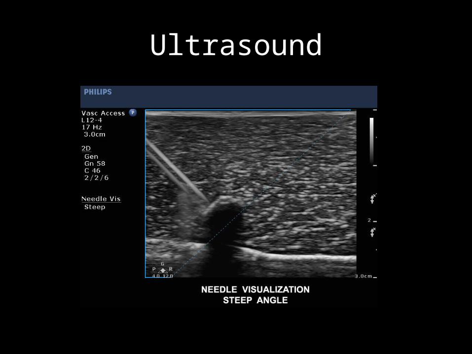
Ultrasound

Ultrasound
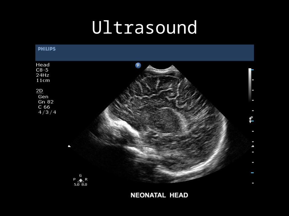
Ultrasound
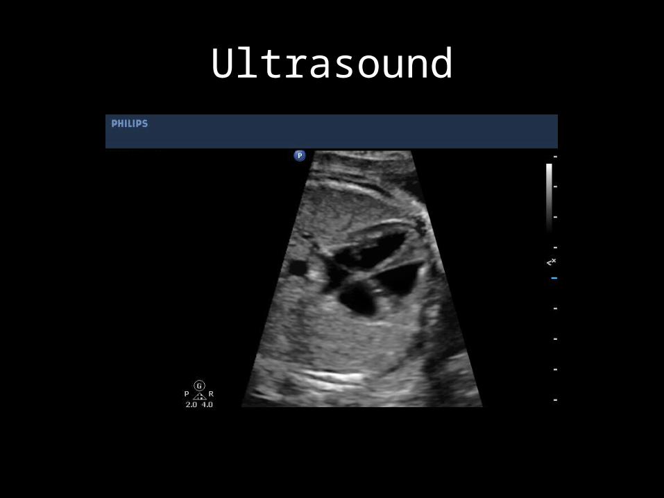
Ultrasound
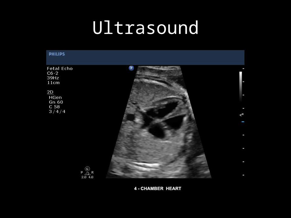
Ultrasound

Ultrasound
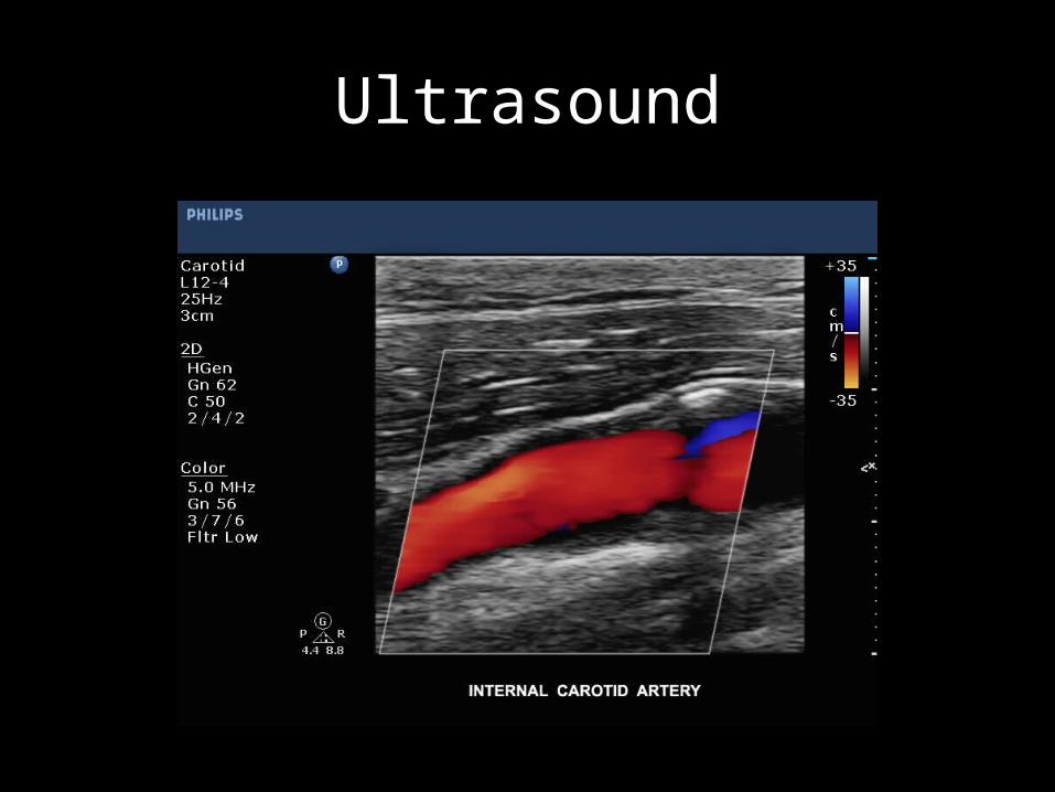
Ultrasound
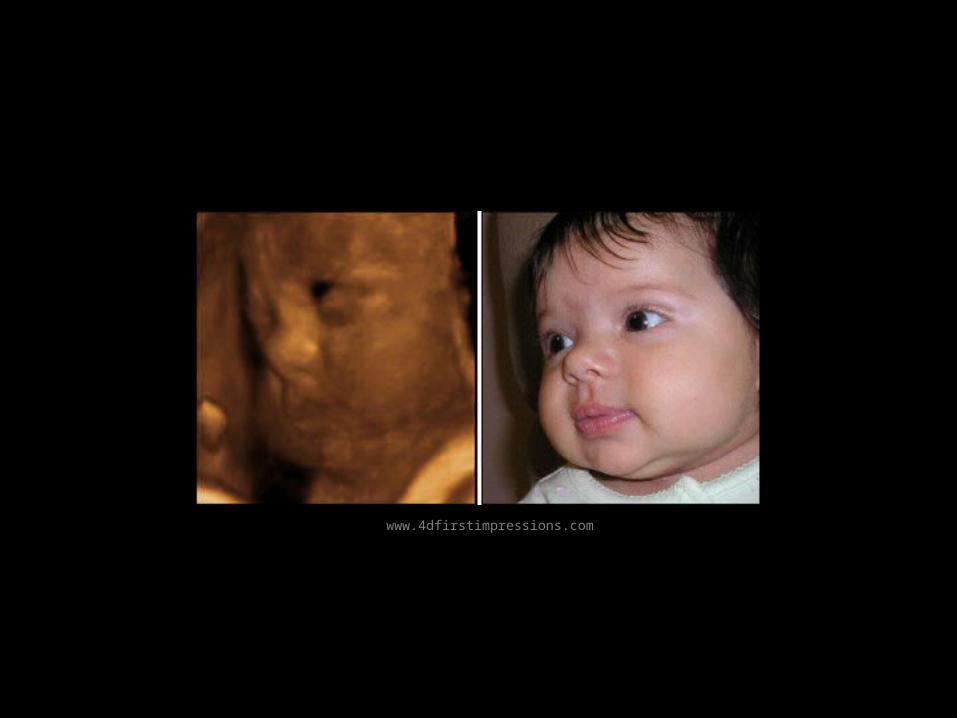
www.4dfirstimpressions.com
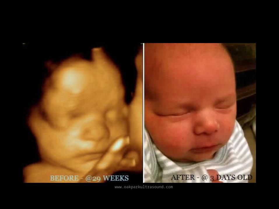
www.oakparkultrasound.com
www.firstglimpseultrasound.com


![PET/ CT [Positron Emission Tomography]](https://img.pdfslide.net/doc/110x75/56d6bf451a28ab30169592f3/pet-ct-positron-emission-tomography.jpg)


